See discussions, stats, and author profiles for this publication at: https://www.researchgate.net/publication/313786807
Influence of vaginal lactoferrin administration
on amniotic fluid cytokines and its role against
inflammatory...
Article in Journal of Inflammation · December 2017 DOI: 10.1186/s12950-017-0152-9 CITATIONS0
READ1
8 authors, including: Some of the authors of this publication are also working on these related projects: The role of Chlamydia in Ocular LymphomaView project The Great Epidemics of Infectious Diseases
View project Silva Seraceni IRCCS Ospedale Infantile Burlo Garofolo 67 PUBLICATIONS 469 CITATIONS SEE PROFILE Alessandro Trentini University of Ferrara 32 PUBLICATIONS 173 CITATIONS SEE PROFILE Fortunato Vesce University of Ferrara 92 PUBLICATIONS 695 CITATIONS SEE PROFILE Carlo Contini University of Ferrara 171 PUBLICATIONS 939 CITATIONS SEE PROFILE
All content following this page was uploaded by Fortunato Vesce on 22 February 2017.
The user has requested enhancement of the downloaded file. All in-text references underlined in blue are added to the original document and are linked to publications on ResearchGate, letting you access and read them immediately.
R ES EAR CH
Open Access
Influence of vaginal lactoferrin
administration on amniotic fluid cytokines
and its role against inflammatory
complications of pregnancy
Martina Maritati
1, Manola Comar
2, Nunzia Zanotta
3, Silva Seraceni
2, Alessandro Trentini
5, Fabrizio Corazza
4,
Fortunato Vesce
6*and Carlo Contini
1Abstract
Background: An altered amniotic cytokine profile has been reported in inflammatory pregnancy complications with a leading role for IL-6, a marker of the foetal systemic inflammatory response. Up to this date there is no exhaustive information neither on the foetal cytokine balance nor on the best method for its modulation. We aimed to evaluate the influence of vaginal lactoferrin administration on amniotic fluid concentration of 47 cytokines, chemokines and growth factors.
Methods: Sixty women undergoing genetic amniocentesis were enrolled in an open-label clinical trial. 300 mg of vaginal lactoferrin (Florence, Italy) were randomly administered to obtain 3 groups: A, 20 untreated patients; B and C (20 patients in each) respectively treated 4 and 12 h before amniocentesis. Cytokines, chemokines and growth factors concentrations were quantified by a magnetic bead Luminex multiplex immunoassays panel technology. Data analysis was performed with the software Stata (v. 13.1) and GraphPad Prism (v. 5). Group comparisons were performed using Kruskal–Wallis followed by Mann–Whitney U tests, with Bonferroni correction for multiple comparisons. A p < 0.05 was considered significant.
Results: Among the 47 tested mediators, 24 (51.06%) were influenced by lactoferrin. 11 (23.4%), showed a highly significant difference (p <0.001); among these IL-9, IL-15, IFN-γ, IP-10, TNF-α, IL-1α and MCP-3 underwent a down-regulation, while IL-17 and FGF-basic, G-CSF, GM-CSF an up-regulation. Difference between group C and both B and A was small for IL-15, IP-10, IL-1α, MCP-3, while it was negligible for IL-9, IFN-γ and TNF-α. IL-17 and the 3 growth factors were strongly enhanced in B and C groups. IL-17, FGF-basic and GM-CSF showed increasing concentrations in both B and C groups, while G-CSF resulted up-regulated only in group C. Significance was intermediate (p < 0.01) for the down regulated IL-2RA, IL-12p40 and IFNα2 (6.38%) while it was small for 10 mediators (21.27%) 7 of which (IL-2, IL-4, eotaxin, PDGF-BB, RANTES, IL-18 and MIF) down-regulated and 3 (MCP-1, IL-3, and SDF-1α) up-regulated.
Conclusion: Lactoferrin down-regulates 17 pro-inflammatory amniotic mediators while up-regulating 7 anti-inflammatory amniotic mediators, 5 of which definitively belonging to an anti-anti-inflammatory profile. These findings open to clinical investigation on its use against inflammatory complications of pregnancy.
Keywords: Amniotic fluid, Lactoferrin, Pregnancy, Mediators, Cytokines, Inflammatory complications, Abortion, Amniocentesis
* Correspondence:[email protected]
6Section of Obstetrics and Gynaecology, Department of Morphology, Surgery
and Experimental Medicine, University of Ferrara, I 44 100 Ferrara, Italy Full list of author information is available at the end of the article
© The Author(s). 2017 Open Access This article is distributed under the terms of the Creative Commons Attribution 4.0 International License (http://creativecommons.org/licenses/by/4.0/), which permits unrestricted use, distribution, and reproduction in any medium, provided you give appropriate credit to the original author(s) and the source, provide a link to the Creative Commons license, and indicate if changes were made. The Creative Commons Public Domain Dedication waiver (http://creativecommons.org/publicdomain/zero/1.0/) applies to the data made available in this article, unless otherwise stated.
Background
Successful pregnancy depends on an orderly production of trophoblast mediators aimed at modulating maternal adaptation, progressively increasing uterine-placental per-fusion to draw substances for foetal growth. This task must be directly performed by the foetus, counteracting the natural reaction against the trophoblast induced uter-ine blood vessel rupture, that in any other tissue but uterus should activate inflammatory changes leading to coagulation and smooth muscle contraction. Such a foetal capacity must be even stronger in the presence of mater-nal pathologic conditions linked with inflammation, like rheumatic disease, coagulation, and thrombophilia. With regard to maternal immunity and related mediators, it has been reported that Th1-type is incompatible with success-ful pregnancy, while Th2-type is protective [1].
As for the foetal role, there is a large body of data on normal trophoblast function [2] as well as evidence indi-cating that foetal chromosomal abnormalities are charac-terized by alteration of some mediators of inflammation and coagulation which may explain the high incidence of early abortion linked with aneuploidy [3, 4]. Such evidence reinforces the concept of a direct foetal control on normal gestational processes.
Furthermore, an altered amniotic cytokine profile has been reported in pregnancy complications such as pre-mature delivery and chorioamnionitis with a leading role for IL-6, a marker of the foetal systemic inflammatory response [5]. In this regard, once recognized that an in-creased concentration of IL-6 in the foetal compartment cannot be considered a benign condition, attempts were made to reduce it by administering antibiotic [6, 7] as well as by lactoferrin (LF) administration [8]. Up to this date there is no exhaustive information neither on the foetal cytokine balance nor on the best method for its modulation, when clinically justified. Therefore, with the aim to contribute to a better understanding of the foetal mediators of physiological pregnancy, we analysed mid-trimester amniotic fluid (AF) concentration of a 47 cyto-kines panel, in basal condition as well as upon assump-tion of LF, an 80 kDa iron binding glycoprotein of the transferrin family, component of the mammalian innate immune system provided with anti-inflammatory and antimicrobial properties [9].
Methods
We performed an open-label clinical study enrolling 60 pregnant patients undergoing genetic amniocentesis at the 16th gestational week at the Obstetric Unit of Ferrara University from March 2011 to March 2012. The inclu-sion criteria were: singleton physiological pregnancy and maternal age as the only indication to foetal karyotyping. The exclusion criteria were: assumption of drugs inter-fering with the immune system, previous miscarriages,
pregnancy at risk for maternal or foetal disease. The re-search was carried out in accordance with the ethical principles of the Declaration of Helsinki. The Local Ethics Committee approved the design of this study and all pregnant women gave their written informed consent. Moreover, a questionnaire was administered to the patients in order to check for any complications (vaginal bleeding, uterine contraction, rupture of the membranes) within 7 days following the procedure.
Our protocol entails using a 22-gauge needle provided with a 24-gauge tip to reduce the diameter of the hole in the amniotic membrane and the consequent risk of AF leakage. Eligible patients were randomly assigned in a 1:1:1 ratio with a random number table to receive a vaginal compound containing 300 mg of LF (Difesan, Progine, Italy) to obtain 3 groups:
! group A: 20 untreated patients;
! group B: 20 patients treated 4 h before
amniocentesis;
! group C: 20 patients treated 12 h before
amniocentesis.
Based on a previous study, the compound was admin-istered in the pharmaceutical form of a tablet by means of an applicator deeply inserted into the vaginal route.
A total amount of 20 ml AF was withdrawn for karyo-type analysis, microbiological culture, α-fetoprotein (18 ml) and cytokines analysis.
Fresh AF specimens (1 ml) were centrifuged at 3000 g,
4 °C for 10 min, aliquoted and stored at −80 ° C until
analysis. In supernatant specimens diluted (1:4) and tested in triplicate, the chemokines and cytokines concentrations were quantified by a magnetic bead Luminex multiplex immunoassays panel technology (47 analytes) (Bio-Plex, BIORAD Laboratories, Milan, Italy), as previously de-scribed [10]. The samples were treated following manu-factures’ instructions, using the Bio-Plex array reader (Luminex, Austin, TX) to determine the cytokines levels expressed as concentration (pg/mL) by the Manager software. The validation of the Bioplex platform has been performed in our laboratory using the sandwich ELISA assay as the reference [11].
Data analysis was performed with the software Stata (v. 13.1) and GraphPad Prism (v. 5). Group comparisons were performed using Kruskal–Wallis followed by Mann– Whitney U tests, with Bonferroni correction for multiple comparisons. A p < 0.05 was considered significant.
Results
The mean maternal age in the whole study population was 37.5 ± 2.1 years (minimum 35, maximum 42). This parameter was not different among the 3 groups (p = 0.5)
and distributed as follows: 37.8 (±2.4), 37.7 (±2.3) and 37.1 (±2.1) years, for groups A, B and C, respectively.
A normal karyotype was registered in all cases. The AF microbiological cultures were negative in all the tested samples. No complications were registered within 7 days after amniocentesis, and the course of pregnancy was normal in all patients, ending in spontaneous deliv-ery at term. Among the 47 tested mediators, 24 (51.06%) were influenced by LF administration although with dif-ferent levels of statistically significant difference. In par-ticular, 11 (23.4%), showed a highly significant difference (p <0.001); among these, 7 mediators (IL-9, IL-15, IFN-γ, IP-10, TNF-α, IL-1α, MCP-3) underwent a down-regulation, 4 (IL-17 and 3 factors FGF-basic growth, G-CSF, GM-CSF) an up-regulation. The statistical difference between group C and both B and A was small for IL-15, IP-10, IL-1α, MCP-3, while it was negligible for IL-9, IFN-γ and TNF-α. (Table 1, Fig. 1). As for the strongly up-regulated ones, IL-17 and 3 growth factors (FGF-basic, G-CSF, GM-CSF) were enhanced
in groups B and C. In particular IL-17, FGF-basic and GM-CSF showed increasing concentrations in both groups B and C, while G-CSF resulted up-regulated in group C only (Table 1, Fig. 2).
Three of the mediators analysed (6.38%) 2RA, IL-12p40 and IFNα2 were down-regulated and showed an intermediate statistical significance (p < 0.01).
Finally, 10 mediators (21.27%) were influenced with a weakly significance (p < 0.05). Among these, 7 (IL-2, IL-4, eotaxin, PDGF-BB, RANTES, IL-18 and MIF) were down-regulated and the remaining 3 (MCP-1, IL-3, and SDF-1α) up-regulated (Table 1).
The analysis by ELISA confirmed the concentration trend observed with the BIO-plex Luminex XMap analysis. Although quantitative differences were found between ab-solute values for the cytokines obtained by Luminex and ELISA assays are found, the relative values are comparable and the two methods have shown similar trends, as also reported in recent literature [12–14].
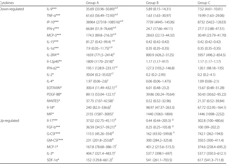
Table 1 Amniotic fluid concentration of mediators showing statistically significant difference in the three groups of examined patients
Cytokines Group A Group B Group C
Down-regulated IL-9*** 35.69 (33.96–50.89)a,d 5.89 (0.15–14.31) 7.52 (4.61–10.01)
TNF-α*** 61.63 (56.49–72.93)a,d 1.63 (1.63–30.97) 19.99 (1.63–29.06) IP-10*** 38964 (27318–108516)a,d 7739 (4945–14536) 8732 (5422–12633) IFN-γ*** 66.84 (51.9–76.64)a,d 24.7 (17.66–44.11) 27.7 (12.88–47.51) MCP-3*** 174.3 (89.8–216.3)a,d 28.63 (22.13–44.32) 30.49 (23.79–41.70) IL-15*** 81.27 (0.42–99.4)a,e 0.42 (0.42–0.42) 0.42 (0.42–0.42) IL-1α*** 7.9 (0.35–11.75)c,d 0.35 (0.35–0.35) 0.35 (0.35–0.35) IL-2RA** 1659 (771,5–2414)e 800.9 (426.2–3125) 5957 (496.2–854.5) Il-12p40** 1809 (1170–2518)e 1.17 (1.17–917) 1.17 (1.17–1.17) IFN-α2** 195.1 (128.9–233.1)c,e 127.3 (103.2–146.8) 120.1 (98.18–135) IL-2* 30.04 (0.2–35.02)c,f 0.2 (0.2–2.95) 0.2 (0.2–4.1) IL-4* 1.97 (0.06–2.6)c 0.06 (0.06–1.475) 1.09 (0.06–2.1) EOTAXIN* 300.4 (11.49–432.1)c,f 6.01 (0.48–23.2) 15.67 (0.48–31.28) PDGF-BB* 89.13 (53.04–122.1)c 39.86 (30.24–70.64) 50.43 (30.62–95.22) RANTES* 37.75 (7.07–42.58)c 0.52 (0.52–32.96) 21.37 (0.52–39.84) Il-18* 240 (82.3–336.6)f 98.97 (47.37–263.3) 67.72 (52.95–164.1) MIF* 2155 (1587–3005)c 1440 (1063–1804) 1446 (1098–2232) Up-regulated Il-17*** 37.02 (32.75–45.11)d 0.44 (0.44–205.5)g 302.8 (100–480.6) FGF-b*** 36.59 (34.57–39.21)d 0.25 (0.25–105.8)g 100 (99–202.2) G-CSF*** 115.5 (45.26–354)d 162 (43.92–599.8)h 742.1 (362–1943) GM-CSF*** 231 (201.8–253.8)b 300 (284.2–325.8) 350.5 (300–411.4) MCP-1* 167.8 (78.68–386–7)f 401.2 (215.6–515.5) 374.6 (258.4–695.2) IL-3* 404.7 (321.4–483.7)c 537.7 (398.1–697) 537.7 (350.3–612.1) SDF-1α* 152 (129.8–661.2)f 541 (261.1–703.5) 617 (541.3–711.8)
Data are given as medians (quartiles) (pg/ml); ***p < 0.001; **p < 0.01; *p < 0.05 Group A vs Group B:ap < 0.001;bp < 0.01;cp < 0.05
Group A vs Group C:dp < 0.001;ep < 0.01;fp < 0.05
Discussion
Given the key-role of foetal mediators in the regulation of pregnancy, a better knowledge of their normal profile and their possible pharmacologic modulation is needed. As we previously demonstrated that vaginal LF adminis-tration significantly down-regulates amniotic IL-6 in women undergoing genetic amniocentesis [8], we found it noteworthy to extend our observation to other cyto-kines, chemokines and growth factors in basal condition as well as upon the action of the glycoprotein.
By analysing our data, 24 out of 47 (51.06%) tested mediators were significantly influenced by LF. Among the group with the highest statistical significance (p < 0.001) comprising of 11 analytes, 7 were down-regulated (IL-9, IL-15, IFN-γ, IP-10, TNF-α, IL-1α, MCP-3) while the remaining 4 (IL-17 FGF-basic, G-CSF, GM-CSF) were up-regulated. Most of the down-regulated ones belong to the pro-inflammatory type. Although IL-9 is more frequently associated with allergy, its role in Th1-mediated inflam-mation has been recently reported. As for IL-15, it is
Fig. 1 Differences among strongly down-regulated mediators in the three groups. Legend: Data are given as medians (quartiles) (pg/ml); ***p < 0.001; **p < 0.01; *p < 0.05
Fig. 2 Differences among strongly up-regulated mediators in the three groups. Legend: Data are given as medians (quartiles) (pg/ml); ***p < 0.001; **p < 0.01; *p < 0.05
present in the amniochorion and decidua, and increased amniotic concentration was found in preterm labour [15]. Furthermore, the increased amniotic level of the trad-itional inflammatory cytokines IL-1a, TNF-α, IFN-γ as well as of IL-1beta, IL-4, IL-6, IL-8 and MCP-1 are be-lieved to represent a marker for identification of the pa-tients at risk of preterm delivery [16]. Together with physiology of gestation, the above mediators also control foetal health. In fact, it has been reported that an in-creased level of pro-inflammatory cytokines TNF-α, IL-1, IL-6, IL-11, as well as VEGF, TGF-α and TGF-β, may damage the foetal capillary endothelium and alveolar epi-thelium, resulting in hyaline membrane formation. This evidence demonstrates that the cause of hyaline mem-brane disease is not prematurity per se, but rather inflam-matory conditions leading to both premature delivery and foetal disease.
Accordingly, the inflammatory role of IL-1 has also been experimentally documented on sheep by intra-amniotic injection of the recombinant ovine cytokine, which induces a strong elevation of the intestinal mRNA levels for IFN-γ, TNF-α, IL-4 and IL-10. This increased pro-inflammatory state is associated with a decreased level of the intestinal fatty acid binding protein, disrup-tion of the tight juncdisrup-tional protein ZO-1 and loss of ileum mucosal barrier [17]. Only two chemokines were found to be strongly down-regulated, namely IP-10 and MCP-3.
Of note, the mean levels of IP-10 we measured are al-most 5 times higher than in previous reports [18]. The observed discrepancy may be due to the different meth-odology used, since Luminex assay showed higher abso-lute cytokine values than ELISA, with a good correlation and a similar trend between the two assays. (12-13-14). Nevertheless, since IP-10 is a pro-inflammatory chemo-kine, the high value recovered in the control group (A) may be related to a physiological response to asymptom-atic infection, not diagnosed by routine microbiological culture. In fact, as shown in our study, IP-10 was found to be strongly down-regulated after LF intake, a glycopro-tein with anti-inflammatory and antimicrobial properties.
Increased amniotic fluid level of IP-10 has been re-ported in presence of foetal Down Syndrome, suggesting a link with the increased abortion rate observed in an-euploid pregnancy [19]. Accordingly, a possible role in adverse events of pregnancy, was suggested for other inflammatory cytokines in foetal aneuploidies [3, 4]. Fi-nally, high levels of TNF-α and MCP-3 have recently been observed in AF of women with preterm birth [20]. Such evidence suggests a possible beneficial role for both chemokines down-regulated by LF.
Among the strongly up-regulated mediators there were 3 growth factors, FGF-basic, G-CSF, GM-CSF and the cytokine IL-17. The latter belongs to a complex family
including six more members, namely IL-17/IL-17A, IL-17B, IL-17C, IL-17D, IL-17E/IL-25, produced by several different cell types and primarily prompting Th2-and Th9-type responses. Some of these components activate IL-4, IL-5 and IL-13 expression, as well as eo-sinophil recruitment. In particular, IL-17 has been linked to inflammatory conditions of pregnancy such as chorioamnionitis leading to preterm delivery [21]. Although our data showed an up-regulation of IL-17 12 h after LF intake when compared to the control group thus suggesting a possible detrimental effect of the treat-ment, it is noteworthy that at 4 h the difference in the cytokine level was no longer visible. It should be noted that the main effects of LF intake, in terms of statistical significance, are observed at 4 h for most of the tested cy-tokines, as demonstrated before [8]. Thus, this possible discrepancy between the results observed at 4 h and at 12 h might be attributable to sampling characteristics.
As for the 3 growth factors evaluated in our study, lit-tle is known about their behaviour during pregnancy. FGF-basic, has been reported to stimulate the growth of cultured amniotic fluid cells, without harmful effects on chromosomes [22]. GM-CSF is a hematopoietic cytokine provided with neurotrophic and neuroprotective functions for which specific pregnancy-stage and organ modulation have been reported in mice foetuses [23]. Finally, G-CSF has been reported to improve pregnancy rate, reducing re-current miscarriage in assisted reproduction [24].
With regard to the group with intermediate statistical significance, IL-2RA, IL-12p40 and IFN-α2 were all down-regulated. High levels of the first two are linked with pre-eclampsia [25] recurrent miscarriage [26] and preterm birth of growth retarded neonates [27]. Down-regulation of IFN-α2 may possibly be beneficial considering it be-longs to the same IFN-γ family. In particular, IFN type I (IFN-α) promotes the expression of T lymphocyte recep-tors for IL-12, the primary inducer of TH1 population.
The group which showed the lowest statistical signifi-cance (p < 0.05), includes 10 mediators, among which 7 down-regulated, namely IL-2, IL-4, IL-18, EOTAXIN, RANTES, MIF and PDGF-BB, and 3 up-regulated, i.e. IL-3, SDF-1, MCP-1.
IL-2 and IL-4 belong to the specific immune response of T lymphocytes to protein antigens. Physiologically, they regulate lymphocyte differentiation and growth and activate macrophages and eosinophils, thus producing local tissue damage such as granulomatous inflammation. As for their role during pregnancy, only the receptor for IL-4 (IL-4r), but not the levels of IL-4 and IL-2 have been reported to increase in pre-eclampsia [28].
IL-18, mainly produced by macrophages, belongs to the innate immunity, activating NK and T cells, as well as stimulating IFN-γ production. Its specific function in pregnancy has not been investigated yet, but considering
the above-mentioned effects, its decrease following LF administration may be beneficial.
RANTES and Eotaxin are both produced by Th2, al-though respectively responsible for inflammation and allergy. Provided with pro-invasive function, they are released in the mucosal secretion, needing to be pre-cisely balanced, as an excessive amount can affect the outcome of pregnancy [29, 30]. It is therefore worthy of note that LF is able to reduce their amniotic concentra-tion. The same appears to be true for the MIF produced by the trophoblast at the implantation site, and re-ported to be linked to pre-eclampsia [31].
With regard to PDGF-BB, this factor is included among the factors regulating the endometrial stromal cell motility at the implantation site [32]. Down-regulation of this fac-tor following LF administration needs extensive clinical investigation.
Among the slightly up-regulated mediators, we found the last three molecules: IL-3, SDF-1 and MCP-1. The first one is a haematopoiesis promoting factor, whose ma-ternal serum level increases as a function of the trimester [33]; the second compound, SDF-1, is a CXC-chemokine expressed by human trophoblast, enhancing VEGF release and possibly facilitating spiral arteries remodelling [34].
The small increase of MCP-1 is more difficult to clas-sify. As reported above, LF induced a highly significant down-regulation of the same chemokine family member MCP-3 and therefore a similar action on MCP-1 was ex-pected. This may be due to the 2 molecules sharing only 70% of the same structure or the distinct receptor usage and spectrum of action: MCP-3 binds to CCR1, CCR2, and CCR3 receptors [33] while MCP-1 binds only CCR2. Accordingly, MCP-3 only partially overlaps MCP-1 action, for instance, the first is active on eosinophils and dendritic cells, which are not affected by MCP-1. Based on such evi-dence, so far it has not been possible to hypothesize any clinical implication for the small increase of MCP-1 in comparison with the strong decrease of MCP-3.
The maternal immune system undergoes a significant modulation during pregnancy. Historically, it is believed that such adaptation is needed to induce tolerance to-wards the foetal antigens derived from the inherited pa-ternal genome. However, it became clear that the so called ‘rejection’ of the product of conception does not derive from an antigen-antibody reaction, but rather, even simpler, from an inflammatory one. Indeed, successful pregnancy appears to be assured throughout a delicate balance of pro- and anti-inflammatory cytokines, chemo-kines and prostanoids, modulating the maternal response towards foetal invasion, but preserving at the same time the capacity to prevent infections from a multitude of po-tential pathogens. Although maternal mediators shift to-wards acceptance has not been completely understood, it is known that an anti-inflammatory profile is essential for
successful pregnancy, while an inflammatory one can lead to serious complications, including abortion [1], preterm labour, infection and perinatal death [35].
Modulation of maternal immune system is a task of the foetus itself.
Such a logical conviction derives not only from the knowledge of mediators produced by trophoblast [2], but also from studies indicating that foetal aneuploidy -which is by definition linked with pregnancy loss - is characterized by imbalance of cytokines, peptides and prostanoids known to modulate maternal immune re-sponse and vascular changes. Indeed, at this regard, while it is admitted that amniotic fluid cytokines may also be maternal [36], those produced in vitro by cyto-trophoblast can only be foetal. They represent the tool by which the foetus modulates maternal response, and their imbalance must be considered a feature of the foetus itself [3, 4, 37, 38].
Conversely, since the maternal reaction can also be in-fluenced by pre-existing conditions, the outcome of pregnancy will ultimately depend on the comparison of the foetal ability with the maternal response.
From an anatomy perspective, it can be accepted that vaginally administered LF is absorbed to enter the ma-ternal circulation and it is then transferred to the foetal compartment through the placental barrier, like any other glycoprotein such as maternal antibodies. Not only does ‘Foetal compartment’ mean amniotic fluid, but also foetal body. Therefore, the modulation of amniotic me-diator levels can reflect the action of LF on either the foetus or the foetal adnexa.
Obviously, amniotic inflammatory and/or coagulation mediator imbalance can also derive from the mother, es-pecially in a number of chronic connective tissue diseases and other genetic conditions. However, knowing that the trophoblast and the foetus itself do release their own mediators, in the absence of any maternal disease, their imbalance should be interpreted as a direct foetal re-sponsibility. As for the purpose of our study, given the role of the foetal mediators in the control of maternal response, it is noteworthy that LF is able to modulate their balance within the foetal compartment.
The limits of our study were above all the small sam-ple size which needs to be further increased. Secondly, the design of the study didn’t allow to establish the best timing and dosage of LF able to modulate amniotic cyto-kines to avoid inflammatory complication of pregnancy.
To this purpose, a longitudinal approach would be useful, although these preliminary data represent a good starting point to achieve further experimental results.
Conclusions
Overall our data shows that vaginal LF administration down-regulates 17 AF pro-inflammatory cytokines while
up-regulating 7 mediators, 5 of which definitively be-longing to an anti-inflammatory profile. Such a finding reinforces our earlier assertion on a possible protective role of this glycoprotein opening to a more extensive clinical investigation on its use against inflammatory complications of pregnancy.
Abbreviations
AF:Amniotic fluid; LF: Lactoferrin Acknowledgements
Not applicable. Funding Not applicable.
Availability of data and materials
All data generated or analysed during this study are included in the present article.
Authors’ contributions
Martina Maritati, Fortunato Vesce and Carlo Contini equally contributed to the design of the study and the writing of the manuscript; Manola Comar contributed to the revision of the article and its significant intellectual content; Silva Seraceni and Alessandro Trentini contributed to the elaboration of the statistical analysis and interpretation of data; Nunzia Zanotta, and Fabrizio Corazza contributed to the collection of patient’s data and the creation of graphics. All authors reviewed, revised and provided final approval of this manuscript before submission.
Competing interests
The authors declare that they have no competing interests. Consent for publication
Not applicable.
Ethics approval and consent to participate
Ethical approval for this study was obtained from the Regional Ethical Committee. The research was carried out in accordance with the ethical principles of the Declaration of Helsinki.
Written informed consent was obtained from all subjects before the study. Author details
1Section of Infectious Diseases, Department of Medical Sciences, University of
Ferrara, Ferrara, Italy.2Institute for Maternal and Child Health – IRCCS “Burlo
Garofolo”, Trieste, Italy.3Department of Medical Sciences, University of
Trieste, Trieste, Italy.4Obstetrics and Gynaecology Unit Hospital of Cento,
Ferrara, Italy.5Section of Medical Biochemistry, Molecular Biology and
Genetics, Department of Biomedical and Specialist Surgical Sciences, University of Ferrara, Ferrara, Italy.6Section of Obstetrics and Gynaecology,
Department of Morphology, Surgery and Experimental Medicine, University of Ferrara, I 44 100 Ferrara, Italy.
Received: 24 September 2016 Accepted: 7 February 2017 References
1. Pazos M, Sperling RS, Moran TM, Kraus TA. The influence of pregnancy on systemic immunity. Immunol Res. 2012;54(1–3):254–61.
2. Lunghi L, Ferretti ME, Medici S, Biondi C, Vesce F. Control of human trophoblast function. Reprod Biol Endocrinol. 2007;5:6.
3. Vesce F, Scapoli C, Giovannini G, Tralli L, Gotti G, Valerio A, et al. Cytokine imbalance in pregnancies with foetal chromosomal abnormalities. Hum Reprod. 2002;17(3):803–8.
4. Vesce F, Scapoli C, Giovannini G, Piffanelli A, Geurts-Moespot A, Sweep FC. Plasminogen activator system in serum and amniotic fluid of euploid and aneuploid pregnancies. Obstet Gynecol. 2001;97(3):404–8.
5. Romero R, Gomez R, Ghezzi F, Yoon BH, Mazor M, Edwin SS, et al. A fetal systemic inflammatory response is followed by the spontaneous onset of preterm parturition. Am J Obstet Gynecol. 1998;179(1):186–93.
6. Vesce F, Buzzi M, Ferretti ME, Pavan B, Bianciotto A, Jorizzo G, et al. Inhibition of amniotic prostaglandin E release by ampicillin. Am J Obstet Gynecol. 1998;178(4):759–64.
7. Vesce F, Pavan B, Lunghi L, Giovannini G, Scapoli C, Piffanelli A, et al. Inhibition of amniotic interleukin-6 and prostaglandin E2 release by ampicillin. Obstet Gynecol. 2004;103(1):108–13.
8. Vesce F, Giugliano E, Bignardi S, Cagnazzo E, Colamussi C, Marci R, et al. Vaginal Lactoferrin Administration before genetic amniocentesis decreases amniotic interleukin-6 levels. Gynecol Obstet Invest. 2014;77(4):245–9. 9. González-Chávez SA, Arévalo-Gallegos S, Rascón-Cruz Q. Lactoferrin:
structure, function and applications. Int J Antimicrob Agents. 2009;33:301. 10. Zanotta N, Tornesello NM, Annunziata C, Stellato G, Buonaguro FM, Comar M.
Candidate soluble immune mediators in young women with high risk of human Papillomavirus infection: high expression of chemokines promoting angiogenesis and cell proliferation. PLoS One. 2016;11(3):e0151851. 11. Comar M, Zanotta N, Bonotti A, Tognon M, Negro C, Cristaudo A, Bovenzi M.
Increased levels of C-C chemokine RANTES in asbestos exposed workers and in malignant mesothelioma patients from an hyperendemic area. PLoS One. 2014;9:e104848.
12. Leng SX, McElhaney JE, Walston JD, Xie D, Ferdarko NS, Kuchel GA. ELISA and multiplex technologies for cytokine measurement in inflammation and aging research. J Gerontol A Biol Sci Med Sci. 2008;63(8):879–84. 13. Codorean E, Nichita C, Albulescu L, Raducan E, Popescu ID, Lonita AC,
Albulescu R. Correlation of XMAP and ELISA cytokine profiles; development and validation for immunotoxicological studies in vitro. Roum Arch Microbiol Immunol. 2010;69:13–9.
14. Tighe P, Negm O, Todd I, Fairclough L. Utility, reliability and reproducibility of immunoassay multiplex kits. Methods. 2013;61:23–9.
15. Fortunato SJ, Menon R, Lombardi SJ. IL-15, a novel cytokine produced by human fetal membranes, is elevated in preterm labor. Am J Reprod Immunol. 1998;39(1):16–23.
16. La Sala GB, Ardizzoni A, Capodanno F, Manca L, Baschieri MC, Soncini E, et al. Protein microarrays on midtrimester amniotic fluids: a novel approach for the diagnosis of early intrauterine inflammation related to preterm delivery. Int J Immunopathol Pharmacol. 2012;25(4):1029–40.
17. Wolfs TG, Kallapur SG, Polglase GR, Pillow JJ, Nitsos I, Newnham JP, et al. IL-1α mediated chorioamnionitis induces depletion of FoxP3+ cells and ileal inflammation in the ovine fetal gut. PLoS One. 2011;6(3):e18355. 18. Gervasi MT, Romero R, Bracalante G, Erez O, Dong Z, Hassan SS, Yeo L,
Yoon BH, Chaiworaponqsa T. Midtrimester amniotic fluid concentrations of interleukin-6 and interferon-gamma-inducible protein-10: evidence for heterogeneity of intra-amniotic inflammation and associations with spontaneous early (<32 weeks) and late (>32 weeks) preterm delivery. J Perinat Med. 2012;40(4):329–43.
19. Laudanski P, Zbucka-Kretowska M, Charkiewicz K, Wolczynski S, Wojcik D, Charkiewicz R. Maternal plasma and amniotic fluid chemokines screening in fetal Down syndrome. Mediators Inflamm. 2014;2014:835837.
20. Menon R, Bhat G, Saade GR, Spratt H. Multivariate adaptive regression splines analysis to predict biomarkers of spontaneous preterm birth. Acta Obstet Gynecol Scand. 2014;93(4):382–91.
21. Ito M, Nakashima A, Hidaka T, Okabe M, Bac ND, Ina S, et al. A role for IL-17 in induction of an inflammation at the fetomaternal interface in preterm labour. J Reprod Immunol. 2010;84(1):75–85.
22. Chettur L, Christensen E, Philip J. Stimulation of amniotic fluid cells by fibroblast growth factor. Clin Genet. 1978;14(4):223–8.
23. Matsumoto A, Hatta T, Ono A, Hashimoto R, Otani H. Stage-specific changes in the levels of granulocyte-macrophage colony-stimulating factor and its receptor in the biological fluid and organ of mouse foetuses. Congenit Anom. 2011;51(4):183–6.
24. Cavalcante MB, Costa Fda S, Barini R, Araujo Júnior E. Granulocyte colony-stimulating factor and reproductive medicine: a review. Iran J Reprod Med. 2015;13(4):195–202.
25. Medeiros LT, Peraçoli JC, Bannwart-Castro CF, Romão M, Weel IC, Golim MA, et al. Monocytes from pregnant women with pre-eclampsia are polarized to a M1 phenotype. Am J Reprod Immunol. 2014;72(1):5–13.
26. Giannubilo SR, Landi B, Pozzi V, Sartini D, Cecati M, Stortoni P, et al. The involvement of inflammatory cytokines in the pathogenesis of recurrent miscarriage. Cytokine. 2012;58(1):50–6.
27. Lindner U, Tutdibi E, Binot S, Monz D, Hilgendorff A, Gortner L. Levels of cytokines in umbilical cord blood in small for gestational age preterm infants. Klin Padiatr. 2013;225(2):70–4.
28. Taylor BD, Tang G, Ness RB, Olsen J, Hougaard DM, Skogstrand K, et al. Mid-pregnancy circulating immune biomarkers in women with preeclampsia and normotensive controls. Pregnancy Hypertens. 2016;6(1):72–8.
29. Sharma S, Godbole G, Modi D. Decidual control of trophoblast invasion. Am J Reprod Immunol. 2016;75(3):341–50.
30. Brou L, Almli LM, Pearce BD, Bhat G, Drobek CO, Fortunato S. Dysregulated biomarkers induce distinct pathways in preterm birth. BJOG. 2012;119(4):458–73. 31. Rolfo A, Giuffrida D, Nuzzo AM, Pierobon D, Cardaropoli S, Piccoli E, et al.
Pro-inflammatory profile of preeclamptic placental mesenchymal stromal cells: new insights into the etiopathogenesis of preeclampsia. PLoS One. 2013;8(3):e59403.
32. Schwenke M, Knöfler M, Velicky P, Weimar CH, Kruse M, Samalecos A, et al. Control of human endometrial stromal cell motility by PDGF-BB, HB-EGF and trophoblast-secreted factors. PLoS One. 2013;8(1):e54336. 33. Holtan SG, Chen Y, Kaimal R, Creedon DJ, Enninga EA, Nevala WK, et al.
Growth modeling of the maternal cytokine milieu throughout normal pregnancy: macrophage-derived chemokine decreases as inflammation/ counterregulation increases. J Immunol Res. 2015;2015:952571.
34. Rzepka R, Dołęgowska B, Rajewska A, Kwiatkowski S. On the significance of new biochemical markers for the diagnosis of premature labour. Mediators Inflamm. 2014;2014:251451.
35. Chow SS, Craig ME, Jones CA, Hall B, Catteau J, Lloyd AR, et al. Differences in amniotic fluid and maternal serum cytokine levels in early midtrimester women without evidence of infection. Cytokine. 2008;44(1):78–84. 36. Zaretsky MV, Alexander JM, Byrd W, Bawdon RE. Transfer of inflammatory
cytokines across the placenta. Obstet Gynecol. 2004;103(3):546–50. 37. Gessi S, Merighi S, Stefanelli A, Mirandola P, Bonfatti A, Fini S, Sensi A, Marci R,
Varani K, Borea PA, Vesce F. Downregulation of A(1) and A(2B) adenosine receptors in human trisomy 21 mesenchymal cells from first-trimester chorionic villi. Biochim Biophys Acta. 2012;1822(11):1660–70.
38. Vesce F, Farina A, Jorizzo G, Tarabbia C, Calabrese O, Pelizzola D, Giovannini G, Piffanelli A. Raised level of amniotic endothelin in pregnancies with fetal aneuploidy. Fetal Diagn Ther. 1996;11(2):94–8.
• We accept pre-submission inquiries
• Our selector tool helps you to find the most relevant journal
• We provide round the clock customer support
• Convenient online submission
• Thorough peer review
• Inclusion in PubMed and all major indexing services
• Maximum visibility for your research Submit your manuscript at
www.biomedcentral.com/submit
Submit your next manuscript to BioMed Central
and we will help you at every step:
Maritati et al. Journal of Inflammation (2017) 14:5 Page 8 of 8
View publication stats View publication stats