Development/Plasticity/Repair
A Signaling Mechanism Coupling Netrin-1/Deleted
in Colorectal Cancer Chemoattraction to SNARE-Mediated
Exocytosis in Axonal Growth Cones
Tiziana Cotrufo,
1* Francesc Pe´rez-Brangulí,
1* Ashraf Muhaisen,
1Oriol Ros,
1Rosa Andre´s,
1Thomas Baeriswyl,
4Giulia Fuschini,
1Teresa Tarrago,
2Marta Pascual,
1Jesu´s Uren˜a,
1Joan Blasi,
3Ernest Giralt,
2Esther T. Stoeckli,
4and Eduardo Soriano
11Developmental Neurobiology and Regeneration Unit, Institut de Recerca Biome`dica (IRB) Barcelona, Parc Cientific de Barcelona, Centro de Investigacio´n Biome´dica en Red sobre Enfermedades Neurodegenerativas (Instituto de Salud Carlos III), Department of Cell Biology, University of Barcelona, E-08028 Barcelona, Spain,2Design, Synthesis, and Structure of Peptides and Proteins, IRB Barcelona, Parc Cientific de Barcelona, Department of Organic Chemistry, University of Barcelona, E-28028 Barcelona, Spain,3Department of Experimental Pathology, University of Barcelona, E-08028 Barcelona, Spain, and4Institute of Molecular Life Sciences, University of Zurich, CH-8057 Zurich, Switzerland
Directed cell migration and axonal guidance are essential steps in neural development. Both processes are controlled by specific guidance cues
that activate the signaling cascades that ultimately control cytoskeletal dynamics. Another essential step in migration and axonal guidance is the
regulation of plasmalemma turnover and exocytosis in leading edges and growth cones. However, the cross talk mechanisms linking guidance
receptors and membrane exocytosis are not understood. Netrin-1 is a chemoattractive cue required for the formation of commissural pathways.
Here, we show that the Netrin-1 receptor deleted in colorectal cancer (DCC) forms a protein complex with the t-SNARE (target SNARE) protein
Syntaxin-1 (Sytx1). This interaction is Netrin-1 dependent both
in vitro and in vivo, and requires specific Sytx1 and DCC domains. Blockade of
Sytx1 function by using botulinum toxins abolished Netrin-1-dependent chemoattraction of axons in mouse neuronal cultures. Similar
loss-of-function experiments in the chicken spinal cord
in vivo using dominant-negative Sytx1 constructs or RNAi led to defects in commissural axon
pathfinding reminiscent to those described in Netrin-1 and DCC loss-of-function models. We also show that Netrin-1 elicits exocytosis at growth
cones in a Sytx1-dependent manner. Moreover, we demonstrate that the Sytx1/DCC complex associates with the v-SNARE (vesicle SNARE)
tetanus neurotoxin-insensitive vesicle-associated membrane protein (TI-VAMP) and that knockdown of TI-VAMP in the commissural pathway
in the spinal cord results in aberrant axonal guidance phenotypes. Our data provide evidence of a new signaling mechanism that couples
chemotropic Netrin-1/DCC axonal guidance and Sytx1/TI-VAMP SNARE proteins regulating membrane turnover and exocytosis.
Introduction
Netrin-1 is a chemoattractive and chemorepellent cue required
for the formation of commissural pathways throughout the brain
and spinal cord (Kennedy et al., 1994; Serafini et al., 1994, 1996;
Fazeli et al., 1997; Barallobre et al., 2000). Deleted in colorectal
cancer (DCC), DSCAM (Down syndrome cell adhesion
mole-cule), and the UNC5 family of proteins are Netrin-1 receptors
that mediate chemoattraction and chemorepulsion, respectively
(Keino-Masu et al., 1996; Fazeli et al., 1997; Hong et al., 1999; Ly
et al., 2008). Netrin-1 signaling activates several kinases (Forcet et
al., 2002; Ren et al., 2004) and the responses to Netrin-1 are
regulated by cyclic nucleotides and the activation of L-type Ca
2⫹channels (Song and Poo, 2001; Nishiyama et al., 2003).
More-over, Netrin-1 signaling is linked to cytoskeletal rearrangement
(Del Río et al., 2004; Dent et al., 2004), and it was recently shown
that asymmetrical
-actin translation mediates responses to
Netrin-1 (Leung et al., 2006; Tcherkezian et al., 2010).
Synaptic vesicle exocytosis is controlled by a complex protein
machinery. Neurotransmitter secretion depends on the assembly
of the soluble N-ethylmaleimide-sensitive factor attachment
pro-tein receptor (SNARE) complex formed by the propro-teins Syntaxin
1 (Sytx1), synaptosomal-associated protein of 25 kDa (SNAP25)
[plasma membrane target SNAREs (t-SNAREs)], and
vesicle-associated membrane protein 2 (VAMP2)/Synaptobrevin [vesicle
SNARE (v-SNARE)] (Rizo and Su¨dhof, 2002; Jahn and Scheller,
2006; Su¨dhof and Rothman, 2009), as well as on the participation of
Received June 10, 2011; revised July 27, 2011; accepted Aug. 12, 2011.
Author contributions: T.C., F.P.-B., E.T.S., and E.S. designed research; T.C., F.P.-B., A.M., O.R., R.A., T.B., G.F., T.T., M.P., J.U., J.B., E.G., and E.T.S. performed research; J.B. contributed unpublished reagents/analytic tools; T.C., F.P.-B., A.M., O.R., R.A., T.B., G.F., T.T., M.P., J.U., J.B., E.G., and E.T.S. analyzed data; T.C., F.P.-B., E.T.S., and E.S. wrote the paper.
This work was supported by grants from the Ministerio de Ciencia e Innovacio´n (MICINN), La Caixa Foundation, Marato´ de TV3 Foundation, Fondo de Investigacio´n Sanitaria (Instituto de Salud Carlos III), and Caixa Catalunya Foundation (awardedtoE.S.,J.U.,andM.P.),fromtheMICINN(J.B.andE.G.),andfromtheNationalSwissFoundation(E.T.S.).Wethank M. Popoff (Paris, France), T. Galli (Paris, France), M. Fukuda (RIKEN), E. Stein (Yale University, New Haven, CT), and J. Rizo, T. Su¨dhof, and I. Dulubova (University of Texas Southwestern Medical Center, Dallas, TX) for reagents. We thank Hagar Lock and Maica Lo´pez for technical support, Fernando Aguado for valuable discussion, and Robin Rycroft and Tanya Yates for editorial assistance.
The authors declare no competing financial interests. *T.C. and F.P.-B. contributed equally to this work.
Correspondence should be addressed to Dr. Eduardo Soriano, Institut de Recerca Biome`dica Barcelona, Baldiri Reixac 10, E-08028 Barcelona, Spain. E-mail: [email protected].
DOI:10.1523/JNEUROSCI.3018-11.2011
other proteins, such as the small GTPases Rabs, Complexins,
Synap-totagmins, MUNC-18, and Rab3a-interacting molecule (RIM)
(Verhage et al., 2000; Castillo et al., 2002; de Wit et al., 2009;
Maxi-mov et al., 2009). SNARE proteins are essential for
calcium-regulated exocytosis in neurons, and for a variety of cellular
processes involving membrane fusion events (Rizo and Su¨dhof,
2002; Jahn and Scheller, 2006; Su¨dhof and Rothman, 2009). In
ad-dition to expressing the machinery required for axonal guidance, the
growth cone is believed to be a major site of exocytosis, thereby
allowing the extension of axons. In fact, many proteins involved in
exocytosis have been localized in developing growth cones (Igarashi
et al., 1997; Martinez-Arca et al., 2001; Sabo and McAllister, 2003;
Alberts et al., 2006; Tojima et al., 2007; Pfenninger, 2009). It is
con-ceivable that axonal guidance may rely on the precise control of
exocytosis at select regions of the growth cone. For instance,
che-moattractant guidance cues may lead to increased exocytosis,
thereby resulting in membrane extension in a given direction of
growth. Recent studies have shown that PKA (protein kinase A)
activation recruits DCC to the plasma membrane and enhances
axon chemoattraction to Netrin-1 (Bouchard et al., 2004; Moore et
al., 2008). Indeed, recent studies showed that local increases of Ca
2⫹result in asymmetric exocytosis in the growth cone (Tojima et al.,
2007) and that clathrin-mediated endocytosis is essential for
Sema-phorin 3A-induced chemorepulsion (Tojima et al., 2010). Here, we
studied the molecular cross talk between Netrin-1 chemoattraction
and SNARE proteins. We show that the DCC receptor associates
specifically with the SNARE proteins Sytx1 and tetanus
neurotoxin-insensitive vesicle-associated membrane protein (TI-VAMP) and
that this association is Netrin-1 dependent. Furthermore,
interfer-ence with Sytx1 function blocks Netrin-1-mediated
chemoattrac-tion of axons.
Materials and Methods
Materials. Botulinum neurotoxin type A (BoNT/A) was generated as
described previously (Blasi et al., 1993a). BoNT/C1 was generously pro-vided by M. R. Popoff (Institut Pasteur, Paris, France). Tetanus neuro-toxin (TeNT) was purchased from Calbiochem. Rat pRc/CMVSytx1A was cloned as described previously (Paradisi and Mehlen, 2010). Full-length Sytx1A or truncated DNAs (Sytx1ACYT, Sytx1AHabc, Sytx1AH3TM, Sytx1AH3, and Sytx1ATM) were cloned into EcoRI sites in pEGFPC1 fusion mammalian protein expression vector (Clontech). The truncated fragments were obtained by PCR amplification of rat Sytx1A using the following primers: 5⬘-GAATTCTATGAAGGACCGAA C-3⬘ and 5⬘-GGGCCCCTACGTGGTCCG-3⬘ for Sytx1AHabc, 5⬘-GAAT
TCTATGATCATCATGGA-3⬘ and 5⬘-GGGCCCGATCTATCCAAAGA
T-3⬘ for Sytx1AH3TM, 5⬘-ATGAAGGACCGAACCCGA-3⬘ and 5⬘-CT ACTTCTTCCTGCGTGC-3⬘ for Sytx1ACYT, 5⬘-ATGATCATCATGGA CTCCAGC-3⬘ and 5⬘-CTACTTCTTCCTGCGTGCCTT-3⬘ for Sytx1AH3, and 5⬘-ATGATCATGATCATCATTTGC-3⬘ and 5⬘-CTAGATCTATTCAA AGATGCC-3⬘ for Sytx1ATM. The Sytx1AH3TM fragment was also cloned in the EcoRI site in pIRES2EGFP mammalian protein expression vector (Clontech).
pCDNA3-hDCC was generously provided by E. Stein (Yale University, New Haven, CT). The truncated fragments were obtained by PCR am-plification of human DCC using the following primers: 5⬘-TGCACCCG ACGC-3⬘and5⬘-AAAGGCTGAGCCTG-3⬘forDCCT1-P3,5⬘-AACAGC AACCTG-3⬘ and 5⬘-AAAGGCTGAGCCTG-3⬘ for DCCTM-P3, 5⬘-AA CAGCAACCTG-3⬘ and TGTTGGCACAGA-3⬘ for DCCTM-T3, 5⬘-AACAGCAACCTG-3⬘ and 5⬘-TGGTGCCTCCTCGC-3⬘ for DCCTM-T2, 5⬘-ATGTGCACCCGACGCTCTTTCA-3⬘ and 5⬘-TTCAATATTTTT CATCTC-3⬘ for DCCT1-P1, 5⬘-ATGTGCACCCGACGCTCTTTCA-3⬘ and 5⬘-TCGGAGGTCCTTCTG-3⬘ for DCCT1, and 5⬘-ATGCCCCCTG ATCTTTGGATC-3⬘ and 5⬘-AAAGGCTGAGCCTG-3⬘ for DCCP1-P3. Truncated DCC DNAs (DCCT1-P3, DCCTM-P3, DCCTM-T3, DCCTM-T2, DCCT1-P1, DCCT1, DCCP1-P3) were cloned into HindIII sites of the pEGFPC3 expression vector (Clontech).
TI-VAMPpCMV6 was obtained from Origene. pEF-BOS-SNAP25-FLAG and pEF-BOS-VAMP2-FLAG plasmids were kindly provided by Dr. M. Fukuda (RIKEN, Wako, Saitama, Japan).
Animals. OF1 embryos and postnatal and adult mice (Iffa Credo) were
used. The mating day was considered as embryonic day 0 (E0), and the day of birth, as postnatal day 0 (P0). Adult heterozygous mice carrying a mutation in the netrin-1 gene, provided by M. Tessier-Lavigne (Rocke-feller University, New York, NY), were crossed, and the offspring were genotyped as described previously (Serafini et al., 1996). Adult mice were killed in accordance with National and European regulations.
Production of recombinant Netrin-1. Netrin-1-conditioned media was
prepared by culturing HEK293 cells that had been stably transfected with pCEP4-rNetrin-1c-myc or with pCEP451 at 60 – 80% confluence. Cell culture medium was collected after 2 d, filtered, and concentrated through a 0.20m NC filter (Millipore). The presence of rNetrin-1-myc in the media was checked by Western blot (WB) with an␣-Myc antibody (1:1000; Roche). Netrin-1- and control-conditioned media were used at a dilution of 1:15/20.
Cell transfection. HEK293 or COS-1 cells were plated at 40 – 60%
conflu-ence the day before transfection. Transfection was performed using Lipo-fectamine Plus reagent (Invitrogen). Cells were transfected with different combinations of DNAs. For immunoprecipitation assays, 3– 4g of DNA per plate was used in 90 mm culture plates. For the immunofluorescence analyses, 0.4g of DNA per well was used in 24-well culture dishes.
Dissociated and explant cultures. The hippocampus of E15 mouse
em-bryos was dissected out to obtain primary neuronal cultures. After tryp-sin (Invitrogen) and DNase (Roche Diagnostics) treatments, tissue was dissociated by gentle sweeping. Cells were cultured for 2– 4 d onto
poly-D-lysine-coated dishes in Neurobasal medium containingL-glutamine
and B27 supplement (Invitrogen). For biochemical analyses, cells were plated at a concentration of 5– 8 million cells per 90 mm plate. For immunofluorescence, 50,000 cells were plated onto coverslips. Primary cultures were incubated with Netrin-1-containing or control media, for 15–30 min, or with BDNF (50 ng/ml; Promega). Cultures were also treated with 25 nMBoNT/A, 15 nMBoNT/C1, or 2 nMTeNT, 2–3 h before
the addition or Netrin-1 or BDNF.
To obtain hippocampal explants, brains (E15) were sectioned using a tissue chopper. Small tissue explants (300 – 400m thick) were obtained from the CA1/CA3 regions (Barallobre et al., 2000). Tissue explants were cocultured at a distance (500 – 600m) with aggregates of HEK293 cells stably transfected with a pCEP4-rNetrin-1c-myc construct or with con-trol cells. Explants and cell aggregates were embedded in collagen gels and cultured for 2 d. Hippocampal explants were cultured in Neurobasal medium plus 0.54% glucose, 0.032% NaH2CO3,L-glutamine, 10% horse serum, and supplemented with B27. Cultures were treated with 25 nM
BoNT/A, 15 nMBoNT/C1, or 2 nMTeNT.
Immunoprecipitation assays and immunoblots. Embryonic, postnatal,
and adult brains, as well as primary cultures and transiently transfected HEK293 or COS-1 cells were lysed in hypotonic buffer [150 mMNaCl, 50
mMTris, pH 7.2, 2 mMEDTA, 1% Triton X-100, protease inhibitors
(Complete; Mini Protease Inhibitor Cocktail Tablets; Roche)]. For the immunoprecipitation assays, 100 –200g of total protein per sample were used.
Brain and primary culture lysates were incubated overnight at 4°C with mouse␣-DCC (1:100; Oncogene), ␣-Sytx1 (HPC-1 clone; 1:500– 1000; Sigma-Aldrich),␣-SNAP25 (SMI-81 clone; 1:500; Sternberger-Meyer),␣-VAMP2 (1:500; Synaptic Systems), ␣-TI-VAMP (clone 158.2; 1:500; Abcam), or␣-Sytx 4 (1:500; Synaptic Systems) in immunoprecipi-tation buffer (10 mMTris, pH 7, 140 mMNaCl, 1.5 mMEDTA).
Trans-fected HEK293 or COS-1 cell homogenates were incubated with␣-DCC, ␣-Sytx1, ␣-TI-VAMP, ␣-myc (1:500; Roche), ␣-HA (1:500; Roche), or ␣-GFP (1:1000; Invitrogen) antibodies. All the assays were performed using protein G-Sepharose beads (Sigma-Aldrich) for 2 h at 4°C.
After three washes with buffer (10 mMTris, pH 7, 500 mMNaCl, 1 mM
EDTA, 1 mMEGTA, 1% Triton X-100, 0.5% NP-40), SDS-sample buffer was added to the beads, and the proteins were analyzed by SDS-PAGE and WB. Proteins were transferred onto nitrocellulose membranes, which were blocked with 5% nonfat dry milk in Tris-HCl-buffered saline (TBS) containing 0.1% Tween 20, and incubated overnight at 4°C with
mouse␣-Sytx1 (1:1000), mouse ␣-DCC (1:100), ␣-SNAP25 (1:1000), ␣-VAMP2 (1:4000), rabbit ␣-Sytx 4 (1:1000), ␣-TI-VAMP (1:500), ␣-HA (1:1000), or rabbit ␣-GFP (1:2000) antibodies. After incubation with secondary antibodies, blots were developed following the enhanced chemiluminescence (ECL) method (GE Healthcare). Sytx1A and 1B iso-forms were analyzed by urea/SDS-PAGE, and the WBs were probed with the␣-Sytx1 antibody. WBs were quantified (in arbitrary units; GelPro Analyzer 3.1; Media Cybernetics). To normalize the data, we represented the ratios between the amount of coimmunoprecipitated protein (e.g., Sytx1) and the immunoprecipitating protein (DCC) (expressed as per-centage association, respect to controls).
Sytx1A expression and purification and affinity pull-down assays.
Teta-nus toxin heavy chain (HC-TeTx) and prolyl oligopeptidase (POP) were obtained by expression in Escherichia coli and affinity purification using a His tail fusion as reported previously (Gil et al., 2001; Tarrago et al., 2006). E. coli Rosetta BL21 (DE3) competent cells were transformed with pET28aHisSytx1A plasmid (gift from Dr. J. Rizo-Rey, Dallas, TX). The autoinduction protocol was used to produce the expression of the re-combinant protein (Studier, 2005). Briefly, the inoculated cultures were grown at 37°C during 2 h, and afterward the temperature was decreased to 16°C; the autoinduction incubation was performed for 30 h. Cells were harvested (3500⫻ g, 15 min, 4°C) and the pellet was suspended in sus-pension buffer (50 ml) (50 mMTris-HCl, pH 8, 300 mMNaCl, 5 mM
imidazole, 0.5% Triton X-100, protease inhibitors without EDTA). After sonication, the sample was centrifuged (40,000⫻ g, 30 min, 4°C), and the supernatant was used immediately for His-Sytx1A purification. An A¨ KTA explorer FPLC system was used for purification (GE Healthcare). The supernatant was applied at a flow of 1 ml/min to a HiTrapQuelating column (5 ml) (GE Healthcare). The column was washed with suspen-sion buffer until the absorbance at 280 nm returned to basal level. The column was then rinsed with 5 vol of washing buffer (50 mMTris-HCl,
pH 8, 300 mMNaCl, 50 mMimidazole, 0.5% Triton X-100, protease
inhibitors without EDTA). The elution was performed with 3 vol of elution buffer (50 mMTris-HCl, pH 8, 300 mMNaCl, 500 mMimidazole,
0.5% Triton X-100, protease inhibitors without EDTA). Fractions (4 ml) were collected during the entire elution.
The presence of His-Sytx1A was checked in all fractions by SDS-PAGE and stained with Biosafe Coomassie Stain G-250 (Bio-Rad). Positive fractions were collected and desalted using a PD-10 column (GE Health-care) using final buffer (50 mMTris-HCl, pH 8, 300 mMNaCl, 0.5%
Triton X-100, protease inhibitors without EDTA). His-Sytx1A was quan-tified with the Bio-Rad Protein Assay (Bio-Rad) using BSA as standard. Pure His-Sytx1A was kept at 4°C.
The cytosolic fragment of DCC (GST-DCCCYT) and MUNC18a (GST-Munc18) cloned in pGEX-KD and pGEX-4T-1 vectors, respec-tively, were expressed in E. coli Rosetta BL21 (DE3) competent cells after induction with 1 mMIPTG (isopropyl--D-thiogalactoside). Cells were centrifuged at 6000⫻ g for 20 min, and the pellet was resuspended in ice-cold phosphate buffer (20 mM), pH 7.5, with 250 mMNaCl and 1%
Triton X-100. Cells were disrupted by sonication and centrifuged at 12,000⫻ g for 30 min. The supernatants were incubated overnight at 4°C with glutathione-Sepharose 4B beads (GE Healthcare). After incubation, beads were washed three times and kept in the same buffer at 4°C before used.
Forebrain lysates and lysates from DCC-transfected HEK293 cells (or untransfected cells) were obtained as above. Forebrain protein extracts (2 mg of total protein) were mixed with 500g of His-Sytx1A. The mixture was incubated on a rocking platform at 4°C for 14 h. Then, 200l of Chelating Sepharose Fast Flow bead suspension (GE Healthcare) was equilibrated with 2 vol of 50 mMNiSO4and 2 vol of binding buffer (50
mMTris-HCl, pH 8, 300 mMNaCl, 5 mMimidazole, 0.5% Triton X-100, protease inhibitors without EDTA). Equilibrated beads were added to the mixture and incubated at 4°C for 30 min. Protein– bead complexes were centrifuged at 2000⫻ g for 30 s, the supernatant was removed, and the pellet was washed twice with binding buffer and twice with washing buffer (binding buffer plus 50 mMimidazole). Bound proteins were re-leased by addition of 50l of binding buffer containing 500 mM
imida-zole, incubation for 2 min on ice, and centrifugation at 2000⫻ g for 1 min to remove the beads. The supernatant was collected and resolved on a 7%
SDS-PAGE, transferred onto a nitrocellulose membrane, and detected with␣-DCC antibody (Oncogene). As a positive control for Sytx1A-interacting proteins, the membranes were incubated with␣-SNAP25 antibodies (data not shown) (Synaptic Systems). The same protocol was used for HEK293 cells transfected with DCC or for untransfected cells. To remove EDTA and DTT that could interfere with the pull down, the extracts were washed four times using a Microcon device YM-10, MWCO 10,000 Da (Millipore). Extracts (100l) were incubated with 500g of His-Sytx1A as previously described. Controls were performed by incubation with 500g of HC-TeTx or POP, or without adding re-combinant proteins.
In other experiments, rat brains from P1 animals were homogenized in ice-cold 20 mMHEPES/KOH, pH 7.2, 120 mMpotassium glutamate,
20 mMpotassium acetate with 1% Triton X-100, 10g/ml leupeptin, and
10g/ml aprotinin and centrifuged at 20,000 ⫻ g for 15 min. Supernatants were incubated with Talon metal affinity resin beads (BD Biosciences) cou-pled or not to His6-Syntaxin 1A-full length, Syntaxin 1A-H3-TM fragment, or with gluthathione-Sepharose beads coupled to GFP, GST-Munc18a, or GST-DCCcyt. After an overnight incubation at 4°C, the beads were washed three times with homogenization buffer. After adding SDS-PAGE sample buffer to the pellets, samples were boiled for 5 min, processed for SDS-PAGE, and transferred to nitrocellulose membranes. Western blot was performed using a monoclonal antibodies against DCC (BD Biosciences Pharmingen) and HRP-coupled rabbit anti-mouse as secondary antibody. Membranes were developed using the ECL method.
In other experiments, P0 brain extracts were incubated with Ni2⫹ affinity resin beads (BD Biosciences) coupled or not to His-Sytx1A pro-tein. After an overnight incubation at 4°C, the beads were washed three times with homogenization buffer. After adding SDS-PAGE sample buf-fer to the pellets, samples were boiled for 5 min, processed for SDS-PAGE, and transferred to nitrocellulose membranes. WB was performed using a monoclonal antibody against DCC as primary antibody. Membranes were developed using the ECL method. For the in vitro transcription and translation of Sytx1A using reticulocyte extracts, [35S]methionine-labeled Sytx1A was obtained using the TNT kit (Promega). Two to 4 l of [35S]methionine-labeled Sytx1A were used for pull-down assays, which were performed essentially as described above, except that GST-DCCCYT coupled to glutathione-Sepharose 4B beads (GE Healthcare) was used, and that after SDS-PAGE, gels were stained with Coomassie Blue R250, destained, dried, and exposed to a storage phosphor screen (GE Healthcare). Screens were scanned and analyzed with a phosphorimager (Bio-Rad). For competition experiments, nonlabeled His-Sytx1A (2– 4g), purified as above, was added together with the [35S]methionine-labeled Sytx1A.
Surface plasmon resonance analyses. The interaction between DCC and
Sytx1A was analyzed using the BIAcore technology. This approach is based on changes in surface plasmon resonance (SPR) signals from chips coated with carboxymethylated dextran to which the proteins or peptides can be covalently cross-linked (Johnsson et al., 1991; Fivash et al., 1998). The putative ligands were flowed at a range of concentrations into the chamber containing the chips, and signals proportional to changes in the refractive index due to increased protein concentration were recorded. In this study, we used a BIAcore T1000 apparatus (Biacore) and CM5 sensor chips. The buffer used for continuous flow was HBS-EP (10 mMHEPES,
pH 7.4, 150 mMNaCl, 3.0 mMEDTA, 0.05% surfactant P20).
GST-DCCCYT fragment (or control GST protein) was immobilized by acti-vation of the CM5 sensor chip with 50 mMN-hydroxysuccinimide and
200 mMN-ethyl-N⬘-(3-diethylaminopropyl)-carbodiimide for 7 min at a
flow rate of 5 ml/min. The protein was injected at a concentration of 1 mg/ml in 50 mMHEPES, pH 7.5, for 15 min, and finally the surface was
deactivated by injection of 1Methanolamine, pH 8.5, for 7 min. Purified
His-Sytx1A, resuspended in 50 mMHEPES, pH 7.4, and 300 mMNaCl, was injected at a range of concentrations (10 nMto 30M) into the previously immobilized GST-DCCCYT fragment. The sensorgrams were analyzed using the BIAevaluation software, version 4.1 (Biacore). The equilibrium dissociation constants (KDvalues) were calculated either from steady-state binding data or from kinetic parameters (konand koff). A peptide corresponding to T1 region of DCC (CTRRSSAQQRKKRATH SAGKRKGSQKDLR) was purchased from Invitrogen and immobilized by activation with 50 mMN-hydroxysuccinimide and 200 mM
N-ethyl-N⬘-(3-diethylaminopropyl)-carbodiimide for
8 min at a flow rate of 5 ml/min, followed by injection of the peptide (20 min) at a concen-tration of 1 mg/ml in 50 mMHEPES, pH 7.5,
and by 8 min of deactivation with 1M
ethanol-amine, pH 8.5. A control peptide correspond-ing to the P3 region of DCC (EQMASLEGLM KQLNA) was used as a negative control. Purified His-Sytx1A was injected at a range of concentrations (10 nMto 30M), and the
bind-ing data were analyzed as described above.
BODIPY staining. For staining membrane
vesicles, hippocampal explants were cultured as above and labeled with BODIPY ceramide (Invitrogen) for 30 min at room temperature and then chased for 2.5–3 h at 37°C (Pagano et al., 1991; Paglini et al., 2001). Explants were then treated with Netrin-1-containing or con-trol media for 15, 30, or 60 min. Cultures were also treated with 25 nMBoNT/A, 15 nMBoNT/
C1, or 2 nMTeNT, 2–3 h before the addition of
Netrin-1. Explants were fixed for 1 h at 4°C with 2% (w/v) paraformaldehyde in PBS, washed with PBS, and mounted with Mowiol. Confocal images of axonal growth cones were acquired using a TCS SP2 confocal micro-scope. Data were collected from at least three independent experiments, and an average of 40 growth cones for each condition was analyzed for BODIPY staining.
Immunohistochemistry. Hippocampal explants
were immunostained with an antibody against neuron-specific classIII-tubulin (clone TUJ-1; 1:4000). Explants were incubated with a bio-tinylated secondary antibody and streptavi-din-HRP complex, followed by reaction with diaminobenzimidine and H2O2, or processed for immunofluorescence. Hippocampal primary cultures were stained with mouse␣-DCC (1: 500; Oncogene),␣-Sytx1 (1:1000), ␣-SNAP25 (1:500),␣-VAMP2(1:500),␣-TI-VAMP(1:500), followed by Alexa 568- and Alexa 488-coupled antibodies.
Quantification of explants. Quantification of
explants was as described previously (Yee et al., 1999; Schiavo et al., 2000; Pozas et al., 2001). For hippocampal explants, the density of axons in 50m stripes was counted in the proximal, distal, and lateral quadrants. The proximal/dis-tal ratio (attraction index) was also calculated (Pozas et al., 2001). Data were collected from at least three independent experiments (three to seven experiments per condition). The number of cultures quantified is shown in the figures.
Quantification of immunofluorescence exper-iments. Confocal images of axonal growth
cones were acquired sequentially to ensure there was no bleed-through of signal between channels, using a TCS SP2 confocal micro-scope (Leica Microsystems) (63⫻ objective, 1.4 NA). Single section images were analyzed using the built-in Leica confocal software (LCS) multicolor program package. Briefly, a scatter plot of pixel intensities (1–225) of the two channels was built and a region extending along the midline (X⫽ Y) of the plot was con-sidered as “yellow” (overlap between the two channels). Pixel intensities⬍40 were
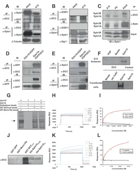
consid-Figure 1. Sytx1coassociateswiththeDCCreceptorinbraintissueandintransfectedcells.A,DCCandSytx1immunoprecipitationofE15 and adult forebrain homogenates. DCC immunoprecipitation yields coimmunoprecipitation of Sytx1 in both samples (top panels). Sytx1 immunoprecipitation yields coassociation of DCC (bottom panels). Anti-III-tubulinantibodieswereusedasloadingcontrols.B,DCCand Sytx1immunoprecipitationassaysdonotresultincoimmunoprecipitationofEgr1.LevelsofEgr1proteinlysatesareshown(bottom).C,DCC associates with A and B isoforms of Sytx1. Forebrain homogenates were immunoprecipitated and immunocomplexes were subjected to urea/SDS-PAGE. DCC coimmunoprecipitates two Sytx1 bands corresponding to Sytx1A and 1B (top panel). D, E, Coimmunoprecipitation experiments in HEK293 cells. DCC immunoprecipitation results in coassociation with Sytx1A, visualized either with GFP (D) or anti-Sytx1 (E) antibodies. The reverse immunoprecipitation with anti-GFP or anti-anti-Sytx1 antibodies also reveals DCC (D, E). Note that the efficiency of the immunoprecipitation with anti-GFP antibodies is consistently higher than when using anti-DCC antibodies, which yields low recovery of proteins in this condition. F, DCC affinity pull-down experiments with purified His-Sytx1A. Incubation with anti-DCC antibody reveals DCC in the beads coupled to His-Sytx1A, but not in control beads. G, Sytx1A was transcribed and translated in vitro in the presence of [35S]Met, and incubated with glutathione-Sepharose beads coupled to GST-DCCCYTor GST-MUNC18a. After SDS-PAGE, gels were exposed to a storage phosphor screen. [35S]Met-Sytx1A binds to DCCCYT(as well as to MUNC18a), but not to empty beads. Binding of [35S]Met-Sytx1A to DCCCYTis decreased in the presence of nonradioactive Sytx1A (Sytx1A*). H, Sensorgram showing binding between the GST-DCCCYTdomain and His-Sytx1A. Increasing concentrations of His-Sytx1A were injected into a chip where the GST-DCCCYTwas cross-linked; the interaction was recorded as SPR changes [in response units (RU)]. Increasing concentrations of His-Sytx1A yielded higher responses. I, Plot of the steady-state response (in RU) between GST-DCCCYTand a range of concentrations of His-Sytx1A (red) or between control GST and His-Sytx1A (black). No binding is detected when GST protein was cross-linked, whereas a saturable response is observed when GST-DCCCYTwas immobilized. J, Pull-down experiments in which brain extracts (P0) were passed through Ni
2⫹-affinity columns to which recombinant His-Sytx1A or His-Sytx1AH3TM proteins were coupled. Western blot analyses show coprecipitation of DCC with similar efficiencies in both cases. No coprecipitation of DCC is detected when extracts are incubated with control GST-GFP or GST-MUNC18a columns, whereas strong coprecipitation was observed with GST-DCCcyt. The high DCC signal in GST-DCCcyt samples is probably due to multimerization of the DCC receptor through the P3 domain. K, Sensorgram showing binding between the T1DCC peptide and purified His-Sytx1A. The peptide was immobilized as described in Material and Methods, and then increasing concentrations of His-Sytx1A were injected. Responses (in RU) increase at increasing concentrations of His-Sytx1A as a function of the time (in seconds). L, Plot of the steady-state response between T1 peptide and His-Sytx1A (red) or between a control peptide and His-Sytx1A (black). While no binding is detected when the control peptide is cross-linked, specific binding is observed when T1 peptide is immobilized.
ered as background and omitted from the study. Regions of interest (growth cones) were drawn on the images, and overlapped pixels were divided by the area of these regions of interest. Colocalization values were normalized to the intensity levels of red (DCC) and green (Sytx1A) chan-nels, to refer colocalization to total protein content. We quantified DCC/ Sytx1 colocalization in the entire growth. Similar experiments were performed with SNAP25 and VAMP2 antibodies. For the calculation of membrane/growth cone ratios in DCC immunofluorescence, the signal intensity at the surface of growth cones (the outermost signals, defined as a region comprised from the outline up to 400 nm inside the growth cone) were referred to the signals detected inside the growth cones. At least five separate experiments (usually seven to eight) were performed for each condition. The number of axonal growth cones counted is shown in the figures.
In vivo analysis of commissural axon pathfinding. The analysis of com-missural axon trajectories was performed as described previously (Stoeckli and Landmesser, 1995; Fazeli et al., 1997). In brief, fertilized eggs were windowed on the third day of incubation. Extra-embryonic membranes were removed to access the spinal cord in ovo. The plasmids pIRESEGFP, Sytx1AH3TMpIRESEGFP, or those encoding other fusion proteins (Sytx1AH3pIRESEGFP, Sytx1ATMpIRESEGFP) under the control of the-actin promoter in PBS (100 ng/l) were injected into the central canal using glass capillaries. For visualization and control of in-jection quality and quantity, 0.04% trypan blue was added. For electro-poration, we used platinum electrodes connected to a BTX square wave electroporator. Electrodes were positioned parallel to the longitudinal axis of the lumbosacral spinal cord of the chicken embryo, as detailed previously (Perrin and Stoeckli, 2000; Bourikas et al., 2005). Five pulses of 26 V and 50 ms duration with a 1 s interpulse interval were applied.
The same parameters were used for the trans-fection of dsRNA derived from Sytx1A, TI-VAMP, or neuron– glia cell adhesion molecule (NgCAM). dsRNA was produced by in vitro transcription as described previously (Pekarik et al., 2003). For the production of dsRNA de-rived from Sytx1A, NgCAM, or TI-VAMP, we used ESTs obtained from Geneservices (Bourikas et al., 2005). dsRNA was injected in PBS (250 ng/l) and 0.04% trypan blue. After electroporation, eggs were sealed with a glass coverslip and melted paraffin or Scotch tape and incubated for another 2 d.
Embryos were killed between stages 25 and 26 (Hamburger and Hamilton, 1992). The spi-nal cord was removed from the embryo, opened at the roof plate (“open-book” prepa-ration), and fixed in 4% paraformaldehyde for 30 – 60 min.
Alternatively, 250-m-thick transverse slices were cut from fixed embryos. In both cases, the trajectories of commissural axons at the lum-bosacral level of the spinal cord were visualized by the application of the lipophilic dye Fast DiI (dissolved at 5 mg/ml in methanol; Invitrogen) to the cell bodies. Care was taken to exclusively label the dorsal-most population of commis-sural neurons to avoid confusion with more ventral populations that have distinct path-finding behavior. For the quantification of the axonal pathfinding phenotype (see Figs. 8 K, 9K ), we used open-book preparations. After visualizing a subset of dorsal commissural ax-ons by dye tracing, we assessed the percentage of fibers that extended from the dorsal spinal cord but that failed to enter and cross the floor plate. We classified the phenotype as weak when between 20 and 50% of the fibers failed to reach the contralateral floor plate border, and as strong when 50% or more fibers were af-fected. It is important to emphasize that the proportion of cells that are successfully transfected by this in ovo electro-poration procedure is on average 60% (Perrin and Stoeckli, 2000). Therefore,⬃40% of the cells are expected to be wild type and to cross the floor plate normally.
Statistics. Data were expressed as means⫾ SEM and were analyzed
statistically using ANOVA (Statgraphics Plus 5.1).
Results
The DCC receptor associates with the t-SNAREs Sytx1A/1B
Axonal guidance receptor function is modulated by interaction
with other membrane proteins, including guidance receptors
(Hong et al., 1999), and SNARE protein functions and exocytosis
essentially operate through complex and multiple protein
inter-actions (see above) (Verhage et al., 2000; Castillo et al., 2002; Jahn
and Scheller, 2006; de Wit et al., 2009; Su¨dhof and Rothman,
2009). We therefore hypothesized that there is a cis-direct
inter-action between the Netrin-1 receptor DCC and the plasmalemma
t-SNAREs SNAP25 and Sytx1 and that this interaction might
influence exocytosis. In preliminary experiments on brain
ly-sates, DCC pull-down assays coimmunoprecipitated Sytx1 (Fig.
1). In contrast, coimmunoprecipitation assays on brain lysates or
transfected cells revealed that DCC does not interact with the
SNAREs VAMP2 and SNAP25, or with Sytx4, a SNARE involved
in protein traffic (Fig. 2). This observation suggested that the
canonical Sytx1 SNARE partners (SNAP25 and VAMP2) were
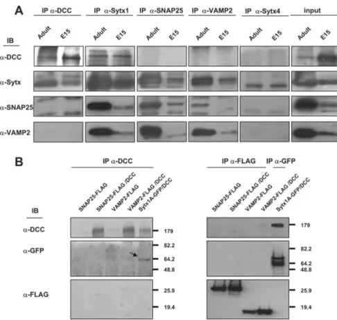
Figure 2. The DCC receptor does not coassociate with SNAP25, VAMP2, or Syntaxin 4. A, Coimmunoprecipitation experiments in brain lysates (E15 and adult). Whereas DCC and Sytx1 coimmunoprecipitate, the DCC receptor does not coassociate with SNAP25, VAMP2, or Sytx 4 in brain lysates. The Sytx antibody used for the immunoblot was an anti-Sytx1A antibody in all cases, except in the column immunoprecipitated with anti-Sytx 4 antibodies. Inputs are shown to the right. B, Coimmunoprecipitation experiments in HEK293 cells transfected with pCMVDCC and SNAP25-FLAG, VAMP2-FLAG or Sytx1A-EGFP DNAs. DCC immunoprecipitation results in coassociation with Sytx1A visualized with anti-GFP antibodies, but not with SNAP25 or VAMP2 (FLAG antibodies). The reverse immunoprecipitation with anti-FLAG antibodies did not reveal DCC coimmunoprecipitation.
not associated with the DCC–Syntaxin 1
complexes (see also below).
We thus focused on the association of
DCC and Sytx1. DCC antibodies
immu-noprecipitated in brain lysates a band of
35 kDa, which corresponded to Sytx1A
and 1B isoforms (WB with the HPC-1
Mab; Fig. 1 A) (Ruiz-Montasell et al.,
1996). A 180 kDa band corresponding to
DCC was identified after
immunopre-cipitation with anti-Sytx1 antibodies
(Fig. 1 A). When DCC or Sytx1
immuno-precipitates were revealed with control
antibodies (Fig. 1 B), no signal was
de-tected. Furthermore,
immunoprecipita-tion with beads alone or with irrelevant
antibodies (anti-GFP or anti-Egr1) did
not reveal coassociation (data not shown).
Because Sytx1A and 1B are difficult to
resolve in conventional SDS-PAGE,
im-munoprecipitates were run in urea/
SDS-PAGE. These experiments showed
that Sytx1A and 1B isoforms
coimmuno-precipitated with DCC, but with distinct
developmental profiles (Fig. 1C).
To-gether, these results indicate an in vivo
as-sociation of Sytx1A/1B and DCC in the
developing and adult brain.
To corroborate the interaction of DCC
and Sytx1A, we performed several
ex-periments. First, in
coimmunoprecipi-tation assays in HEK293 or COS-1 cells
transfected with constructs of both
pro-teins (pRcCMVDCC or Sytx1AEGFP),
Sytx1AEGFP was detected by WB in lysates
immunoprecipitated with DCC
anti-bodies. Pull-down assays with anti-GFP or
anti-Sytx1 antibodies revealed DCC
pro-tein. No coimmunoprecipitation was
de-tected when cells were transfected with
either Sytx1AEGFP or DCC DNA alone
(Fig. 1D,E). These data indicate that Sytx1A
and DCC coassociate after expression
in non-neuronal cells. Second, affinity
pull-down experiments with purified
His-Sytx1A were performed. Bacterially
ex-pressed His-Sytx1A or control His-tagged
proteins (POP; HC-TeTx) were purified
and incubated with either forebrain lysates
or extracts from HEK293 cells transfected
with DCC. Protein complexes were eluted
and fractions were labeled with
␣-DCC
an-tibodies. DCC was present in the samples
incubated with Sytx1A-FL, both in extracts
from brain tissue and in DCC-transfected
cells, but not in beads coupled to control
proteins (Fig. 1F).
To provide evidence of direct binding of
Sytx1A to DCC, Sytx1A was transcribed and translated in vitro using
reticulocyte lysates, in the presence of [
35S]Met, and incubated with
glutathione-Sepharose beads coupled to GST-DCC
CYT,
GST-MUNC18a, or control beads (Fig. 1G). Bound proteins were eluted
and revealed by autoradiography. The results showed binding of
[
35S]Met-Sytx1A to DCC
CYT
(as well as to MUNC18a), but not to
empty beads. Furthermore, the binding of [
35S]Met-Sytx1A to
DC-C
CYTwas decreased in the presence of nonradioactive Sytx1A (Fig.
1G). To further demonstrate the direct interaction between Sytx1A
and DCC, we applied the BIAcore technique, in which purified
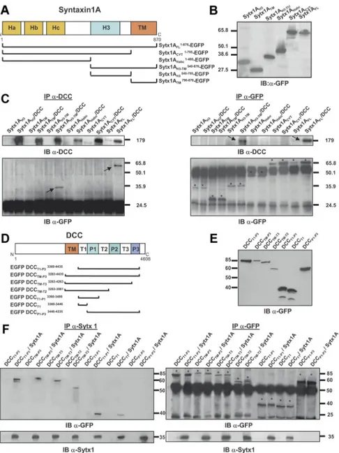
GST-Figure 3. Characterization of protein regions required for Sytx1A/DCC interaction. A, Diagram summarizing Sytx1A domains and the truncated Sytx1AEGFP chimeras generated. B, Expression of the several Sytx1AEGFP DNAs in HEK293 cells, showing that they generate proteins of the appropriate Mrbetween 65 and 27 kDa, as revealed by immunoblotting with anti-GFP antibodies. C, Coimmunoprecipitation experiments in HEK293 cells cotransfected with the distinct Sytx1AEGFP constructs together with pCMV or pCMVDCC. DCC immunoprecipitations (200g) were revealed by immunoblotting with anti-DCC or anti-GFP antibodies (left panel). Cells cotransfected with pCMVDCC and Sytx1AFLEGFP or Sytx1AH3TMEGFP show positive coimmunoprecipitation of Sytx1A fusions. The reverse immunoprecipitation assays with anti-DCC antibodies also show coimmunoprecipitation with DCC, exclusively when cells are cotransfected with Sytx1AFLEGFP or Sytx1AH3TMEGFP DNAs (arrows in top right panel). The bands corresponding to the distinct Sytx1AEGFP chimeras are labeled with asterisks in the bottom right panel label. D, Diagram summarizing the DCC domains and the truncated EGFPDCC chimeras generated. E, Western blot, revealed with an anti-GFP antibody, demonstrating appropriate Mr(between 90 and 40 kDa) of the distinct EGFPDCC chimeras expressed in HEK293 cells. F, Coimmunoprecipitation experiments in HEK293 cells cotransfected with the distinct EGFPDCC constructs together with pRcCMV or pRcCMVSytx1A DNAs. Sytx1 immunoprecipitations (200g) were revealed by immunoblotting with anti-GFP or anti-Sytx1 antibodies (left panel). All the cells cotransfected with pRcCMVSytx1A and the EGFPDCC constructs show coimmunoprecipitation of EGFP-tagged DCC chime-ras (arrows), except when cells are cotransfected with EGFPP1-P3DCC DNA. The reverse immunoprecipitation assays with anti-GFP antibodies also reveal coimmunoprecipitation with Sytx1A (arrows in bottom right panel) in all cotransfected cells, except in those transfected with EGFPP1-P3DCC DNA. The bands corresponding to the distinct Sytx1AEGFP chimeras are labeled with asterisks in the top right panel.
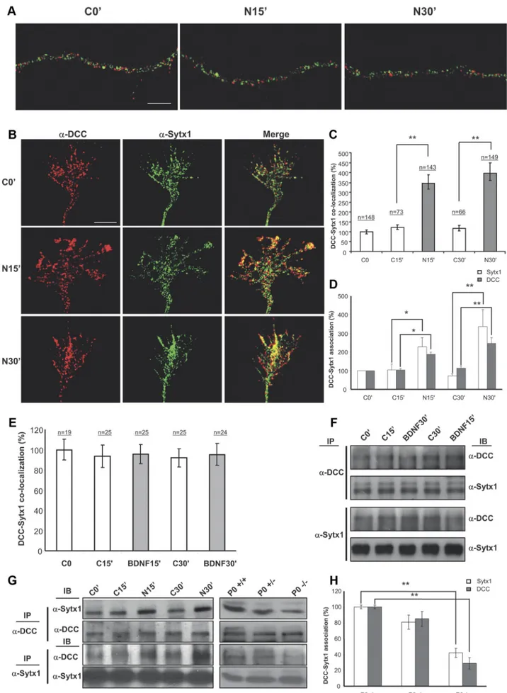
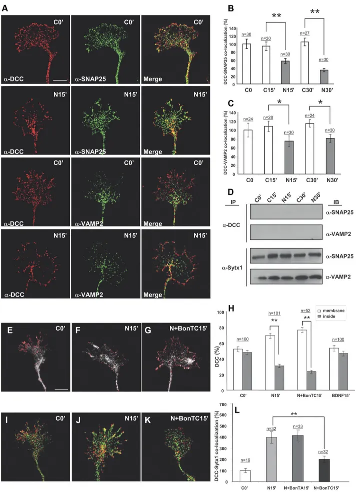
Figure 4. Netrin-1, but not BDNF, triggers DCC mobilization and DCC/Sytx1 colocalization. A, Confocal images of hippocampal axonal shafts treated with Netrin-1-conditioned media for 0, 15, and 30 min, immunolabeled for DCC (red) and Sytx1A (green). Note that DCC and Sytx1A do not colocalize in axonal shafts in either condition. B, Confocal images of (Figure legend continues.)
DCC
CYTprotein was immobilized on a CM5 sensor chip by EDC/
NHS coupling. Solutions containing different concentrations of
purified His-Sytx1A (10 n
Mto 30
M) were passed through the
chamber containing the chip, and binding was analyzed according to
the SPR signal obtained in real time. We found a specific interaction
between the two proteins, which was not detected when control GST
protein was immobilized or when an empty chip was used (Fig.
1H,I). Quantitative evaluation of five independent experiments
showed that the two molecules interacted with a K
Dof
⬃2.4 ⫾ 0.7
10
⫺6M(Fig. 1H,I) (k
on, 1.8
⫻ 10
4M⫺1s
1; k
off, 0.033 s
⫺1).
To-gether, these biochemical and biophysical experiments show
that recombinant DCC and Sytx1 interact directly in vitro.
The transmembrane-H3 region of Sytx1A and the T1 region
of DCC are required for Sytx1A/DCC interaction
We next attempted to identify the domains of Sytx1 required for
its interaction with DCC. Sytx1A contains a transmembrane
do-main (TM) and a cytosolic tail containing the H3 (SNARE) motif
and the N-terminal Habc domain (Fig. 3A) (Sutton et al., 1998;
Dulubova et al., 1999). While the H3 domain interacts with other
SNARE proteins, the Habc region modulates the formation of the
SNARE complex (Misura et al., 2000). We generated several
truncated Sytx1A constructs to which EGFP was fused at the
N-terminal region (Fig. 3B). Constructs were transfected in
com-bination with DCC in HEK293 cells, and lysates were processed
for immunoprecipitation. DCC immunoprecipitation yielded
coimmunoprecipitation of EGFP-tagged full-length Sytx1A (Fig.
3C, left panel). Cotransfection with either the Sytx1ACYTEGFP
or the Sytx1AHabc EGFP constructs did not reveal
coassocia-tion. In contrast, DCC antibodies coimmunoprecipitated the
Sytx1AH3TMEGFP chimera (Fig. 3C, arrows), but no
coimmu-noprecipitation was detected after transfection with DNAs
en-coding for Sytx1ATM EGFP protein. Transfection with
Sytx1AH3EGFP yielded a very weak coassociation (Fig. 3C, left
panel). The reverse experiments, using
␣-GFP antibodies to
im-munoprecipitate EGFP-tagged Sytx1A chimeras, and
immuno-blotting with
␣-DCC antibodies, yielded identical results as
above (Fig. 3C, right panel). Thus, only when cells were
cotrans-fected with DCC and Sytx1AFLEGFP or Sytx1AH3TMEGFP was
the DCC protein pulled down (Fig. 3C, arrows). These results
reinforce the notion that DCC coassociates with Sytx1A and
demonstrates that the H3TM region of Sytx1A is required for the
interaction with DCC.
DCC contains an extracellular domain, a single
transmem-brane domain, and a cytosolic tail with three proline-rich regions
(P1, P2 and P3) separated by three tails (regions T1, T2 and T3;
Fig. 3D). Because Sytx1A lacks an extracellular domain, and the
H3TM region of Sytx1A is required for association with DCC,
we hypothesized that Sytx1A and DCC interact through the
transmembrane region and/or cytosolic tail of DCC. We
gen-erated five DCC chimeras tagged with EGFP at the N terminus
(Fig. 3E): the entire DCC cytosolic tail and transmembrane
re-gion (EGFPDCCTM-P3), successively deleted P3–P1 domains
(EGFPDCCTM-T3, EGFPDCCTM-T2, EGFPDCCTM-T1), and
an EGFPDCCT1-P3 chimera lacking the transmembrane region.
These EGFPDCC chimeras expressed proteins of the appropriate
size (Fig. 3E) and were cotransfected with Sytx1A. When lysates
were pulled down with
␣-Sytx1 antibodies and the
immunocom-plexes were probed with
␣-GFP antibodies, Sytx1A interacted
with the cytosolic DCC tail both in the presence and in the
ab-sence of the TM region (EGFPDCCTM-P3 and EGFPDCCT1-P3
constructs; Fig. 3F, left panel). Sytx1A/DCC
coimmunoprecipi-tation was also detected when the P3 or the P2–P3 domains were
deleted, but no coimmunoprecipitation was detected in cells
transfected with the EGFPDCCP1-P3 DNA, demonstrating
that this region is not required for interaction with Sytx1A
(Fig. 3F ). We generated an additional chimera containing the
T1 region (EGFPDCCT1). Sytx1A coimmunoprecipitated
with EGFPDCCT1-P1 and EGFPDCCT1 proteins, indicating
that the T1 region of DCC is sufficient for interaction with Sytx1A
(Fig. 3F ). Identical results were obtained in the reverse
coimmu-noprecipitation experiments (i.e., immucoimmu-noprecipitation of
EGF-PDCC chimeras, followed by immunoblotting with the HPC-1
␣-Sytx1 antibody). Only when the T1 region of DCC was deleted
did the DCC/Sytx1A interaction not occur (Fig. 3F, right panel).
Our findings indicate that the region of DCC required for its
interaction with Sytx1A is the T1 region.
These conclusions were further strengthened by pull-down
experiments showing that purified
CYTDCC recruits truncated
Sytx1AH3TM protein (the Sytx1A region responsible for binding
to DCC), and by BIAcore experiments, in which an immobilized
T1 peptide (the 29 aa DCC region responsible for the interaction)
was also found to bind purified Sytx1A (Fig. 1 J–L). We detected
specific binding between the T1 peptide and His-Sytx1A with a
K
Dof
⬃3.9 ⫾ 1.1 10
⫺6M(k
on, 2.9
⫻ 10
4M⫺1s
1; k
off, 0.065 s
⫺1).
Thus, DCC and Sytx1A interact through specific protein
domains.
Netrin-1 regulates the association of DCC and Sytx1
To determine whether Netrin-1 regulates the association of DCC
and Sytx1, we incubated hippocampal cultures with Netrin-1 or
control-conditioned medium, and then measured overlapping
signals in growth cones by immunofluorescence. Interestingly,
Sytx1 and DCC were localized in distinct vesicle-like organelles in
cell bodies and along the shafts of developing axons (Fig. 4A).
Weak DCC/Sytx1 colocalization was detected along the leading
edges of axonal growth cones (Fig. 4 B). These data indicate that
the DCC receptor and Sytx1 are transported in different vesicles
along axons but may interact in distal growth cones of developing
4
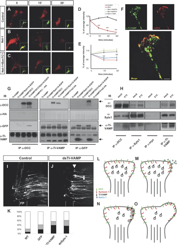
(Figure legend continued.) hippocampal growth cones treated with Netrin-1-conditioned media for 0 –30 min, immunolabeled for DCC and Sytx1. Note increased DCC/Sytx1 colocaliza-tion in growth cones incubated with Netrin-1. C, Quantificacolocaliza-tion of DCC/Sytx1 colocalizacolocaliza-tion signals in hippocampal growth cones (expressed as percentage of DCC/Sytx1 colocalization over total Sytx1 signals) in cultures treated with Netrin-1-conditioned (black bars) or control-conditioned (white bars) media. D, Quantification of DCC/Sytx1 coimmunoprecipitation in hip-pocampal cultures treated with Netrin-1. Sytx1 immunoprecipitation reveals an increase in coassociated DCC in neurons treated with Netrin-1 (black bars). Immunoprecipitation with anti-DCC antibodies shows a marked increase in Sytx1 signals (white bars). E, Quantification of DCC/Sytx1 colocalization signals in hippocampal growth cones (expressed as percentage of DCC/Sytx1 colocalization over total Sytx1 signal) in cultures treated with BDNF (gray bars) or control (white bars). Note that BDNF does not increase colocalization of DCC and Sytx1. F, Western blots from hippocampal cultures treated with BDNF for 15–30 min and immunopre-cipitated with anti-DCC or anti-Sytx1A antibodies. Immunoblots reveal that BDNF does not increase the coassociation of DCC with Sytx1A. G, Western blots from hippocampal cultures treated with Netrin-1-conditioned (N) or control-conditioned (C) media for 0 –30 min (left), and immunoprecipitated with anti-DCC (top panel) or anti-Sytx1 (bottom panel) antibodies. Immunoblots reveal increased association of DCC and Sytx1 in neuronal cultures treated with Netrin-1. Immunoprecipitations of brain lysates from newborn wild-type, heterozygous and homozygous netrin-1 mutant mice reveal decreased DCC/Sytx1 association in the null mutants (right). Note decreased coimmunoprecipitation of Sytx1 (top) and DCC (middle) in null-mutant brains. H, Quantification of DCC and Sytx1 coimmunoprecipitations in homogenates from new-born wild-type, heterozygous and homozygous netrin-1 mutant mice. Immunoprecipitations with either anti-DCC (white bars) or anti-Sytx1 (black bars) antibodies reveal a marked reduc-tion in DCC/Sytx1 coassociareduc-tion in netrin-1-null mutants. Significant differences are labeled by asterisks (*pⱕ 0.05; **p ⱕ 0.001). Scale bar: A, B, 3m. Error bars indicate SEM.
Figure 5. Netrin-1 does not trigger coassociation of DCC with the SNAREs SNAP25 and VAMP2. A, Confocal images of hippocampal growth cones treated with Netrin-1-conditioned media for 0 and 15 min, immunolabeled for DCC and the SNAREs SNAP25 and VAMP2. Note low colocalization signals of DCC and SNAP25 or VAMP2, both in control conditions and after (Figure legend continues.)
neurons. Growth cones treated with Netrin-1 for 15–30 min
ex-hibited a strong increase in DCC/Sytx1 in signal overlapping
compared with neurons incubated with control medium (Fig.
4 B, C). In contrast, the neurotrophin BDNF, which does not bind
DCC, did not increase DCC/Sytx1 colocalization, indicating that
the increase in DCC/Sytx1 colocalization was specifically
trig-gered by Netrin-1 (Fig. 4 E).
To substantiate these findings, lysates of neuronal cultures
incubated with Netrin-1 were immunoprecipitated with DCC or
Sytx1 antibodies. Immunoprecipitation with the former revealed
higher levels of Sytx1 in cultures incubated with Netrin-1 for 15
and 30 min than in control cultures. The reverse
immunoprecipi-tations with anti-Sytx1 antibodies also showed higher DCC
pro-tein levels after incubation with Netrin-1 (Fig. 4 D, G). Incubation
of hippocampal cultures with control media or BDNF did not
increase the DCC/Sytx1 coassociation (Fig. 4 E, F ).
To determine whether the DCC/Sytx1 interaction was
depen-dent on Netrin-1 in vivo, we analyzed lysates from wild-type and
netrin-1-deficient mice (Serafini et al., 1996). The amount of
DCC protein that was immunoprecipitated with Sytx1
anti-bodies was severely diminished in netrin-1-deficient forebrains in
comparison with wild-type brains. A similar reduction was
ob-served when DCC immunoprecipitates were analyzed for Sytx1
by immunoblotting (Fig. 4G,H ).
Interestingly, and in agreement with the above data (Fig. 2),
cultured neurons treated with Netrin-1 did not trigger increases
in the coassociation of DCC with the SNAREs SNAP25 and
VAMP2, as detected by both confocal microscopy and
coimmu-noprecipitation (Fig. 5A–D). Thus, DCC did not appear to bind
VAMP2 and SNAP25 in any of the tested conditions, including
incubation with the DCC ligand Netrin-1. We therefore conclude
that Netrin-1 specifically promotes the interaction of DCC and
Sytx1 in growth cones of cultured neurons.
It has been shown that Netrin-1 triggers membrane exposure
of DCC-containing vesicles in growth cones (Bouchard et al.,
2004; Moore et al., 2008). First, we corroborated that incubation
with Netrin-1, but not with control media or BDNF (which acts
via TrkB receptors), caused the mobilization of DCC from the
inside of growth cones to the cell membrane (Fig. 5E–H )
(Bouchard et al., 2004). Moreover, we found that treatment with
BoNT/C1 did not alter the DCC mobilization induced by
Netrin-1. Incubation of hippocampal cultures with BoNT/C1
gave a yield of Sytx1 degradation (data not shown) similar to that
in Figure 6 E. In contrast, and interestingly, incubation of
hip-pocampal growth cones with BoNT/C1 prevented the increase in
DCC/Sytx1 colocalization induced by Netrin-1 (Fig. 5J–L). This
observation indicates that the release of DCC to the membrane is
independent of Sytx1 (Fig. 5E–H ). This is consistent with the
observation that DCC and Sytx1 do not colocalize in the same
vesicles along axonal shafts and in growth cones (see above).
Together, these findings indicate that, although the DCC/Sytx1
interaction occurs in the absence of Netrin-1, this guidance cue
triggers a specific increase in the association of these proteins.
BoNT/C1 blocks Netrin-1-induced chemoattraction of
hippocampal axons
To study whether Sytx1 is required for Netrin-1-dependent
che-moattraction, we first used botulinum toxins, metalloproteases
that inhibit the exocytosis of synaptic vesicles and
neurotransmit-ter release by cleaving specific SNARE proteins. While BoNT/A
exclusively cleaves SNAP25, BoNT/C1 cleaves Sytx1 and SNAP25
(Blasi et al., 1993a,b; Schiavo et al., 2000). We used hippocampal
explants cultured in collagen gels with Netrin-1-expressing cells
to examine whether t-SNARE protein cleavage by botulinum
tox-ins interferes with Netrin-1-induced axonal guidance. Cleavage
of target t-SNARE proteins in our culture model was confirmed
by WB (Fig. 6 E). The amount of cleaved SNARE proteins seen in
our culture conditions was similar to that described in previous
studies, including mature neurons, and the relatively small
amount of Sytx1 cleaved by BoNT/C1 is believed to be due to the
fact that Sytx1 protein forming part of SNARE complexes is
pro-tected against cleavage (Blasi et al., 1993a,b; Schiavo et al., 2000).
We next showed that botulinum toxins did not affect the
secre-tion of Netrin-1 in HEK293 cells stably transfected with a
Netrin-1 expression vector (Fig. 6 F). Thus, hippocampal
ex-plants (Barallobre et al., 2000) were cocultured with aggregates of
control or Netrin-1-expressing cells, in the presence of
botuli-num toxins.
While explants cultured with control cells showed radial
ax-onal growth, those cultured with Netrin-1-expressing cells
exhib-ited axonal attraction (Fig. 6 A, B,G). Chemoattraction was
maintained when hippocampal explants were cocultured with
Netrin-1 in the presence of BoNT/A (Fig. 6C,G). In contrast,
Netrin-1-dependent chemoattraction was impaired in explants
incubated with BoNT/C1, which resulted in radial axonal growth
(Fig. 6 D, G). Quantification of axonal growth by comparing the
density of outgrowing axons in the proximal and distal quadrants
supported these observations (Fig. 6G).
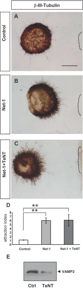
We also treated hippocampal explants with TeNT, which
spe-cifically cleaves the v-SNARE VAMP2 [Fig. 7E (Blasi et al.,
1993b)]. Incubation of explants with this toxin did not alter
Netrin-1-induced chemoattraction of hippocampal axons (Fig.
7), in agreement with the nondetectable coassociation of DCC
with VAMP2 (Figs. 2, 5). Together with the above results, the data
suggest that, while the SNAREs SNAP25 and VAMP-2 are
dis-pensable for Netrin-1-signaling, Sytx1 is specifically required for
Netrin-1-dependent axonal guidance.
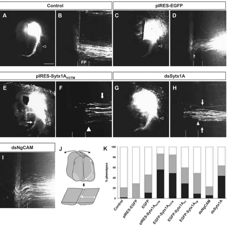
Sytx1A is required for commissural axon pathfinding in the
chicken spinal cord in vivo
The requirement for functional Sytx1 in Netrin-1-mediated
axonal guidance in vivo was analyzed in the chicken spinal
cord, where commissural dorsal axons extend toward the floor
4
(Figure legend continued.) incubation with Netrin-1. B, C, Quantification of DCC/SNAP25 and DCC/VAMP2 colocalization signals in hippocampal growth cones (expressed as percentage of DCC/SNARE colocalization over total SNARE signals) in cultures treated with Netrin-1-conditioned (black bars) or control-Netrin-1-conditioned (white bars) media. Note decreased DCC/SNARE colocalization signals after Netrin-1 treatment. D, Western blots from hippocampal cultures treated with Netrin-1- (N) or control- (C) conditioned media for 0 –30 min, and immunopre-cipitated with anti-DCC or anti-Sytx1A antibodies. Immunoblots reveal no coimmunoprecipi-tation of DCC with the SNAREs SNAP25 and VAMP2 after incubation with DCC. Note coimmunoprecipitation of Sytx1A with SNAP25 and VAMP2. E–G, Confocal images of hip-pocampal growth cones treated with Netrin-1-conditioned media for 0 and 15 min, and in the presence of BoNT/C1. Cultures were immunolabeled for DCC and stained with phalloidin. Note that the mobilization of DCC to the axonal membrane after incubation with Netrin-1 (F) is not altered by BoNT/C1 incubation (G). H, Quantification of DCC signals in the periphery of and inside growth cones treated with Netrin-1-conditioned media for 0, 15, and 30 min, showing mobilization of DCC to the axonal membrane after incubation with Netrin-1. Whereas no DCC mobilization is detected after incubation with BDNF, treatment with BoNT/C1 does not alter DCC mobilization. I–L, Confocal images of control growth cones (I) and cones incubated with Netrin-1-conditioned media for 15 min (J), and with Netrin-1/BoNT/C1 (K). Cultures were im-munolabeled for DCC (red) and Sytx1 (green). Note that the increase in DCC/Sytx1 colocalization in J is blocked after incubation with BoNT/C1 (K). L, Histograms illustrating DCC/Sytx1 colocal-ization in several experimental conditions. Significant differences are labeled by asterisks (*pⱕ 0.05, **pⱕ 0.001). Scale bar: A, 3m. Error bars indicate SEM.
plate in a Netrin-1- and DCC-dependent manner (Serafini et al.,
1996; Fazeli et al., 1997). To assess the relevance of Sytx1A, we first
expressed dominant-negative constructs (pIRES-Sytx1A
H3TMEGFP
or the fusion Sytx1A
H3TMEGFP) in commissural neurons at the time
when they start to extend axons. After 2 d, the trajectory of
commis-sural axons was visualized in transverse slices or in whole-mount
preparations of the spinal cord, the so-called
“open-book preparations” (Fig. 8J), by the
injection of Fast DiI (Perrin and Stoeckli,
2000). In untreated control spinal cords,
virtually all commissural axons had crossed
the floor plate. After entering the floor plate
and crossing the midline, these axons turned
into the longitudinal axis of the spinal cord
and extended rostrally, while maintaining
contact with the floor plate border (Fig.
8A,B). However, and in contrast to
com-missural axons from embryos transfected
with the control pIRES-EGFP construct,
those transfected with the
dominant-negative pIRES-Sytx1A
H3TMEGFP failed to
cross the floor plate and stalled before or at
the floor plate border (Fig. 8C–F). In fact,
in pIRES-Sytx1A
H3TMEGFP-electroporated
spinal cords, many fibers failed to reach the
floor plate and extended along the lateral
edge of the spinal cord (Fig. 8E, arrowhead).
The same results were obtained with the
Sytx1A
H3TMEGFP fusion protein both
qual-itatively (data not shown) and
quantita-tively (Fig. 8K). While embryos expressing
the Sytx1A
TMEGFP fusion protein did not
show a significant increase in the failure of
commissural axons to reach and cross the
floor plate, expression of Sytx1A
H3EGFP
re-sulted in a mild phenotype (Fig. 8K).
As an independent approach to interfere
with Sytx1A function in commissural axon
pathfinding, we used in ovo RNAi (Pekarik
et al., 2003). Downregulation of Sytx1A in
commissural neurons by electroporation of
dsRNA derived from Sytx1A gave similar
results to the perturbation of Sytx1A
func-tion by dominant-negative Sytx1A variants.
Thus, downregulation of Sytx1A by in ovo
RNAi resulted in the failure of commissural
axons to reach and cross the floor plate (Fig.
8G,H,K). As a control, we used dsRNA
de-rived from NgCAM, a cell adhesion
mole-cule of the Ig superfamily that is expressed in
dorsal commissural neurons (Pekarik et al.,
2003). Perturbation of NgCAM
func-tion interfered with the fasciculafunc-tion of
commissural axons but not with their
midline crossing (Fig. 8 I, K ). In
sum-mary, we found strong interference with
commissural axon pathfinding toward
and across the floor plate after in vivo
perturbation of Sytx1A function either
by expressing dominant-negative variants
of Sytx1A or by its knockdown using in ovo
RNAi. Moreover, the phenotypes observed
here are reminiscent of those described in
loss-of-function models of the Netrin-1 and DCC genes (Serafini et
al., 1996; Fazeli et al., 1997).
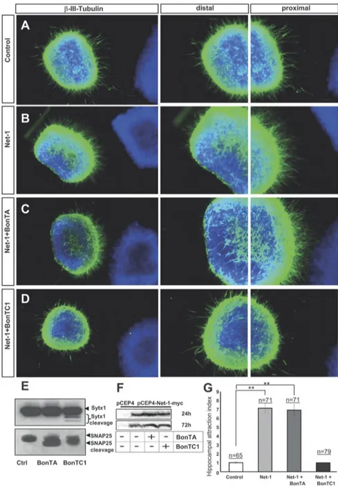
Netrin-1 triggers exocytosis in growth cones
We next studied whether Netrin-1 regulates exocytosis in growth
cones through a DCC/Sytx1 interaction. For this purpose,
hip-Figure 6. BoNT/C1 blocks Netrin-1-mediated chemoattraction of hippocampal axons. A–D, Examples of hippocampal explants cocultured in collagen gels with control or Netrin-1-expressing cell aggregates. Explants cocultured with control cells show a radial growth (A). Explants cocultured with Netrin-1-secreting cells exhibit chemoattraction toward the Netrin-1 source (B). Explants cocultured with Netrin-1-expressing cells and BoNT/A also display chemoattraction (C). Netrin-1-dependent chemoattraction is blocked in hippocampal explants cultured with BoNT/C1 (D). Low-power images are shown in the first row of panels. High-power magnifications of the proximal and distal quadrants are illustrated in the second and third rows. E, Western blots of protein samples from lysates of hippocampal explants cultured with BoNT/A, BoNT/C1, or left untreated (Ctrl), probed with␣-Sytx1 and ␣-SNAP25 antibodies. Incubation with BoNT/A results in cleavage of SNAP25 and treatment with BoNT/C1 cleaves both SNAP25 and Sytx1. F, Western blots of conditioned media from control HEK293 cells or cells stably transfected with pCEP4-Netrin-1-myc cultured for 24 – 48 h in the presence or absence of BoNT/A or BoNT/C1, probed with an␣-Myc antibody to detect Netrin-1 in the culture media. The Western blot did not show any differences in the secretion of Netrin-1 in the presence of botulinum toxins. G, Histograms illustrating proximal/distal ratios (attraction index) in hippocampal explants in different culture conditions. Significant differences are labeled by asterisks (**pⱕ 0.001). Scale bar: A, 300m. Error bars indicate SEM.
pocampal cultures were prelabeled with BODIPY ceramide (a
membrane, lipid marker that preloads vesicular compartments)
(Pfenninger et al., 2003; Pfenninger, 2009), and then incubated
with Netrin-1 at a range of times. The decrease in fluorescence in
BODIPY ceramide-preloaded growth cones reflects exocytosis
(Pfenninger et al., 2003). While incubation with control media
did not result in a decrease of BODIPY ceramide signals in
growth cones, treatment with Netrin-1 led to a substantial
reduc-tion of intracellular BODIPY ceramide fluorescence, thereby
in-dicating release of prelabeled vesicles (Fig. 9 A, B,D). Moreover,
incubation with BoNT/C1, 30 min before Netrin-1 stimulation,
abolished the decrease in fluorescence, thus suggesting blockade
of vesicle release (Fig. 9C,E). In contrast, incubation of growth
cones with BoNT/A or TeNT did not decrease the vesicle release
induced by Netrin-1 (Fig. 9E). These observations strongly
sug-gest that Netrin-1 triggers local exocytosis in growth cones and
that Sytx1 is required for these events.
The v-SNARE TI-VAMP interacts with DCC and is required
for commissural axonal guidance in vivo
In addition to the t-SNARE Sytx1, vesicle exocytosis and
mem-brane fusion require the formation of a protein complex with
v-SNAREs. As described above, we tested whether DCC
coasso-ciates with the classical neural SNARES that regulate
neurotrans-mitter release. However, coimmunoprecipitation assays revealed
that DCC does not interact with SNAP25, VAMP2, or Sytx4 (a
SNARE involved in protein traffic) (Figs. 2, 5). This observation
is in agreement with the finding that BoNT/A and TeNT, toxins
that block SNAP25 and VAMP2, respectively, did not affect
Netrin-1/DCC-dependent guidance (Figs. 6, 7).
The v-SNARE TI-VAMP is selectively enriched in growth
cones and has been proposed to mediate exocytotic events
in-volved in neurite growth (Alberts et al., 2006). We thus studied
whether TI-VAMP forms part of the DCC/Sytx1 complex.
Cul-tured hippocampal neurons exhibited colocalization of DCC and
TI-VAMP, which was conspicuous at the edges of the growth
cones (Fig. 9F ). Expression of DCC and TI-VAMP constructs in
EBNA293 cells followed by coimmunoprecipitation and WB
showed that these two proteins interact in vitro after heterologous
expression, both in the presence or absence of Sytx1 (Fig. 9G). In
contrast, TI-VAMP did not coassociate with Frizzled 2, another
membrane receptor (Fig. 9G). Furthermore, DCC and TI-VAMP
coimmunoprecipitation experiments from E15 and adult brain
lysates demonstrated that DCC interacts with TI-VAMP in vivo
(Fig. 9H ). In agreement with the low expression of TI-VAMP in
adult tissue, DCC/TI-VAMP interaction was diminished in adult
brain. These two proteins also interacted with Sytx1, whereas
control immunoprecipitations with an irrelevant antibody in
brain lysates were negative (Fig. 9H ). These findings show that, in
contrast to VAMP2, the vesicular membrane protein TI-VAMP
be-longs to the DCC/Sytx1 complex, suggesting that this v-SNARE
me-diates Netrin-1-dependent guidance.
To examine this possibility, we studied the impact of
TI-VAMP downregulation in the development of
Netrin-1-dependent commissural pathway in the chicken spinal cord.
TI-Figure 7. TeNT incubation does not alter Netrin-1-induced chemoattraction. A–C, Examples of hippocampal explants cocultured with control or Netrin-1-expressing cell aggregates in the presence of TeNT. Explants were immunolabeled forIII-tubulin. Explants cocultured with Netrin-1-secreting cells show strong chemoattraction (B). Explants cocultured with Netrin-1-secreting cells and TeNT also show strong chemoattraction (C). D, Histogram illus-trating proximal/distal ratios (attraction index) in a range of conditions. Hippocampal explants
4
cultured with Netrin-1-expressing cells in the presence or absence of TeNT show chemoattrac-tive patterns of axonal growth. E, Western blots of protein samples from lysates of hippocampal explants cultured with TeNT, or left untreated (Ctrl), probed with␣-VAMP2 antibody. Incuba-tion with TeNT cleaves VAMP2. Significant differences are labeled by asterisks (**pⱕ 0.001). Scale bar: A, D, 150m. Error bars indicate SEM.