1
Index
Summary
2
Chapter I.
General Introduction
4
Chapter II.
Original Research Work:
Epigenetic signature: implications for mitochondrial quality
control in human aging
29
Conclusive Remarks
66
Appendix.
2
Summary
During my PhD program, my work has been addressed to the study of the role of epigenetic modifications in aging and in age-related phenotypes.
Epigenetics is the study of changes in gene expression that do not involve changes to the underlying DNA sequence. These changes affect cellular phenotypic expression by regulating relative gene expression levels. They are a common and natural process in living cells and are tightly controlled by pre-programmed mechanisms. Epigenetics modifications can be influenced by multiple factors including environmental conditions, lifestyle, nutrition, use of drugs, disease state and age.
Patterns of DNA methylation, the best known and characterized epigenetic modification, change during aging; indeed, with increasing aging, genome-wide methylation levels decrease, meanwhile genomic regions, including CpG islands, become more methylated. Analyses of the above patterns provided new perspectives for establishing powerful biomarkers of human aging which have the potential to generate accurate prediction not only of the chronological but also of the biological age. The first section of the PhD thesis consists in a comprehensive overview of the general features of DNA methylation and its implication in age and age-related diseases. The topic is addressed referring to the methylation patterns established not only at nuclear but also at mitochondrial genome level. In addition, the influence of a number of environmental factors on the above patterns is also discussed. In the second section, an original research work, carried out in order to identify novel biomarkers of aging, is reported. In this work, methylation status of nuclear genes involved in mitochondrial fusion, fission, biogenesis and mitophagy, fundamental components of the mitochondrial quality control process, was investigated in subjects of different ages of the Calabrian population. The methylation levels of RAB32 and RHOT2 genes were significantly associated with age and, in particular, those of
RAB32 even with the risk of developing disability. The study, therefore, led to the identification of
two new biomarkers for both chronological and biological aging.
In the Appendix, research works already published are reported. The first one concerns the correlation between DNA methylation and nutrition during lifetime. Global DNA methylation profiles were examined in different tissues of rats of different ages, fed with a standard and hypocaloric diet, and their association with aging and nutrition was evaluated. The results obtained have shown that tissue-specific variations in methylation levels occur during aging and that nutrition influences the state of global DNA methylation during the course of life. The hypocaloric diet seems to influence more strongly the epigenetic status of the offspring when administered during the maternal pre-gestational period compared to the gestation and lactation period. Therefore, changes in the global DNA methylation status represent an epigenetic mechanism by which age and nutrition intersect each other
3
and, in turn, influence the plasticity of aging. The second one is a review on the current advances in mitochondrial epigenetics studies and the increasing indication of mtDNA methylation status as an attractive biomarker for peculiar physiological and pathological phenotypes. It comes from the increasing evidence on the fact that, similarly to nuclear DNA, also mtDNA is subject to methylation and hydroxymethylation and these modifications are influenced by multiple environmental factors.
4
Chapter I.
5
The term epigenetics was introduced by the British embryologist and developmental biologist Conrad Waddington in the early 1940 to explain unclear features of development. He defined epigenetics as ‘‘the branch of biology which studies the causal interactions between genes and their products which bring the phenotype into being’’. Waddington was attracted in the interaction between environmental stimuli and genotype during development and proposed the concept of “epigenetic landscape” (Waddington 1942 and 1957; Goldberg et al., 2007; Allis and Jenuwein, 2016; Pinel et al., 2017). Over the following decades, the meaning of the word epigenetics has changed and several definitions were formulated but, currently, it refers to mitotically or meiotically heritable phenotypic changes which are not derived from underlying DNA sequence change. Therefore, epigenetics indicates several changes influencing gene expression that are not wrote in the genome but can be inherited (Rakyan and Beck, 2006; Allis and Jenuwein, 2016). These changes are carried out by different mechanisms including DNA methylation, histone modifications and non-coding RNA (ncRNA) which are involved in gene transcription control.
Epigenetic patterns are established at pre-conceptional and gestational level but they undergo variations during life starting from intrauterine environment in response to internal, environmental and stochastic factors (Rakyan and Beck, 2006; Whitelaw and Whitelaw, 2006; Fraga, 2009; D’Aquila et al., 2013; Kanherkar et al., 2014; Meloni and Testa, 2014; Pal and Tyler, 2016). Precisely this flexibility makes the epigenome the means for the organism to adapt in response to different stimuli such as nutrition, seasonal changes, psychological state, social interactions, therapeutic drugs, physical exercise (Kanherkar et al., 2014). Therefore, epigenetic changes are often considered as bridge between genome and environment in the definition of phenotype (Norouzitallab et al., 2018). In the last decade, many evidences have suggested that aging, which is deeply influenced by genetics, environment and by their interaction may be influenced by (and at the same time influences) epigenetics.
Here, DNA methylation and its involvement in aging and age related phenotypes are reviewed.
DNA methylation
DNA methylation represents the most prevalent epigenetic modification in all kingdoms of life and consists in a covalent transfer of methyl group to the aromatic ring of the DNA nitrogenous base (Barros et al., 2009; Illingworth and Bird, 2009; Kanherkar et al., 2014).
6
The C5-methylcytosine (5-mC) is the canonical methylated base in eukaryotes, the N6-methyladenosine (m6A) is the dominant modification in bacteria, meanwhile the N4-methylcytosine (4-mC) is very common in bacteria but absent in mammals. Albeit it has also been hypothesized the presence of 6mA in eukaryotic genomes, its minimal levels are detectable only by highly sensitive methods (Schübeler, 2015; Luo et al., 2015; Sánchez-Romero et al., 2015; Luo et al., 2016; Wu et al., 2016; Zhu et al., 2018).
In vertebrates, methylation mostly occurs at the cytosines followed by guanine residues (CpG methylation), although recent data report the presence of methylation in embryonic stem cells and neurons at sites other than CpGs (non-CpG methylation), mainly in CpA context, likely regulating cell type-specific functions (Lister et al., 2009; Patil et al., 2014; Pinney, 2014).
Notably, methylated CpGs are predominantly located into intergenic and intronic CpG-poor regions and repetitive sequences, such as interspersed and tandem repeats, most of which derived from transposable elements. Unmethylated CpG dinucleotides are, instead, concentrated in CpG-rich regions, termed CpG islands (CGIs), which are, on average, 1000 base pairs long and show an elevated G+C base composition and little CpG depletion (Gardiner-Garden and Frommer, 1987; Goldberg et al., 2007; Deaton and Bird, 2011; Moore et al., 2013). Approximately, CpG islands have been demonstrated to be associated with 70% of the annotated gene promoters, including all housekeeping genes, a number of tissue-specific genes and developmental regulator genes (Larsen et al., 1992; Saxonov et al., 2006; Zhu et al., 2008; Maunakea et al., 2010; Jones, 2012; Moore et al., 2013; Norouzitallab et al., 2018).
Methylation patterns come from the activity of enzymes belonging to the family of DNA methyltransferases (DNMTs), which transfer a methyl group from S-adenosyl-L-methionine (SAM) to deoxy-cytosine. In particular, DNMT1 is involved in the maintenance of the DNA methylation during cell division by acting on hemi-methylated CpG sequences, and DNMT3a and 3b are both responsible of the de novo establishment of DNA methylation (Okano et al., 1999; Dan and Chen, 2016; Lyko, 2018).
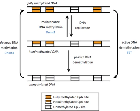
The dynamic regulation of the genome is determined by the balance between events of DNA methylation and demethylation (Figure 1). The latter process includes both the loss of 5mC during the replication (passive demethylation), induced by down-regulation of DNMT enzymes, inhibition of their activity or decreased levels of SAM, and the active removal of 5-mC (active demethylation) resulting in the formation of 5-hydroxymethylcytosine (5-hmC), considered to date the sixth base of DNA and a novel epigenetic mark (Branco et al., 2011; Guo et al., 2014; Saitou et al., 2012; Sadakierska-Chudy et al., 2015). Discovered for the first time in mouse and frog brain by Penn et al., recently the presence of 5-hmC has been reported in different tissues and cells and considered as an
7
intermediate of the oxidation of 5-mC by Ten-eleven translocation (TET)-family of methyl-cytosine dioxygenases (Hotchkiss, 1948; Griffith and Mahler, 1969; Penn et al., 1972; Naveh-Many et al., 1981; Waechter and Baserga, 1982; Tahiliani et al., 2009; Koh, 2011; Wu et al., 2017).
The earliest observations of the DNA methylation function date back to transfection experiments and microinjections of methylated sequences demonstrating that it induces gene silencing and that, in cultured cell lines, silent genes, can be activated following treatment with the demethylating agent 5-azacytidine (Jones 1985a and 1985b; Keshet et al., 1985; Yisraeli et al., 1988; Kass et al., 1993; Yan et al., 2014; Seelan et al., 2018).
Figure 1. DNA methylation model. DNMT1 carries out its action during cell replication performing the so-called maintenance DNA
methylation: the enzyme acts on hemimethylates CpG sites to restore DNA methylation status on the newly formed strand after cell division by copying pre-existing methylation patterns. On the other hand, DNMT3A and DNMT3B are de novo methyltransferases and function on unmethylated DNA by introducing methylation in only one of the two DNA strands at most CpG sites, generating the ideal hemimethylated sites for DNMT1. The active demethylation is performed by TETs (Jeltsch and Jurkowska, 2014).
Genome-wide studies of the methylome have highlighted that methylation patterns are cell-type specific and their effects are influenced by the position of methylated cytosines: if located adjacent to transcription factor binding sites, they block initiation, through either recruiting specific factors acting as gene expression repressors or by inhibiting the binding of activators, meanwhile in body gene they may either stimulate transcription elongation, thus hypothesizing a their role on splicing, or impede the alternative promoters activation (Watt and Molloy, 1988; Boyes and Bird, 1991; Singal and Ginder, 1999; Clouaire and Stancheva, 2008; Sasai and Defossez, 2009; Jones, 2012; Yin et al., 2017). Methylation in repeat regions such as centromeres is important for chromosomal stability, for
8
example chromosome segregation at mitosis, and is also likely involved in the suppression of the expression of transposable elements and thus to have a role in genome stability. Recently, the role of methylation in altering the activities of enhancers, insulators and other regulatory regions has been described. CpG islands methylation of the transcription start sites is associated with long-term silencing, see chromosome X inactivation, imprinting, genes expressed predominantly in germ cells and some tissue-specific genes (Moore et al., 2013; Huang et al., 2014; Allis and Jenuwein, 2016). Recently, a more complex epigenetic landscape is emerging, as demonstrated by the role played by the mitochondrial genome (mtDNA) in regulating intracellular DNA methylation as well as by the evidence reporting that, similarly to nuclear genome, also mtDNA is subject to CpG and non-CpG methylation and hydroxymethylation. Although the first attempt to identify traces of methylation within mtDNA dates back to the early 1970, for many years the epigenetic modification of mtDNA was controversial. Only recently, the advent of more innovative and sensitive techniques has allowed the discovery of DNMTs members in mitochondrial protein fractions and unequivocally identified the presence of methylation within the mitochondrial control region (D-loop) and some genes (ND1, ND2, ND6, Cytb, COI, 12SRNA, 16SRNA). Several hypothesis have been formulated to explain the functional role of mtDNA methylation, including the processing of mitochondrial polycistronic primary transcript and the regulation of the affinity of TFAM binding (Bellizzi et al., 2013)
DNA methylation and aging
Aging is a slow and gradual decline process of functional abilities that makes individuals more susceptible to environmental phenomena and diseases, and leads to a reduction in the probability of survival and finally to death (Johnson et al., 1999; Kirkwood, 2005; Sebastiani et al., 2012).
Aging affects all living organisms but lifespan is characteristic of each species. Moreover, among the various populations and within them there is considerable variability as regards the way and the quality of aging. This heterogeneity has largely been described as resulting from a complex interaction among genetics, environmental e stochastic factors and, more recently, epigenetic alterations have been included (Montesanto et al., 2012; D’Aquila et al., 2013). These alterations, by regulating gene expression, influence not only most of hallmarks of aging (genomic instability, telomere attrition, loss of proteostasis, deregulated nutrient sensing, mitochondrial dysfunction, cellular senescence, stem cell exhaustion, altered intercellular communication) but, at the same time, themselves because they are subjected to dynamic changes during lifetime, a phenomenon described as epigenetic drift (López-Otín et al., 2013; Li and Tollefsbol, 2016; Pal and Tyler, 2016). During
9
early embryogenesis, genomic DNA undergoes reprogramming processes including genome wide demethylation and de novo methylation leading to the re-establishment of DNA methylation patterns in the progeny that will be maintained in the somatic cells throughout the lifespan. After birth, although global DNA methylation patterns are quite stable, stochastic and environmental stimuli (ROS, inflammation, diet, stress, trauma) as well as the failure of the epigenetic machinery may induce random changes at certain loci, leading to a loss of phenotypic plasticity among individuals (Jones et al., 2015; Zampieri et al., 2015). Indeed, during each cell division, aberrant DNA methylation patterns accumulates over time contributing to epigenetic drift and creating an epigenetic mosaicism that may allow for the selection of biological defects that may lead to cancer and other age-related diseases (Amodio et al., 2017). Support for these evidence comes mainly from studies carried out in mono- and di-zygotic twin pairs in which a gradual age-related divergence in epigenetic marks was observed in monozygotics (Martin et al., 2005; Lipman and Tiedje, 2006; Kaminsky et al., 2009; Bell and Spector, 2011; Tan et al., 2013; Mendelsohn et al., 2017). DNA methylation drift comes from non-directional changes occurring during aging and involves both hypermethylation and hypomethylation events. Recently, Slieker and coll. identified several age-related Variably Methylated Position (aVMPs) exhibiting high variability in their methylation status and are associated with the expression of genes involved in DNA damage and apoptosis (Slieker et al., 2016). During aging, epigenetic drift also deeply influences the function of aged stem cells by limiting their plasticity and their differentiation potential that ultimately results in the exhaustion of the stem cell pool and in the selective growth advantage in other stem cells, which leads to clonal expansion and local hyper-proliferation (Teschendorff et al., 2010; Issa, 2014; Li and Tollefsbol, 2016).
Recently, several studies reported the presence of directional and non-stochastic changes occurring over time within clusters of consecutive CpG sites throughout the whole genome, referred as age-Differentially Methylated Regions (a-DMRs) (Rakyan et al., 2010; Li and Tollefsbol, 2016; Bacalini et al., 2017). Hundreds of hyper- and hypo-methylated a-DMR have been identified in multiple tissues and replicated in independent samples. A number of these aDMRs were located within 500 bp of the transcriptional start sites. Literature data agree to consider them associated with biological mechanisms involved in aging and longevity. Ashapkin et al., assume that most hyper-aDMRs represent epigenetic perturbations inherent to the aging per se, while hypo-aDMRs may be correlated to modifications associated both with aging per se and age-dependent modifications in relative proportions of the blood cell subtypes (Bell et al., 2012; Ashapkin et al., 2017).
Candidate genetic loci undergoing profound epigenetic changes with age and in age-related diseases have been progressively characterized. Global genomic DNA hypomethylation is especially evident at repetitive sequences, to a greater extent at Alu and HERV-K sequences, contributing to the increase
10
of genome instability as well as at specific promoter regions of some genes including ITGAL (Integrin alpha-L) and IL17RC (Interleukin 17 Receptor C) (Vijg and Dollé, 2007; Bollati et al., 2009; Zhang et al., 2009; Jintaridth and Mutirangura, 2010; Wei et al., 2012). By whole-genome bisulfite sequencing (WGBS), Heyn et al. compared the DNA methylation state of more than 90% of all CpGs present in the genome between newborn and nonagenarian/centenarian samples. A significant loss of methylated CpGs was found in the centenarian vs newborn DNAs. This was observed for all chromosomes and concerned all genomic regions such as promoters, exonic, intronic and intergenic regions. Most of these changes were focal and the aged genome was consequently less homogeneously methylated with respect to the newborn due to the age-dependent epigenetic drift (Heyn et al, 2012).
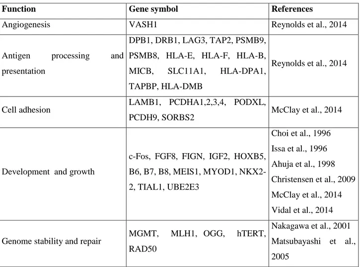
Besides to this extensive hypomethylation, the promoter regions of specific genes are subjected to a gradual increase of DNA methylation across lifespan (Table 1). In most of cases, the observed hypermethylation was associated to the transcriptional silencing suggesting that with increasing age there is an epigenetic turning off of these genes.
Figure 2 represents DNA methylation variations occurring during aging within interspersed repeats and genes.
Function Gene symbol References
Angiogenesis VASH1 Reynolds et al., 2014
Antigen processing and
presentation
DPB1, DRB1, LAG3, TAP2, PSMB9, PSMB8, HLA-E, HLA-F, HLA-B,
MICB, SLC11A1, HLA-DPA1,
TAPBP, HLA-DMB
Reynolds et al., 2014
Cell adhesion LAMB1, PCDHA1,2,3,4, PODXL,
PCDH9, SORBS2 McClay et al., 2014
Development and growth
c-Fos, FGF8, FIGN, IGF2, HOXB5, B6, B7, B8, MEIS1, MYOD1, NKX2-2, TIAL1, UBE2E3 Choi et al., 1996 Issa et al., 1996 Ahuja et al., 1998 Christensen et al., 2009 McClay et al., 2014 Vidal et al., 2014
Genome stability and repair MGMT, MLH1, OGG, hTERT,
RAD50
Nakagawa et al., 2001 Matsubayashi et al., 2005
11
Silva et al., 2008 Christensen et al., 2009 Madrigano et al., 2012
Ion channel GRIA2, KCNJ8, RYR2 McClay et al., 2014
Metabolism
AGPAT2, ATP13A4, COX7A1,
CRAT, ECRG4, ELOVL2, EPHX2, GAD2, LEP, MGC3207, MGEA5,
SLC38A4, SLC22A18, SNTG1, STAT5A Rönn et al., 2008 Bell et al., 2012 Madrigano et al., 2012 McClay et al., 2014 Gentilini et al., 2012
Immune response CD4, INFG, TNF NOD2, PTMS Madrigano et al., 2012
McClay et al., 2014 Signal transduction ARL4A, DLC1, GPR128, GRIA2, LAG3, MYO3A, PRR5L, PTPRT, TFG, TRAF6, TRHDE Bell et al., 2012 McClay et al., 2014
Stress response HSPA2 McClay et al., 2014
Transcription factors
ARID5B, BICC1, ESR1, FOXP1, HIPK2, LHX5, MLF2, NFIA, NOD2, POU4F3, RARB, TBX4, TBX20, TRPS1, WT1, ZBTB1, ZEB2, ZNF827 Gaudet et al., 2009 Christensen et al., 2009 Bell et al., 2012 Reynolds et al., 2014 McClay et al., 2014 Tumor suppression
APC, CASP8, CHD1, GSTP1, HIC1, LOX, LSAMP, N33, P16INK4A, RASSF1, RUNX3, SOCS1, TIG1, DAPK1, hMLH1, p16, Ahuja et al., 1998 Fujii et al., 1998 Cody et al., 1999 Dammann et al., 2000 Virmani et al., 2001 Waki et al., 2003 Sutherland et al., 2004 So et al., 2006 Nishida et al., 2008 Yuan et al., 2008 Christensen et al., 2009 McClay et al., 2014
12 Figure 2. A Graphical representation of DNA methylation patterns changes in young (A) an old people (B). Active and repressed
transcription at transcription start sites are indicated in ligth blue and red arrows respectively. Black lines indicate the methylation status of DNA from different people where white circles represent unmethylated CpGs, black circles represent methylated CpGs. Age-associated DMRs are highlighted in blue (hypomethylated regions) and red (hypermethylated regions), respectively. (Zampieri et al., 2015).
Epigenome-wide association studies (EWAS) identified the so-called “clock CpGs”, namely a large set of CpG markers whose methylation status is measured in order to construct quantitative models effective in predicting the age of cells, tissues or organs, referred as epigenetic age or DNAm age. DNAm age not only reflects the chronological, but also the biological age and, thus, these biomarkers would, on one hand, facilitate the differentiation of individuals who are of the same chronological age yet have variant aging rates, on the other define a panel of measurements for healthy aging and, even further, predict life span (Bellizzi et al., 2012a and 2012b, D’Aquila et al., 2017 and 2018). Figure 3 depicts the existing relationship among chronological age, biological age and epigenome.
13
Figure 3. In the picture are indicated chronological age (x-axis) and biological age (y-axis), respectively. Individuals with the same chronological age may display different phenotypes due to their biological age (Benayoun et al., 2015).
Multiple candidate loci have been selected showing linear correlation of methylation status with biological age. Among these, the loci ELOVL2 (ELOVL fatty acid elongase 2), FHL2 (Four and a half LIM domains 2), CCDC102B (coiled-coil domain containing 102B), C1orf132 (chromosome 1
open reading frame 132) are the most thoroughly evaluated markers of age in various human tissues
(Garagnani et al., 2012, Freire-Aradas et al., 2016). More recently, D’Aquila et al., reports the identification of RAB32 and RHOT2 genes as potential biomarkers of chronological and biological age. Being two genes involved in mitochondrial quality control, this evidence provide further confirm to the role of mitochondrial functions during age (in Chapter I: Original Research Work).
Starting from Bocklandt et al, which described the first age estimator model by using DNA samples from saliva, a series of epigenetic clocks were developed by analyzing DNA methylation marks in single and multiple tissues (Bocklandt et al., 2011). Currently, Hannum and Horvath clocks represent the most robust recognized models showing both a high age correlation (R>0.9) and low mean error of the age prediction (4.9 and 3.6 years, respectively). The first model was developed only in blood, while the second, designed matching data from 51 healthy tissues, such as blood, cerebellum, occipital cortex, buccal, colon, adipose, liver, lung, saliva, and cell types, including CD4 T and immortalized B cells, results compatible with different technological platforms and used in a wide range of studies (Hannum et al., 2013; Horvath, 2013).
14
Both models are also able to predict all-cause mortality independent of several risk factors including smoking, alcohol use, education, body mass index and comorbidities (Marioni et al., 2015; Christiansen et al., 2016; Perna et al., 2016). More recently, DNAmAge biomarkers which also consider clinical measures of physiological dysregulations have been developed. One of these, referred as DNAmPhenoAge (phenotypic age estimator), constructed by generating a weighted average of 10 clinical characteristic, such as albumin, creatinine, glucose and C-reactive proteins, and then analysed by regression analysis against DNA methylation levels in blood, proved to be effective in predicting mortality, health span, disease risk and in various measures of comorbidity (Levine et al, 2018). Age acceleration, that is an estimated DNAmAge higher than chronological age was registered for several age-related diseases including Down syndrome (Horvath et al., 2015a and 2015b), Alzheimer’s and Parkinson’s diseases (Levine et al., 2015; Horvath et al., 2015a and 2015b), HIV-infection (Boulias et al., 2016, Rickabaugh et al., 2015), frailty (Breitling et al., 2016), diabetes and cancer (Zheng et al., 2016; Bacalini et al., 2017).
Lastly, a significant number of reports also evidence a correlation between mitochondrial DNA methylation with aging. The first of them dates to 1983, when a decrease of mtDNA methylation was observed in aged cultured fibroblasts (D’Aquila et al., 2017). More recently, although methylation levels of the mitochondrial D-loop region resulted not associated with aging, high methylation levels (>10%) of one CpG site located within the MT-RNR1 gene were observed more frequent in old women with respect to youngers (Bellizzi et al., 2013; D’Aquila et al., 2015). Furthermore, the non-canonical CpG methylation patterns, such as non-CpG and hydroxymethylation, are deregulated during aging, potentially leading to downstream changes in transcription and cellular physiological functions. A global non-CpG methylation decrease with age has been described. 5-hmC content significantly decreases in some tissues, including blood and liver, and is negatively correlated with aging, in association with low mRNA expression levels of TET1 and TET3 (Truong et al., 2015). In a contrasting, mouse cerebellum and hippocampus show an increase of 5hmC levels with aging which can be prevented by caloric restriction (Szulwach et al., 2012; Chouliaras et al., 2012). A decrease of mitochondrial DNA levels of 5-hmC during aging was observed in frontal cortex but not in the cerebellum (Dzitoyeva et al., 2012). An increase in 5hmC signals was observed in genes activated in old mice with respect to young ones demonstrating that 5hmC is acquired in developmentally activated genes (Szulwach et al., 2012). What is more, age-related non-overlapping 5-mCand 5hmC pattern have been observed (Kochmanski et al., 2018).
Considering that epigenetic marks induce profound changes in the gene expression and contribute to the cellular and organismal phenotypic plasticity during lifetime, it is evident that dysregulations of epigenetic patterns may contribute to age-related diseases, including cancer, diabetes, cardiovascular
15
and neurodegenerative diseases. These changes represent potential disease biomarkers. In all of these diseases, methylome-wide association studies (MWAS) have identified characteristic methylome signatures and brought to light as alteration in DNA methylation are hallmarks often coincident with those observed in aging. Example of this overlapping come from the observation that DNA hypomethylation, prevalently at repetitive DNA elements, and locus specific hypermethylation of tumour-suppressor genes (p53, p21, p16, TIG1, and RB1), oncogenes (cMYC and TERT), genes involved in type 2 diabetes (COX7A1, PRDX2, IRS1 and KCNJ11), genes involved in Alzheimer’s disease (APP, PS1 and BACE1) generally occur in the above diseases as in aging thus causing aberrant gene expression (Brunet et al, 2014). Changes in the levels of 5mC and DNMTs, DNMT1 and DNMT3a have been detected in neuronal mitochondria from patients with amyotrophic lateral sclerosis (ALS) suggesting that motor neurons can engage epigenetic mechanisms involving DNMT
upregulation and increased DNA methylation to drive apoptosis (Wong et al., 2013).Silva et al. found
that patients with Alzheimer disease (AD) had a higher methylation frequency of hTERT compared to elderly controls (Silva et al, 2008). Repetitive LINE-1 elements were also reported to be significantly hypermethylated in AD patients with respect to healthy controls. AD patients have been further characterized by a decrease in brain SAM levels, and temporal neocortex neuronal nuclei were found to be hypomethylated in a patient with AD compared to his non-AD monozygotic twin (Mastroeni et al., 2009; Bollati et al., 2011). Guarasci et al. have recently observed that individuals affected by Down Syndrome exhibit dysregulated mtDNA methylation patterns in D-loop region with respect to healthy individuals (manuscript in preparation).
It follows that epigenetic based drugs which reverse aberrant DNA methylation profiles may be considered effective in the assessment and development of epigenetic-based treatments (Shenouda et al., 2009).
Environmental factors and epigenetics aging
Environmental factors, including chemicals, pollutants, diet, drugs, infectious, trauma, and psycho-social and socio-economic status have been associated with DNA methylation changes in aging and age-related diseases (Huidobro et al., 2013; Obata et al., 2015). These changes can be induced in the individual in each period of the life, from the in utero period to the elderly and are relevant with respect to the shift from the healthy to the diseased statuses of an individual with aging, due to the increased chance to encounter environmental insults or to accumulate their effects during aging. Epidemiological evidence suggests that maternal environmental exposure to stimuli result in
16
epigenetic changes, since DNA methylation patterns at DMRs are established before gastrulation, which occur early in development and play an important role in susceptibility to disease in later life. It was observed that energy-rich, protein-deficient, micronutrient-deficient and/or methyl donor-rich diets during pregnancy induce modifications of methylation profile in mothers which, in turn, can be transmitted to next generation thus regulating in offspring long term metabolic processes which contribute to age phenotypes and age-related disease (Vickers, 2014; Lillycrop et al., 2015; Park et al., 2017). An emblematic example of the trans-generational relationship between food and epigenetic modifications is represented by The Dutch Honger winter (1944-1945) family studies in which adult health outcomes in relation to exposure to famine prior to conception or at specific periods of gestation was analyzed. It emerged that prenatal exposure to the famine is associated with increased prevalence of overweight, hypertension, and coronary heart disease, meanwhile maternal famine exposure around the time of conception has been related to prevalence of major affective disorders, antisocial personality disorders, schizophrenia, decreased intracranial volume, and congenital abnormalities of the central nervous system (Lumey et al., 2007; Stein et al., 2009). More recently, Guarasci et al. reported that the differences in global DNA methylation among different tissues are magnified in 96 weeks old rats fed with low calorie diet. Moreover, the low-calorie diet appears to affect the offspring's epigenetic status more strongly if administered during the maternal
pre-gestational period than the pre-gestational and lactation time (Guarasci et al., 2018).Caloric restriction,
the decrease in nutrient intake above the level of starvation and below what an organism would consume ad libitum, is one of the most consistent means of increasing life span across a spectrum of organisms. Both caloric intake and DNMT3A play a role in neuronal aging, in which DNMT3A contribute to the formation of memory and synaptic and neuronal plasticity (Miller and Sweatt, 2007). It was found that Dnmt3a-immunoreactivity increases with age in the CA3 and CA1-2 hippocampal regions of mice and that reducing caloric intake by 50% attenuates this increase. An age-related increase in 5mC occurs in these regions as well as the hippocampal dentate gyrus and is also attenuated by CR.63 Similarly, 5hmC content increases with age in all three of these regions, and CR opposes this age-related increase in the CA3 region, further implicating methylomics in hippocampal aging (Chouliaras et al., 2011; Johnson et al., 2012).
In this context, different studies have also demonstrated the role of nutrition in molecular mechanisms related to onset and progression of neurodegenerative diseases, such as Alzheimer and Parkinson diseases, psychiatric disorders and dementia. Transgenic mouse model of Alzheimer disease treated with a diet deficient of vitamin B12, B6 and folate showed a decrease of SAM/SAH ratio leading to impaired methylation potential, DNMTs inhibition and DNA demethylase stimulation, PSEN1 promoter hypomethylation, PSEN1 overexpression, increased amyloid processing and deposition in
17
senile plaques and, finally, cognitive impairment. The supplementation with SAM was able to restore control-like conditions in AD mice or even to partially revert the Alzheimer-like phenotype (Fuso et al., 2012).
Lastly, an increase of pollutant, metal and pesticides exposure, stress, trauma were significantly associated with increase in DNAm-age and in Horvath DNAm-age (Dihigra). Glucocorticoids, a class of endocrine signaling hormones which includes cortisol are a component of the biological response to stress. Notably, 85 of the 353 loci that comprise the Horvath epigenetic clock are located near glucocorticoid receptor elements, and 110 loci showed altered DNA methylation after exposure to dexamethasone, a glucocorticoid receptor agonist (Zannas et al., 2015). Intra-uterine exposure to arsenic alters DNA methylation in offspring which may result in a higher risk of disease in later life likely by influencing the generation of reactive oxygen species (ROS), which causes oxidative DNA damage, binding and inhibition of arsenic metabolites to enzymes, and perturbation of key signaling pathways (Rossman et al., 2011.).
Similar to nuclear DNA methylation, occurrence of the abnormal mtDNA methylation is often depending on different factors, such as diseases, environment, drugs, and food (Gao et al., 2017). It was observed that fructose consumption induce metabolism disorders by stimulating hepatic mtDNA-encoded gene expression through epigenetic changes in mtDNA. Lower mtDNA D-loop methylation levels were found in the blood patients with late-onset Alzheimer’s disease patients compared to the blood of normal controls. In addition, insulin resistance was associated with DNA methylation in mitochondrial NADH dehydrogenase 6 and D loop-region (Stoccoro et al., 2017). Alcohol exposure was reported modulates levels of DNMT enzymes (Mandal et al., 2017; Miozzo et al., 2018).
18
References
1. Ahuja N, Li Q, Mohan AL, Baylin SB, Issa JP. Aging and DNA methylation in colorectal mucosa and cancer. Cancer Research. 1998; 58(23):5489-94.
2. Allis CD, Jenuwein T. The molecular hallmarks of epigenetic control. Nat Rev Genet 2016;17: 487–500. 3. Amodio N, D'Aquila P, Passarino G, Tassone P, Bellizzi D. Epigenetic modifications in multiple
myeloma: recent advances on the role of DNA and histone methylation. Expert Opinion on Therapeutic Targets. 2017; 21:91–101.
4. Ashapkin VV, Kutueva LI, Vanyushin BF. Aging as an Epigenetic Phenomenon. Curr Genomics. 2017; 18:385–407.
5. Bacalini MG, D’Aquila P, Marasco E, Nardini C, Montesanto A, Franceschi C, Passarino G, Garagnani P, Bellizzi D. The methylation of nuclear and mitochondrial DNA in ageing phenotypes and longevity. Mech Ageing Dev. 2017; 165:156–161.
6. Barros SP, Offenbacher S. Epigenetics: connecting environment and genotype to phenotype and disease. J Dent Res. 2009;88: 400–408.
7. Bell JT, Spector TD. A twin approach to unraveling epigenetics. Trends in Genetics. 2011; 27:116–125. 8. Bell JT, Tsai PC, Yang TP, Pidsley R, Nisbet J, Glass D, Mangino M, Zhai G, Zhang F, Valdes A, Shin
SY, Dempster EL, Murray RM, Grundberg E, Hedman AK, Nica A, Small KS; MuTHER Consortium, Dermitzakis ET, McCarthy MI, Mill J, Spector TD, Deloukas P. Epigenome-wide scans identify differentially methylated regions for age and age-related phenotypes in a healthy ageing population. PLoS Genetics. 2012; 8(4): e1002629.
9. Bellizzi D, D’aquila P, Giordano M, Montesanto A, Passarino G. Global DNA methylation levels are modulated by mitochondrial DNA variants. Epigenomics 2012a; 4:17–27.
10. Bellizzi D, D’Aquila P, Scafone T, Giordano M, Riso V, Riccio A, Passarino G. The control region of mitochondrial DNA shows an unusual CpG and non-CpG methylation pattern. DNA Res 2013; 20:537– 547.
11. Bellizzi D, D'Aquila P, Montesanto A, Corsonello A, Mari V, Mazzei B, Lattanzio F, Passarino G. Global DNA methylation in old subjects is correlated with frailty. Age. 2012b; 34:169–179.
12. Benayoun BA, Pollina EA, Brunet A. Epigenetic regulation of ageing: linking environmental inputs to genomic stability. Nat Rev Mol Cell Biol. 2015 Oct;16(10):593-610.
13. Bocklandt S, Lin W, Sehl ME, Sánchez FJ, Sinsheimer JS, Horvath S, Vilain E. Epigenetic predictor of age. PLoS One. 2011; 6:14821.
14. Bollati V, Galimberti D, Pergoli L, Dalla Valle E, Barretta F, Cortini F, Scarpini E, Bertazzi PA, Baccarelli A. DNA methylation in repetitive elements and Alzheimer disease. Brain Behav Immun. 2011 Aug; 25(6):1078-83.
15. Bollati V, Schwartz J, Wright R, Litonjua A, Tarantini L, Suh H, Sparrow D, Vokonas P, Baccarelli A. Decline in genomic DNA methylation through aging in a cohort of elderly subjects. Mechanisms of
19 Ageing and Development. 2009; 130:234–9.
16. Boulias K, Lieberman J, Greer EL. An Epigenetic Clock Measures Accelerated Aging in Treated HIV Infection. Mol Cell. 2016 Apr 21;62(2):153-155.
17. Boyes J, Bird A. DNA methylation inhibits transcription indirectly via a methyl-CpG binding protein. Cell. 1991; 64:1123–1134.
18. Branco MR, Ficz G, Reik W. Uncovering the role of 5-hydroxymethylcytosine in the epigenome. Nat Rev Genet. 2011; 13:7–13.
19. Breitling LP, Saum KU, Perna L, Schöttker B, Holleczek B, Brenner H. Frailty is associated with the epigenetic clock but not with telomere length in a German cohort. Clin Epigenetics. 2016 Feb 26;8:21. 20. Brunet A, Berger SL. Epigenetics of Aging and Aging-related Disease. J Gerontol A Biol Sci Med Sci.
2014; 69: S17–S20.
21. Choi EK, Uyeno S, Nishida N, Okumoto T, Fujimura S, Aoki Y, Nata M, Sagisaka K, Fukuda Y, Nakao K, Yoshimoto T, Kim YS, Ono T. Alterations of c-fos gene methylation in the processes of aging and tumorigenesis in human liver. Mutation Research. 1996; 354(1):123 -8.
22. Chouliaras L, van den Hove DL, Kenis G, Dela Cruz J, Lemmens MA, van Os J, Steinbusch HW, Schmitz C, Rutten BP. Caloric restriction attenuates age-related changes of DNA methyltransferase 3a in mouse hippocampus. Brain Behav Immun. 2011 May; 25(4):616-23.
23. Chouliaras L, van den Hove DL, Kenis G, Keitel S, Hof PR, van Os J, Steinbusch HW, Schmitz C, Rutten BP. Age-related increase in levels of 5-hydroxymethylcytosine in mouse hippocampus is prevented by caloric restriction. Curr Alzheimer Res. 2012; 9:536–544.
24. Christensen BC, Houseman EA, Marsit CJ, Zheng S, Wrensch MR, Wiemels JL, Nelson HH, Karagas MR, Padbury JF, Bueno R, Sugarbaker DJ, Yeh RF, Wiencke JK, Kelsey KT. Aging and environmental exposures alter tissue-specific DNA methylation dependent upon CpG island context. PLoS Genetics. 2009; 5(8): e1000602.
25. Christiansen L, Lenart A, Tan Q, Vaupel JW, Aviv A, McGue M, Christensen K. DNA methylation age is associated with mortality in a longitudinal Danish twin study. Aging Cell. 2016; 15:149–154. 26. Clouaire T, Stancheva I. Methyl-CpG binding proteins: specialized transcriptional repressors or
structural components of chromatin? Cellular and Molecular Life Sciences. 2008; 65:1509–1522. 27. Cody DT, Huang Y, Darby CJ, Johnson GK, Domann FE. Differential DNA methylation of the p16
INK4A/CDKN2A promoter in human oral cancer cells and normal human oral keratinocytes. Oral Oncology. 1999; 35(5):516-22.
28. Dammann R, Li C, Yoon JH, Chin PL, Bates S, Pfeifer GP. Epigenetic inactivation of a RAS association domain family protein from the lung tumour suppressor locus 3p21.3. Nature Genetics. 2000; 25(3):315-9.
29. Dan J, Chen T. Genetic Studies on Mammalian DNA Methyltransferases. Advances in Experimental Medicine and Biology. 2016; 123–945.
20 2018; 10:7–8.
31. D'Aquila P, Giordano M, Montesanto A, De Rango F, Passarino G, Bellizzi D. Age-and gender-related pattern of methylation in the MT-RNR1 gene. Epigenomics. 2015; 7:707–716.
32. D'Aquila P, Montesanto A, Guarasci F, Passarino G, Bellizzi D. Mitochondrial genome and epigenome: two sides of the same coin. Frontiers in Bioscience (Landmark Edition). 2017; 22: 888-908.
33. D'Aquila P, Montesanto A, Mandalà M, Garasto S, Mari V, Corsonello A, Bellizzi D, Passarino G. Methylation of the ribosomal RNA gene promoter is associated with aging and age-related decline. Aging Cell. 2017; 16:966–975.
34. D'Aquila P, Rose G, Bellizzi D, Passarino G. Epigenetics and aging. Maturitas. 2013; 74:130–136. 35. Deaton AM, Bird A. CpG islands and the regulation of transcription. Genes Dev. 2011; 25:1010–1022. 36. Dzitoyeva S, Chen H, Manev H. Effect of aging on 5-hydroxymethylcytosine in brain mitochondria.
Neurobiology of Aging. 2012; 33:2881–2891.
37. Fraga MF; Genetic and epigenetic regulation of aging. Curr Opin Immunol. 2009 Aug; 21(4):446-53. 38. Freire-Aradas A, Phillips C, Mosquera-Miguel A, Girón-Santamaría L, Gómez-Tato A, Casares de
Cal M, Álvarez-Dios J, Ansede-Bermejo J, Torres-Español M, Schneider PM, Pośpiech E, Branicki W, Carracedo Á, Lareu MV. Development of a methylation marker set for forensic age estimation using analysis of public methylation data and the Agena Bioscience EpiTYPER system. Forensic Sci Int Genet. 2016 Sep;24:65-74.
39. Fujii H, Biel MA, Zhou W, Weitzman SA, Baylin SB, Gabrielson E. Methylation of the HIC-1 candidate tumor suppressor gene in human breast cancer. Oncogene. 1998; 16(16):2159-64.
40. Fuso A, Nicolia V, Ricceri L, Cavallaro RA, Isopi E, Mangia F, Fiorenza MT, Scarpa S. S -adenosylmethionine reduces the progress of the Alzheimer-like features induced by B-vitamin deficiency in mice. Neurobiol Aging. 2012 Jul;33(7):1482.e1-16.
41. Gao D, Zhu B, Sun H, Wang X. Mitochondrial DNA Methylation and Related Disease. Adv Exp Med Biol. 2017;1038:117-132.
42. Garagnani P, Bacalini MG, Pirazzini C, Gori D, Giuliani C, Mari D, Di Blasio AM, Gentilini D, Vitale G, Collino S, Rezzi S, Castellani G, Capri M, Salvioli S, Franceschi C. Methylation of ELOVL2 gene as a new epigenetic marker of age. Aging Cell. 2012 Dec;11(6):1132-4.
43. Gardiner-Garden M, Frommer M. CpG islands in vertebrate genomes. J. Mol. Biol. 1987. 196: 261– 282. 44. Gaudet MM, Campan M, Figueroa JD, Figueroa JD, Yang XR, Lissowska J, Peplonska B, Brinton
LA, Rimm DL, Laird PW, Garcia-Closas M, Sherman ME. DNA hypermethylation of ESR1 and PGR in breast cancer: pathologic and epidemiologic associations. Cancer Epidemiology, Biomarkers & Prevention. 2009; 18(11):3036-43.
45. Gentilini D1, Mari D, Castaldi D, Remondini D, Ogliari G, Ostan R, Bucci L, Sirchia SM, Tabano S, Cavagnini F, Monti D, Franceschi C, Di Blasio AM, Vitale G. Role of epigenetics in human aging and longevity: genome-wide DNA methylation profile in centenarians and centenarians' offspring. Age (Dordr). 2013 Oct;35(5):1961-73.
21 46. Goldberg AD, Allis CD, Bernstein E. Epigenetics: A Landscape Takes Shape. Cell. 2007; 128:635–638 47. Griffith JS, Mahler HR. DNA ticketing theory of memory. Nature. 1969; 223:580–582.
48. Guarasci F, D'Aquila P, Mandalà M, Garasto S, Lattanzio F, Corsonello A, Passarino G, Bellizzi D. Aging and nutrition induce tissue-specific changes on global DNA methylation status in rats. Mechanisms of Ageing and Development. 2018; 174: 47–54.
49. Guo F, Li X, Liang D, Li T, Zhu P, Guo H, Wu X, Wen L, Gu TP4, Hu B, Walsh CP, Li J, Tang F, Xu GL. Active and passive demethylation of male and female pronuclear DNA in the mammalian zygote. Cell Stem Cell. 2014; 15:447–459.
50. Hannum G, Guinney J, Zhao L, Zhang L, Hughes G, Sadda S. Genome-wide methylation profiles reveal quantitative views of human aging rates. Mol Cell. 2013; 49:359–367.
51. Heyn H, Li N, Ferreira HJ, Moran S, Pisano DG, Gomez A, Diez J, Sanchez-Mut JV, Setien F, Carmona FJ, Puca AA, Sayols S, Pujana MA, Serra-Musach J, Iglesias-Platas I, Formiga F, Fernandez AF, Fraga MF, Heath SC, Valencia A, Gut IG, Wang J, Esteller M. Distinct DNA methylomes of newborns and centenarians. Proc Natl Acad Sci. 2012; 109:10522–10527.
52. Horvath S, Garagnani P, Bacalini MG, Pirazzini C, Salvioli S, Gentilini D, Di Blasio AM, Giuliani C, Tung S, Vinters HV, Franceschi C. Accelerated epigenetic aging in Down syndrome. Aging Cell. 2015a Jun;14(3):491-5.
53. Horvath S, Pirazzini C, Bacalini MG, Gentilini D, Di Blasio AM, Delledonne M, Mari D, Arosio B, Monti D, Passarino G, De Rango F, D'Aquila P, Giuliani C, Marasco E, Collino S, Descombes P, Garagnani P, Franceschi C. Decreased epigenetic age of PBMCs from Italian semi-supercentenarians and their offspring. Aging (Albany NY). 2015b Dec;7(12):1159-70.
54. Horvath S. DNA methylation age of human tissues and cell types. Genome Biol. 2013;14(10): R115. 55. Hotchkiss RD. The quantitative separation of purines, pyrimidines, and nucleosides by paper
chromatography. The Journal of Biological Chemistry. 1948; 175(1):315-32.
56. Huang B, Jiang C, Zhang R. Epigenetics: The language of the cell? Epigenomics 2014; 6:73–88. 57. Huidobro C, Fernandez AF, Fraga MF. Aging epigenetics: Causes and consequences. Mol Aspects Med
2013; 34:765–781.
58. Illingworth RS, Bird AP. CpG islands–’a rough guide. FEBS Letters. 2009; 583: 1713–1720. 59. Issa JP. Aging and epigenetic drift: a vicious cycle. J Clin Invest 2014; 124:24–29.
60. Jeltsch, A and Jurkowska, RZ. New concepts in DNA methylation. Trends Biochem Sci. 2014 Jul;39(7):310-8.
61. Jintaridth P, Mutirangura A. Distinctive patterns of age-dependent hypomethylation in interspersed repetitive sequences. Physiological Genomics. 2010; 41:194–200.
62. Johnson AA, Akman K, Calimport SRG, Wuttke D, Stolzing A, de Magalhães JP. The Role of DNA Methylation in Aging, Rejuvenation, and Age-Related Disease. Rejuvenation Res. 2012 Oct; 15(5): 483–494.
22 64. Jones MJ, Goodman SJ, Kobor MS. DNA methylation and healthy human aging. Aging Cell. 2015;
14:924–932.
65. Jones PA. Altering gene expression with 5-azacytidine. Cell. 1985b; 40:485–486.
66. Jones PA. Effects of 5-azacytidine and its 2’-deoxyderivative on cell differentiation and DNA methylation. Pharmacol Ther. 1985a; 28:17–27.
67. Jones PA. Functions of DNA methylation: Islands, start sites, gene bodies and beyond. Nat Rev Genet. 2012; 13:484–492.
68. Kaminsky ZA, Tang T, Wang SC, et al. DNA methylation profiles in monozygotic and dizygotic twins. Nat Genet. 2009; 41:240–245.
69. Kanherkar RR, Bhatia-Dey N, Csoka AB. Epigenetics across the human lifespan. Frontiers in Cell and Developmental Biology. 2014; 2:49.
70. Kass SU, Goddard JP, Adams RL. Specific methylation of vector sequences inhibits transcription from the SV40 early promoter. Biochem Soc Trans. 1993; 21:9
71. Keshet I, Yisraeli J, Cedar H. Effect of regional DNA methylation on gene expression. Proc Natl Acad Sci U S A 1985; 82:2560–4.
72. Kirkwood TBL. Understanding the odd science of aging. Cell 2005; 120:437–447.
73. Kochmanski J, Marchlewicz EH, Cavalcante RG, Sartor MA, Dolinoy DC. Age-related Epigenome-wide DNA Methylation and Hydroxymethylation in Longitudinal Mouse Blood. Epigenetics 2018; 1-14. 74. Koh KP, Yabuuchi A, Rao S, Huang Y, Cunniff K, Nardone J, Laiho A, Tahiliani M, Sommer CA,
Mostoslavsky G, Lahesmaa R, Orkin SH, Rodig SJ, Daley GQ, Rao A. Tet1 and Tet2 regulate 5-hydroxymethylcytosine production and cell lineage specification in mouse embryonic stem cells. Cell Stem Cell. 2011; 8:200–213.
75. Larsen F, Gundersen G, Lopez R, Prydz H. CpG islands as gene markers in the human genome. Genomics. 1992; 13:1095–1107.
76. Levine ME, Lu AT, Bennett DA, Horvath S. Epigenetic age of the pre -frontal cortex is associated with neuritic plaques, amyloid load, and Alzheimer's disease related cognitive functioning. Aging (Albany NY). 2015 Dec;7(12):1198-211.
77. Levine ME, Lu AT, Quach A, Chen BH, Assimes TL, Bandinelli S, Hou L, Baccarelli AA, Stewart JD, Li Y, Whitsel EA, Wilson JG, Reiner AP1, Aviv A, Lohman K, Liu Y, Ferrucci L, Horvath S. An epigenetic biomarker of aging for lifespan and healthspan. Aging (Albany NY). 2018; 10:573–591. 78. Li Y, Tollefsbol TO. Age-related epigenetic drift and phenotypic plasticity loss: Implications in
prevention of age-related human diseases. Epigenomics. 2016; 8:1637–1651.
79. Lillycrop KA, Burdge GC. Maternal diet as a modifier of offspring epigenetics. J Dev Orig Health Dis. 2015; 6:88–95.
80. Lipman T, Tiedje LB. Epigenetic Differences Arise During the Lifetime of Monozygotic Twins. MCN, Am J Matern Nurs. 2006; 31:204.
23 81. Lister R, Pelizzola M, Dowen RH, Hawkins RD, Hon G, Tonti-Filippini J, Nery JR, Lee L, Ye Z, Ngo QM, Edsall L, Antosiewicz-Bourget J, Stewart R, Ruotti V, Millar AH, Thomson JA, Ren B, Ecker JR. Human DNA methylomes at base resolution show widespread epigenomic differences. This was the first report of a human methylome at single-base resolution. Nature. 2009; 462:315–322.
82. López-Otín C, Blasco MA, Partridge L, Serrano M, Kroemer G. The hallmarks of aging. Cell. 2013; 153:1194–1217.
83. Lumey LH, Stein AD, Kahn HS, van der Pal-de Bruin KM, Blauw GJ, Zybert PA, Susser ES. Cohort profile: the Dutch Hunger Winter families study. Int J Epidemiol 2007; 36:1196–204.
84. Luo GZ, Blanco MA, Greer EL, He C, Shi Y. DNA N(6)-methyladenine: a new epigenetic mark in eukaryotes? Nat Rev Mol Cell Biol 2015; 16: 705–10.
85. Luo GZ, Wang F, Weng X, Chen K, Hao Z, Yu M, Deng X, Liu J, He C. Characterization of eukaryotic DNA N(6)-methyladenine by a highly sensitive restriction enzyme-assisted sequencing. Nat Commun. 2016 Apr 15;7:11301.
86. Lyko F. The DNA methyltransferase family: a versatile toolkit for epigenetic regulation. Nature Reviews Genetics. 2018; 19:81–92.
87. Madrigano J, Baccarelli A, Mittleman MA, Sparrow D, Vokonas PS, Tarantini L, Schwartz J. Aging and epigenetics: longitudinal changes in gene-specific DNA methylation. Epigenetics. 2012; 7(1):63-70.
88. Mandal C, Halder D, Jung KH, Chai YG. Gestational Alcohol Exposure Altered DNA Methylation Status in the Developing Fetus. Int J Mol Sci. 2017 Jul; 18(7): 1386
89. Marioni RE, Shah S, McRae AF, Chen BH, Colicino E, Harris SE, Gibson J, Henders AK, Redmond P, Cox SR, Pattie A, Corley J, Murphy L, Martin NG, Montgomery GW, Feinberg AP, Fallin MD, Multhaup ML, Jaffe AE, Joehanes R, Schwartz J, Just AC, Lunetta KL, Murabito JM, Starr JM, Horvath S, Baccarelli AA, Levy D, Visscher PM, Wray NR, Deary IJ. DNA methylation age of blood predicts all-cause mortality in later life. Genome Biol 2015; 16:25.
90. Martin GM. Epigenetic drift in aging identical twins. Proceedings of the National Academy of Science of the United States of America. 2005; 102: 10413–10414.
91. Mastroeni D, McKee A, Grover A, Rogers J, Coleman PD. Epigenetic differences in cortical neurons from a pair of monozygotic twins discordant for Alzheimer's disease. PLoS One. 2009 Aug 12; 4(8):e6617.
92. Matsubayashi H, Sato N, Brune K, et al. Age- and disease-related methylation of multiple genes in nonneoplastic duodenum and in duodenal juice. Clinical Cancer Research. 2005; 11(2 Pt 1):573-83. 93. Maunakea AK, Nagarajan RP, Bilenky M, Ballinger TJ, D'Souza C, Fouse SD, Johnson BE, Hong C, Nielsen C, Zhao Y, Turecki G, Delaney A, Varhol R, Thiessen N, Shchors K, Heine VM, Rowitch DH, Xing X, Fiore C, Schillebeeckx M, Jones SJ, Haussler D, Marra MA, Hirst M, Wang T, Costello JF. Conserved role of intragenic DNA methylation in regulating alternative promoters. Nature. 2010; 466:253–257.
24 94. McClay JL, Aberg KA, Clark SL, Nerella S, Kumar G, Xie LY, Hudson AD, Harada A, Hultman CM, Magnusson PK, Sullivan PF, Van Den Oord EJ. A methylome-wide study of aging using massively parallel sequencing of the methyl-CpG-enriched genomic fraction from blood in over 700 subjects. Human Molecular Genetics. 2014 ;23(5):1175-85.
95. Meloni M and Testa G. Scrutinizing the epigenetics revolution. Biosocieties. 2014 Nov;9(4):431-456. 96. Mendelsohn AR, Larrick JW. Epigenetic Drift Is a Determinant of Mammalian Lifespan. Rejuvenation
Res. 2017; 20:430–436.
97. Miller CA, Sweatt JD. Covalent modification of DNA regulates memory formation. Neuron. 2007 Mar 15; 53(6):857-69.
98. Miozzo F, Arnould H, de Thonel A, Schang AL, Sabéran-Djoneidi D, Baudry A, Schneider B, Mezger V. Alcohol exposure promotes DNA methyltransferase DNMT3A upregulation through reactive oxygen species-dependent mechanisms. Cell Stress Chaperones. 2018 Jan;23(1):115-126.
99. Montesanto A, Dato S, Bellizzi D, Rose G, Passarino G. Epidemiological, genetic and epigenetic aspects of the research on healthy ageing and longevity. Immun Ageing. 2012; 9:6.
100. Moore LD, Le T, Fan G. DNA Methylation and Its Basic Function. Neuropsychopharmacology. 2013; 38:23–38.
101. Nakagawa H, Nuovo GJ, Zervos EE, Martin EW Jr, Salovaara R, Aaltonen LA, de la Chapelle A. Age-related hypermethylation of the 5' region of MLH1 in normal colonic mucosa is associated with microsatellite-unstable colorectal cancer development. Cancer Research. 2001; 61(19):6991 -5. 102. Naveh-Many T, Cedar H. Active gene sequences are undermethylated. Proceedings of the National
Academy of Science of the United States of America. 1981; 78(7): 4246–4250.
103. Nishida N, Nagasaka T, Nishimura T, Ikai I, Boland CR, Goel A. Aberrant methylation of multiple tumor suppressor genes in aging liver, chronic hepatitis, and hepatocellular carcinoma. Hepatology. 2008; 47(3):908-18.
104. Norouzitallab P, Baruah K, Vanrompay D, Bossier P. Can epigenetics translate environmental cues into phenotypes? The Science of the Total Environment. 2018; 647: 1281–1293.
105. Obata Y, Furusawa Y, Hase K. Epigenetic modifications of the immune system in health and disease. Immunol Cell Biol. 2015; 93:226–232.
106. Okano M, Bell DW, Haber DA, Li E. DNA methyltransferases Dnmt3a and Dnmt3b are essential for de novo methylation and mammalian development. Cell. 1999; 99:247–57.
107. Pal S, Tyler JK. Epigenetics and aging. Sci Adv. 2016; 2:1600584.
108. Park JH, Kim SH, Lee MS, Kim MS. Epigenetic modification by dietary factors: Implications in metabolic syndrome. Molecular Aspects of Medicine. 2017; 54:58-70.
109. Patil V, Ward RL, Hesson LB. The evidence for functional non-CpG methylation in mammalian cells. Epigenetics. 2014; 9(6):823–828.
110. Penn NW, Suwalski R, O'Riley C, Bojanowski K, Yura R. The presence of 5-hydroxymethylcytosine in animal deoxyribonucleic acid. Biochem J. 1972; 126:781–790.
25 111. Perna L, Zhang Y, Mons U, Holleczek B, Saum KU, Brenner H. Epigenetic age acceleration predicts cancer, cardiovascular, and all-cause mortality in a German case cohort. Clin Epigenetics. 2016; 8:64. 112. Pinel C, Prainsack B, McKevitt C. Markers as mediators: A review and synthesis of epigenetics
literature. BioSocieties. 2018; 13, Issue 1:276–303.
113. Pinney SE. Mammalian Non-CpG Methylation: Stem Cells and Beyond. 2014;3(4):739–751
114. Rakyan VK, Beck S. Epigenetic variation and inheritance in mammals. Curr Opin Genet Dev. 2006 Dec;16(6):573-7.
115. Rakyan VK, Down TA, Maslau S, Andrew T, Yang TP, Beyan H, Whittaker P, McCann OT, Finer S, Valdes AM, Leslie RD, Deloukas P, Spector TD. Human aging-associated DNA hypermethylation occurs preferentially at bivalent chromatin domains. Genome Res. 2010; 20:434–9.
116. Reynolds LM, Taylor JR, Ding J, Lohman K, Johnson C, Siscovick D, Burke G, Post W, Shea S, Jacobs DR Jr, Stunnenberg H, Kritchevsky SB, Hoeschele I, McCall CE3, Herrington D, Tracy RP, Liu Y. Age-related variations in the methylome associated with gene expression in human monocytes and T cells. Nature Communications. 2014; 5:5366.
117. Rickabaugh TM, Baxter RM, Sehl M, Sinsheimer JS, Hultin PM, Hultin LE, Quach A, Martínez -Maza O, Horvath S, Vilain E, Jamieson BD. Acceleration of age-associated methylation patterns in HIV-1-infected adults. PLoS One. 2015 Mar 25;10(3):e0119201.
118. Rönn T, Poulsen P, Hansson O, Holmkvist J, Almgren P, Nilsson P, Tuomi T, Isomaa B, Groop L, Vaag A, Ling C. Age influences DNA methylation and gene expression of COX7A1 in human skeletal muscle. Diabetologia. 2008;51(7):1159-68.
119. Rossman TG, Klein CB. Genetic and epigenetic effects of environmental arsenicals. Metallomics. 2011 Nov; 3(11):1135-41.
120. Sadakierska-Chudy A, Kostrzewa RM, Filip M. A Comprehensive View of the Epigenetic Landscape Part I: DNA Methylation, Passive and Active DNA Demethylation Pathways and Histone Variants. Neurotox Res. 2015; 27:84–97.
121. Saitou M, Kagiwada S, Kurimoto K. Epigenetic reprogramming in mouse pre-implantation development and primordial germ cells. Development. 2012; 139:15–31.
122. Sánchez-Romero MA, Cota I, Casadesús J. DNA methylation in bacteria: from the methyl group to the methylome. Current Opinion in Microbiology. 2015; 25: 9–16.
123. Sasai N, Defossez PA. Many paths to one goal? The proteins that recognize methylated DNA in eukaryotes. Int J Dev Biol. 2009; 53:323–334.
124. Saxonov S, Berg P, Brutlag DL. A genome-wide analysis of CpG dinucleotides in the human genome distinguishes two distinct classes of promoters. Proceedings of the Natural Academy of Sciences of the United States of America. 2006; 103(5):1412-1417.
125. Schübeler D. Function and information content of DNA methylation. Nature. 2015; 517:321–326. 126. Sebastiani P, Solovieff N, Dewan AT, Walsh KM, et al. Genetic signatures of exceptional longevity in
26 127. Seelan RS, Mukhopadhyay P, Pisano MM, Greene RM. Effects of 5-Aza-2’-deoxycytidine (decitabine)
on gene expression. Drug Metab Rev. 2018; 50:193–207.
128. Shenouda SK, Alahari, SK. MicroRNA function in cancer: oncogene or a tumor suppressor? Cancer Metastasis Reviews. 2009; 28:369.
129. Silva PN, Gigek CO, Leal MF, et al. Promoter methylation analysis of SIRT3, SMARCA5, HTERT and CDH1 genes in aging and Alzheimer's disease. Journal of Alzheimer’s Disease. 2008; 13(2):173-6.
130. Singal R, Ginder GD. DNA methylation. Blood. 1999; 93:4059–70.
131. Slieker RC, van Iterson M, Luijk R, Beekman M, Zhernakova DV, Moed MH, Mei H, van Galen M, Deelen P, Bonder MJ, Zhernakova A, Uitterlinden AG, Tigchelaar EF, Stehouwer CD, Schalkwijk CG, van der Kallen CJ, Hofman A, van Heemst D, de Geus EJ, van Dongen J, Deelen J, van den Berg LH, van Meurs J, Jansen R, 't Hoen PA, Franke L, Wijmenga C, Veldink JH, Swertz MA, van Greevenbroek MM, van Duijn CM, Boomsma DI; BIOS consortium, Slagboom PE, Heijmans BT. Age-related accrual of methylomic variability is linked to fundamental ageing mechanisms. Genome Biol. 2016; 7:191. 132. So K, Tamura G, Honda T, Homma N, Waki T, Togawa N, Nishizuka S, Motoyama T. Multiple
tumor suppressor genes are increasingly methylated with age in non-neoplastic gastric epithelia. Cancer Science. 2006; 97(11):1155-8.
133. Stein AD, Pierik FH, Verrips GHW, Susser ES, Lumey LH. Maternal exposure to the Dutch famine before conception and during pregnancy: quality of life and depressive symptoms in adult offspring. Epidemiology 2009; 20:909–15.
134. Stoccoro A, Siciliano G, Migliore L, Coppedè F. Decreased Methylation of the Mitochondrial D -Loop Region in Late-Onset Alzheimer's Disease. J Alzheimers Dis. 2017;59(2):559-564.
135. Sutherland KD, Lindeman GJ. Choong DY, e Wittlin S, Brentzell L, Phillips W, Campbell IG, Visvader JE. Differential hypermethylation of SOCS genes in ovarian and breast carcinomas. Oncogene. 2004; 23(46):7726-33.
136. Szulwach KE, Li X, Li Y, Song CX, Wu H, Dai Q, Irier H, Upadhyay AK, Gearing M, Levey AI, Vasanthakumar A, Godley LA, Chang Q, Cheng X, He C, JinP. 5-hmC–mediated epigenetic dynamics during postnatal neurodevelopment and aging Keith. Nat Neurosci. 2012; 14:1607–16.
137. Tahiliani M, Koh KP, Shen Y, Pastor WA, Bandukwala H, Brudno Y, Agarwal S, Iyer LM, Liu DR, Aravind L, Rao A. Conversion of 5-methylcytosine to 5-hydroxymethylcytosine in mammalian DNA by MLL partner TET1. Science. 2009; 324:930–5.
138. Tan Q, Christiansen L, Thomassen M, Kruse TA, Christensen K. Twins for epigenetic studies of human aging and development. Ageing Res Rev. 2013; 12:182–187.
139. Teschendorff AE, Menon U, Gentry-Maharaj A, Ramus SJ, Weisenberger DJ, Shen H, Campan M, Noushmehr H, Bell CG, Maxwell AP, Savage DA, Mueller-Holzner E, Marth C, Kocjan G, Gayther SA, Jones A, Beck S, Wagner W, Laird PW, Jacobs IJ, Widschwendter M. Age-dependent DNA methylation of genes that are suppressed in stem cells is a hallmark of cancer. Genome Res 2010;
27 20:440–446.
140. Truong TP, Sakata-Yanagimoto M, Yamada M, Nagae G, Enami T, Nakamoto-Matsubara R, Aburatani H, Chiba S. Influence of Age-Dependent Decrease of DNA Hydroxymethylation in Human T Cells. J Clin Exp Hematop. 2015; 55:1–6.
141. Vickers MH. Early life nutrition, epigenetics and programming of later life disease. Nutrients. 2014; 6:2165–2178.
142. Vidal AC, Benjamin Neelon SE, Liu Y, Maternal stress, preterm birth, and DNA methylation at imprint regulatory sequences in humans. Genetics & Epigenetics. 2014; 6:37 -44.
143. Vijg J, Dollé ME. Genome instability: cancer or aging? Mechanisms of Ageing Development. 2007; 128:466–468.
144. Virmani AK, Rathi A, Sathyanarayana UG, Padar A, Huang CX, Cunnigham HT, Farinas AJ, Milchgrub S, Euhus DM, Gilcrease M, Herman J, Minna JD, Gazdar AF. Aberrant methylation of the adenomatous polyposis coli (APC) gene promoter 1A in breast and lung carcinomas. Clinical Cancer Research. 2001; 7(7):1998-2004.
145. Waddington CH. The epigenotype. Endeavour. 1942;1:18–20.
146. Waddington CH. The Strategy of the Genes; a Discussion of Some Aspects of Theoretical Biology. Allen & Unwin, London (1957).
147. Waechter DE, Baserga R. Effect of methylation on expression of microinjected genes. Proc. Natl. Acad. Sci. U. S. A. 1982; 79:1106–1110.
148. Waki T, Tamura G, Sato M, Motoyama T. Age-related methylation of tumor suppressor and tumor-related genes: an analysis of autopsy samples. Oncogene. 2003; 22(26):4128-33.
149. Watt F, Molloy PL. Cytosine methylation prevents binding to DNA of a HeLa cell transcription factor required for optimal expression of the adenovirus major late promoter. Genes Dev 1988; 2:1136–1143. 150. Wei L, Liu B, Tuo J, Shen D, Chen P, Li Z, Liu X, Ni J, Dagur P, Sen HN, Jawad S, Ling D, Park S, Chakrabarty S, Meyerle C, Agron E, Ferris FL 3rd, Chew EY, McCoy JP, Blum E, Francis PJ, Klein ML, Guymer RH, Baird PN, Chan CC, Nussenblatt RB. Hypomethylation of the IL17RC promoter associates with age-related macular degeneration. Cell Reports. 2012; 2(5): 1151-8.
151. Whitelaw NC and Whitelaw E. How lifetimes shape epigenotype within and across generations. Hum Mol Genet. 2006 Oct 15; 15 Spec No 2:R131-7.
152. Wong M, Gertz B, Chestnut BA, Martin LJ. Mitochondrial DNMT3A and DNA methylation in skeletal muscle and CNS of transgenic mouse models of ALS. Front Cell Neurosci. 2013 Dec 25;7:279.
153. Wu TP, Wang T, Seetin MG, Lai Y, Zhu S4, Lin K1, Liu Y1, Byrum SD, Mackintosh SG, Zhong M, Tackett A, Wang G, Hon LS, Fang G, Swenberg JA, Xiao AZ. DNA methylation on N(6)-adenine in mammalian embryonic stem cells. Nature. 2016; 532:329–333.
154. Wu X, Zhang Y. TET-mediated active DNA demethylation: mechanism, function and beyond. Nature Reviews Genetics. 2017; 18:517–534.
28 155. Yan X, Ehnert S, Culmes M, Bachmann A, Seeliger C, Schyschka L, Wang Z, Rahmanian-Schwarz A, Stöckle U, De Sousa PA, Pelisek J, Nussler AK. 5-azacytidine improves the osteogenic differentiation potential of aged human adipose-derived mesenchymal stem cells by DNA demethylation. PLoS One. 2014; 9:90846.
156. Yin Y, Morgunova E, Jolma A, Kaasinen E, Sahu B, Khund-Sayeed S, Das PK, Kivioja T, Dave K, Zhong F, Nitta KR, Taipale M, Popov A, Ginno PA, Domcke S, Yan J, Schübeler D, Vinson C, Taipale J. Impact of cytosine methylation on DNA binding specificities of human transcription factors. Science. 2017; 356(6337).
157. Yisraeli J, Frank D, Razin A, Cedar H. Effect of in vitro DNA methylation on beta-globin gene expression. Proc Natl Acad Sci 1988; 85:4638–4642.
158. Yuan Y, Qian ZR, Sano T, Asa SL, Yamada S, Kagawa N, Kudo E. Reduction of GSTP1 expression by DNA methylation correlates with clinicopathological features in pituitary adenomas. Modern Pathology. 2008; 21(7):856-65.
159. Zampieri M, Ciccarone F, Calabrese R, Franceschi C, Bürkle A, Caiafa P. Reconfiguration of DNA methylation in aging. Mechanisms of Ageing and Development. 2015; 151: 60-70.
160. Zannas AS, Arloth J, Carrillo-Roa T, Iurato S, Röh S, Ressler KJ, Nemeroff CB, Smith AK, Bradley B, Heim C, Menke A, Lange JF, Brückl T, Ising M, Wray NR, Erhardt A, Binder EB, Mehta D. Lifetime stress accelerates epigenetic aging in an urban, African American cohort: relevance of glucocorticoid signaling. Genome Biol. 2015;16:1–12.
161. Zhang Z, Deng C, Lu Q, Richardson B. Age-dependent DNA methylation changes in the ITGAL (CD11a) promoter. Mech Ageing Dev. 2002; 123:1257–1268
162. Zheng YC, Ma JL, Liu Y, Liu HM. Writers and Erasers of Histone Lysine methylation with Clinically Applied Modulators: Promising Target for Cancer Therapy. Curr Pharm Des. 2016;22(39):5943-5947.
163. Zhu J, He F, Hu S, Yu J. On the nature of human housekeeping genes. Trends in Genetics. 2008; 24:481– 484.
164. Zhu S, Beaulaurier J, Deikus G, Wu TP, Strahl M, Hao Z, Luo G, Gregory JA, Chess A, He C, Xiao A, Sebra R, Schadt EE, Fang G. Mapping and characterizing N6-methyladenine in eukaryotic genomes using single-molecule real-time sequencing. Genome Res. 2018 Jul;28(7):1067-1078.
29
Chapter II.
Original Research Work:
Epigenetic signature: implications for mitochondrial
quality control in human aging
30
Epigenetic signature: implications for mitochondrial quality control in human aging
Patrizia D’Aquila*, Alberto Montesanto*, Francesco De Rango, Francesco Guarasci, Giuseppe Passarino§, Dina Bellizzi§
Department of Biology, Ecology and Earth Sciences, University of Calabria, 87036 Rende, Italy
*these authors contributed equally to this work
§Correspondence to: Dina Bellizzi, Department of Biology, Ecology and Earth Sciences, University
of Calabria, 87036 Rende, Italy. Phone: +390984492930, E-mail: [email protected] and
Giuseppe Passarino, Department of Biology, Ecology and Earth Sciences, University of
Calabria,87036Rende, Italy. Phone: +390984492932, E-mail: [email protected]
E-mail addresses of all the authors:
PDA: [email protected] AM: [email protected] FDR: [email protected] FG: [email protected] GP: [email protected] DB: [email protected]
Keywords: DNA methylation, aging biomarkers, epigenetics biomarkers, mitochondrial quality
31
Abstract
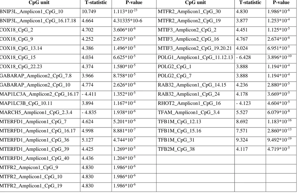
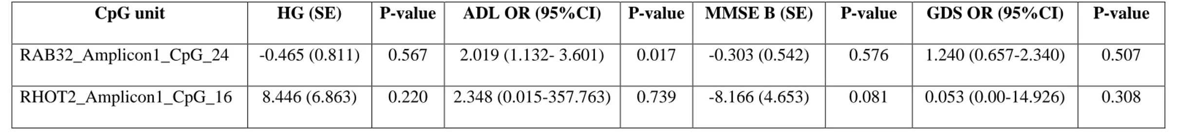
Maintenance of functional mitochondria is essential to prevent damage leading to aging and diseases. What is more, the research of biomarkers of aging is focusing on better predicting functional capability along the lifetime beyond chronological age. Aim of this study was to identify novel CpG sites the methylation of which might be correlated to the chronological and biological age. We performed methylation analyses of the CpG sites in candidate genes involved in mitochondrial biogenesis, mitophagy, fusion, and fission, all key quality control mechanisms to ensure maintenance of healthy mitochondria and homeostasis during aging, using DNA samples from two independent datasets composed by 381 and 468 differently-aged individuals, respectively. Twelve potential CpG predictors resulted associated with aging in the discovery dataset. Of these, two sites located within
RAB32 and RHOT2 genes were replicated in the second dataset. What is more, individuals exhibiting
methylation levels of the RAB32 CpG site higher than 10% were observed more prone to disability than people with lower levels.
These results seem to provide the first evidence that epigenetic modifications of genes involved in mitochondrial quality control occur over time according to the aging decline, and may then represent potential biomarkers of both chronological and biological age.