Università della Calabria
Dipartimento di Farmacia e
Scienze della Salute e della Nutrizione
Dottorato di Ricerca in
MEDICINA TRASLAZIONALE
XXX Ciclo
MED 04/ Patologia Generale
Role ofERa/NOTCH4 axis in sustaining stemness in
breast cancer cells
/
Supervisore: Ch.mo Prof.
Andò
Dottoranda: Dott.ssa Elena Spina
2
Table of Contents
Abstract
Pag.3
Introduction
Pag.4
Materials and Methods
Pag.7
Cell culture Pag.7
Plsmids Pag.7
Mammosphere culture Pag.7
Flow cytometry (FACS) Pag.8
Reverse transcription and real-time reverse transcriptase PCR assays Pag.8
Immunoblot analysis Pag.10
Transmigration assays Pag.10
Transfections assays Pag.10
In vivo experiments Pag.11
Statistical analyses Pag.11
Results
Pag.12
HBD-ESR1 mutations in MCF-7, T47D, ZR75 cell lines Pag.12 Breast cancer stem cells activity (BCSCs) activity is enriched by
Y537S-ERα mutation
Pag.13
BCSCs activity in Y537S-ERα expressing cells is dependent on Notch Signaling
Pag.17
Phosphorylation at Serine 118 residue of ERαis involved in stemness in mutant-expressing cells
Pag.23
Y537N and D538G-ERα mutations influence BCSC phenotype Pag.25 MCF-7/CRISPR-Cas9 knock-in of Y537S-ERα increases tumor
initiation capability of breast cancer cells
Pag.28
Discussion
Pag.31
References
Pag.34
Abstract
Abstract
Early detection and new therapeutic strategies have improved breast cancer patient outcome and survival rates in the last years. However, breast cancer still remains the second leading cause of cancer-related deaths among women worldwide, and approximately 30% of patients eventually experience a tumor relapse.
Treatment failure is mainly due to metastatic process and resistance to conventional therapy. Over the past decade it has been established the existence of a subpopulation of cancer stem cell (CSC) within breast cancers that is responsible for tumor initiation, progression and resistance to endocrine therapies. It is well known the “driving role” of oestrogens and its receptor alpha (ERα), in development and progression of breast cancer disease, but still unknown their role in regulating breast CSCs (BCSCs).
In the past few years, several studies revealed the presence of gain-of-function mutation in ESR1, gene encoding for ERα, in metastatic breast cancer patients after long-term endocrine therapies treatment. Particularly, Y537N, Y537S and D538G are the most frequent “hot spot” mutations within ERα hormone-binding domain (HBD) that lead to ligand-independent ERα activity and consequently, resistance to endocrine therapy. Here, we studied how HBD-ESR1 mutations might account for a mechanism of metastatic process and endocrine resistance, sustaining stem cell-like phenotype.
As experimental model, we used breast cancer cell lines expressing wild-type and HBD-ESR1 mutations. Our results, using in vivo and in vitro experiment (mammosphere-forming assay and CD44+/CD24- phenotype analysis) have suggested an enrichment of BCSCs activity by HBD-ESR1 mutations, that seems to be sustained by Notch4 signaling through constitutive hyper phosphorylation of Serine 118 residue of ERα that has been demonstrated related to stem cell phenotype and tumor initiation, in mutant-expressing cells. Experiments conducted using CRISPR-Cas9 knock-in of Y537S-ERα mutation confirmed the role of this mutation in tumor initiation and progression as obtained using HBD-ESR1 stable clones.
We propose a potential novel role of HBD-ESR1 mutations in sustaining BCSCs activity, that could have clinical relevance, suggesting new molecular biomarker and target to aim better therapeutic strategies for ERα-positive breast cancer metastatic patients.
Introduction
Introduction
Breast cancer is the major endocrine-related cancer in the human population and the second leading cause of cancer death in women after lung cancer [Miller KD, et al. 2016]. It is a heterogeneous disease that includes a variety of tumors with different morphological and clinical features, as a result of complex interactions between different signaling pathways and genetic and epigenetic alterations [Polyak K. 2011]. Although advances in early prevention and treatment, about 30% of all women still experience relapse within 2 to 5 years after treatment and metastatic breast cancer (MBC) remains a largely incurable disease [Siegel RL, et al.2016].
Accumulating evidence has established the presence of a subpopulation of cancer cells with stem-cell like characteristics called Cancer Stem Cells (CSCs) within the tumor. Al-Hajj and colleagues were the first to identify a subpopulation of breast cancer stem cells (BCSCs), defined by a CD44+/CD24-/low phenotype, which was able to generate tumors in immunosuppressed NOD/SCID mice [Al-Hajj M, et al. 2003]. BCSCs are defined by their ability to initiate tumor, to undergo self-renew and to differentiate into the non-self-renewing cells, which constitute the tumor bulk [Visvader JE, et al. 2008] [O’Brien CS, et al.2011]. Evidence suggests that this high tumorigenic cell population contribute critically to tumor recurrence and metastasis after chemo- and endocrine therapy [Li X, et al. 2008] [Creighton CJ, et al. 2009] (Fig. 1).
Figure 1: CSCs and resistance to conventional therapies. Current cancer treatments target
the bulk of tumor cell population but not CSCs. Following therapy, the treatment-resistant CSCs remain and are able to re-populate the tumor.
Introduction
Considerable attention has been directed to target CSCs through evolutionarily pathways that mediate various stem cell properties, such as self-renewal, cell fate decisions, survival, proliferation, and differentiation [O'Brien CA, et al.2010]. Among them, Wnt, Hedgehog and Notch signalings are important CSCs’ regulators, are becoming the focus of development therapeutic strategy [Rizzo P, et al.2008] [Takahashi-Yanaga F, et al.2010] [Merchant A, et al.2010].
Although redundant evidence about the driving role of oestrogens in development of breast cancer, sustaining the growth of breast cancer cells expressing functional oestrogen receptor alpha (ERα) [Hilton HN, et al. 2017], the role of oestrogens in the regulation of BCSCs remains unclear.
Previous studies have demonstrated a direct regulation of normal and cancer stem cell activity by oestrogens [Clarke RB, et al.1997] [Cheng G, et al.2004] [Ma R, et al. 2017]. On the other side, it has been shown that oestrogens can influence BCSCs activity by inducing the secretion of paracrine growth factors from ERα-positive cells through paracrine mechanism [Mallapell S, et al. 2006] [Fillmore CM, et al. 2008] [Harrison H, et al. 2013] [Sun Y, et al. 2014] [Simões BM, et al. 2015].
But regarding the effects of oestrogens on BCSCs activity, contradictory effects have been demonstrated. Simões BM, et al. have shown that oestrogen was able to reduce the self-renewal capacity of MCF-7 BCSCs by promoting differentiation through down-regulation of embryonic stem cell genes NANOG, OCT4, and SOX2 [Simões BM, et al. 2011]. Other studies, have proved that oestrogens induced the expansion of the CD44+/CD24-/low cancer stem cells population by cross-talk with EGFR, FGFR, Gli1 and Notch signaling [Fillmore CM, et al. 2010] [Harrison H, et al. 2013] [ Sun Y, et al. 2014]. Preclinical and clinical investigations have reported loss of ERα in less than 25% of MBCs [Johnston SR, et al. 1997] [Yao ZX, et al. 2014] [Hoefnagel LD, et al. 2012], while the majority of metastatic breast tumors which are no responsive to endocrine therapies, still maintain ERα expression suggesting its role in metastatic disease [Nardone A, et al 2015].
In the last years, several “hot spot” mutations harboring in the residue 536-538 of the Hormone Binding Domain of ERα (HBD-ESR1), Y537N (tyrosine to arginine), Y537S (tyrosine to serine) and D538G (aspartate to glycine), have been detected in a large cohort of metastatic BC patients, and are associated with shorter overall survival (OS) and progression-free survival (PFS) [Spoerke JM, et al. 2016] [Gelsomino L, et al. 2016] [Takeshita T, et al. 2017] [Clatot F, et al. 2017]. These mutations map to the loop
Introduction
connecting α-helices 11 and 12, which play a crucial role in determining interactions with coactivators and corepressors and, therefore, the respective agonist or antagonist effect of the ligand (Fig. 2) [Fanning SW, et al. 2016].
Consequently, HBD-ESR1 mutations have been shown to confer constitutive ligand-independent activation of ERα, suggesting that these naturally-occurring mutations leads to a stabilized agonist state that resists inhibition by anti-oestrogens [Li S, et al. 2013].
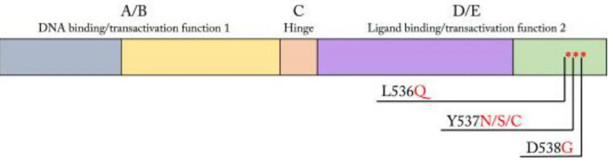
Figure 2. Schematic diagram of ESR1. The ESR1 gene encodes the nuclear receptor protein
ERα, containing domains for DNA binding, transactivation functions 1 and 2, and ligand binding. The most common ESR1 mutations result in variation of the hormone binding domain at residues 536, 537, and 538, modified from Button B, et al. 2016.
It has been already demonstrated that ERα mutations confer an increased phosphorylation of the receptor in the three main serine residues (118, 167, 3 05) [Barone I, et al.2009] [Giordano C, et al.2010], particularly HBD-ESR1 mutations display a constitutive phosphorylation of S118 responsible to modulate the ligand-independent activity of the receptor. These features allow cells to become resistant to adjuvant therapy agents such as tamoxifen, fulvestrant or aromatase inhibitors, conferring enhanced metastatic potential and/or aggressive biological attributes to breast cancer [Toy W, et al.2014, 2017] [Gelsomino L, et al.2016].
Based on these observations the aim of this study was to define the role of HBD-ESR1 mutations in influencing BCSCs activity. Our findings could assess HBD-HBD-ESR1 mutations as novel predictive biomarkers for tumor recurrence and help to discover new potential therapeutic approaches to target CSCs. Genomic screenings of HBD-ESR1 mutations might be warranted to design personalized therapies that will be the most valuable clinical decision to improve BC patient outcome.
Materials and Methods
Materials and Methods
Cell culture
MCF-7, ZR75 and T47D stable clones were generated as previously described [Gelsomino L, et al.2016]. MCF-7 cells were grown in MEM, T47D and ZR75 cells (generously obtained from Dr. Marc Lippman) in RPMI-1640 supplemented with 10% fetal bovine serum (FBS, Invitrogen), 2mmol/L-glutamine, and 1mg/ml penicillin-streptomycin (Life Technologies), 1% Eagle’s none ssential amino acids. All stable clones were maintained in puromycin (1μg/μl). MCF-7/CRISPR-Cas9 knock-in of Y537S-ERα were gently provide by Prof.Suzanne Fuqua (Baylor College of Medicine, Houston, Texas).
Plasmids
The ER constructs were generated using QuickChange Site-Directed Mutagenesis (Stratagene, La Jolla, CA) in pYFP-ERα. The primer sequences were 118A: 5’-GCAGGAAAGGCGCCAGCTGCG-3’. Sequences were verified using Sanger Sequencing. The luciferase reporter plasmid Hes1-Luc (−467 to +46 of the Hes1 promoter with the luciferase gene) and the plasmid encoding dominant-negative MAML-1 (DN-MAML-MAML-1) were gently provided by Prof. Maggiolini (University of Calabria, Italy).
Mammosphere Culture
All stable clones were grown in monolayer and then were disaggregated enzymatically using Trypsin-EDTA(1X) and mechanically using a 25 G needle for four times to obtain a single cell suspension. After, 2000 cells/well were plated in 6 well ultra-low attachment plates provided by Corning in a serum-free DMEM-F12, supplemented with B27, 20ng/ml human epidermal growth factor (EGF), 1mg/ml penicillin-streptomycin. All treatments, XAV-939 (Tocris), Ciclopamine (Calbiochem), DAPT(Sigma), RO4929097(Roche), PD98059 (Calbiochem) were added at the beginning of the experiments. Mammospheres (≥50µm diameter) were counted after 5 days using a microscope fitted with a graticule. Mammosphere forming efficiency (MFE) was calculated as the number of mammospheres formed (≥50μm diameter) divided by the number of cells plated out and expressed as a percentage. Primary mammospheres were
Materials and Methods
collected, enzymatically dissociated, plated at the same seeding density used in the primary generation to obtain secondary mammospheres for 5 days. Mammospheres forming efficiency (MFE) was calculated as number of mammospheres per well/number of cells seeded per well and reported as fold versus control. Mammospheres self-renewal (MSR) was calculated as a proportion of the first generation MFE:
Total first generation mammospheres
Total second generation mammospheres × First generation MFE
Flow Cytometry (FACS)
Mammospheres were dispersed to obtain single-cell suspension. Cells were washed in PBS with 2,5% BSA and stained with FITC anti-human CD44 and PE anti-human CD24 (BD Biosciences,), according to the supplier’s protocol. Flow cytometric analysis was performed on a FACScan and acquisition was performed with WinDI software (Becton Dickinson).
Reverse transcription and real-time reverse transcriptase PCR assays
Total RNA was extracted from cells using TRIzol reagent. Real time PCR was assed using SYBR Green Universal PCR Master Mix (Biorad). Each samples were normalized on its GAPDH mRNA content. Primers used for the amplification of CD44, SMAD4, OCT-4, SOX-2, SOX-4, SOX-9, BMI-1, MAPK6, YES-1, CDH2, VIM, YAP-1, ERRB2, NOTCH1, NOTCH2, NOTCH3, NOTCH4 are listed in Table 1.
GENE Forward Primer 5’ – 3’ Reverse Primer 5’ – 3’
CD44 CCTTTGATGGACCAATTACCATAAC TCAGGATTCGTTCTGTATTCTCCTT
SMAD4 GGAGCTCATCCTAGTAAATG GACGGGCATAGATCACATGA
OCT4 AGCGACTATGCACAACGAGA CCATAGCCTGGGTACCAAA
SOX-2 GCACATGAACGGCTGGAGCAACG TGCTGCGAGTAGGACATGCTGTAGG
Materials and Methods
Table 1. Sequence of primers
The relative gene expression levels were normalized to a calibrator that was chosen to be the WT-ERα expressing cells. Final results were expressed as n-fold differences in gene expression relative to GAPDH rRNA and calibrator, calculated using the DDCt method as follows: nfold = 2_(DCtsample_DCtcalibrator), where DCt values of the sample and calibrator were determined by subtracting the average Ct value of the GAPDH rRNA reference gene from the average Ct value of the different genes analyzed.
GENE Forward Primer 5’ – 3’ Reverse Primer 5’ – 3’
SOX-9 AACGCCGAGCTCAGCAAGA TTCTTGTGCTGCACGCGCA
BMI GTGCTTTGTGGAGGGTACTTCAT TACACGTTTTACAGAAGGAATGTAGAC
MAPK6 GTCGGAGAAGTCCCGTTGTATC GTCGGAGAAGTCCCGTTGTATC
YES-1 CCTCGAGAATCTTTGCGACTAGA CCATTCCATGTTCCCATCCA
CDH2 ACAGTGGCCACCTACAAAGG CCGAGATGGGGTTGATAATG
VIM GAGAACTTTGCCGTTGAAGC GCTTCCTGTAGGTGGCAATC
YAP-1 GCCGGAGCCCAAATCC GCAGAGAAGCTGGAGAGGAATG
ERRB2 CACCTACAACACAGACACGTTTGA GCAGACGAGGGTGCAGGAT
NOTCH1 GTGACTGCTCCCTCAACTTCAAT CTGTCACAGTGGCCGTCACT
NOTCH2 CACCCCAGCTGCTACTCACA GCCAACCCAGCCTGCAT
NOTCH3 CCTGTCTTCCTGGGTTTGAG CAGAACTGGCCTGTGCACTC
NOTCH4 CCAACCCTGCGATAATGCGAG AGTCATCCGTTGAGACCCTGC
Materials and Methods
Immunoblot analysis
Cells were lysate in 500μl of 50 mM Tris-HCl, 150 mM NaCl, 1% NP-40, 0.5% sodium deoxycholate, 2 mM sodium fluoride, 2 mM EDTA, 0.1% SDS, containing a mixture of protease inhibitors (aprotinin, phenylmethylsulfonyl fluoride, and sodium orthovanadate) for total protein extraction. Equal amounts of proteins were resolved on 10% SDS-polyacrylamide gel, transferred to a nitrocellulose membrane and probed with specific antibodies as NOTCH4(Novus Biologicals), JAG1(Santa Cruez), HES1(Novus Biologicals), DLL1(Abcam), DLL3(Abcam), pERα serine 118 (Cell Signaling) and ERα (Santa Cruz). To ensure equal loading, all membranes were stripped and incubated with anti-GAPDH (Santa Cruz) antibody. The antigen-antibody complex was detected by incubation of the membranes with peroxidase-coupled goat anti-mouse or goat anti-rabbit antibodies and revealed using the ECL System. Blots are representative of three independent experiments.
Transmigration Assays
Mammospheres derived MCF-7 WT-ERα and Y537S-ERα cells were placed in the upper compartments of Boyden Chamber (8μm membranes, Corning). Regular media was used as chemoattractant and after 24h invaded cells were fixed with 4% paraformaldehyde and stained with DAPI (Sigma). Migration was quantified by viewing five separate fields/membrane (10X magnification) and expressed as mean numbers of migrated cells. Data represent three independent experiments, assayed in triplicate.
Transfection assays
For luciferase assays, cells were plated into 24-well plates with 500 μl of regular growth medium/well the day before transfection. Transfection was performed using lipofectamine reagent (Invitrogen) as recommended by the manufacturer, in a mixture containing 0.5 μg of reporter plasmid and TK Renilla luciferase plasmid (25 ng/well) used to normalize the efficiency of the transfection. Firefly and Renilla luciferase activities were measured using a Dual Luciferase kit (Promega, Milan, Italy) according to the manufacturer's recommendations. The normalized relative light unit values obtained from MCF-7 WT-ERα cells were set as one-fold induction upon which the activity induced was calculated. MCF-7 WT-ERα and Y537N, Y537S, D538G-ERα were cultured in regular media and transfected with an empty vector or a plasmid encoding for the
Materials and Methods
dominant-negative of MAML-1 (DN-MAML-1) or for S118A-ERα using lipofectamine reagent. After 24h cells were plated as mammospheres culture to evaluate MFE.
In vivo experiments
To evaluate tumor initiation capacity of MCF-7 WT-ERα and CRISPR/CAS-9 Y537S-ERα cells, NOD SCID GAMMA (NSG) mice, the model of choice for cancer xenograft and stem cell biology studies, were injected subcutaneously with cells in mammosphere media mixed 1:1 with Matrigel. 60-days slow release estrogen pellets will be implanted sub-cutaneously into mice 2 days before cell injection. Serial limiting dilution implantation of cells (10000; 1000; 100;10) will be used to perform calculation of tumor initiation analysis. After 60 days will be assessed the 95% Confidence Intervals for the frequency of active cells employing Extreme Limiting Dilution Analysis (ELDA) software [Simões BM, et al.2015] [Hu Y, et al.2009].
Statistical analyses
Each datum point represents the mean±s.d. of three different experiments. Data were analyzed by Student’s t test using the GraphPad Prism 7 software.
Results
Results
HBD-ESR1 mutations in MCF-7, T47D, ZR75 cell lines
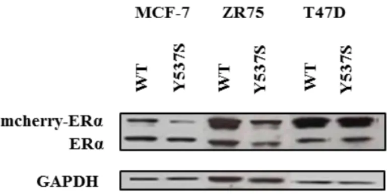
Several point mutations in the hormone binding domain (HBD) of ESR1 have been identified in metastatic tumor samples from patients with ERα-positive breast cancer after treatment with anti-oestrogen therapy. These mutations are rare in primary untreated tumors [Robinson DR, et al. 2013] [Toy W, et al. 2013]. Previously, we generated ERα-positive breast cancer cell lines, MCF-7, T47D and ZR75, stably infected with wild-type (WT) ESR1 or the most frequent HBD-ESR1 mutant Y537N, Y537S or D538G (m-cherry-tagged exogenous receptor, 95kDa) [Gelsomino L, et al. 2016]. These stable clones co-express endogenous and exogenous ERα receptor to better represent the clinical situation were wild-type receptor is often co-expressed with the mutant one. It has been demonstrated using in vitro and in vivo assay, that Y537S compared to D538G mutants, were more oestrogen-independent in term of proliferation, and were not fully inhibited by fulvestrant despite dosing to higher levels than are typically achieved in the clinic [Toy W, et al. 2017]. Thus, as experimental model we decided to use MCF-7, T47D and ZR75 expressing Y537S-ERα (Fig. 3).
Figure 3. MCF-7, ZR75 and T47D stable infected with WT-ERα and Y537S-ERα. Western
Blot analysis of breast cancer cell lines stable infected with WT-ERα and Y537S-ERα. GAPDH, was used as loading control.
Results
Breast Cancer Stem Cells (BCSCs) activity is enriched by Y537S-ERα
mutation
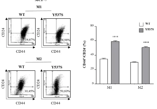
To determine the role of HBD-ESR1 mutations in influencing breast cancer stem cell (BCSC) activity, MCF-7 stable pools expressing wild-type (WT) ERα and the most frequently occurring clinical ESR1 mutation Y537S-ERα were grown as mammospheres [Dontu G et al. 2003] [Shaw FL, et al. 2012]. Thus, we performed fluorescence-activated cell sorting (FACS) analysis to evaluate BCSC subpopulation in relation to CD44 and CD24 expression. As expected we evidenced an enrichment of CD44+/CD24 -subpopulation in mammosphere derived-cells from wild-type and mutated ERα cells lines compared to the cells grown in adherent conditions (data not shown). Interestingly, MCF-7 Y53MCF-7S-ERα mammosphere-derived cells showed a significant higher percentage of CD44+/CD24- cells compared to WT-ERα mammosphere-derived cells both in primary (M1, 58.98% in Y537S-ERα cells versus 34.67% in WT-ERα cells) and in secondary generation (M2, 50.60% in Y537S-ERα cells versus 30.45% in WT-ERα cells) as reported in Fig. 4.
Figure 4. CD44+/CD24- phenotype in MCF-7 Y537S-ERα mammosphere-derived cells.
MCF-7 WT-ERα and Y537S-ERα cells were grown in non-adherent conditions (M1, first generation of mammospheres; M2 second generation of mammospheres) and subjected to FACS analysis to evaluate CD24 and CD44 expression. Representative plots of FACS analysis (left panel). Percentage of CD44+/CD24- subpopulation (right panel). The values represent the means ± s.d. of three different experiments each performed in triplicate; ****p ≤ 0.0001.
Results
Accordingly, mutant-expressing cells exhibited increased mRNA level of several genes strongly associated with stem cell phenotype, such as CD44, SMAD4, OCT4, SOX2,
SOX4, SOX9, BMI1, MAPK6, YAP1, CDH2, VIM, YES-1 and ERBB2 (Fig. 5).
Figure 5. Stem-related gene expression in MCF-7 Y537S-ERα mammosphere-derived cells.
Total RNA was isolated from M1 MCF-7 WT-ERα and Y537S-ERα and converted to cDNA. Real Time RT-PCR was performed for a subset of genes in M1 MCF-7 WT-ERα and Y537S-ERα cells. Each samples was normalized to its GAPDH mRNA content. The values represent the means ± s.d. of three different experiments each performed in triplicate; *p ≤0.05; **p ≤0.01; ***p ≤ 0.001; ****p ≤ 0.0001.
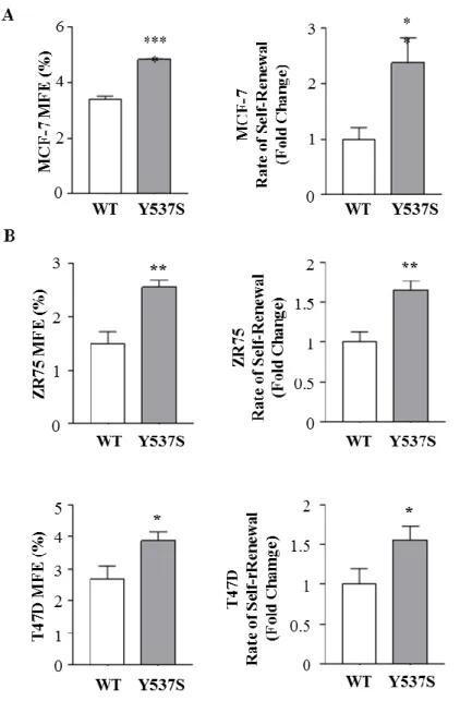
We found an increase in mammosphere-forming efficiency (MFE) in mutant expressing cell lines compared to wild-type (Fig. 6A left panel). Moreover, we also evaluated the ability to form secondary mammospheres (renewal, SR) observing a higher self-renewal capacity in expressing Y537S-ERα breast cancer cell lines (Fig. 6A right panel). To extend our results we used also ZR75 and T47D stably expressing both WT-ERα and Y537S-ERα. We observed in both cell lines that Y537S-ERα mammosphere-derived cells showed higher MFE (Fig. 6 B left panel) as well as an increased SR (Fig. 6 B right panel) compared to WT-ERα demonstrating that Y537S-ERα mutation affected BCSC activity independently of cellular background.
Results
Figure 6. Effects of HBD Y537S-ERα mutation on MFE and SR in breast cancer cells. A) Mammospheres Forming Efficiency (MFE) (left panel) and self-renewal (right panel) of
MCF-7 WT-ERα and Y537S-ERα clones. B) Mammospheres Forming Efficiency (MFE) (left panel) and self-renewal (right panel) of ZR75, T47D WT-ERα and Y537S-ERα clones. The values represent the means ± s.d. of three different experiments each performed in triplicate; *p ≤0.05; **p ≤0.01; ****p ≤ 0.0001.
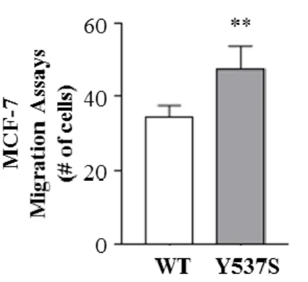
Since migratory potential is one of the most important features for BCSCs, we performed boyden chambers transmigration assays in MCF-7 WT-ERα and Y537S-ERα mammosphere-derived cells. The results clearly demonstrated that the expression of the mutation resulted in an augmented cell migration (Fig. 7).
Results
16
Figure 7. Migratory potential in Y537S-ERα MCF-7. Transmigration assays of M1 MCF-7
WT-ERα and Y537S-ERα. The values represent the means ± s.d. of three different experiments each performed in triplicate; **p ≤0.01;
Collectively these data demonstrated that Y537S-ERα mutation promotes stem cells activity in breast cancer cells.
Results
17
BCSCs activity in Y537S-ERα expressing cells is dependent on Notch
Signaling
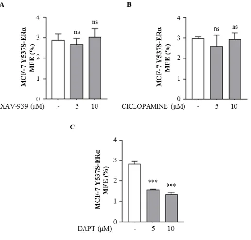
Wnt/β-catenin, Sonic Hedgehog and Notch signaling, the main stem cell regulatory pathways, are frequently deregulated in a number of malignancies, including breast cancer [Koury J, et al. 2017]. Thus, in order to explore which of these pathways may sustain BCSCs activity in mutant-expressing cells, we tested the effects of specific inhibitors of Wnt/β-catenin (XAV-939), Sonic Hedgehog (Ciclopamine) and Notch (N-[N-(3,5-Difluorophenacetyl)-L-alanyl]-S-phenylglycine t-butyl ester, DAPT) pathways on MFE. We observed that XAV-939 and Ciclopamine did not affect MFE (Fig. 8A and B) while DAPT significantly decreased the number of mammospheres in mutant-expressing cells (Fig. 8C).
Figure 8. MFE of MCF-7 Y537S-ERα in the presence of Wnt/β-catenin, Sonic Hedgehog and Notch inhibitors. MFE of MCF-7 WT-ERα and Y537S-ERα treated with XAV-939 (5μM
and 10μM, A), Ciclopamine (5μM and 10μM, B), DAPT (5μM and 10μM, C) The values represent the means ± s.d. of three different experiments each performed in triplicate; ns, not significant, ***p ≤ 0.001.
Results
18
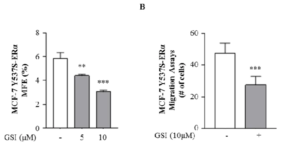
To further confirm these data, we used a different Notch inhibitor RO4929097 (GSI), a potent and selective small molecule inhibitor of gamma-secretase that is currently in phase 1 study in patients with solid tumors [NCT Number: NCT01131234]. Notch inhibition can be achieved inhibiting the formation of NICD, responsible of Notch activity, using gamma-secretase inhibitor to prevent the final cleavage step of the precursor form of Notch [Olsauskas-Kuprys R et al. 2013]. Remarkably, GSI also decreased MFE (Fig. 9A) and strongly reduced the migratory activity in mutant-expressing cells (Fig. 9B).
Figure 9. GSI inhibitor effects in MCF-7 Y537S-ERα. A) MFE of MCF-7 WT-ERα and
Y537S-ERα treated with RO4929097 (GSI, 5μM and 10μM). B) Transmigration assays of M1 MCF-7 WT-ERα and Y537S-ERα grown in non-adherent conditions, in the presence or absence (-) of GSI 10μM. The values represent the means ± s.d. of three different experiments each performed in triplicate; **p ≤0.01; ***p ≤ 0.001.
The same inhibitory effects of GSI on MFE have been also detected in ZR75 Y537S-ERα stable clones (Fig.10), confirming a positive interaction between Notch signaling and Y537S-ERα mutation. These results demonstrated that the blockade of Notch pathway selectively reduced the stemness potential of breast cancer cells displaying Y537S-ERα mutation.
Results
19
Figure 10. MFE of ZR75 WT-ERα and Y537S-ERα treated with GSI. ZR75 WT-ERα and
Y537S-ERα were grown as mammospheres in non-adherent conditions, in the presence or absence (-) of GSI 10μM. The values represent the means ± s.d. of three different experiments each performed in triplicate; *p ≤0.05; ***p ≤ 0.001.
Notch signaling plays a key role in cell biology (stem cell maintenance, cell differentiation, cellular homeostasis), angiogenesis, tumor formation and cell fate decision [Guo S, et al.2011] [Brzozowa-Zasada M, et al.2016] [Lamy M, et al.2017]. Its activation derived by contact between signal-sending cell expressing Notch ligand and signal-receiving cell expressing Notch receptor. In mammals have been identified four Notch transmembrane proteins receptors (NOTCH1, 2, 3 and 4) that can bind five ligands (DLL-1, 3 and 4 and JAG-1 and 2). After the binding, Notch receptor undergoes to proteolytic cleavage by ADAM-family protease (S2 cleavage) and then by presenilin-γ-secretase complex (S3 cleavage) that induces a release of the Notch intracellular domain (NICD). NICD can translocate to the nucleus and interact with RBP-Jk/CBF-1 transcription factor complex mediating the transcription of target genes which are transcriptional repressor of the HES (Hairy and Enhancer Of Split) (HES1-7) and HEY (Hairy/Enhancer-Of-Split Related With YRPW Motif Protein) subfamilies (HEY1, HEY2, HEYL) [Iso T, et al. 2003] [Miele L. 2006].
Thus, to better explore the role of Notch pathway in driving MCF-7 Y537S-ERα stem cell activity, we first assessed mRNA level of Notch1, Notch2, Notch3 and Notch4 receptors by real time RT-PCR. Our data showed a higher expression of these genes in mutant expressing-cells compared to WT-ERα cells (Fig. 10A, left panel). Several studies reported Notch4 as a crucial player in BCSCs activity and its elevated expression significantly correlated with poor patient survival among BC patients [Simões BM, et
Results
20
al.2015] [Lombardo Y, et al.2014]. We found using immunoblot analysis in mutant-expressing cells that Notch4 and its intracellular domain (Notch4ICD) were up-regulated along with its ligands, JAG1, DLL1, DLL3 and its primary target gene HES1 (Fig. 10A right panel). The increased mRNA levels of Notch receptor isoforms, concomitantly with the activation of Notch4 signaling pathway have been also revealed in ZR75 Y537S-ERα stable clones as reported in Fig. 10B.
Figure 10. Notch expression and signaling in Y537S-ERα MCF-7 and ZR75. Real Time
RT-PCR for NOTCH1, NOTCH2, NOTCH3, NOTCH4 receptors in M1 MCF-7 (A) and ZR75 (B) WT-ERα and Y537S-ERα clones (left panel). NOTCH4 full length, NOTCH4ICD, JAG1, DLL1, DLL3 and HES1 protein expression levels revealed by immunoblot analysis of total cellular extracts of M1 MCF-7 WT-ERα and Y537S-ERα clones (right panel). GAPDH, was used as loading control. Numbers represent the average fold change versus MCF-7 WT-ERα normalized
Results
21
for GAPDH. The values represent the means ± s.d. of three different experiments each performed in triplicate; ns, not significant *p≤0.05; **p≤0.01; ***p ≤ 0.001; ****p ≤ 0.0001.
Since HES1 is one of the main Notch target genes and it is considered as a read-out of NOTCH activation [Borggrefe T, et al. 2009], we also evaluated the HES1 transcriptional activity by HES1 Luciferase reporter assay. As shown in Fig. 11 HES1 transcriptional activity resulted higher in Y537S-ERα clones compared to WT-ERα expressing cell, both transfected with HES1 promoter, confirming the Notch pathway activation.
Figure 11. HES1 transcriptional activity in MCF-7 Y537S-ERα. HES1-luciferase reporter
assay in MCF-7 WT-ERα and Y537S-ERα. The values represent the means ± s.d. of three different experiments each performed in triplicate; ***p ≤ 0.001.
Additionally, to further sustain a possible role of Notch pathway in mutant-expressing cells we evaluated MFE in MCF-7 clones transfected with a dominant-negative of Notch transcriptional coactivator, Master-mind like 1 (DN-MAML-1) [Wu L, et al. 2002][Pupo M, et al.2014]. The presence of DN-MAML1 affected MFE only in mutant expressing cells, while no changes have been found in WT-ERα stable clones (Fig. 12).
Results
22
Figure 12. MFE is reduced in the presence of Notch dominant-negative coactivator. MFE in
MCF-7 WT-ERα and Y537S-ERα cells transiently transfected with either an empty vector (-) or a dominant negative for MAML-1 (DN-MAML-1). The values represent the means ± s.d. of three different experiments each performed in triplicate; ns, not significant, ***p < 0.001.
These data demonstrated that Notch pathway might sustain BCSC activity in mutant-expressing cells.
Results
23
Phosphorylation at serine 118 residue of ERα is involved in stemness in
mutant-expressing cells
It has been reported that HBD-ESR1 mutant-expressing cells showed high ERα serine 118 (S118) phosphorylation level and consequently a constitutive transactivation of the receptor [Toy W, et al.2014]. In addition, it has been demonstrated that increased levels of S118 are linked to the tumor initiating ability of SOX2-overexpressing cells [Vazquez-Martin A, et al. 2013]. Thus, we investigated whether phosphorylation at S118 might be potentially involved in stemness activity of mutant-expressing cells. First, we performed immunoblot analysis to evaluate phosphorylation levels of ERα at S118 in mammospheres derived from WT-ERα and mutant-expressing cells. Our data demonstrated that mutant expressing cells showed higher phosphorylation level of S118 compared to WT-ERα cells (Fig. 13A). Since serine residue 118 is one of the main effectors of Mitogen Activated Protein Kinase (MAPK), to confirm also the involvement of this pathway in sustain stemness in mutant-expressing cells we performed immunoblot analysis [Catalano S, et al.2004]. We discovered a hyper phosphorylation of MAPK in mammospheres derived from mutant-expressing cells (Fig. 13B).
Figure 13. Hyper phosphorylation at S118 of ERα in MCF-7 Y537S-ERα. A) Phosphorylation
levels of serine residue 118 (pERα Ser118) and total level of ERα protein expression revealed by immunoblot analysis of total cellular extracts of M1 MCF-7 WT-ERα and Y537S-ERα clones.
B) Phosphorylation (pMAPKThr202/Tyr204) and total level of MAPK protein expression revealed by immunoblot analysis of total cellular extracts of M1 MCF-7 WT-ERα and Y537S-ERα clones. GAPDH, was used as loading control.
Results
24
To better elucidate the role of S118, in our experimental model, we transfected S118A-ERα construct, a plasmid containing a serine to alanine substitution at residue 118. In the presence of S118A ERα we observed in Y537S-ERα cells a reduction in phosphorylation level of S118 and concomitantly decreased expression of Notch4 and Notch4ICD, JAG-1 and HES-1 (Fig. 14A) suggesting a possible crosstalk existing between ERα and Notch4 in supporting stemness in mutant clones. Indeed, cells transfected with S118A-ERα showed a significant reduction in MFE in mutant-expressing cells (Fig. 14B).
Figure 14. Serine Residue 118 in Y537S-ERα modulates stemness activity. A) MCF-7
WT-ERα and Y537S-WT-ERα cells were transiently transfected with either an empty vector (-) or S118A-ERα plasmid (S118A-S118A-ERα). NOTCH4 full length, NOTCH4ICD, HES1, pERα Ser118 and ERα
protein expression levels revealed by immunoblot analysis of total cellular extracts of MCF-7 WT-ERα and Y537S-ERα cells. GAPDH, was used as loading control. Numbers represent the average fold change versus MCF-7 ERα normalized for GAPDH. B) MFE in MCF-7 WT-ERα and Y537S-WT-ERα cells transiently transfected with either an empty vector (-) or S118A-WT-ERα plasmid (S118A-ERα). The values represent the means ± s.d. of three different experiments each performed in triplicate; ns, not significant, ***p < 0.001.
All these results demonstrated that the hyper phosphorylation of S118 is required for Notch4 activation in maintaining stem cell phenotype in mutant-expressing cells.
Results
25
Y537N and D538G-ERα mutations influence BCSCs phenotype
In order to extend our knowledge of HBD-ESR1 mutations in regulating stemness, we investigated the impact of other two more frequent mutations, named Y537N- and D538G-ERα in BCSCs activity. Thus, we used as experimental models other two different previously established MCF-7 stable clones expressing Y537N-ERα and D538G-ERα mutations respectively [Gelsomino L, et al.2016] (Fig.15).
Figure 15. MCF-7 stable clones expressing Y537N and D538G-ERα. Western Blot analysis
of MCF-7 stable infected with WT, Y537N and D538G-ERα. GAPDH, was used as loading control.
In agreement with the results obtained in Y537S-ERα mutant-expressing cells, we observed in both clones an increased MFE as well as SR compared to WT-ERα expressing cells (Fig. 16A and B).
Figure 16. MFE and SR in Y537N and D538G-ERα MCF-7 cells. A) MFE of MCF-7
Results
26
The values represent the means ± s.d. of three different experiments each performed in triplicate; p*≤0.05; **≤0.01.
Moreover, these mutant-expressing cells displayed higher levels of S118 phosphorylation and increased Notch activation compared to WT-ERα expressing cells (Fig. 17A) and consequently a reduced MFE either in the presence of S118A-ERα plasmid or by using the Notch inhibitor, GSI (Fig. 17B and C).
Figure 17. Phosphorylation at S118 in Y537N-ERα and D538G-ERα sustains stemness. A)
Phosphorylation levels pERα 118, total ERα, NOTCH4 full length and NOTCH4ICD protein
expression levels revealed by immunoblot analysis of total cellular extracts of M1 MCF-7 WT-ERα and Y537N-WT-ERα and D538G-WT-ERα clones. GAPDH, was used as loading control. Numbers represent the average fold change versus MCF-7 WT-ERα normalized for GAPDH. B) MFE of MCF-7 WT-ERα, Y537N-ERα and D538G-ERα clones transiently transfected with either an
Results
27
empty vector (-) or S118A-ERα or treated with GSI 10μM. The values represent the means ± s.d. of three different experiments each performed in triplicate; ns, not significant **p≤0.01; ***p ≤ 0.001.
All together our data highlight a crucial role for both S118 and Notch4 pathway in influencing BCSCs activity in the more frequent mutations harboring in the HBD of ERα.
Results
28
MCF-7/CRISPR-Cas9 knock-in of Y537S-ERα increases tumor initiation capability of breast cancer cells
To further confirm the role of Y537S-ERα mutation in tumor initiation and progression, we generated Y537S-ERα knock-in MCF-7 using CRISPR-Cas9 genome editing technique.
CRISPR-Cas9-mediated genomically introduction of Y537S mutation in the encoded
ESR1 gene, would facilitate direct comparison of isogenic wild-type and mutant breast
cancer cells. As expected, MCF-7/CRISPR-Cas9 knock-in of Y537S-ERα cells showed a higher MFE and SR compared to parental MCF-7 cells (Fig.19A and B) and treatment with GSI reduced MFE confirming the involvement of Notch signaling (Fig.19C).
Figure 19. MFE and SR in CRISPR/CAS-9 knock-in Y537S-ERα MCF-7 cells. A) MFE of
parental MCF-7 and MCF-7/CRISPR-Cas9 knock-in of Y537S-ERα. B) Self Renewal (SR) of parental 7 and 7/CRISPR-Cas9 knock-in of Y537S-ERα. C) MFE of parental
MCF-Results
29
7 and MCF-7/CRISPR-Cas9 knock-in of Y537S-ERα treated with GSI 10μM. The values represent the means ± s.d. of three different experiments each performed in triplicate; *p≤0.05; **≤0.01; ****p ≤ 0.0001.
One major hallmark of cancer stem cells is the ability to initiate tumors “in vivo”. To determine the frequencies of CSC in Parental and MCF-7/CRISPR-Cas9 knock-in of Y537S-ERα cells “in vivo”, we performed limiting-dilution xenograft experiments. Four groups with four mice each were formed for each cell line. We implanted progressively smaller number of tumor cells (10,100,1000,10000 cells) into NSG mice for each group, and they were followed over a period of 60 days. Extreme Limiting Dilution Analysis (ELDA) revealed a 5-fold enrichment in tumor initiating cell frequency in Y537S-1 compared to MCF-7 Parental cells. Particularly, the estimated frequency of CSC in in MCF7-Parental was 1 in 1335 cells, whereas that in Y537S-1 was 1 in 253 (P = 0.00273) (Table 2).
All together these data strongly demonstrated an important role of the Y537S-ERα mutation in supporting BCSC activity displaying a potential role in sustaining tumor initiation and metastatic disease progression.
Results
30
Table 2. Number of cell injected, numbers of mice and those positive for tumor growth are shown
for MCF-7 parental and YS-1. Tumor-initiating cell (TIC) frequency and 95% confidence intervals (CI) demonstrating the highly significant difference in TICs using Chi-sqd to test in Parental and YS-1 cells.
Discussion
31
Discussion
Oestrogen receptor alpha (ERα)-positive tumors represent the most common form of breast cancer (BC) (~70% in newly diagnosed breast cancers) [Ariazi EA, et al. 2006]. ERα and its cognate ligand oestrogen are considered the “driving forces” in development and progression of BC disease. Therapeutic targeting of oestrogen signaling and biosynthesis is the mainstay therapy used in both early and metastatic breast cancers. However, patients after benefit initial endocrine therapy, may experience tumor recurrence. In the last years, understanding the mechanisms involved in metastatic process and resistance to conventional therapy, has become the prior aim, to prevent the development of secondary tumors and to improve the clinical outcome of these patients. In recent years a better knowledge of tumor heterogeneity has led to the discovery of small subpopulation within the tumor, characterized by high tumorigenic ability and by stem cell-like properties of self-renewal, differentiation and development of (malignant) tissue. This has permitted to define tumor initiating cells referred as “cancer stem cell (CSC)”. CSCs have been shown to contribute directly or indirectly to the generation of metastasis since their highly-resistant feature to conventional cancer therapies. So far, interest in targeting cancer stemness is becoming priority.
Research attention has been focused also on recurrent hormone-binding domain (HBD)-ESR1 mutations as mechanism enrolled in metastatic disease and therapy resistance [Robinson DR, et al.2013] [Jeselsohn R, et al.2017]. HBD-ESR1 mutations were found in metastatic breast cancer patients. Some studies sustained that these mutations evolved under the pressure of endocrine treatment in metastatic breast cancer patients who received long-term anti-oestrogen therapies, and they are rarely found in treatment-naïve ERα-positive breast cancers others described the presence of HBD-ESR1 mutations in primary tumors proposing that cells might acquire these genomic alterations or that those arise from a rare single clone [Angus L, et al.2016] [Gelsomino L, et al. 2016].
Since HBD-ESR1 mutations and CSCs are pioneering factors in metastatic disease-progression, it could be important to provide a therapeutic strategy that can strongly block their action. Therefore, based on these observations we questioned whether HBD-ESR1 mutations might account for sustain stem cell phenotype.
Discussion
32
Firstly, we used as experimental model breast cancer cell lines, MCF-7, ZR75 and T47D, expressing Y537S-ERα, the most prevalent ESR1 point mutation that has been found in metastatic breast cancer.
We demonstrated that cells expressing Y537S-ERα mutation showed higher BCSCs activity comparing to WT-ERα expressing cells in term of mammosphere forming efficiency (MFE), self-renewal (SR) and migratory potential along with increased mRNA level of the genes mostly up-regulated in stemness.
Hedgehog, Notch and Wnt pathways play a key role in normal mammary gland biology (stem-cell maintenance, development, proliferation) and cell fate, and dysregulation of these pathways could be responsible for breast cancers [Rizzo P, et al.2008] [Takahashi-Yanaga F, et al.2010] [Merchant A, et al.2010]. Thus, we investigated which of these pathways was correlated with BCSCs activity in mutant-expressing cells. Our results demonstrated a pivotal role of Notch4 signaling pathway to maintain stem cell phenotype in Y537S-ERα expressing cells. MFE of mutant-expressing cells was exclusively down-regulated by two different Notch inhibitors, DAPT and GSI.
De-regulation of the Notch pathway is reported in the pathogenesis of breast cancer in mice and humans [Gallahan D, et al.1987] [Dievart A, et al.1999] [Farnie G, et al.2007] and has been linked to treatment resistance [Lombardo Y, et al.2010] [Magnani L, et al.2014] [Simoes BM, et al.2015]. Particularly, Notch4 signaling has also been implicated in the regulation of BCSCs [Harrison H, et al. 2010] [Farnie G, et al.2007]. Our results revealed hyper-activation of Notch4 pathway in Y537S-ERα expressing cells.
The role of ERα in supporting breast cancer growth has been fully elucidated, but its involvement in BCSC activity is still controversial. Researchers usually described stem cell population as ERα-negative cells, but some studies revealed that ERα signaling activation could directly or in a paracrine manner sustains stem cell compartments [Fillimore CM, et al.2010]. Moreover, Vazquez-Martin group demonstrated the impact of non-genomic oestrogen-signaling activation in stemness validating the hypothesis that increased phosphorylation at serine 118 enhanced tumor initiation ability in SOX2-overexpressing cells [Vazquez-Martin A, et al.2013]. It has been reported that HBD-ESR1 mutations are characterized by hyper activation of the receptor and showed a constitutively phosphorylation of serine 118 [Toy W, et al.2017] [Harrod A, et al.2016]. Thus, here we demonstrated that BCSCs retain ERα expression and the constitutive phosphorylation in serine 118 that resulted necessary to sustain Notch activation in maintaining stemness in mutant-expressing cells.
Discussion
33
In order to extend our knowledge of HBD-ESR1 mutations in regulating stemness, we investigated the impact of other two more frequent mutations, named Y537N- and D538G-ERα in BCSC activity. We demonstrated that Y537N and D538G influence stemness potential, in fact cells expressing these mutations, exhibited hyper phosphorylation of serine 118, concomitantly with activation of Notch4 signaling pathway which is responsible to maintain higher MFE and SR in mutant-expressing cells. Finally, to further confirm the role of Y537S-ERα mutation in tumor initiation and progression, we generated Y537S-ERα knock-in MCF-7 using CRISPR-Cas9 genome editing technique. As expected, MCF-7/CRISPR-Cas9 knock-in of Y537S-ERα cells showed a higher MFE and SR compared to parental MCF-7 cells, dependent by Notch signaling. Since, hallmark of CSCs is the ability to initiate tumors in vivo, we demonstrated that Y537S-ERα mutation displayed a potential role in sustaining tumor initiation, using tumor dilution “in vivo” assay. Frequency of CSCs in MCF-7 Parental was 1 in 1335 cells, whereas Y537S-1 was 1 in 253, suggesting enrichment in tumor-initiation ability.
In conclusion, we suggested for the first time that HBD-ESR1 mutations might account for a mechanism of metastatic process and endocrine resistance, sustaining stem cell-like phenotype. Constitutive hyper phosphorylation of ERα Ser118, induced by HBD-ESR1 mutations, is the mainstay of Notch4 activation, which maintained stemness.
Our discoveries might help to define the role of HBD-ESR1 mutations, and reassess them as a predictive and prognosis marker. Our work consistently will impact breast cancer outcome, in fact here we propose Notch pathway as a valuable therapeutic target in patients affected by BCs that exhibit HBD-ESR1 mutations in order to eradicate BCSCs that are refractory to anti-estrogens with the aim to prevent tumor progression and metastatic disease.
References
34
References
Al-Hajj M, Wicha MS, Benito-Hernandez A, Morrison SJ, Clarke MF. Prospective identification of tumorigenic breast cancer cells. Proceedings of the National Academy of Sciences of the United States of America. (2003) 100 (7):3983–3988.
Angus L, Beije N, Jager A, Martens JW, Sleijfer S. ESR1 mutations: Moving towards guiding treatment decision-making in metastatic breast cancer patients. Cancer Treat Rev. 2017 Jan; 52:33-40.
Ariazi EA, Ariazi JL, Cordera F, Jordan VC. Estrogen receptors as therapeutic targets in breast cancer. Curr Top Med Chem. 2006;6(3):181-202.
Barone I, Cui Y, Herynk MH, Corona-Rodriguez A, Giordano C, Selever J, Beyer A, Andò S, Fuqua SA. Expression of the K303R estrogen receptor-alpha breast cancer mutation induces resistance to an aromatase inhibitor via addiction to the PI3K/Akt kinase pathway. Cancer Res. 2009 Jun 1;69(11):4724-32.
Belandia B, Powell SM, García-Pedrero JM, Walker MM, Bevan CL, Parker MG. Hey1, a mediator of notch signaling, is an androgen receptor corepressor. Mol Cell Biol. 2005 Feb;25(4):1425-36.
Bhuvanalakshmi G, Basappa, Rangappa KS, Dharmarajan A, Sethi G, Kumar AP, Warrier S.Breast Cancer Stem-Like Cells Are Inhibited by Diosgenin, a Steroidal Saponin, by the Attenuation of the Wnt β-Catenin Signaling via the Wnt Antagonist Secreted Frizzled Related Protein-4. Front Pharmacol. 2017 Mar 20; 8:124.
Borggrefe T, Oswald F. The Notch signaling pathway: transcriptional regulation at Notch target genes. Cell Mol Life Sci. 2009 May;66(10):1631-46.
Brzozowa-Zasada M, Piecuch A, Dittfeld A, Mielańczyk Ł, Michalski M, Wyrobiec G, Harabin-Słowińska M, Kurek J, Wojnicz R. Notch signalling pathway as an oncogenic factor involved in cancer development. Contemp Oncol (Pozn). 2016;20(4):267-72.
Button B, Ho Park B. ESR1 mutations: Pièce de résistance. Genes & Diseases Volume 3, Issue 2, June 2016, Pages 124-129.
Catalano S, Mauro L, Marsico S, Giordano C, Rizza P, Rago V, Montanaro D, Maggiolini M, Panno ML, Andó S.Leptin induces, via ERK1/ERK2 signal, functional activation of estrogen receptor alpha in MCF-7 cells. J Biol Chem. 2004 May 7;279(19):19908-15.
Cheng G, Weihua Z, Warner M, Gustafsson JA. Estrogen receptors ER alpha and ER beta in proliferation in the rodent mammary gland. Proc Natl Acad Sci U S A 2004, 101:3739–3746. Clarke RB, Howell A, Potten CS, Anderson E. Dissociation between steroid receptor expression and cell proliferation in the human breast. Cancer Res. 1997 Nov 15;57(22):4987-91.
Clatot F, Perdrix A, Sefrioui D, Sarafan-Vasseur N, Di Fiore F. Clinical relevance of ESR1 circulating mutations detection in hormone receptor positive metastatic breast cancer. Bull Cancer. 2017 Oct 9. pii: S0007-4551(17)30234-5.
References
35
Creighton CJ, Li X, Landis M, Dixon JM, Neumeister VM, Sjolund A, Rimm DL, Wong H, Rodriguez A, Herschkowitz JI, Fan C, Zhang X, He X, Pavlick A, Gutierrez MC, Renshaw L, Larionov AA, Faratian D, Hilsenbeck SG, Perou CM, Lewis MT, Rosen JM, Chang JC. Residual breast cancers after conventional therapy display mesenchymal as well as tumor-initiating features. Proc Natl Acad Sci U S A. 2009 Aug 18;106(33):13820-5.
Diévart A, Beaulieu N, Jolicoeur P. Involvement of Notch1 in the development of mouse mammary tumors. Oncogene. 1999 Oct 28;18(44):5973-81.
Dontu G, Jackson KW, McNicholas E, Kawamura MJ, Abdallah WM, Wicha MS. Role of Notch signaling in cell-fate determination of human mammary stem/progenitor cells. Breast Cancer Res. 2004;6(6): R605-15.
Fanning SW, Mayne CG, Dharmarajan V, Carlson KE, Martin TA, Novick SJ, Toy W, Green B, Panchamukhi S, Katzenellenbogen BS, Tajkhorshid E, Griffin PR, Shen Y, Chandarlapaty S, Katzenellenbogen J, Greene GL. Estrogen receptor alpha somatic mutations Y537S and D538G confer breast cancer endocrine resistance by stabilizing the activating function-2 binding conformation. Elife. 2016 Feb 2;5. pii: e12792.
Farnie G, Clarke RB. Mammary stem cells and breast cancer role of Notch signalling. Stem Cell Rev. 2007 Jun;3(2):169-75.
Fillmore CM, Kuperwasser C. Human breast cancer cell lines contain stem-like cells that self-renew, give rise to phenotypically diverse progeny and survive chemotherapy. Breast Cancer Res. 2008;10(2):R25.
Fillmore CM, Gupta PB, Rudnick JA, Caballero S, Keller PJ, Lander ES, Kuperwasser C. Estrogen expands breast cancer stem-like cells through paracrine FGF/Tbx3 signaling. Proc Natl Acad Sci U S A 2010, 107:21737–21742.
Gallahan D, Callahan R. Mammary tumorigenesis in feral mice: identification of a new int locus in mouse mammary tumor virus (Czech II)-induced mammary tumors. J Virol. 1987 Jan;61(1):66-74.
Gelsomino L, Gu G, Rechoum Y, Beyer AR, Pejerrey SM, Tsimelzon A, Wang T, Huffman K, Ludlow A, Andò S, Fuqua SAW. ESR1 mutations affect anti-proliferative responses to tamoxifen through enhanced cross-talk with IGF signaling. Breast Cancer Res Treat. 2016 Jun;157(2):253-265.
Giordano C, Cui Y, Barone I, Ando S, Mancini MA, Berno V, Fuqua SA. Growth factor-induced resistance to tamoxifen is associated with a mutation of estrogen receptor alpha and its phosphorylation at serine 305. Breast Cancer Res Treat. 2010 Jan;119(1):71-85.
Guo S, Liu M, Gonzalez-Perez RR. Role of Notch and its oncogenic signaling crosstalk in breast cancer. Biochim Biophys Acta. 2011 Apr;1815(2):197-213.
Harrison H, Farnie G, Howell SJ, Rock RE, Stylianou S, Brennan KR, Bundred NJ, Clarke RB. Regulation of breast cancer stem cell activity by signaling through the Notch4 receptor. Cancer Res. 2010 Jan 15;70(2):709-18.
References
36
Harrison H, Simoes BM, Rogerson L, Howell SJ, Landberg G, Clarke RB. Oestrogen increases the activity of oestrogen receptor negative breast cancer stem cells through paracrine EGFR and Notch signalling. Breast Cancer Res 2013, 15: R21.
Harrod A, Fulton J, Nguyen VTM, Periyasamy M, Ramos-Garcia L, Lai CF, Metodieva G, de Giorgio A, Williams RL, Santos DB, Gomez PJ, Lin ML, Metodiev MV, Stebbing J, Castellano L, Magnani L, Coombes RC, Buluwela L, Ali S. Genomic modelling of the ESR1 Y537S mutation for evaluating function and new therapeutic approaches for metastatic breast cancer. Oncogene. 2017 Apr 20;36(16):2286-2296.
Hilton HN, Clarke CL, Graham JD. Estrogen and progesterone signalling in the normal breast and its implications for cancer development. Mol Cell Endocrinol. 2017 Aug 26. pii: S0303-7207(17)30433-1.
Hoefnagel LD, Moelans CB, Meijer SL, van Slooten HJ, Wesseling P, Wesseling J, Westenend PJ, Bart J, Seldenrijk CA, Nagtegaal ID, Oudejans J, van der Valk P, van Gils CH, van der Wall E, van Diest PJ. Prognostic value of estrogen receptor α and progesterone receptor conversion in distant breast cancer metastases. Cancer. 2012 Oct 15;118(20):4929-35.
Hu Y, Smyth GK. ELDA: extreme limiting dilution analysis for comparing depleted and enriched populations in stem cell and other assays. J Immunol Methods. 2009 Aug 15;347(1-2):70-8. Iso T, Kedes L, Hamamori Y. HES and HERP families: multiple effectors of the Notch signaling pathway. J Cell Physiol. 2003 Mar;194(3):237-55.
Jeselsohn R, De Angelis C, Brown M, Schiff R. The Evolving Role of the Estrogen Receptor Mutations in Endocrine Therapy-Resistant Breast Cancer. Curr Oncol Rep. 2017 May;19(5):35. Johnston SR, Lu B, Dowsett M, Liang X, Kaufmann M, Scott GK, Osborne CK, Benz CC. Comparison of estrogen receptor DNA binding in untreated and acquired antiestrogen-resistant human breast tumors. Cancer Res. 1997 Sep 1;57(17):3723-7.
Koury J, Zhong, Hao. Targeting Signaling Pathways in Cancer Stem Cells for Cancer Treatment. Stem Cells Int. 2017; 2017:2925869.
Lamy M, Ferreira A, Dias JS, Braga S, Silva G, Barbas A. Notch-out for breast cancer therapies. N Biotechnol. 2017 Aug 22. pii: S1871-6784(16)32490-6.
Li S, Shen D, Shao J, Crowder R, Liu W, Prat A, He X, Liu S, Hoog J, Lu C, Ding L, Griffith OL, Miller C, Larson D, Fulton RS, Harrison M, Mooney T, McMichael JF, Luo J, Tao Y, Goncalves R, Schlosberg C, Hiken JF, Saied L, Sanchez C, Giuntoli T, Bumb C, Cooper C, Kitchens RT, Lin A, Phommaly C, Davies SR, Zhang J, Kavuri MS, McEachern D, Dong YY, Ma C, Pluard T, Naughton M, Bose R, Suresh R, McDowell R, Michel L, Aft R, Gillanders W, DeSchryver K, Wilson RK, Wang S, Mills GB, Gonzalez-Angulo A, Edwards JR, Maher C, Perou CM, Mardis ER, Ellis MJ. Endocrine-therapy-resistant ESR1 variants revealed by genomic characterization of breast-cancer-derived xenografts. Cell Rep. 2013 Sep 26;4(6):1116-30. Li X, Lewis MT, Huang J, Gutierrez C, Osborne CK, Wu MF, Hilsenbeck SG, Pavlick A, Zhang X, Chamness GC, Wong H, Rosen J, Chang JC. Intrinsic resistance of tumorigenic breast cancer cells to chemotherapy. J Natl Cancer Inst. 2008 May 7;100(9):672-9.
References
37
Lombardo Y, Faronato M, Filipovic A, Vircillo V, Magnani L, Coombes RC. Nicastrin and Notch4 drive endocrine therapy resistance and epithelial to mesenchymal transition in MCF7 breast cancer cells. Breast Cancer Res. 2014 Jun 11;16(3): R62.
Ma R, Karthik GM, Lövrot J, Haglund F, Rosin G, Katchy A, Zhang X, Viberg L, Frisell J, Williams C, Linder S, Fredriksson I, Hartman J. Estrogen Receptor β as a Therapeutic Target in Breast Cancer Stem Cells. J Natl Cancer Inst. 2017 Mar 1;109(3):1-14.
Magnani L, Stoeck A, Zhang X, Lanczky A, Mirabella AC, Wang TL, Gyorffy B, Lupien M: Genome-wide reprogramming of the chromatin landscape underlies endocrine therapy resistance in breast cancer. Proc Natl Acad Sci U S A (2013) Apr 16; 110: E1490–E1499.
Mallepell S, Krust A, Chambon P, Brisken C.Paracrine signaling through the epithelial estrogen receptor alpha is required for proliferation and morphogenesis in the mammary gland. Proc Natl Acad Sci U S A. 2006 Feb 14;103(7):2196-201.
Merchant A, Matsui W. Targeting Hedgehog - a cancer stem cell pathway. Clin Cancer Res 2010; 16:3130–40.
Miele L. Notch signaling. Clin Cancer Res. 2006 Feb 15;12(4):1074-9.
Miller KD, Siegel RL, Lin CC, Mariotto AB, Kramer JL, Rowland JH, Stein KD, Alteri R, Jemal A. Cancer treatment and survivorship statistics, 2016.mCA Cancer J Clin. 2016 Jul;66(4):271-89. Nardone A, De Angelis C, Trivedi MV, Osborne CK, Schiff R. The changing role of ER in endocrine resistance. Breast. 2015 Nov;24 Suppl 2: S60-6.
O'Brien CA, Kreso A, Jamieson C. Cancer stem cells and self-renewal. Clin Cancer Res 2010; 16:3113–20.
O’Brien CS, Farnie G, Howell SJ, Clarke RB. Breast cancer stem cells and their role in resistance to endocrine therapy. Hormones Cancer (2011) Apr, 2:91–103.
Olsauskas-Kuprys R, Zlobin A, Osipo C. Gamma secretase inhibitors of Notch signaling. Onco Targets Ther. 2013 Jul 23; 6:943-55.
Polyak K. Heterogeneity in breast cancer. J Clin Invest, 2011. 121(10): p. 3786-8.
Pupo M, Pisano A, Abonante S, Maggiolini M, Musti AM. GPER activates Notch signaling in breast cancer cells and cancer-associated fibroblasts (CAFs). Int J Biochem Cell Biol. 2014 Jan; 46:56-67.
Rizzo P, Osipo C, Foreman KE, Golde TE, Osborne BA, Miele L. Rational targeting of Notch signaling in cancer. Oncogene 2008; 27:5124–3.
Robinson DR, Wu YM, Vats P, Su F, Lonigro RJ, Cao X, Kalyana-Sundaram S, Wang R, Ning Y, Hodges L, Gursky A, Siddiqui J, Tomlins SA, Roychowdhury S, Pienta KJ, Kim SY, Roberts JS, Rae JM, Van Poznak CH, Hayes DF, Chugh R, Kunju LP, Talpaz M, Schott AF, Chinnaiyan AM. Activating ESR1 mutations in hormone-resistant metastatic breast cancer. Nat Genet. 2013 Dec;45(12):1446-51.
References
38
Shaw FL, Harrison H, Spence K, Ablett MP, Simões BM, Farnie G, Clarke RB. A detailed mammosphere assay protocol for the quantification of breast stem cell activity. J Mammary Gland Biol Neoplasia. 2012 Jun;17(2):111-7.
Siegel RL, Miller KD, Jemal A. Cancer statistics, 2016.CA Cancer J Clin. 2016 Jan-Feb;66(1):7-30.
Simões BM, Piva M, Iriondo O, Comaills V, López-Ruiz JA, Zabalza I, Mieza JA, Acinas O, Vivanco MD. Effects of estrogen on the proportion of stem cells in the breast. Breast Cancer Res Treat. 2011 Aug;129(1):23-35.
Simões BM, O'Brien CS, Eyre R, Silva A, Yu L, Sarmiento-Castro A, Alférez DG, Spence K, Santiago-Gómez A, Chemi F, Acar A, Gandhi A Howell A1, Brennan K, Rydén L, Catalano S6, Andó S, Gee J, Ucar A, Sims AH, Marangoni E, Farnie G, Landberg G, Howell SJ, Clarke RB. Anti-estrogen Resistance in Human Breast Tumors Is Driven by JAG1-NOTCH4-Dependent Cancer Stem Cell Activity. Cell Rep. 2015 Sep 29;12(12):1968-77.
Spoerke JM, Gendreau S, Walter K, Qiu J, Wilson TR, Savage H, Aimi J, Derynck MK, Chen M, Chan IT, Amler LC, Hampton GM, Johnston S, Krop I, Schmid P, Lackner MR. Heterogeneity and clinical significance of ESR1 mutations in ER-positive metastatic breast cancer patients receiving fulvestrant. Nat Commun. 2016 May 13; 7:11579.
Sun Y, Wang Y, Fan C, Gao P, Wang X, Wei G, Wei J. Estrogen promotes stemness and invasiveness of ER-positive breast cancer cells through Gli1 activation. Mol Cancer. 2014 Jun 3; 13:137.
Takahashi-Yanaga F, Kahn M. Targeting Wnt signaling: can we safely eradicate cancer stem cells? Clin Cancer Res 2010; 16:3153–62.
Takebe N, Miele L, Harris PJ, Jeong W, Bando H, Kahn M, Yang SX, Ivy SP. Targeting Notch, Hedgehog, and Wnt pathways in cancer stem cells: clinical update. Nat Rev Clin Oncol. 2015 Aug;12(8):445-64.
Takeshita T, Yamamoto Y, Yamamoto-Ibusuki M, Tomiguchi M, Sueta A, Murakami K, Omoto Y, Iwase H. Comparison of ESR1 Mutations in Tumor Tissue and Matched Plasma Samples from Metastatic Breast Cancer Patients. Transl Oncol. 2017 Jul 31;10(5):766-771.
Toy W, Shen Y, Won H, Green B, Sakr RA, Will M, Li Z, Gala K, Fanning S, King TA, Hudis C, Chen D, Taran T, Hortobagyi G, Greene G, Berger M, Baselga J, Chandarlapaty S. ESR1 ligand-binding domain mutations in hormone-resistant breast cancer. Nat Genet. 2013 Dec;45(12):1439-45.
Toy W, Weir H, Razavi P, Lawson M, Goeppert AU, Mazzola AM, Smith A, Wilson J, Morrow C, Wong WL, De Stanchina E, Carlson KE, Martin TS, Uddin S, Li Z, Fanning S, Katzenellenbogen JA, Greene G, Baselga J, Chandarlapaty S. Activating ESR1 Mutations Differentially Affect the Efficacy of ER Antagonists. Cancer Discov. 2017 Mar;7(3):277-287. Vazquez-Martin A, Cufí S, López-Bonet E, Corominas-Faja B, Cuyàs E, Vellon L, Iglesias JM, Leis O, Martín AG, Menendez JA. Reprogramming of non-genomic estrogen signaling by the stemness factor SOX2 enhances the tumor-initiating capacity of breast cancer cells. Cell Cycle. 2013 Nov 15;12(22):3471-7.
References
39
Visvader JE, Lindeman GJ. Cancer stem cells in solid tumours: accumulating evidence and unresolved questions. Nat Rev Cancer. (2008) Oct; 8(10):755-68.
Wu L, Sun T, Kobayashi K, Gao P, Griffin JD. Identification of a family of mastermind-like transcriptional coactivators for mammalian notch receptors. Mol Cell Biol. 2002 Nov;22(21):7688-700.
Yao ZX, Lu LJ, Wang RJ, Jin LB, Liu SC, Li HY, Ren GS, Wu KN, Wang DL, Kong LQ. Discordance and clinical significance of ER, PR, and HER2 status between primary breast cancer and synchronous axillary lymph node metastasis. Med Oncol. 2014 Jan;31(1):798. doi: 10.1007/s12032-013-0798-y.
Yun J, Pannuti A, Espinoza I, Zhu H, Hicks C, Zhu X, Caskey M, Rizzo P, D’Souza G, Backus K, Denning MF, Coon J, Sun M, Bresnick EH, Osipo C, WuJ, Strack PR, Tonetti DA, Miele L: Crosstalk between PKCalpha and Notch-4 in endocrine-resistant breast cancer cells. Oncogenesis (2013) Aug 5; 2: e6.
NCT Number: NCT01131234 Gamma-Secretase Inhibitor RO4929097 and Cediranib Maleate in Treating Patients with Advanced Solid Tumors (www. clinicaltrials.gov).