U
NIVERSITÀ DI
P
ISA
Dottorato di Ricerca in Chirurgia, Biotecnologie
ed Immunologia dei Trapianti
Presidente: Prof. F. Mosca
Tesi di Dottorato
“Glycaemic control with Mesenchymal Stem Cells and
Endothelial Progenitor Cells in an experimental model
of pancreatic islet transplantation”
Tutore Candidato
Prof. ssa Biancamaria Longoni Dr. ssa Sara Antonini
Settore Scientifico Disciplinare MED/18 Ciclo Accademico 2009-2011
INDEX
ABSTRACT ... 5
1. INTRODUCTION... 7
1.1 Diabetes Mellitus ... 7
1.2 Pancreas Transplantation... 9
1.3 Rejection and immunosoppressive treatment within pancreas transplantation... 11
1.3.1 Early immunosuppression ... 13
1.3.2 Maintenance therapy... 13
1.4 Pancreatic islet transplantation ... 15
1.5 Islets of Langerhans ... 21
1.5.1 Architecture of Pancreatic Islets ... 21
1.5.2 Physiological site for pancreatic islet transplantation... 22
1.5.3 Murine models of islet transplantation... 25
1.5.4 Clinical outcomes of islet transplantation... 25
1.5.5 What happens to islet structure after an islet transplantation... 27
1.5.6 Angiogenic features of intra-islet cells ... 30
1.6 Endothelial Progenitor Cells ... 32
1.6.1 Origin of Endothelial Progenitor Cells ... 34
1.6.2 Homing of Endothelial Progenitor Cells... 36
1.6.3 EPCs and diabetes: impaired function of EPCs and mechanisms involved ... 39
1.6.4 Mobilization of EPCs into the circulation ... 41
1.6.5 EPCs in the clinical practice ... 42
1.7 Mesenchymal Stem Cells ... 46
1.7.1 Mesenchymal Stem Cells and islets ... 51
1.7.2 Mesenchymal Stem Cells and Endothelial Progenitor Cells ... 54
1.7.3 Mesenchymal Stem Cells – Endothelial Progenitor Cells – Pancreatic Islets ... 56
1.7.4 Mesenchymal Stem Cells and revascularization ... 57
2. AIM OF THE THESIS ... 59
3. MATERIALS AND METHODS... 60
3.1 Animals ... 60
3.2 Pancreatic Islets... 60
3.2.1 Pancreas isolation, digestion and islet culture... 60
3.2.2 Pancreatic islet viability assay ... 62
3.2.3 Islet quality assessment by Dithizone ... 63
3.3 Mesenchymal Stem Cells ... 64
3.3.1 MSCs isolation, expansion and characterization ... 64
3.3.2 Immunophenotyping ... 64
3.3.3 Differentiation of MSCs... 65
3.3.4 Osteogenic differentiation ... 65
3.3.5 Adipogeneic differentiation ... 66
3.4 Endothelial Progenitor Cells ... 66
3.4.2 Flow cytometric assay ... 67
3.4.3 Dil-AC-LDL uptake ... 67
3.5 In vitro experiments... 67
3.5.1 In vitro angiogenesis assay... 67
3.5.2 Monitoring cell growth rate ... 68
3.6 In vivo experiments... 68
3.6.1 Experimental Transplant groups ... 69
3.6.2 Pancreatic islet, MSCs and EPCs transplantation... 69
3.7 Assessment of Graft Function ... 70
3.8 Intraperitoneal glucose tolerance test (IPGTT)... 71
3.9 Ex vivo experiments... 71
3.9.1 Total RNA extraction and characterization... 71
3.9.2 Reverse transcription... 73
3.9.3 Real-time quantitative RT-PCR ... 74
3.10 Statistical analysis and image performing ... 76
4. RESULTS ... 77
4.1 In vitro experiments... 77
4.1.1 Expansion and characterization of rat MSCs... 77
4.1.2 Expansion and characterisation of rat EPCs ... 80
4.2 In vivo experiments... 83
4.2.1 Portal vein syngeneic and allogeneic transplantation of 700 IE... 84
4.2.2 Portal vein allogeneic transplantation of 500,000 EPCs alone ... 85
4.2.3 Portal vein syngeneic and allogeneic transplantation of 700 IE + 500,000 EPCs ... 86
4.2.4 Portal vein transplantation of 700 IE + 500,000 MSCs... 90
4.2.5 Comparison between syngeneic portal vein transplantation of 700 IE + 500,000 EPCs and 700 IE + 500,000 MSCs ... 91
4.2.6 Intra peritoneal Glucose Tolerance Test (IPGTT) ... 92
4.3 Ex vivo experiments... 96
4.3.1 Gene expression in liver tissues in IE and IE + EPCs transplantation by RT-PCR... 96
4.3.2 Comparison of gene expression in liver tissues in IE + MSCs and IE + EPCs transplantation by RT-PCR ... 97
5. DISCUSSION ... 100
6. REFERENCES... 110
7. ACKNOWLEDGEMENTS... 121
ABSTRACT
Insulin-Dependent Diabetes Mellitus (IDDM or type 1) is an autoimmune, chronic disease characterised by hyperglycaemia, resulting from an inflammatory infiltration of the islets of Langerhans. The selective destruction of β-cells leads to a lower insulin secretion from the endocrine pancreas. The mainstay treatment for IDDM patients is chronic insulin injection. Although insulin therapy has dramatically reduced mortality from diabetes, patients often incur in complications such as nephropathy, neuropathy, angiopathy and retinopathy. Moreover, patients are risk severe and sometimes fatal hypoglycaemic events.
Pancreas transplantation is currently the only therapeutic approach to restore normoglycaemia and maintain long-term glucose homeostasis; moreover, this procedure improves patients’ quality of life.
An alternative to the replacement of the whole pancreas is the transplantation of islet of Langherans cells, isolated from donor pancreata and infused into the recipient’s liver via the portal vein.
Compared to solid organ transplantation, the advantages of islet transplantation consist in a relatively simple surgical procedure with low incidence of peri-operative risks. Nevertheless, the particular structure of pancreatic islets resulted in being injured after the isolation procedure.
However, the recurrence of immune response after transplantation and the diabetogenic and growth-stunting side effects of immunosuppressants are major challenges to the application of islet transplantation.
In the last decade many studies have demonstrated the efficacy of cell therapy either with Mesenchymal Stem Cells (MSCs) or Endothelial Progenitor Cells (EPCs) treatment when co-transplanted with pancreatic islets. The first type of cells were reported to modulate the immune response in an allogeneic transplant, preventing the graft-versus-host disease (GVHD) and also improving graft function in the long-term by maintaining glucose homeostasis. The EPCs showed to have strong revascularization properties in several diseases, such as cardiovascular disorders, atherosclerosis and diabetes.
This thesis aims to investigate the role of MSCs and EPCs in prolonging graft survival of pancreatic islet transplantation in a chemically induced rat model of
type 1 diabetes in order to prolong the graft function and reach normoglycaemic levels in the long-term.
We used a rat model to investigate the effect of MSCs and EPCs in combination with islets of Langerhans (700 IE + 500,000 EPCs and 700 IE + 500,000 MSCs) in a syngeneic and an allogeneic diabetic-induced rat model which underwent pancreatic islet transplantation via the portal vein. These types of transplants were compared with islet alone treatment (700 IE), either syngeneic or allogeneic.
We obtained the reversal of the diabetic status in animals up to 6 months after the transplant when they had received islets and EPCs, and up to 75 days post transplant when they had received islets and MSC therapy. The glycaemic values were also confirmed by intraperitoneal glucose tolerance test measures for animals transplanted with IE and EPCs either in syngeneic or allogeneic models.
From data obtained from molecular biology assays on ex vivo liver tissues deriving from transplanted animals, we observed that a regulation in the revascularization and angiogenesis genes (VEGF-A, ANG-1, PECAM-1, SDF-1) occurred. Thus, EPCs could act through a regulatory mechanism as shown by their angiogenic gene expression.
These data suggested that both MSCs and EPCs were able to revascularize pancreatic islets and improve the syngeneic graft survival up to a complete healing in our diabetic animal model.
1. INTRODUCTION
1.1 Diabetes Mellitus
Diabetes mellitus is a chronic disease characterised by hyperglycaemia, which is caused by lower insulin secretion from the endocrine pancreas or by insulin resistance of peripheral tissue.
According to data available from WHO, about 346 million people worldwide have diabetes, in 2004 an estimated 3.4 million people died from the consequences of high blood sugar, and more than 80% of diabetes deaths occur in low- and middle-income countries. Furthermore, it is predicted that diabetes deaths will double between 2005 and 2030 (http://www.who.int/diabetes/en/index.html).
Two main types of diabetes mellitus are currently known: type 1 (Insulin-Dependent Diabetes Mellitus, IDDM) and type 2 (Non-Insulin-Dependent Diabetes Mellitus, NIDDM).
The first type is an autoimmune disease that mainly affects young people and is caused by the presence of an inflammatory infiltrate in islets of Langerhans and a selective destruction of pancreatic beta cells which provokes insulin deficiency1.
NIDDM generally has its onset in persons of over 40 years of age; it is untied up to immune mechanisms and relates to cellular insulin-resistance by peripheral tissues2. Furthermore, it is linked to major metabolic dysfunctions such as obesity, altered insulinic secretion and functionality, and also high endogenous glucose levels. Insulin resistance is the basis of the disease, nevertheless high blood glucose levels derive from a combination of reduced insulin secretion. The latter can be due to a failure in β cells functionality, a chronic exposition to fatty acids and hyperglycaemia3. Type 2 diabetes is the most common form of diabetes mellitus as approximately 80% of diabetic patients are affected by NIDDM. Due to changes in human behaviour, increasing body mass index, obesity and life style, there is a rising incidence of the disease4. Type 2 diabetes develops with a minor tolerance of glucose in the blood after a progressive increase of insulin resistance deriving from tissues. Insulin resistance is balanced at the beginning of the onset by an increase in the functionality of β cells (from an hyperplasia of β cells) and it is subsequently characterized by a loss of the mass in these (the same) cells.
Only 5-10% of diabetic population is affected by type 1 diabetes, although it represents the main social and health problem. As the disease rapidly develops, young people are mostly affected and there are long-term consequences due to serious complications with the cardiovascular, nephritic and nervous system5.
Nowadays, 5 million people in the world are affected by IDDM and millions of new cases are being registered with severe diabetic complications. In contrast to type 2 diabetes, IDDM is connected to chromosome 6 gene modification in correspondence of the Human leukocyte antigen region (HLA), responsible for gene codification of class II Major Histocompatibility Complex, (MHC-II), which is able to bind and present antigen to T-helper lymphocytes. This triggers the immunological response. In the HLA-D region, either DR3 or DR4 alleles in DR gene class are present in the majority of people affected by IDDM. The only factors that cannot lead to the income of the disease are genetic ones; several environmental factors, difficult to be revealed, play a key role in its appearance. The most probable and suspected factors are viruses, such as enterovirus6, rotavirus7 and rubella8. In type 1 diabetes, the genetic alteration of MHC-II molecules of the HLA system compromises the identification T cells-mediated; activated clones of T cells identify and destroy β cells with a consequent severe insulin deficiency. These clones are able to escape thymus check during the process of tolerance induction and, once released into the blood flow, can be activated and destroy self antigens.
More than 30 decades ago it was observed that the antibodies directed against the sections of pancreatic islets were present in the serum of patients affected by type 1 diabetes. These antibodies were named ICA, “islet cell antibodies”, directed against islet cells. The main identified auto-antigens was the isoform 65 of glutamic acid decarboxylase (GAD65)9, the transmembrane protein that belongs to the protein tyrosine phosphatase family (IA-2)10 and the endogenous insulin (IAA)11. In the pre-diabetic phase, auto-antibodies directed against islet cells, ICA, are the most useful for predicting the disease. In most cases, in patients who developed diabetes later, ICA levels were present since they were 5 years old12. This implies that the autoimmune process remains in a sub-clinical phase for many years and clinical signs are not evident until 80% of β-cells are destroyed. In the early phase of diagnosis, about 90% of patients affected by IDDM have a high auto-antibody level (Type 1A DM); the remaining 10%, who have no level of auto-antibodies in their serum, develop type 1 diabetes in adult age, the maturity-onset diabetes of the young (MODY). This can be the
result of a dysfunction in β-cells with a dominant autosomic inheritance (Type 1B DM)13.
Some patients affected by type 1 diabetes exhibit an insulin resistance typical of type 2 diabetes which can lead to neurological and vascular diseases14. There is an important controversial overlap between type 1 and type 2 diabetes: about 10% of patients affected by type 2 diabetes have a high level of auto-antibodies in the blood15.
Long-term studies have shown that tight control of blood glucose either through conventional intensive insulin treatment, self-monitoring and the patient’s food education, significantly prevents the onset of the disease and delays the development of chronic complications associated with it16. Nevertheless, in these patients severe hypoglycaemic events can occur which, in some cases, can become fatal17.
A new and promising approach to improve glucose homeostasis is based on insulin pump and insulin secreting devices, but the development of reliable glucose sensory technology at present remains a limiting factor.
1.2 Pancreas Transplantation
Pancreas transplantation is at present the only therapy able to restore normoglycemia and maintains long-term glucose homeostasis18. The first pancreas transplant was performed in 1996 in combination with a kidney transplant in a patient affected by IDDM with terminal renal insufficiency; it proved to re-establish normoglycemic conditions without endogenous insulin injection. Nevertheless, primitive surgical techniques reported a high level of morbidity, graft difficulty and a low survival rate amongst patients19. Improvements in surgical techniques, the introduction of immunosuppressive therapies and the monitoring of rejection grade and organ functionality after transplantation reduced patients’ morbidity and prolonged graft survival20.
Three distinctive types of pancreas transplantation have been identified: simultaneous kidney-pancreas transplantation (SPK), pancreas after kidney transplantation (PAK), in patients who have already received a kidney transplant because of severe renal insufficiency, and a pancreas transplantation alone (PTA). Different kinds of surgical approaches can be estimated with their results and risks. About 25,000 patients throughout the world undergo pancreas transplantation, because
it improves quality of life and regresses several diabetic complications21. Simultaneous kidney-pancreas transplantation is considered to be standard treatment for patients (selected on main criteria) with type 1 diabetes and end stage renal insufficiency22. Numerous studies show the beneficial effects on survival, quality of life and the reversal of further complications related to diabetes in these patients23.
Simultaneous kidney-pancreas transplantation can indeed solve the metabolic and renal problem together, requiring no further treatment for insulin-dependence and no need for dialysis treatment (also in cases where the transplant is executed before entering into the dialysis). Another advantage is that possible rejection can be diagnosed early related to functional and structural modifications by the kidney. In this case, the right surgery is able to recover the kidney, and, above all, the pancreas.
Though pancreas transplantation can lead to insulin independence (in the first year after transplantation), more than 80% of patients who have received transplants have been exposed to numerous and severe post-operative risks due to the complexity of surgical methodology24, such as graft vessel thrombosis, intra-abdominal haemorrhages, graft organ pancreatitis, and pancreatic fistulas development etc., requiring subsequent laparotomy and producing further graft loss. As a matter of fact some aspects of pancreas transplantation have to be improved, such as the role of surgical procedures, the selection criteria of the recipients, the correct immunosuppressive therapy and the individual impact of both metabolic and genetic immunological factors on survival and graft functionality.
Post-operative complications limit the wide diffusion of pancreas transplantation, which is a surgical procedure with high morbidity and occasional mortality25.
Because of surgical-related risks and immunosuppressive treatment, pancreas transplantation is limited to patients affected by type 1 diabetes with severe clinical complications; in such cases the severity of the disease justifies the risks related to the transplant procedure. Finally, pancreas transplantation is not considered a valid alternative for young patients who do not have apparent complications.
1.3 Rejection and immunosoppressive treatment within pancreas transplantation
In order to avoid the functional deficit of a living organ, patients are exposed to organ transplantation.
Transplantation is a procedure involving cells, tissues or organs sampling from an individual (donor) with the graft being re-transferred to a different one (recipient). Based on both donor and recipient identity, the following types of transplantation can be identified:
Autotransplantation: the transfer of graft tissue from one part of the body to another in the same individual;
Isotransplantation: organs or tissues from a donor are transplanted in a genetically identical recipient (i.e.homozygous twin);
Allotransplantation: the most common clinically performed transplant whereby a donor gives an organ to a genetically different recipient;
Xenotransplantation: a tissue or an organ from a donor is transferred to a different species recipient (maximum genetical variability).
The introduction of a foreign organ or foreign tissue into the organism can lead to the development of rejection, due to the recipient’s immunological system identifying the graft donor tissue as foreign cells which either give a humoral or cellular response, with consequential graft loss. The intensity and time required to verify a rejection grade depends on the donor and recipient genetic compatibility. If there is perfect compatibility (auto- or iso-transplantation), no rejection grade will develop. Furthermore it is compulsory to prevent rejection infiltration by selecting the best donor possible and the right immunosuppressive treatment.
Several rejection grades have been classified according to the histopathological characteristics and the time gap between the operation and the onset of rejection.
Hyperacute rejection focuses on haemorrhage and vessels thrombosis: it begins in the early minutes after transplantation, particularly after donor vessel anastomosis with recipient vessels. This causes the host’s preformed circulating antibodies to bind to the antigens expressed on endothelial cells of the graft.
This link activates the complement system with further alterations on graft vessel walls, thus favouring the development of vessels thrombosis. Endothelial cells have been induced to secrete high molecular weight types of vonWillebrand factor,
which are able to mediate adhesion and platelet aggregation. All of these phenomena contribute to thrombosis and vessel closure and graft tissue also has an irreversible ischemic impairment. Hyperacute rejection is mainly due to G immunoglobulin, IgG, which has turned into proteic alloantigens due to the development of foreign MHC molecules after a blood transfusion, prior to the graft procedure or repeated pregnancies. If their levels are low, the phenomenon develops slowly, over several days, before establishing itself.
Acute rejection occurs one week after transplantation mediated by T lymphocytes, macrophages and antibodies inducing parenchymal or vascular impairment.
The delayed onset of this type of rejection is due to the time required for T effector lymphocytes to differentiate themselves and for the production of antibodies to mediate the reaction26, 27. They can induce the direct lysis of grafted cells or cytokines production by recruiting and activating inflammatory cells responsible for tissue necrosis28. Acute rejection appears in vascularized grafts, just as in kidney grafts, mediated by CD4+ and CD8+ lymphocytes often detected by acute inflammation through the microvasculature: the first targets of this process are endothelial cells29. The graft infiltrate present in acute cellular rejection is mainly made up of graft alloantigens specific CD8+ T cells30. CD4+ T lymphocytes can exert their role through cytokines production and the induction of a delayed type hypersensitivity (DTH) reaction; these cells alone can induce an acute rejection31.
Over a long period of time a chronic rejection is characterized by graft tissue fibrosis with normal structural loss32. Nowadays the “graft arteriosclerosis” or “accelerated arteriosclerosis” is recognized as the main cause of graft failure and appears as an arterial vessel occlusion due to smooth muscle cell proliferation of the intima vessel. This condition is an expression of a delayed hypersensibility reaction in the organ parenchyma where lymphocytes, activated by alloantigens expressed on endothelial cells, induce macrophages to secrete growth factors to smooth muscle cells. Therefore, macrophages activation and subsequent mesenchymal stem cell growth factor production, just like platelet-derived growth factor (PDGF) which stimulates proliferation of fibroblasts and synthesis of collagen, are typical fibrosis-based rejection grade factors. Without the right immunosuppressive treatment, the transplanted organ gives rise to increasing impairment mediated by the recipient’s immunological system.
In the last forty decades, the development of immunosuppressive drugs have changed solid organ transplantation (the kidney, heart, lung, liver and pancreas) into a
clinical routine procedure. Increased short-term graft survival is due to better rejection prevention and its more efficacious treatment in cases where the process appears.
Immunosuppressive treatment can induce its effects by acting through the activation of T cells, cytokines production or clonal espansion. Immunosuppression standard protocols consist of early therapies and maintenance to prevent acute rejection and predict short cycles of more aggressive therapies for the treatment of further acute rejection events.
1.3.1 Early immunosuppression
Very strict immunosuppressive protocols are applied in the immediate post-operative days when the grade of rejection is at its maximum. Most patients’ initial therapy consists of high drug doses used in maintenance therapy, even though in some cases potent antibodies anti-T cells are administered. The latter protocol is mainly used in subjects with higher risk of acute rejection (children, sensitive patients who have had previous transplants and transfusion or multiple pregnancies).
Standard induction therapy consists of a 7-14 days cycle with polyclonal anti-lymphocytes antibodies or anti-CD3 antibodies. These drugs successfully decrease the incidence of acute rejection and its extent, though their long-term use is not advised since they can increase severe infections and malignancies.
In early therapy, the efficacy of several clinical trials has shown that some human monoclonal antibodies (basiliximab, daclizumab) are active towards interleukin-2 receptor (IL-interleukin-2) on T cell activated surfaces, whose inhibition causes a specific immunosuppressive effect.
1.3.2 Maintenance therapy
The immunosuppressive state of maintenance in grafted patients can be achieved by the right combination of immunosuppressive drugs. This protocol reduces side effects and blocks several molecular mechanisms involved in T cells activation. The standard combination is generally made up of corticosteroids, calcineurin inhibitors and antiproliferative agents.
Transplant characteristics such as donor/recipient HLA compatibility rate, the organ to be transplanted and the patient’s state of health, all influence the choice of
and doses of immunosuppressive drugs. Actual immunosuppressive protocols, used in pancreas transplantation require a combined use of drugs: tacrolimus and mycophenolate mofetil in 80% of cases or cyclosporine and mycophenolate mofetil in 20% of cases. Cyclosporine and tacrolimus belong to calcineurin inhibitors; they both bind to cytoplasmatic receptors (cyclophyllin and FKBP-12) and resulted as calcineurin-inactivating complexes, a T cells receptor signalling pathway enzyme, preventing the IL-2 gene transcription and relative T lymphocytes-dependent activation. The introduction of Cyclosporine A in graft treatment has significantly increased graft survival even though the drug has severe side effects such as acute and chronic nephrotoxicity, hypertension, hepatotoxicity, neurotoxicity and hyperlypidemia etc.33. In order to minimize cyclosporine toxic effects and at the same time maintain an adequate immunosuppressive effect, its dose has to be monitored and personalized for each patient.
Tacrolimus is a highly nephrotoxic, neurotoxic and diabetogenic drug, but in response to cyclosporine it reduces hypertension, hyperlipidemia, hirsutism and gingival hypertrophy cases34.
Mycophenolate mofetil is a micophenolate semisinthetic acid derivative, which acts through the de novo selective synthesis block of purins and several lymphocytes responses; among the various collateral effects, severe gastrointestinal outcomes and an increasing infectious risk by cytomegalovirus were present.
The combination of tacrolimus and mycophenolate mofetil has largely substituted that of cyclosporine and mycophenolate mofetil due to evidence of a rejection grade reduction and minor side effects.
The use of sirolimus in different concentrations, in association with other immunosuppressants has led to great advantages in grafted patients.
Sirolimus (Rapamycin) is an immunosuppressive drug which is able to inihibit T cells response towards cytokines stimuli, by blocking IL-2 receptor intracellular signalling and inhibiting T lymphocytes development. Sirolimus binds to the same intracellular protein over which the tacrolimus acts (FKBP-12) and is often used together with tacrolimus or cyclosporine. Its use can reduce an associated drug quality with a consequent decrease in side effects.
Among the various drug combinations, tacrolimus/sirolimus is the most frequent, followed by sirolimus-cyclosporin, tacrolimus-sirolimus-mycophenolat
mofetil and mycophenolate mofetil-sirolimus; sirolimus is used alone in only very few cases35.
In some transplant centres, corticosteroids continue to be used in immunosuppressive treatments but in most cases it has been a real challenge to turn towards drug free protocols in order to minimize glucose intolerance, dyslipidemia and osteoporosis.
Calcineurin inhibitors can also show these side effects, including the β-cell damage within islets because of apoptosis36. Recent times have seen an increase of patients treated with antibodies, mostly those receiving a non-combined pancreas transplant or simultaneous kidney-pancreas transplant37. In 90% of cases the treatment entails a combination of several antibodies but occasionally just one alone is used38.
The most involved anti-lymphocytes agent is the monoclonal antibody OKT3, together with polyclonal treatments, such as anti-thymocyte globulin (ATG). T anti-lymphocytes antibodies treatment (monoclonal or polyclonal) can begin immediately after the post-operative period or during the pre-operative protocol in order to create an immunoprotective condition within the organism.
1.4 Pancreatic islet transplantation
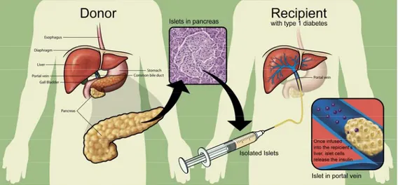
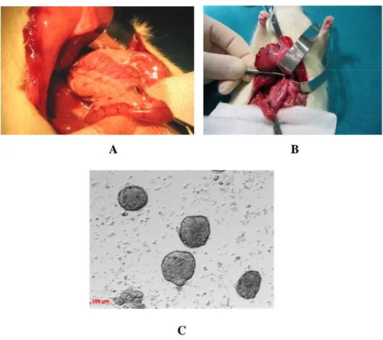
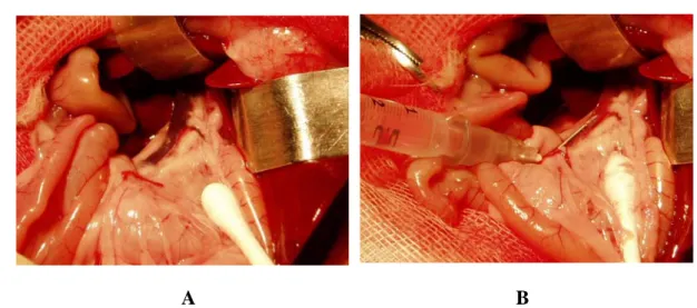
The islets of Langerhans are a promising alternative to whole organ transplantation; they are collected from a donor’s pancreas and injected through the portal vein into a recipient’s liver (figure 1).
Figure 1. Islet transplantation. Islets were purified and infused via the portal vein of diabetic recipients, once they were extracted and isolated from the pancreas.
In 1893 the English surgeon W.Williams attempted to transplant the extract of sheep pancreas into a 15-year-old boy with a severe form of diabetes, marking the first pioneeristic xenograft transplant39.
More than 50 years later, in 1967, Paul Lacy suggested that a better solution to exogenous insulin treatment was the transplant of pancreatic islets40. Later, in 1972, Lacy successfully reversed diabetes for the first time in a murine model of chemically induced diabetes through a pancreatic islet infusion41.
It was only in 1986 that an automated method for islet isolation with a sufficient number of islets to be transplanted was introduced into the clinical setting, and in 1989 the Paul Lacy group exploited this method to reverse diabetes and obtain insulin independence after human islet transplantation42. Unfortunately the graft failed a few days after surgery, perhaps due to an inadequate immunosuppressive regimen.
Until 1990, none of the above-mentioned transplants were successful until the group from Pittsburg, USA, performed the first series of human islet allograft, prolonging insulin independence through steroid-free immunosuppressants treatment (FK506). The patients remained insulin independent up to 5 years after the transplant. The long-term survival of intrahepatic human islets was confirmed by liver biopsies, revealing that allogeneic human islets were successfully engrafted in the hepatic microenvironment43.
In 1996 in Giessen, Germany, there was an improvement in peri-transplant management as well as the immunosuppression results in 100% initial islet-graft function and 40% insulin independence after one year.
In 1999 a very successful protocol was achieved in clinical trials by the Edmonton group which combined islet transplantation with the use of Rapamicyn by implementing a steroid-free protocol. The result was a series of transplants with insulin independence in 7/7 human islet-cell transplant recipients within the first year44. All patients received pancreatic islets from 2 donors (only in one case 4 donors were necessary). The Edmonton protocol used a combination of immunosuppressive drugs, including Daclizumab (Zenapax), Sirolimus (Rapamune) and Tacrolimus (Prograf). Daclizumab was given intravenously right after the transplant and then discontinued, while Sirolimus and Tacrolimus, the two main drugs that maintain the immune system preserved from the destruction of the transplanted islets, must be administered throughout the patients’ life44.
The difference between the Edmonton protocol and previous ones was a convenient number of high purity level islets, but, most of all, a less diabetogenic and steroid-free use of immunosuppressive drugs.
Islet Transplant Registry reported more than 450 allogeneic pancreatic islet transplantations between 1974 and 1999 in patients affected by type 1 diabetes, but less than 10% reached insulin independence within one year, though 28% of patients secreted high levels of C peptide. Several factors contributed to the failure, such as a poor transplanted islet mass, inadequate anti acute rejection treatment, diabetes autoimmune recurrence and extremely toxic immunosuppressive drugs such as cyclosporine and glucorticoids. The clinical introduction in the Edmonton protocol led to progress in pancreatic islets transplantation, with the general acceptance of islet transplantation as a feasible clinic therapy, especially for the treatment of patients affected by type 1 diabetes with developed frequent hypoglycaemic episodes.
A further study in the University of Alberta on a 66-year-old patient showed that 82% of patients who received a transplant remained insulin independent up to one year after transplantation; this percentage decreases as the years go by: up to 70% in the second year and 50% in the third year. In spite of this, many patients continued to produce sufficient amounts of insulin in order to reduce hypoglycaemic risky episodes; therefore, islet transplantation mostly became efficacious in glucose level control.
Similar results were produced by other studies involving islets which were kept in culture before transplantation up to three days (2001, Miami, USA).
The National Institute of Health, Immune Tolerance Network (ITN) set up a multicentre trial of islet transplantation. The Collaborative Islet Transplant Registry (CITR) was established in 2001 by the National Institute of Diabetes and Digestive and Kidney Diseases (NIDDK).
The Registry was created with the aim of collecting, analyzing and comprehensively communicating data from all islet/beta cell transplantation centres in the US, Canada and some European and Australian centres to guide transplant centres in developing and refining islet/beta cell transplant protocols.
A close network of collaboration between initiatives and programs of the National Institutes of Health, the Juvenile Diabetes Research Foundation International (JDRFI) and other health centres, such as the Health Care Finance Administration-HCFA, the Health Resources and Services Administration (HRSA), and the Food and
Drug Administration (FDA), ensure that the outcome measures used by CITR are appropriate, standardized, and relevant.
The last annual scientific report, specifically 2010 CITR (7th) Annual Report, presents data from the past ten years, namely the large majority of the islet transplant programs active in 1999-2009 (www.citregistry.org). The following data refer to the last decade and shows that 571 allogeneic islet transplant recipients (481 islet transplant alone, 90 islet after or simultaneous kidney) received 1,072 infusions from 1,187 donors. The mean age of recipients who received an islet allograft transplant rose from 42 to 49 years and the mean duration of diabetes grew from 26 to 32 years over the decade. The donors, on the other hand, rose from 42 to 44.8 years of age; about 58% of them were male, deceased due to cerebrovascular accidents/strokes.
The pancreas were processed better and faster; in addition the islet product showed that the mean total IE rose from 417 to 463 per infusion and the β-cells growth amount from 217 to 335. Nevertheless, the immunosuppressive regimen revealed a change only using IL2R antagonists to a replacement or supplementation including T-cell depletion with or without TNF antagonist in over 80% of the transplants in the last three years.
The maintenance of the immunosuppressors was achieved by a calcineurin-inhibitor and a combination of inosine monophosphate dehydrogenase-calcineurin-inhibitor (IMPDH). The overall results demonstrate that there is 65% of insulin independence in the first year post-infusion, increasing up to 75% by the second year. Moreover the long-term graft function significantly improved, i.e. in 2004-2007 the insulin independence lasted for a longer time than in 1999-2003. By observing the global data of CITR, it can be concluded that over the last decade the allograft recipients exhibited benefit with relatively low risks, (low levels of infusion-related complications, few events of immunosuppressors-related cancer and death), and islet transplantation was engrafted in recipients over 35 years of age with good glycaemic control. Finally, the use of vaso suppressors and insulin in the donors, as well as the use of T-cell depletion and calcineurin-inhibitors for immunosuppressive maintenance in the pre-operative period, create the best conditions for obtaining improved outcomes (http://www.citregistry.org/).
Islet transplantation offers various advantages compared to the whole organ transplant, such as the fact that it is a relatively simple surgical procedure (mini-invasive
surgery) with consequent low post-operative risks and low morbidity as well as culturing or cryopreserving islets.
The protocols used to handle pancreatic islets for reducing their immunogenicity revealed that a short time of culture was adequate. Furthermore, this allows the recipient’s conditioning to be optimized, with a consequent easier tolerance induction, and less risk of rejection. In addition, the requirement for immunosuppressive drugs treatment was low45.
Islet transplantation is indeed considered a promising strategy for the treatment of diabetes mainly in young patients and children with no evident insurgence of severe complications (i.e. when possibility of whole organ transplant has been discarded)46.
There are some key points to consider in order to perform a successful human pancreatic islet transplant:
- correct islet handling in pre-transplant phase: the cold preservation of the pancreas before islet isolation has to be less than 8 hours. The same procedure has to be executed in as little time as possible in order to obtain maximum yield from a single pancreas and preserve the vitality of the mix to be transplanted47.
- correct number of grafted pancreatic islets: to assess an intervention with a good probability of success, at least 12,000 IE/Kg should be transplanted into a recipient (IE is Islet Equivalent, islet volume converted into the number of islet with a diameter of 150 μ).
On average, a human pancreas contains 300,000 to 1.5 million pancreatic islets and it has been evaluated that only 60% is necessary to maintain a normal glucidic metabolism48.
Nevertheless, at least 2 donors are required in order for pancreatic islets to obtain an insulin independence, but occasionally the same result has been obtained by collecting islets from a single donor49.
- ischemic and nonfunctional processes which can hit transplanted islets: islets have a characteristic microvasculature in a normal pancreas: arterioles are dispersed in capillaries in the centre of the islet, emerge through the islet peripheral area and then drain into the portal system (insulo-acinar portal system)50. This structure is destroyed after the isolation of the islets and their transplantation. During the isolation and culture of the islets, the endothelium loses its differentiation and is able to degenerate.
A rapid revascularization is required to let the islets survive and function after transplantation51. Therefore, the survival of grafted islets most of all depends on endothelial cells and capillaries originating in the implanted organ which creates the new vascular system of the islets. It also has to be considered that the early ischemic period significantly damages the islet centre, which contains the great part of the insulin secreting β-cells52.
It is known that blood vessels inside the transplant begin to be visualized 3-5 days after transplantation, and 7-14 days after transplantation a normal blood flow is re-established.
- adverse effects of actual immunosuppressive protocols: immunosuppressive drugs are essential to prevent the processes of rejection linked to transplantation, even though immunosuppressive protocols based on the use of corticosteroids, cyclosporine and tacrolimus (FK 506) are far from being effective in islet transplantation, because the same drugs can induce diabetes33.
The potential inefficacy of human islet transplantation could be linked to an inadequate number of grafted IE and to technical factors linked to isolation that reduces the vitality and functionality of the islets47.
Moreover, the presence of non-specific inflammation and the onset of rejection processes53 mediated by the immune system can lead to the destruction of grafted islets.
Despite increasing interest in this technique, its clinical application remains limited mainly because of low yield in islet achievement and side effects associated with the immunosuppressive drugs used. Grafted islets appear to have a greater aptitude towards acute rejection to verify autoimmune relapse compared to the whole pancreatic organ transplantation54. In the immediate post-operative period, a loss in grafted islets occurs55; in animal models grafted islets are about 50% less than transplanted ones.
Moreover, it has recently been observed that patients treated with Edmonton protocol occasionally reached graft tolerance due to the homeostatic expansion of autoreactive memory cells which are able to induce rejection56. Though tolerance induction can be induced by using mycophenolate mofetil, the risk of normal growth blockage and child development57 prevent its use, except for cases in which children risk losing their lives because of organ insufficiency or other severe complications due to diabetes58. As a consequence, several aspects of islet transplantation have to be defined in order to introduce this clinical therapy as a routine cure which is valid for the
treatment of type 1 diabetes. Graft survival needs to be improved and side effects linked to the use of immunosuppressive drugs avoided59, by introducing new therapeutic strategies.
Several approaches have been proposed in order to induce tolerance instead of administrate immunosuppressive therapy. These alternatives consist in the infusion of stem cells60 or bone-marrow cells61 or mesenchymal stem cells62 in the pre-transplant phase.
1.5 Islets of Langerhans
1.5.1 Architecture of Pancreatic Islets
The islets of Langerhans are the regions of the pancreas or micro-organs that contain at least four types of endocrine cells: α-cells that produce glucagon, β-cells that produce insulin, δ-cells that produce somatostatin, PP-cells that produce pancreatic polypeptide, and ε-cells that produce ghrelin.
In rodent models the distribution of these cells in the islets is not randomized, but β-cells are located in the inner core of the islets, while the non-β cells form the mantle region. This spatial position within the islets contains several properties so that when insulin secretion is decreased, the organization of islet cells is found to be changed; for example β-cells are bound with non-β-cells63. Since the non-β-cells which are in contact with β-cells show no increase in insulin levels in vitro studies, it is important to observe the that the mantle position of non-β-cells is pivotal for insulin regulation by homologous contacts between β-cells64.
Scientific literature provides evidence that there is no clear agreement on the architecture of human pancreatic islets and their distribution, even though it is accepted that the endocrine components between humans and rodents are not that different and that β-cells are less numerous in humans than in rodents65.
In 1976 Orci and Unger located α- and δ-cells of the human islets in the mantle and grouped against capillary walls within the core of β-cells63. Later Grube proposed that β-cells are located in the islet core and α-cells are arranged at the periphery and along intraislet capillaries66.
During the past three decades it has been a great challenge to identify the distribution of pancreatic islets, but Bosco and his colleagues have recently proposed a
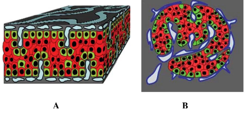
model showing human islet cells distribution in a trilaminar plate (a sandwich structure), where α-cells are in the external layers and β-cells in the internal ones. He also reported no vascular channel in the β-cells in the centre of the structure, but a vascular network was observed at the α-cells level67, (figure 2).
Figure 2 Endocrine cell and vessel organization in human islets. A: α-Cells (green) reside at the periphery of the plate in close contact with vessels (blue). β-Cells (red) reside in the central part of the plate and the developed cytoplasmic extension runs between α-cells reaching the vessels surface. B: The plate with adjacent vessels is folded so that it forms an islet. (Bosco D., Diabetes, 2010)
1.5.2 Physiological site for pancreatic islet transplantation
Recent work by Merani and his colleagues extensively discussed the main role of the site of infusion by means of an animal model and human clinical practice68. Since the liver has been widely studied and has the advantage of a double arterial and venous vascular supply, many of the islets are lost during or immediately after portal vein transplantation. Moreover, the oxygen tension of the parenchyma is lower than that of the pancreas and the liver as a site of implantation is associated with procedure-related complications, including haemorrhage and thrombosis. Patients remain insulin independent immediately after the surgical approach, but the effect is not long lasting69.
Many efforts were made to achieve the best sites for pancreatic islet implantation in order to optimize the engraftment and function of the islets, reduce the number of transplanted islets and decrease immunogenicity.
Islet milieu requires a direct access to oxygen, glucose and other metabolites and need to be free from toxic metabolites and oxygen free radicals. The slow revascularization of the transplanted pancreatic islet is another key issue and is required to have a good vascular supply and high oxygen levels70. The insulin hormone, which is
released by β-cells, should be delivered through the portal vein, as the liver and muscle are primary sites of insulin action, therefore it could be beneficial to transplant the islets in a site that is drained into the liver71.
In addition it is conceivable that the implantation site should be immunoprivileged, thus the pancreatic islets should be infused in a minimal inflammatory reaction site to increase long-term survival and the loss of β-cells should be minimal. Moreover, the implantation site should have easy access to reduce further complications, and the number of islets should be minimal in order to have the lowest transplant volume for multiple donors72.
Merani also reported that the advantages of portal vein infusion of islets of Langerhans were the simple way of injecting them by cannulation of a mesenteric venous tributary during laparotomy or percutaneously using fluoroscopic and ultrasonographic guidance, and also the low requested number of islets73. At the same time, the intraportal site reflects that a reduced function of islets in a model without allorejection and autoimmune diabetes is not suitable for long-term function74. What is more, the instant blood-mediated inflammatory reaction (IBMIR) remains crucial in order for the β-cells to function after transplantation into the vascular site.
The kidney subcapsular site is most commonly used in rodent models and has good results in that diabetes is reversed within a few days. The metabolic engraftment and function of human fetal islet-like clusters transplanted into immunodeficient nude mice under the kidney capsule is better than the lung, liver and spleen from both a morphological and functional perspective75. The disadvantage of the kidney capsule is the scanty blood supply that leads to a low-oxygenated microenvironment for the islets.
Literature provides evidence that only 250 syngeneic subcapsular islets are required for the subcapsular site to reverse streptozotocin-induced diabetes in mice a considerable amount less compared to the 600–800 required for the intraportal site76. Besides the best preserved structure of rodents compared to human islets, it is not easy to lift or infuse islets under the rodent kidney capsule. Finally, the surgical site is very invasive and co-morbidities in the animal recipient population, including diabetic nephropathy, may reduce the hospitality of the kidney.
The spleen, which is a metabolically suitable site for islet transplantation, is an alternative site but it is not as advantageous as the liver. Indeed, there are several
risks of haemorrhage and the improved access of lymphocytes to transplanted tissue in the spleen makes this site unsuitable for islet transplantation77.
Merani investigated other main sites for pancreatic islet transplantation. The pancreas is in theory a good site for supporting the function of the islets in the long-term because of the high partial pressure of oxygen in the tissue. On the other hand, the relatively invasive nature of the isolation procedure and known evidence regarding the autoimmune recurrence of type 1 diabetes make the pancreas a clinically irrelevant site for transplantation68.
Moreover, several papers showed that the intraperitoneal and omental pouch sites are attractive as they provide a large implantation volume and the concurrent use of transplant devices or capsules78. As well as its simple surgical approach, the omentum offers other advantages79, but the large number of islets required for both intraperitoneal and omentum sites and reduced data on long-term survival make these two sites less attractive than others.
Islet transplantation has also been performed into bone marrow of non-diabetic rats and in the os femoris in non-diabetic ones: results reported that there is a persistence of insulin up to 21-30 days post-transplant80. On the other hand, the intramuscular site is very attractive due to its simple accessibility and monitoring, even though this site is associated with a higher frequency of leucocytic infiltration compared to others81. Another site for islet transplantation was the fat pads in the mouse model that showed a simple way of infusion, which was similar to the intramuscular one because of the low required islet number82.
Finally, some immunoprivileged sites, such as the brain, thymus and testis83, were reported because they have shown to confer protection to an allotransplant and these sites are more suitable for transplants of cells. Indeed, they can protect the organism from allorejection or xenorejection without further immunosuppressive therapy.
In conclusion, further research in humans is required before the adoption of any clinical routine, so the careful evaluation of five criteria is suggested (the cellular physiology of the site, the endocrine function for good glycaemic detection, the immunological role of the site, surgical and technical complications due to the chosen site and supporting evidence from studies on large animals) before choosing a clinical application for the alternative site for islet transplantation, even though it is actually indicated that the portal vein remains the main method of infusion68.
1.5.3 Murine models of islet transplantation
A strategy to reduce the graft islet mass for transplantation to a recipient has been investigated in a rat model of islets transplantation. Hara et al has demonstrated that animals transplanted with 1500 syngeneic or allogeneic islets in the portal vein or under the kidney capsule restored to normoglycemia one day after transplantation. The authors reported no significant difference in loss of graft function between syngeneic and allogeneic islet transplantations during the three days after transplantation, neither did they observe any significant difference in deterioration of graft function between portal vein or kidney capsule sites of transplantation84.
Cornolti has recently investigated whether syngeneic islet transplantation can obtain adequate islet function during glucose stimulation by employing a continuous glucose monitoring system in the diabetic rat model. Islet encapsulation through immunoisolation systems requires no further immunosuppressive treatment, thus leading to a new clinical approach. Cornolti’s group has developed two types of islet transplantation: islets immunoisolated in microcapsules inserted in the peritoneal cavity and free islets inserted under the kidney capsule. They observed an homogeneous state of normoglycemia after the two protocols, but the microcapsulated one obtained a better control of glucose sensing and insulin release.
Even though encapsulated islets are separate from the microcirculation with this method, islets can react to high glucose levels in the blood and efficiently release insulin. On the other hand, islets transplanted beneath the kidney capsule showed less viability and function, concluding that despite being nearer to the microcirculation, the host cell reaction was involved85.
1.5.4 Clinical outcomes of islet transplantation
In order to perform human islet transplantation in patients affected by type 1 diabetes, the required clinical indications for hypothetical subjects should be the onset of frequent and severe hypoglycemic events. Moreover, other possible required indications should be severe clinical problems associated with the use of exogenous insulin therapy and an unsuccessful insulin-based therapy to prevent acute complications.
As reviewed by Fiorina et al., the patients that could undergo islet after kidney transplantation were subjects with an end-stage renal disease who also received a kidney transplantation alone or who rejected the pancreas after simultaneous kidney– pancreas transplantation; otherwise, for islet transplant alone, numerous patients should have to be selected86.
Data collected by the CITR revealed that 80% of graft function was successful at 1000 days of follow-up for islet after kidney transplantation, while for islet transplantation alone almost 60% at 1000 days were considered successful86. Several articles reported insulin independence and normalizing glucose homeostasis after an islet transplantation, but only few patients maintained insulin independence in the long term with sustained C-peptide secretion87.
Since insulin is secreted by intrahepatic tissue in grafted patients and cleaved by the liver, Luzi et al. reported that not only a successful islet graft returned to basal hepatic glucose and improved the action of insulin88, but it also achieved a loss of insulin independence, in most cases, by means of long-term partial islet function89.
The positive effect of islet transplantation was also seen in the patients’ survival rate; data regarding the morbidity and the mortality of patients affected by type 1 diabetes were evaluated after the transplantation protocol.
In 2005 Fiorina performed two types of transplants in diabetic patients in an attempt to determine islet transplant improvement with regard to cardiovascular function: a kidney-islet transplant and a kidney alone transplant. The first successful group of patients reached long-term C-peptide secretion, whereas the second group showed a loss of C-peptide secretion 6 months after transplantation. The authors concluded that type 1 diabetic patients with end stage renal disease who received kidney-islet transplantation showed an improvement in aspects of cardiac function for a 3year follow-up in comparison with the group who had received a kidney transplant alone90.
According to available data from clinical trial, it is evident that stringent glucose control can reduce the risk of microvascular complications91, even though no controlled studies have clearly answered the question of whether islet transplantation can halt the progression of long-term diabetic complications. Apart from the difficulties of performing large clinical trials and also other factors, such as the differences in islet isolation procedures, the absence of standardized protocols and the persistence of many regional-based immunosuppressive approaches, the restoration of islet function could in
fact be protective against long-term diabetic complications, as confirmed by a few uncontrolled preliminary studies from Milan, Miami and other groups. Nevertheless, clinical complications largely remain a strong unresolved issue, since the above mentioned cardiovascular function should be kept under strict observation after islet transplantation90.
Patients with type 1 diabetes are also at high risk for macro/microangiopathy. Nephropathy is also considered to be one of the most serious complications of this disease, even though a study on the combined effect of islet transplantation and immunosuppression on kidney function has shown promising results, which are to be confirmed, in patients transplanted with islets alone92. Finally, diabetic retinopathy and complications of the central nervous system have been recently investigated by using islet transplantation as an improved type of treatment.
1.5.5 What happens to islet structure after an islet transplantation
The vasculature of islets cultured in vitro and their subsequent transplantation can dedifferentiate or degenerate from its original architecture. In addition, the immediate days after islet transplantation are critical since this process is characterized by substantial islet cell dysfunction and β-cells death – a more pronounced characteristic in diabetic recipient55. The blood flow of the transplanted islets seems to be lower in diabetic recipients and could be caused by an altered regulation of the blood flow rather than a defect in the revascularization process93.
Furthermore, islet hypoxia is generally thought to be the major reason for the vulnerability of islets in the first few days of engraftment. In 1998 Carlsson demonstrated that oxygen tension immediately after islet transplantation was 50% of that in native islets and slightly lower than that in the adjacent renal cortex. One month after implantation, the exact time when revascularization is likely to be complete, oxygen tension in transplanted islets was lower than after one day. Although the newly born microvessels within the graft are responsible for oxygen transportation, they have a reduced ability to transport oxygen to the islet graft compared to the highly specialized vascular network of the endogenous islets. This means that transplanted islets are exposed to an environment containing a much lower concentration of oxygen compared to the native pancreas94.
Another major issue concerns islet vascularization after their implantation. Islets have less blood flow than surrounding pancreatic exocrine tissue, greater vessel density with greater volume, and intra-islet capillaries are lined by fenestrated endothelial cells95. Indeed, the revascularization of transplanted islets begins within 2–4 days and is completed within 14 days after transplant96.
In various implantation sites it has been observed that grafted islets have reported a decreased vascular density compared to endogenous ones one month after transplantation. In islets implanted into the spleen, as opposed to islets implanted into the liver or kidney, a more pronounced decrease in vascular density is well established, suggesting that the revascularization of transplanted islets is impaired, at least quantitatively, regardless of the implantation site. On the other hand, a large number of blood vessels was found in the connective tissue surrounding the renal subcapsular and intrasplenic islets, so that the predominant location of capillaries in the connective tissue stroma is the immediate vicinity of the endocrine tissue97. This structure was later confirmed by a pancreatic islet graft under the renal capsule rat model where a lymphatic network was shown from one week up to 9-12 months after transplantation, with a major number of capillaries within the connective tissue stroma between the transplanted islets98.
The survival of islets in the graft depends on the diffusion of nutrients and oxygen99 from the surrounding tissue until the revascularization process of the grafts is complete. A part of the revascularization process is likely to depend on the hypoxic activation of genes encoding for angiogenic factors, which lead to new blood vessel formation and improved oxygen delivery100.
An array of peptides including acidic fibroblast growth factor (aFGF), basic acidic fibroblast growth factor (bFGF), tumor growth factor- (TGF) α, TGF-β, PDGF, hepatocyte growth factor (HGF), tumor necrosis factor- (TNF) α, vascular endothelial growth factor (VEGF), angiopoietin (ANG) and interleukin- (IL) 8 were supposed to be the regulators of angiogenesis101. Among these angiogenic factors, the most potent hypoxia-inducible gene that regulates blood vessel growth has been demonstrated to be VEGF, a selective mitogen for endothelial cells in vitro able to promote also vascular permeability in vivo. VEGF is also expressed by pancreatic islets102. A study on VEGF gene expression carried out by Vasir et al. has shown a different pattern on normoglycaemic and diabetic animals after an islet transplantation in the first few days after transplantation. They found a slight increase in the VEGF receptor expression at
days 3 and 5 in the normoglycemic recipients and, at the same time, a decrease in VEGF expression; the beginning of angiogenesis in the grafts could be responsible for this trend. On the contrary, VEGF receptors in the diabetic group at days 5, 7, and 14 showed a marked expression, perhaps in accordance with the concept that vascularization of transplanted islets is delayed by the presence of hyperglycemia, which derives from an increase in local oxygen consumption, as it is known that glucose oxidation in β-cells increases as glucose levels rise103.
Vasir and his colleagues also found that both VEGF isoforms in normal and diabetic recipients at days 1 and 3 after transplantation increased their expressions, suggesting that revascularization had already begun. A parallel increase in the expression of VE-cadherin was also found during the process of revascularization that could in part have been mediated by VEGF. Finally they reported that the diabetic milieu could have impaired the early vascularization of transplanted islets, thus providing an explanation for the less favourable outcomes104.
More recently Golocheikine et al. have studied a model of subcutaneous islet transplantation in matrigel basement membrane matrix to assess the role of indirect proangiogenic growth factors in islet revascularization. The group has studied whether VEGF, a direct agonist capable of activating endothelial cell proliferation and migration, and HFG, a growth factor leading to a blood vessel formation in vivo besides inducing VEGF, could enhance graft survival and function in vivo105. A further confirmation of intercellular adhesion molecules (ICAM) and vascular cell adhesion molecule (VCAM) involvement has also suggested that a cross-talk mechanism between VEGF and HGF could improve the blood vessel formation toward a better vascularization in the graft105.
In order to point out the importance of revascularization after islet transplantation, in 2006 Brissova showed that there is a reciprocal communication endocrine cell–endothelial cell, (figure 3), since early differentiating endocrine cells produced angiogenic factors including VEGF-A and ANG-1. Moreover, it was observed that insulin deficiency in mice with reduced VEGF-A expression was not a result of β-cell dysfunction but an abnormality in the islet vasculature. This was in line with the fact that β-cells and islet microvasculature participate cooperatively in the maintenance of glucose homeostasis. VEGF-A was hence considered to be a key regulator of islet vascularization so that insulin levels in the systemic circulation could be influenced by modulating VEGF-A expression; this suggested that a new mechanism was able to
connect the impairment in islet vascularization with the defect in the release of insulin associated with diabetes106.
Figure 3. Model of pancreatic islet vascularization. After an initial production of angiogenic factors released by early developing islet cells, a recruitment of endothelial cells and their association with developing islet cell clusters occurred. Next, blood flow to small endocrine cell clusters established itself and a concurrent organized structure of islet cell formation occurred. (Brissova M., Diabetes, 2006).
1.5.6 Angiogenic features of intra-islet cells
β-cells in mice are in direct contact with basement membranes of endothelial cells, whereas the intra-islet microvasculature of human islets is surrounded by a double basement membrane107. An extensive capillary branching in the islet core is arranged by one to three arterioles entering an islet.
In this structure, the secreted hormones are forced to exit from the islet through small venules formed from these capillaries. The main characteristic of intra-islet vasculature is the lining of fenestrated endothelial cells108. These fenestrae are thought to be induced by factors such as VEGF-A and angiopoietin-1 both secreted by the islet cells109. It is reported that islet endothelium induce insulin gene expression, stimulating β-cell proliferation and affecting adult β-cell function by secreting several known/unknown paracrine factors110.
Moreover, the islet microvasculature is involved in the regulation of leukocyte recruitment into the islets in type 1 diabetes during the autoimmune destruction of β-cells. During the early phase of type-1 diabetes, islet endothelial cells particularly express surface leucocyte-homing receptors. These receptors can help the
entry of leucocytes into the islets with the consequent destruction of β-cells111. It was recently confirmed that the direction of blood flow within mice islets is from the inner-outer area as well as from the top to the bottom112. This trend implicates that blood can flow from β-cells to non-β-cells thus involving the influence of insulin on the function of non-β-cells. However, in top to bottom blood flow, the cell types do not influence each other in any way.
As assessed by Linn in 2003, endothelial cells contained within the islets required growth factors and were stimulated by FGF-2, VEGF and TNF-α to allow their migration. FGF-2 was indeed found to be an important factor for endothelial proliferation of isolated islets, while VEGF promoted a cord formation. TNF-α was instead able to enhance the effect of VEGF on EC proliferation. The authors demonstrated the significance of donor endothelial cells either in vitro as well as in vivo in the revascularization process. To this aim they observed that the pancreatic islet microvasculature was identified as a target site for the graft rejection process. In addition, this structure was involved in the revascularization process since the donor endothelial cells remained in the graft up to 21 days after transplantation at its periphery and surrounded by renal tissue113.
In order to determine whether intraislet endothelial cells participate in the revascularization and access the structure and composition of blood vessels in the revascularized grafts, Brissova et al used a murine model in which endothelial cells were tagged with lacZ, which was labelled for the mouse endothelial marker PECAM-1 (expressed on the surface of both donor and recipient endothelial cells). Brissova found the existence of two types of blood vessels in the revascularized mouse islet graft: 1) capillaries formed by either donor or recipient endothelial cells directly connected to each other and 2) chimeric blood vessels formed from a mixture of donor and recipient endothelial cells. The chimeric structure was not evident in the human graft. The involvement of both donor and recipient intraislet endothelial cells in the revascularization of transplanted islets was finally confirmed, thus demonstrating that those cells could integrate into the functional vasculature of revascularized islet grafts114.
These data were later confirmed by Nyqvist who stated that intraislet endothelial cells were able to participate in the formation of functional blood vessels within the islet graft vasculature subsequent to transplantation. He observed that the transplantation of freshly isolated islets with a relevant number of endothelial cells, in
contrast to cultured islets, markedly improved their vascularization, thus a preservation of intraislet endothelial cell mass was able to improve the long-term graft function115. Nyqvist recently found that donor islet endothelial cells contributed to the revascularization process of fresh islets and they were also observed in the long-term, but the endothelial cells deriving from donors were not as sufficiently effective as the cultured islets in the reversal of diabetes116.
1.6 Endothelial Progenitor Cells
Endothelial progenitor cells (EPCs) were described for the first time by Asahara et al. as human adult cells involved in postembryonic neovasculogenesis117. Neovasculogenesis, the de novo formation of blood vessels from avascular tissue, differs from neoangiogenesis, the formation of new blood vessels that sprout and grow from pre-existing vessels. Up to Asahara’s findings it was considered that neovasculogenesis only occurred during embryogenesis, within the extra-embryonic yolk sac mesoderm, where primitive haemangioblasts arranged to form islands and tubular structures with further differentiation into blood and vessels118.
The discovery of EPCs led to the new concept that vasculogenesis and angiogenesis could develop at the same time in postnatal life because these cells could differentiate into vascular endothelium, when required, through a mechanism that recapitulates embryonic vasculogenesis. Fadini et al. also reported that EPCs originate in the bone marrow where they are localized in either a quiescent state or a more undifferentiated one.
EPCs can be mobilized from the bone marrow into the peripheral circulation in response to many stimuli, including growth factors and cytokines, released by ischaemic or damaged tissues, as well as during physiologic processes118. When EPCs reside in the blood circulation, they can home specifically towards sites of ischaemia or tissue damage, because of the interaction between soluble chemokines and their receptors expressed on EPCs surface119-124. Once the EPCs have adhered to activated endothelium, they either serve as patches to repair any endothelial denudation or migrate into the surrounding tissue following chemokine gradients. When EPCs are exposed to appropriate environmental stimuli, including growth factors and nutrients, tissue EPCs proliferate and organize themselves into three-dimensional tubular
structures that eventually connect with the pre-existing circulation system and mature to become new blood vessels. EPCs consequently show two main activities in the vascular system: the healing of endothelial damage and the formation of new blood vessels in ischaemic tissues.
Studies with induced experimental mechanical endothelial damage have shown that EPCs are able to repair the vessel wall, re-establishing anatomical and functional endothelial integrity125. The functions of EPCs also include paracrine activity by the secretion of growth factors and cytokines126 and possible trans-differentiation into cardiomyocytes127.
As a result, EPCs can be considered as an integrated component of the cardiovascular system prone to pathological alterations and pharmacological modulation. The inability of EPCs to maintain a normal endothelial homoeostasis and to promote the development of new vessels appears to be the reason for accelerated vascular disease and ageing, with the potential blockage of compensatory angiogenesis, thus favouring the development and progression of ischaemic syndromes. Moreover, EPC alterations have been shown in subjects with no theorical classic cardiovascular risk factors, such as diabetes, hypertension, dyslipidemia, cigarette smoking, obesity and a family history of cardiovascular disease128.
In addition to being altered in the presence of risk factors, EPCs are further impaired in patients with early or advanced atherosclerosis. By using different methods, it has also been demonstrated that levels of EPCs are directly linked with brachial artery flow-mediated dilation, a measure of peripheral endothelial function, regardless of classic risk factors.
A relevant decrease in the level of EPCs represents an advanced atherosclerosis of coronary or peripheral arteries129. This evidence indicates that a drop in EPCs is in accordance with the natural history of atherosclerosis130. At present there is a general agreement concerning EPC levels which provide a measure of the endogenous vascular repair capacity related to atherosclerotic burden and influence of cardiovascular risk118.