UNIVERSITÀ DEGLI STUDI DI ROMA
"TOR VERGATA"
FACOLTA' DI MEDICINA E CHIRURGIA
DOTTORATO DI RICERCA IN
ADVANCED TECHNOLOGY IN REHABILITATION SCIENCES
XXII CICLO DEL CORSO DI DOTTORATO
REDUCTION OF UPPER LIMB FLEXION SPASTICITY IN HEMIPLEGIC
PATIENTS BY THE APPLICATION OF 100 HZ VIBRATION TO THE
TRICEPS BRACHII
Tommaso Sciarra, MD
A.A. 2009/2010
Coordinatore: Prof. Calogero Foti
Index
Index ... 1
Background... 3
Aim of the study ... 4
Materials and Methods ... 4
Patients and treatment ... 4
Statistical analysis... 5
Ethical issues... 5
Results ... 5
Discussion... 6
Neurophysiological basis for the efficacy of vibration treatment ... 6
Possible explanation of the persistent effect of vibration on reducing spasticity... 8
Acknowledgments ... 9
Background
The control of spasticity is a major task in neuro-motor rehabilitation, not only because of the epidemiological importance of cerebrovascular pathologies and the increasing number of survivors with severe sequelae 1(Han DS, 2008) 2(Appelros P, 2009) 3(Agency for Health Care Policy and Research. Post-Stroke Rehabilitation Guideline Panel, 1995) 4(SIGN Scottish Intercollegiate Network, 2002), but also because of the substantial lack of motor improvement in patients with a severe spastic evolution, especially when involving the upper limb 5(Ashford S, 2008) 6(Sommerfeld DK, 2004) 7(Watkins CL, 2002).
Rehabilitation programmes are, therefore, often associated with the use of drugs, given systemically or injected locally, to control spasticity. Although generally effective, when given orally such drugs often induce generalised weakness, drowsiness and sedation 8(Montané, 2004) and, when injected intrathecally, can cause more severe complications 9 (Davidoff RA. 1978)10(Kolaski K, 2007) that sometimes limit their use.
Local treatment of spastic muscles with botulin toxin and phenol 11(Wissel J, 2009) also has some limitations. Although considered a first-line treatment 12(Sheean G, 2009), their cost/benefit ratio, the development of immune-mediated unresponsiveness and some reported side effects, including unexpected loss of strength and diffuse muscle weakness, can limit the use of botulin toxin in daily clinical practice 13(http://www.fda.gov,2009) 14(Elia AE, 2009) 15(Rousseaux M, 2007).
It has been suggested that another possible way to control spasticity is to use physical modalities, such as vibration. However, the clinical reports on the use of vibration in the treatment of spasticity are very few and very old 16 17 18 and only recently was mechanical vibration successfully re-proposed to reduce lower limb spasticity in spastic diplegia 19(Ahlborg L, 2006).
Vibration has been extensively used as a stretch reflex conditioning tool in neurophysiological investigations of spasticity 20(Bishop B, 1975)21(Desmedt JE,1983). Classical neurophysiological experiments have demonstrated that upper limb agonists and antagonists show reciprocal inhibition, that is, the activation of one group induces inhibition of the other 22(Day Bl,1984). It has been demonstrated that this reciprocal inhibition is also induced by vibration 23(Cody FW, 1989) 24 (Cody FW, 1998).
Aim of the study
The aim of this clinical study was to test, in a group of hemiplegic patients, the hypotheses that:
1) the application of selective vibration to the upper limb flexor antagonist (i.e. the triceps brachii) can reduce the spasticity of the flexor agonist (i.e. the biceps brachii);
2) the possible effect of vibration in reducing spasticity can be maintained for longer than the period that the stimulus is applied.
Materials and Methods
Patients and treatment
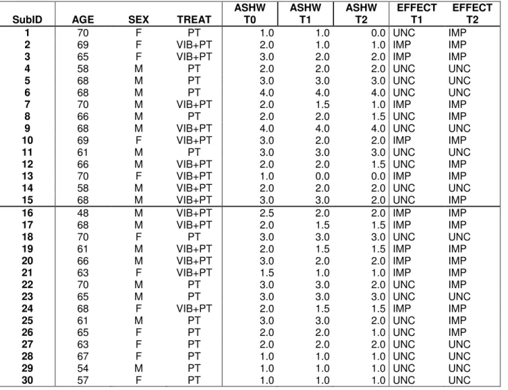
From a group of 60 patients with spastic hemiplegia of the upper limb, 30 patients of either sex and regardless of the affected side were selected for this study following application of the exclusion criteria. The exclusion criteria were: age over 70 years old; lesion present for more than 1 year; age- and educational level-normalised MMSE score below 22 25 (Crum RM, 1993), and the presence of systemic, bone or joint disorders or tumours or changes in either central or peripheral sensitivity able to interfere with the aims of the research. The general characteristics of the patients included in the study are reported in Table 1.
The patients recruited were allocated to two different treatment groups using an automated computer randomisation programme. One group received physiotherapy (PT) and the other physiotherapy plus vibration treatment (VIB+PT). Both groups had daily sessions of standard physiotherapy based mainly on Kabath techniques associated with passive and active exercises to reduce spasticity. For both groups each daily session lasted 45 minutes for 5 days a week (from Monday to Friday) for the 2 weeks of the trial.
Patients in the VIB+PT group, in addition to the daily sessions of physiotherapy, also received treatment with vibration. A pneumatic vibrator powered by compressed air26 (VISS, Italy) was utilized. A 100 Hz vibration thus produced was applied over the belly of the triceps brachii muscle of the spastic side by means of a cup-shaped transducer with a contact surface of 2 cm2 so that the amplitude of vibration was approximately 2 mm with a mean pressure of 250 mBarr. The transducer was kept in place by a non-elastic band wrapped around the arm with a constant contact force of 20-25 Newton. The patients in
the VIB+PT group received vibration sessions of 30 minutes each for 5 consecutive days for 2 weeks at approximately the same time of the day (afternoon).
In both groups the spasticity of the biceps brachii was evaluated using the modified Ashworth scale. The spasticity was evaluated before starting treatment (T0), 48 hours after the fifth session (T1) and 48 hours after the last session of vibration (T2) by a doctor who was unaware of the purpose of the research or the type of treatment applied.
Table 2 reports the timing of the various elements of the research protocol.
Statistical analysis
A descriptive analysis of the distribution frequencies of demographic data was conducted. The primary analyses concerned the effect of the treatments on changes in the patients’ score on the Ashworth scale. The analysis of the effects of treatment was performed using Fisher’s exact test, evaluating the distributions of patients whose score improved and of those whose score remained unchanged in the two treatment groups.
Ethical issues
The protocol followed the Helsinki recommendations on non-therapeutic biomedical research involving human subjects and was approved by the institute’s Ethical Committee. All the subjects received a careful explanation of the aims of the study and methods used and agreed to participate in the study. Before their enrolment they gave signed, informed consent. Subjects were free to withdraw from the study at any time.
Results
An analysis of the demographic characteristics of the patients showed that: (i) there was a higher proportion of men than women, with the overall male to female ratio of 3:2 being conserved in the two treatment groups; (ii) the mean age of the subjects enrolled was 64.7±5.4 years (range 48-70 years) and that this remained equivalent in the two treatment groups (VIB+PT: 65.1±5.8 years, range 48-70; PT: 64.2±5.4 years, range 54-70).
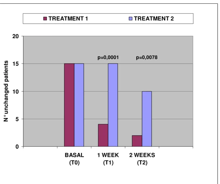
After 1 week of treatment, Fisher’s exact test showed a statistically significant greater improvement in the VIB+PT group (p=0.0001) compared to in the PT group. The results obtained after 1 week of treatment are shown in Table 3.
After 2 weeks of treatment, Fisher’s exact test showed a statistically significant greater improvement in the patients in the VIB+PT group (p=0.0078). The results obtained after 2 weeks of treatment are shown in Table 4. The same results are represented graphically in
Figure 1.
Discussion
The physiopathology of spasticity in hemiplegic patients is not completely understood 27(Nardone A, Schieppati M. 2005) and this lack of complete knowledge is reflected in the field of rehabilitation by the poor control of spasticity. In particular, upper limb rehabilitation of hemiplegic patients is all too often unsatisfactory because of various degrees of spasticity masking any possible motor recovery. Several methods of controlling spasticity are used, including systemic and local administration of antispastic drugs, but their side effects and low cost/benefit ratio can limit their clinical utility 8-15.
The results of this study suggest a possible beneficial utilisation of a vibratory stimulation applied to the antagonist muscle in controlling flexion upper limb spasticity in a group of hemiplegic patients. We demonstrated that a vibratory stimulus of 100 Hz applied to the triceps brachii of a spastic upper limb is able to reduce the spasticity of the flexor agonist, biceps brachii, and, moreover, that this clinically perceivable reduction of spasticity of the biceps brachii extends (for at least 48 hours) beyond the period of application of the vibration, suggesting a reasonable possible role for this strategy in the rehabilitation of spastic hemiplegia.
Neurophysiological basis for the efficacy of vibration treatment
In normal subjects a vibration stimulus has been shown to have several effects. These vary in relationship to the frequency and site of application and also depend on where the effects are studied (i.e. the vibrated muscle or non-vibrated muscle). Vibration increases motor evoked potentials recorded from the vibrated muscle 28(Claus D, 1988) 29(Claus D., 1988) 30(Kossev A., 1999) 31(Rosenkranz K., 2000) 32(Rosenkranz K, 2003) and reduces the intra-cortical inhibition for the same muscle: both of these effects lead to increased
excitability of the spinal motor neuronal pool of that muscle. This excitatory effect along the motor command chain to the vibrated muscle is inverted for adjacent and antagonistic muscles: the application of vibration results in increased intracortical inhibition in the adjacent cortical areas of these non-vibrated muscles 28.
It is known that subjects affected by spastic hemiparesis have increased cortical excitability, as demonstrated by a reduced central silent period, a neurophysiological marker of cortical excitability on the affected side 33 34 35(8-17 Binder, 2009). Vibration applied to a given forearm muscle group has been demonstrated to increase the central silent period of the non-vibrating antagonistic muscles, indicating enhanced inhibition/ reduced excitability of cortico-spinal activity 33(Binder).
Muscle agonists and antagonists are linked by so-called reciprocal inhibition 22 23 24 and therefore increased motor excitability of a given muscle group can lead to decreased motor excitability in its antagonistic muscle group. A combined clinical consequence of this reciprocal inhibition and of the decreased motor excitability of the non-vibrated antagonistic muscle group could be a reduction in flexion spasticity in the upper limb of patients affected by spastic hemiplegic.
Unfortunately, this sound neurophysiological background to a possible therapeutic use of vibration to reduce spasticity has not, so far, been paralleled by extensive clinical application. This can be mainly attributed to a major technical problem related to the vibration devices available for clinical use. Since its earliest applications, vibration was delivered by devices created specifically for laboratory studies and the only devices were cylindrical vibratory devices with an internal eccentric motor 36 37(Hagbart, Casale). The limited supply of these vibration devices and the practical problems with their use have led to an obvious lack of extensive application outside neurophysiology laboratories.
Recently both whole body vibration devices and locally applicable mechano-acoustic vibration devices have become available for clinical use making more extensive clinical studies possible both in normal subjects 26(Casale 2009) and in patients, such as those with cerebral palsy affected by spasticity of the knee extensors 19(Ahlborg 2006). In our study of a group of hemiplegic patients with severe spasticity of the upper limb, we applied localised vibration to the antagonist muscle of the affected limb (i.e. the triceps brachii) and the clinical results supported the neurophysiological rationale of this choice. The physiological explanation of the clinical results of whole body vibration, which acts on both agonist and antagonist muscle groups, is not so clear. Furthermore, we used a 100 Hz
vibration, which seems to be a more appropriate frequency to stimulate highly myelinated afferent than 25-40 Hz used in whole body vibration 36.
Possible explanation of the persistent effect of vibration on reducing spasticity
The second aim of this study was to determine whether 100 Hz vibration can induce a clinical modification of spasticity extending beyond the period of application of the vibration. In our study we found that upper arm flexor spasticity remained reduced, as measured by a clinical score, for 48 hours after the vibration sessions. This was confirmed both at T1 and T2.
It was recently demonstrated that 300 Hz vibration can induce central modifications lasting far longer than the period of application of vibration 26(Casale 2009). The choice of a selective stimulation along with the use of a 100 Hz vibration frequency could account for the early changes obtained with only 10 sessions of 30’ each. Selectivity of the stimulation site and the frequency of vibration chosen could also account for the durable effects on spasticity that we recorded 48 hours after the last vibration session. This could be of relevance for the possible clinical use of vibration.
Some limitations of this study should be acknowledged. The assessment of the effect induced by vibration was limited to the use of the modified Ashworth scale for spasticity 38(Ahlborg) 39(Gregson JM, 1999). However, in this context both the difficulty in finding reliable tests for the upper limb 5(Ashworth), like the simple six-minute walking for the lower limb, 19 and the substantial lack of positive results in the management of the spastic upper limb in hemiplegic patients should be taken into consideration 5-7. Our study should be viewed as providing preliminary evidence of a statistically significant reduction of spasticity of the upper limb induced by 100 HZ vibration applied to the antagonistic muscle. These results should be confirmed by further studies in large numbers of patients, and the research extended by using different vibration frequencies, and possibly by the parallel recording of any improvements in movement, which could be assessed by robotic devices or movement analysis systems.
Acknowledgments
I would like to acknowledge the advice and guidance of Prof. Calogero Foti, chairman. I also thank Dr. Carlo Damiani and Dr. Roberto Casale for the significant help in thinking up, designing, or carrying out the work, without whose knowledge and assistance this study would not have been successful.
Referencies
1 Han DS, Pan SL, Chen SY, Lie SK, Lien IN, Wang TG. Predictors of long-term survival after stroke in Taiwan. J Rehabil Med. 2008;40(10):844-9.
2 Appelros P, Stegmayr B, Terént A. Sex differences in stroke epidemiology: a systematic review. Stroke. 2009 ;40(4):1082-90.
3 Agency for Health Care Policy and Research. Post-Stroke Rehabilitation Guideline Panel. Post-Stroke Rehabilitation. Clinical Practice Guideline no. 16. Rockville, Md. US Department of Health and Human Service, AHCPR, publication 95-0662, Public Health Service 1995.
4 SIGN Scottish Intercollegiate Network. Management of patients with stroke.
Rehabilitation, prevention and management of complications, and discharge planning. A national clinical guideline. Guideline 64. 2002.
http://www.sign.ac.uk/guidelines/published/index.html.
5 Ashford S, Slade M, Malaprade F, Turner-Stokes L. Evaluation of functional outcome measures for the hemiparetic upper limb: a systematic review. J Rehabil Med. 2008; 40(10):787-95.
6 Sommerfeld DK, Eek EU, Svensson AK, Holmqvist LW, von Arbin MH. Spasticity after stroke: its occurrence and association with motor impairments and activity limitations. Stroke. 2004; 35: 134-9.
7 Watkins CL, Leathley MJ, Gregson JM, Moore AP, Smith TL, Sharma AK. Prevalence of spasticity post stroke. Clin Rehabil. 2002; 16:515-22.
8 Montané E, Vallano A, Laporte JR. Oral antispastic drugs in nonprogressive neurologic diseases: a systematic review. Neurology. 2004;63(8):1357-63.
9 Davidoff RA. Pharmacology of spasticity. Neurology. 1978;28 (9 Pt 2):46-51.
10 Kolaski K, Logan LR. A review of the complications of intrathecal baclofen in patients with cerebral palsy. NeuroRehabilitation. 2007;22(5):383-95.
11 Wissel J, Ward AB, Erztgaard P, Bensmail D, Hecht MJ, Lejeune TM, Schnider P, Altavista MC, Cavazza S, Deltombe T, Duarte E, Geurts AC, Gracies JM, Haboubi NH, Juan FJ, Kasch H, Kätterer C, Kirazli Y, Manganotti P, Parman Y, Paternostro-Sluga T, Petropoulou K, Prempeh R, Rousseaux M, Slawek J, Tieranta N. European consensus table on the use of botulinum toxin type A in adult spasticity. J Rehabil Med. 2009; 41(1):13-25.
12 Sheean G. Botulinum toxin should be first-line treatment for poststroke spasticity. J Neurol Neurosurg Psychiatry. 2009;80(4):359.
13 http://www.fda.gov.April 30, 2009 - FDA Requires Boxed Warning for All Botulinum Toxin Products.
14 Elia AE, Filippini G, Calandrella D, Albanese A. Botulinum neurotoxins for post-stroke spasticity in adults: a systematic review. Mov Disord. 2009;24(6):801-12.
15 Rousseaux M, Daveluy W. The risk-benefit of high doses of botulinum toxin injections for muscle spasticity, Ann Readapt Med Phys. 2007;50 Suppl 1:S1-3.
16 Hagbarth KE., Eklund G. The effects of muscle vibration in spasticity, rigidity and cerebellar disorders. J Neurol Neurosurg Psychiat. 1968;31:207-13.
17 Knutsson E, Lindblom U, Odéen I. Reflex facilitation by muscle vibration in the treatment of spastic hemiparesis. Scand J Rehab Med 1970; 2-3: 110-6.
18 Hagbarth KE, Eklund G. The muscle vibrator--a useful tool in neurological therapeutic work. Scand J Rehabil Med. 1969;1(1):26-34.
19 Ahlborg L, Andersson C, Julin P. Whole-body vibration training compared with resistance training: effect on spasticity, muscle strength and motor performance in adults with cerebral palsy. J Rehabil Med. 2006;38(5):302-8.
20 Bishop B. Vibratory stimulation. Part II. Vibratory stimulation as an evaluation tool. Phys Ther. 1975 ;55(1):28-34.
21 Desmedt JE. Mechanisms of vibration-induced inhibition or potentiation: tonic vibration reflex and vibration paradox in man. Adv Neurol. 1983;39:671-83.
22 Day Bl, Marsden CD, Obeso JA, Rothwell JC. Reciprocal inhibition between the muscles of the human forearm. J Physiol 1984;349:519-34.
23 Cody FW, Henley NC, Parker L, Turner G. Phasic and tonic reflexes evoked in human antagonistic wrist muscles by tendon vibration. Electroencephalogr Clin Neurophysiol. 1998;109(1):24-5.
24 Cody FW, Plant T. Electromyographic reflexes evoked in human wrist flexors by tendon extension and by displacement of the wrist joint. J Physiol. 1989;411:379-92.
25 Crum RM, Anthony JC, Bassett SS, Folstein MF Population-based norms for the Mini-Mental State Examination by age and educational level.. JAMA. 1993;269(18):2386-91.
26 Casale R, Ring H, Rainoldi A. High frequency vibration conditioning stimulation centrally
reduces myoelectrical manifestation of fatigue in healthy subjects. J Electromyogr Kinesiol. 2008 Sep 24. [Epub ahead of print]
27 Nardone A, Schieppati M. Reflex contribution of spindle group Ia and II afferent input to leg muscle spasticity as revealed by tendon vibration in hemiparesis. Clin Neurophysiol. 2005 ;116(6):1370-81.
28 Claus D, Mills KR, Murray NM. The influence of vibration on the excitability of alpha motoneurones. Electroencephalogr Clin Neurophysiol. 1988 ;69(5):431-6.
29 Claus D, Mills KR, Murray NM. Facilitation of muscle responses to magnetic brain stimulation by mechanical stimuli in man. Exp Brain Res. 1988;71(2):273-8.
30 Kossev A, Siggelkow S, Schubert M, Wohlfarth K, Dengler R. Muscle vibration: different effects on transcranial magnetic and electrical stimulation. Muscle Nerve. 1999 ;22(7):946- 8.
31 Rosenkranz K, Altenmüller E, Siggelkow S, Dengler R. Alteration of sensorimotor integration in musician's cramp: impaired focusing of proprioception. Clin Neurophysiol. 2000;111(11):2040-5.
32 Rosenkranz K, Rothwell JC. Differential effect of muscle vibration on intracortical inhibitory circuits in humans.J Physiol. 2003; 551( 2):649-60.
33 Binder C, Kaya AE, Liepert J. Vibration prolongs the cortical silent period in an antagonistic muscle. Muscle Nerve. 2009;39(6):776-80
34 Cruz Martínez A, Muñoz J, Palacios F. The muscle inhibitory period by transcranial magnetic stimulation. Study in stroke patients. Electromyogr Clin Neurophysiol. 1998; 38(3):189-92.
35 Alagona G, Delvaux V, Gérard P, De Pasqua V, Pennisi G, Delwaide PJ, Nicoletti F, Maertens de Noordhout A. Ipsilateral motor responses to focal transcranial magnetic stimulation in healthy subjects and acute-stroke patients. Stroke. 2001;32(6):1304-9. 36 Hagbarth KE, Eklund G. The effects of muscle vibration in spasticity, rigidity, and cerebellar disorders. J Neurol Neurosurg Psychiatry. 1968;31(3):207-13.
37 Casale R, Giordano A, Tiengo M. Spinal nociceptive reflex response. Changes in the RaIII nociceptive reflex response and in sciatic pain induced by transcutaneous electric nerve stimulation and vibrations. Minerva Anestesiol. 1985 ;51(5):217-23.
38 Bohannon RW, Smith MB. Interrater reliability of a modified Ashworth scale of muscle spasticity. Phys Ther. 1987;67(2):206-7.
39 Gregson JM, Leathley M, Moore AP, Sharma AK, Smith TL, Watkins CL Reliability of the Tone Assessment Scale and the modified Ashworth scale as clinical tools for assessing poststroke spasticity. Arch Phys Med Rehabil. 1999 ;80 (9):1013-6.
Table 1: Demographic and clinical data of the patients.
SubID AGE SEX TREAT ASHW T0 ASHW T1 ASHW T2 EFFECT T1 EFFECT T2
1 70 F PT 1.0 1.0 0.0 UNC IMP
2 69 F VIB+PT 2.0 1.0 1.0 IMP IMP
3 65 F VIB+PT 3.0 2.0 2.0 IMP IMP
4 58 M PT 2.0 2.0 2.0 UNC UNC
5 68 M PT 3.0 3.0 3.0 UNC UNC
6 68 M PT 4.0 4.0 4.0 UNC UNC
7 70 M VIB+PT 2.0 1.5 1.0 IMP IMP
8 66 M PT 2.0 2.0 1.5 UNC IMP
9 68 M VIB+PT 4.0 4.0 4.0 UNC UNC
10 69 F VIB+PT 3.0 2.0 2.0 IMP IMP
11 61 M PT 3.0 3.0 3.0 UNC UNC
12 66 M VIB+PT 2.0 2.0 1.5 UNC IMP
13 70 F VIB+PT 1.0 0.0 0.0 IMP IMP
14 58 M VIB+PT 2.0 2.0 2.0 UNC UNC
15 68 M VIB+PT 3.0 3.0 2.0 UNC IMP
16 48 M VIB+PT 2.5 2.0 2.0 IMP IMP
17 68 M VIB+PT 2.0 1.5 1.5 IMP IMP
18 70 F PT 3.0 3.0 3.0 UNC UNC
19 61 M VIB+PT 2.0 1.5 1.5 IMP IMP
20 66 M VIB+PT 3.0 2.0 2.0 IMP IMP
21 63 F VIB+PT 1.5 1.0 1.0 IMP IMP
22 70 M PT 3.0 3.0 2.0 UNC IMP
23 65 M PT 3.0 3.0 3.0 UNC UNC
24 68 F VIB+PT 2.0 1.5 1.5 IMP IMP
25 61 M PT 3.0 3.0 2.0 UNC IMP 26 65 F PT 2.0 2.0 1.0 UNC IMP 27 63 F PT 2.0 2.0 2.0 UNC UNC 28 67 F PT 1.0 1.0 1.0 UNC UNC 29 54 M PT 1.0 1.0 1.0 UNC UNC 30 57 F PT 1.0 1.0 1.0 UNC UNC
Table 2. Study protocol. Clinical examination at T-0, T-1 and T-2 were done in the morning, while treatments were always provided in the afternoon.
Days 0 1 2 3 4 5 6 7 8 9 10 11 12 13 14 15 SCREENING ASHWORTH SCALE T0 T1 T2 VIB+PT (TR-1) PT (TR-2)
Table 3: Distribution of the effects after 1 week of treatment (T1).
VIB+PT PT Total
Improved 11 0 10
Unchanged 4 15 20
Total 15 15 30
Table 4: Distribution of the effects after 2 weeks of treatment (T2).
VIB+PT PT Total
Improved 13 5 18
Unchanged 2 10 12
Figure 1: Treatment 1 = VIB+PT, Treatment 2 = PT
0 5 10 15 20 BASAL (T0) 1 WEEK (T1) 2 WEEKS (T2) N ° u nc h an ge d p at ie nt s TREATMENT 1 TREATMENT 2 p=0,0001 p=0,0078