Impact of low-thermal-injury devices on margin status in laryngeal
cancer. An experimental ex vivo study
Giuditta Mannelli
a,⇑, Giuseppe Meccariello
a, Alberto Deganello
a, Vincenza Maio
b, Daniela Massi
b,
Oreste Gallo
aa
First Clinic of Otolaryngology, Department of Surgery and Translational Medicine, University of Florence, Italy
bDivision of Pathological Anatomy, Department of Surgery and Translational Medicine, University of Florence, Viale G.B. Morgagni, 85, 50134 Florence, Italy
a r t i c l e
i n f o
Article history: Received 7 August 2013
Received in revised form 27 September 2013 Accepted 3 October 2013
Available online 23 October 2013
Keywords:
Low thermal injury devices Margin status
HNSCC
Thermal tissue damage
s u m m a r y
Introduction: Status of margins significantly affects disease-free survival. This study examines in ex vivo model the effect of thermal-injury on margins status comparing traditional instrument with several low-thermal-injury devices.
Methods: We conducted a prospective study on 10 excised larynges from patients affected by advanced laryngeal cancer, to assess the thermal-effect due to surgical incisions made at standard distance by using: scalpel, CO2 Laser, harmonic scalpel and electrocautery. Upon histopathological examination, ther-mal damage (Surgical Artifact, SA), tissue lost/retraction (Shrinkage, S), and tissue alterations were com-pared for each instrument.
Results: Low-thermal-injury devices increased SA mean value from 800.7 to 11447.85lm (72%), and S mean value from 2.226 to 2.910 mm (68.4%) (p < 0.05).
Conclusions: The choice of surgical device could influence the histopathological margins status, conse-quently affecting post operative therapeutic strategies and risk of recurrence.
Ó 2013 Elsevier Ltd. All rights reserved.
Introduction
Over the last decades, low-thermal-injury devices use has widely increased in surgical procedures, this is due to their intrin-sic thermal properties which are able to guarantee a simultaneous cut and coagulation at reduced energy, by maintaining the opera-tive field dry and clean with reduced risk of injuries to adjacent structures [1]. These changes in surgical techniques have been accompanied by a progressive extension of conservative surgical indications to more advanced tumor stages, also in head and neck cancer (HNC) patients. Therefore, an accurate assessment of surgi-cal margins status represents a crucial point to achieve both onco-logic radicality and organ function preservation.
This is particularly true especially in laryngeal surgery, where the removal of only 1 mm more of unaffected mucosa, could impair the entire functional results and the chance to avoid demolitive surgery. Nonetheless, neither the frozen section, which is accepted to help surgeons during excision[2], nor definitive histopatholo-gical analysis on margins, are able to guarantee the goal of any
surgical procedure, i.e. total excision of any malignant cells with organ function preservation.
In head and neck oncology, the positivity of surgical margins ranges between 3% and 60% but it is generally around 10%, and it depends upon primary tumor sites, being about 4% in laryngeal cancer surgery[3].
Despite the key role of surgical margins status to define the pro-cedure ‘‘disease-free’’, there is no agreement in definition of posi-tive margins [4]; nonetheless, a classification of ‘‘close’’ or ‘‘positive’’ margins exists and is based on the distance between the closest cancer cells to the line of resection.
When a patient presents a positive-resection margin, approxi-mately in 75% of cases, they will either develop a local recurrence or demonstrate residual tumour upon reoperation[3–5].
Conversely, the presence of a disease-free resection margin does not guarantee that residual tumor is not present within the remaining-host tissue and consequently that recurrence will not supervene. In fact, 25% of patients with negative margins will still go on to develop a recurrence at the primary site[6–9].
Specifically, false-positive margins could be obtained due to: (1) post-operative shrinkage of tissue, (2) inking process, (3) surgeons’ skill, and not (4) appropriately sampling and conservative prac-tices. False-negative margins also represent a serious issue result-ing into under-staged and consequently under-treated disease. According to Slaughter field cancerization theory [10], in HNC
1368-8375/$ - see front matter Ó 2013 Elsevier Ltd. All rights reserved.
http://dx.doi.org/10.1016/j.oraloncology.2013.10.001
⇑Corresponding author. Address: First Clinic of Otolaryngology, Department of Surgery and Translational Medicine, University of Florence, Via Largo Brambilla 3, CAP 50134 Florence, Italy. Tel.: +39 055 7947112; fax: +39 055 7947939.
E-mail address:[email protected](G. Mannelli).
Contents lists available atScienceDirect
Oral Oncology
may have small and multiple foci near the margin which could be rendered unreadable by thermal tissue damage, thus creating the illusion of radical tumor excision. Thus, the wider use of low-ther-mal-injury instruments with their variable thermal damage on ex-cised tissue might have a significative impact on surgical margins status; in this setting, the awareness of quantitative tissue damage variable according to different type of low-thermal-injury devices, may help pathologists in increasing the accuracy of histopatholo-gical assessment and, at least in theory, might explain in part some recurrences in ‘‘disease-free’’ margin patients.
Few studies are reported in literature about the effect of ther-mal tissue injury in head and neck area, and the vast majority of them are based on animal or cadaveric models[11–17]; here, a lar-yngeal model, created using excised larynges, was developed to quantify the effect of thermal injury of different devices on laryn-geal normal mucosa and to predict their effect on margins status and final histopathological report.
Methods Prospective study
The protocol for the prospective controlled clinical study was approved by the Institutional Review Board, and it was conducted in accordance with all accepted standards for human clinical re-search. All patients gave written informed consent prior to study enrollment. Ten consecutive patients, with primary advanced squamous cell laryngeal cancer newly diagnosed, biopsy proven, were treated between January 2010 and December 2011, at our academic tertiary referral center (First Clinic of Otorhinolaryngol-ogy, University of Florence, Azienda Ospedaliero-Universitaria Car-eggi, Italy).
Clinical data of the ten patients are summarized inTable 1. The sites and stage of the tumor have been classified according to the AJCC TNM, 2010[18].
These patients underwent total laryngectomy in general anes-thesia as primary treatment, because of lack of eligibility for laryn-geal preservation protocol. Exclusion criteria were: (1) larynlaryn-geal cancer recurrence, previously treated by surgery or radiotherapy or radiochemiotherapy, (2) larynges widely affected by cancer without showing any areas of mucosa macroscopically upright where it was possible to perform our surgical incisions, and (3) larynges which did not respect the correct conservation process in formalin as laid down in study protocol set out.
Once larynx was excised, it was located on a sterile service tray where, one of us (M.G.), on tumor-free laryngeal mucosa, per-formed four incisions with four different surgical devices. Each incision were made on macroscopically healthy laryngeal mucosa, at a secure distance from the primary tumor, in order to avoid affecting the final histopathologic results. Incisions were distant from each other 5 mm, with a maximum longitudinal length of 1 cm. They were performed by applying a gentle pressure with
the device on the laryngeal surface, in order to obtain the cut of the mucosal layer, only.
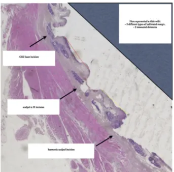
To compare the thermal effects of the three different instru-ments (harmonic scalpel, monopolar electrocautery and CO2 laser) on margin quality, a set of calibrated margins were generated for each tumor-free laryngeal mucosa to provide an artificial margin that would mimic general representation of the tissue quality at the true margin surface. We referred to the scalpel’s incision as the control one; then, calibrated margins were made with each sin-gle thermic device, on its two parallel sides (Fig. 1).
The approximate middle point of each incision distance was lo-cated by bidimensional measurement.
The surgeon used for the incisions: (1) classic scalpel, blade number 15, (2) Flashscanner CO2 laser, Sharplan 780 SurgiTouch TM, 2.29H software, set on super-pulse repeat 0.1–1 on–off and powered 3 Watts, in the respect of normal endoscopic surgery con-ditions, (3) harmonic scalpel, with 55.5 kHz alternative current, with a dissecting tip blade 10 mm long and gently curved, and (4) monopolar electrocautery, with thin tip, with medium cut and coagulation setting.
Tissue generated for this study had no effect on final histopath-ologic diagnosis or margins status interpretation.
Histological preparation of excised incision samples
Excised tissue samples were fixed in 10% neutral buffered for-malin for a minimum of 24 h, followed by paraffin embedding, cut and stained with haematoxylin and eosin. During the embed-ding process, the tissue was orientated such the control calibrated margin (scalpel side) was placed on the central part of each slide, thereby allowing slices to show the relationship of the tissue tran-sitioning from unaffected histology to tissue with thermal injury. Histological levels were cut from each paraffin block, when deemed appropriate. Histopathological evaluation was performed blindly by two pathologists (D.M., V.M.). The study of calibrated margins has been led without invalidating the final histopatholo-gical result.
Definition of thermal injury model looking at generated calibrated margins
There is currently no standardized technique for the evaluation or quantification of thermal injury on histopathological slides in head and neck area. For the current study, we adopted the quanti-tative thermal injury model together with its thermal damage def-initions, suggested by Ruidiaz and colleagues in breast cancer surgery[19]. Accordingly, we identified two zones of fibrocollagen-ous thermal injury (FTI) and four zones of cellular thermal injury (CTI), as previously described according to successive levels of heat-exposure-induced thermal stress on cellular and fibrocollag-enous tissue components. On the basis of their discernable visual characteristics on tissue sections, in terms of differences in staining properties, fibrocollagenous and cellular structure at low and high magnification, we measured the extension of the thermal tissue damage. We identified two zones of fibrocollagenous thermal
Table 1
Clinical data of the prospective study populations.
ID Sex Age (years) Smoke habit (pack/year) Alcohol consumption cTNM pTNM
1 F 61 35 No-Rare cT3N1M0 pT3N2aM0
2 M 65 25 <1 L/die cT3N0M0 pT3N0M0
3 M 80 20 <1 L/die cT4aN0M0 pT4aN0M0
4 F 68 25 >1 L/die cT3N2bM0 pT3N2cM0
5 M 59 30 >1 L/die cT3N0M0 pT3N0M0
6 M 69 20 <1 L/die cT3N2bM0 pT3N2cM0
7 F 65 25 >1 L/die cT4aN2aM0 pT4aN2bM0
8 M 67 25 <1 L/die cT4aN1M0 pT4N2aM0
9 M 75 20 <1 L/die cT4aN2bM0 pT4aN2cM0
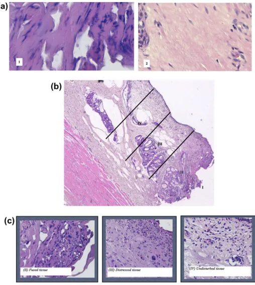
injury (FTI): a zone of undisturbed tissue with well identifiable and normal aspect collage fibres and a zone of collagen denaturation demarcated by a darkening and smoothing of the collagen staining, which is unreadable due to thermic injuries (Fig. 2a). Furthermore, considering cellular thermal injury (CTI), we identified four areas: (I) no cellular structure is identifiable; (II) fused tissue with severe tissue denaturation, few identifiable structures, indistinguishable nuclei, with tissue breakdown; (III) distressed cellular architecture with irregular elongated and spindled nuclei, smudged chromatin, visible distorted fibroblast nuclei, no clear cellular outlines, and no clear distinction between epithelial and stromal components; (IV) undisturbed tissue with no signs of thermal injury, fibroblasts identifiable, clear distinction between epithelial and stromal cells, nuclear ultrastructures and chromatin identifiable (Fig. 2b and c). For quantitative analysis, we referred to Thermal Injury Exten-tion as the sum of thermic damage (Surgical artifact, SA) and sur-gical margins’ retraction (Shrinkage, S)[19].
Measurement of calibrated margins
Slides of calibrated margins of each patient were digitally
scanned at 40 (0.50
l
m per pixel) magnification on a D-Sight(A. Menarini Diagnostics) slide scanner followed by manual histopathological analysis of thermal injury. Measurement of the distance from the control margin (scalpel one) to the closest clearly identifiable cell with distinguishable architecture of the next cali-brated margin created by a thermic device (Fig. 3). Measurements to quantify FTI and CTI were manually performed using the ImageScope Viewer’s measurement tool: (1) distance from the true margin to the end of the collagen denaturation (FTI), (2) distance from the true margin to the fused/distressed boundary (between CTI zones II/III), and (3) distance from the true margin to the distressed/undisturbed boundary (between CTI zones III/IV).
Hypothetical model
In order to apply our technique of margins measurement to real cases, we created a hypothetical retrospective re-evaluation of his-topathological reports.
We assumed the hypothesis that when surgeon cuts a tissue with a low-thermal-injury device, the consequent thermic damage is caused on both sides of the cutting, which implies the presence of SA and S, on both the remaining healthy tissue on patient’s bed side and the resection margin sent to the pathologist. On this set-ting, when we re-evaluated our four historical cases, we started from the assumption to have performed a surgical incision at the middle point of a calibrated distance of 5 mm, equal to the half va-lue of the standard distance of 1 cm proposed in our quantitative model (see ‘‘Prospective study’’ section in Methods), from the more external cancer cell of the nearest malignant cell clusters visible in the histopathological slide (Fig. 4). Therefore, by theorizing that tissue retraction (S) together with loss of readable tissue (SA) could involve about the 50% of both resection margins, which are one on patient surgical field and the other one on surgical specimen, we applied as theoretical visible tissue damage the 50% of the mean value of the amount of S + SA, to our retrospective histopathologic measurements (Table 2).
Statistics
Statistical analysis was performed by STATA (Stata Corporation, College Station, TX, USA). Statistical significance was defined as p < 0.05. Kaplan-Meyer disease-free survival was used to compare results among group A and group B.
Results
Prospective study
Characterization and measurement of SA
Average, instrument-dependent depth of thermal injury to the calibrated-margin, measured from the cauterized surface to the respective boundary, was calculated by evaluating: (1) the depth of the Zone II of fused tissue, and (2) the extension of the Zone III of distressed tissue, per each histologic slides.
Table 2 shows ranges, mean values, and confidence intervals with standard deviations (CI ± DS), which estimate thermal tissue injury created by the three surgical devices (CO2 laser, harmonic scalpel, and monopolar electrocautery) in comparison with tradi-tional cold instrument. Monopolar electrocautery created the wid-est thermal injury, showing a large unreadable tissue boundary whose bidimensional distribution reduced in case of harmonic scalpel use and reached the lowest visible tissue damage by CO2 laser. In fact, fused tissue presented a bidimensional extension which reduced by 45% passing from electrocautery to harmonic scalpel use, and it dropped by 72% by comparing electrocautery and CO2 laser thermic effect. Moreover, the Zone III of distressed tissue, showed a progressive reduction in unreadable tissue due to denaturation affects; it declined by 19% from electrocautery to harmonic scalpel use, and of further 19% by using CO2 laser, with a global difference of 35% between electrocautery and CO2 laser thermal effect.
All of these measurements reported in extenso in Table 2
showed a statistically significant difference (p < 0.0001).
Fig. 5shows the concise representation of thermal injury tissue distribution in accordance with each thermal surgical device. Estimation of calibrated-margin measurements
Average, instrument-dependent shrinkage, that means the mean value of margin retraction which could be measurable, to-gether with ranges and confidence interval with standard devia-tion of each measurement, are summarized inTable 3.
Compared to CO2 laser, whose mean value of shrinkage pre-sents a bidimensional account of 2.09 mm, electrocautery and
Figure 1. Examples of calibrated margins created with different surgical devices on tumor-free laryngeal mucosa. Three types of calibrated margins, performed at a secure distance from the primary tumor (T), far from each other 5 calibrated millimeters, are represented; letter a points at scalpel incision, letter b shows the harmonic scalpel calibrated margin, letter c represents the CO2 laser incision and, letter d represents monopolar electrocautery incision. The primary laryngeal tumor (T) was near or involving true vocal cords (ccvv).
harmonic scalpel produce a higher tissue disruption mean value equal to 2.433 mm and 2.774 mm, respectively (seeFig. 5).
These results were statistically significant if compared to each other: (1) CO2 laser vs. harmonic scalpel (p = 0.0001), (2) CO2 laser vs. electrocautery (p = 0.0172), and (3) harmonic scalpel vs. elec-trocautery (p = 0.0045).
Hypothetical model
By applying our model based on estimation of calibrated-mar-gin measurements (seeTable 3), we critically re-evaluated four representative cases, whose clinical characteristics are summa-rized inTable 4, are described as follows:
Case 1. Final report indicative for close-margin, re-interpreted as false close-margin.
This was a laser cordectomy type II.Fig. 6a shows the distance measured from the inked resection margin to the nearest cancer cell. This distance was 0.4 mm and it was histopathologically inter-preted as close-margin, due to its extent lower than the conven-tional safe margin distance of 1 mm. By our quantitative model,
we have reinterpreted this final result in accordance with thermal CO2 laser properties (Table 2 and 3). Starting from the assumption that a loss of 50% of readable margin occurred (1.45 mm), this close-margin would reach the measure of 1.49 mm, from the sum of 0.4 mm (the measure reported by the pathologist) + 1.45 mm (the 50% of the mean value of surgical tissue distruption in case of CO2 laser use) respectively. According to this hypothesis, this could be a case of false close-margin, i.e. negative one.
Case 2. Final report indicative for close-margin, re-interpreted as false close-margin.
Example of superior resection margin of glottic cancer, per-formed by harmonic scalpel during open partial laryngectomy.
Fig. 6b shows the distance measured from the inked resection mar-gin to the nearest cancer cell. This distance was of 0.9 mm and the pathologist interpreted this margin as close-margin, due to its ex-tent lower than the conventional safe margin distance of 1 mm. By our quantitative model, we have reinterpreted this final result in accordance with thermal harmonic scalpel properties (Tables 2 and 3). We should add to the reported measure of 0.9 mm, the va-lue of 1.113 mm, which represents the 50% of the mean vava-lue of
Figure 2. Histopathologic features of thermal injury: (a) fibrocollagenous tissue (FTI) and cellular tissue (CTI, b and c), and their iconographic representation per each surgical devices used in this study (SA, d). (a) The fibrocollagenous tissue is characterized by: (1) a zone of collagen denaturation demarcated by a darkening and smoothing of the collagen staining, which is unreadable due to electrocoagultion injuries; and (2) a zone of undisturbed tissue with well identifiable and normal aspect collage fibres. (b) Tissue thermal injury induced by harmonic scalpel: Zone I, characterized by extensive charring; Zone II with few identifiable cellular structures and mainly represented by fused tissue; Zone III, has distressed tissue architecture of wispy appearance. Then, Zone IV is undisturbed, with no evidence of thermal cellular artifacts. (c) Dominant characteristics are for each zone represented below. Fused tissue: increased staining uptake, cells not identifiable. Distressed tissue: smudge nuclei, unclear cell outlines, distorted cellular arrangement. Undisturbed tissue: rounded nuclei, visible nuclear substructures, cell types are identifiable.
readable margin in case of harmonic scalpel use. Thus, the true resection margin value would reach a size over the safe standard resection tumor distance. As above, this could be a case of false close-margin, i.e. negative one.
Case 3. Final report indicative for negative-margin, re-interpreted as false negative-margin.
We present a paradigmatic case in which the use of low-ther-mal-injury device could have destroyed a small focus of tumor cells, far from the primary tumor.Fig. 6c, in fact shows the pres-ence of a small area of carcinoma (red circle) far from the primary tumor (T). This area extended for 0.4 mm far from the inked mar-gin. The surgeon using harmonic scalpel partially destroyed this cluster, because of thermal effect. The use of monopolar electro-cautery in place of harmonic scalpel would have resulted in a com-plete reduction of this small area presenting, in accordance with our model, a wider unreadable boundary of 55% than harmonic scalpel (Fig. 6c). Thus, surgical artifacts due to devices can also cre-ate false-negative margins, this may be due to two main mecha-nisms: (1) cell damage of normal tissue resembling cancer cells (artifacts); and more frequently, (2) direct distruction by thermal injury of cancer cell cluster close to the margin.
Case 4. Final report indicative for positive-margin, re-interpreted as false positive-margin.
Here an example of superior resection margin of supraglottic cancer, performed by monopolar electrocautery during open par-tial laryngectomy.
Fig. 6d shows the maximum distance (1.3 mm) because of extensive and massive thermal damage rendering unreadable the full distance between inked resection margin and growth tumor line, the pathologist interpreted this margin as positive. According to our model, if we had a mean thermic distruction tissue of 1.28 mm by monopolar electrocautery, we could have considered this margin as negative (see,Tables 2 and 3).
Discussion
It is common knowledge among surgeons that an oncologic pro-cedure with safe surgical margins represents the best chance in or-der to achieve local control; thus, resection margins status is strictly related to the choice of adjuvant treatment strategies
[20–23].
Nonetheless, in spite of though negative resection margins are reported in literature, a percentage of head and neck cancer pa-tients from 15% to 30% experienced a local tumor recurrence dur-ing the follow-up[7–9,24,25].
The histologically confirmed presence of tumor at the resection margin of a surgical specimen is described in the literature as a po-sitive tumor margin[23].
On the other hand, the significance of the presence of invasive carcinoma near the border of resection remains an issue under dis-cussion[7,24–27].
Figure 3. Example of a histologic slide with three different calibrated margins: classic scalpel with shave number 15, harmonic scalpel and CO2 laser. The yellow line represents the measured distance from the control margin to the harmonic scalpel margin; while, the red line is the distance between control and CO2 laser calibrated margins. (For interpretation of the references to color in this figure legend, the reader is referred to the web version of this article.)
Table 2
Comparison of thermal injury extension represented by the sum of surgical margins’ retraction (S) together with the measurement of the thermic damage (SA) in laryngeal mucosa slides, by instrument.
CO2 laser Harmonic scalpel Electrocautery Zone II Range (lm) 65–154 187–251 280–471 Mean value (lm) 105.71 207.57 377.71 CI ± DS (lm) 79.70– 131.73 ± 28.13 188.36– 226.78 ± 20.77 319.13– 436.30 ± 63.34 Zone III Range (lm) 588–801 650–1023 898–1303 Mean value (lm) 695.00 862.86 1070.14 CI ± DS (lm) 629.30– 760.70 ± 71.04 741.51– 984.21 ± 131.21 935.84– 1204.45 ± 145.22 Figure 4. Hypothesis of retrospective analysis of histopathologic slides. Firstly, we thought of a calibrated distance of 5 mm (black line), measured from the fairest cancer cell of tumor mass (T). Then, we speculated to perform a surgical incision at this standard distance’s middle point (arrow with scalpel’s image).
Table 3
Ranges, mean values and confident interval with standard deviation of measurable distances between calibrated margins.
CO2 laser–scalpel Harmonic scalpel– scalpel Electrocautery– scalpel Range 2.52–3.31 mm 2.02–2.50 mm 2.21–2.87 mm Mean value 2.91 mm 2.226 mm 2.57 mm CI ± DS 2.66887– 3.24141 ± 0.30953 2.08510– 2.36748 ± 0.15266 2.37352– 2.75819 ± 0.20797
There is no consensus on how much normal tissue should be re-moved around a tumor in order to reduce the risk of local recur-rence; it is widely accepted, however, by head and neck surgeons that inadequate excision of a tumor leads to early primary site recurrence; anatomic site in head and neck seems to influence an oncologically safe resection margin [20]. In the larynx, Bocca et al.[28]suggest that a margin of a few millimeters may be en-ough in some areas, whereas in the hypopharynx submucosal spread of 1 cm may occur, thus margins of 2 cm are necessary[29]. Postoperative management of patients with positive surgical margins is another controversial issue. Revision surgery has been shown to be a valid option for such cases. Moreover, it has been suggested that postoperative radiotherapy may protect patients from local recurrence whenever surgical margins are compromised and additional surgery is not feasible; however, it has also been stated that further treatment may compromise functional recovery
[20].
Here, for the first time, we reported an experimental ex vivo model of a quantitative measurement of thermal injury induced by low-thermal-injury devices which may help in improving the accuracy in margin status assessment in laryngeal surgery. An excessive thermal injury may have two potential histopathological outcomes: (a) according to a higher thermal damage, the effective readable distance between the margin and the first line tumor growth can be decreased thus, creating ‘‘close’’ or apparent
‘‘positive margins’’ (false-positive, with indications for re-excision or adjuvant post-operative radiotherapy); (b) thermal injury can destroy small independent malignant or pre-malignant cell islands near the margin,‘‘false-negative’’ ones, (lacking indications for re-excision or adjuvant radiotherapy).
In order to understand better the potential prognostic impact of different surgical devices on status of cancer resection margins, we proposed, for the first time in head and neck district, a quantitative ex vivo model of measuring histopathological artifacts in accor-dance with each thermal device properties. We quantified the de-gree of thermal injury from three different thermal devices (harmonic scalpel, CO2 laser and electrocautery) on calibrated margins created on healthy laryngeal mucosa, and we measured tissue damage considering two principle parameters: thermal in-jury (surgical artifact, SA) and tissue retraction (Shrinkage, S).
Our results show statistical significative differences among hot and cold (control incision) devices, with monopolar electrocautery showing the highest thermal tissue damage (26% more than har-monic scalpel and 45% more than CO2 laser) (p < 0.05). This ther-mal tissue damage turns into unreadable boundaries whose thickness depends on the nature of device. We succeeded in mea-suring, in a bidimensional way, the extension of this thermal in-jury, by looking for SA and S in our histopathological slides, which presented the highest mean value by harmonic scalpel use. Thus, the choice of surgical device might improve the specific margin assessment with a clear implication for post-surgical treat-ment efficacy and prognosis.
In our opinion, pathologists should know device nature, in order to be able to read histologic slides by keeping in mind the different possible influences of each device on margin status.
In this study, we analyzed a limited number of cases and histo-pathological examinations were made by different pathologists, and these represent potential limitations, also because the assess-ment of histological variables of the primary cancer, including measurement of the margins, may vary among pathologists. The
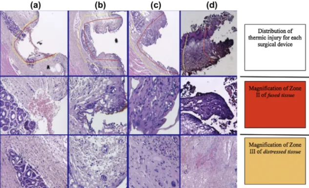
Figure 5. Thermic injury for each surgical device: (a) classic scalpel, (b) CO2 laser, (c) harmonic scalpel, and (d) monopolar electrocautery. Upper row: topographic distribution of thermal injury in the histologic slide, due to the use of different devices. The red line delimits the extension of fused tissue (Zone II); the area between the red line and the yellow line represents the Zone III of distressed tissue. The medium and lower rows show magnifications of zone II and III at higher magnification, respectively. (For interpretation of the references to color in this figure legend, the reader is referred to the web version of this article.)
Table 4
Clinical data of the four representative cases of the hypothetical model. ID Sex Age (years) pTNM Device Margin status 1 F 65 pT1bN0 CO2 laser Close-margin 2 M 60 pT1bN0 Harmonic scalpel Close-margin 3 F 72 pT2N0 Harmonic scalpel Negative-margin 4 M 48 pT3N1M0 Electrocautery Positive-margin
interobserver variations and intraobserver reproducibility are inherent weaknesses of this study design, and can be fully over-come only by prospective studies that incorporate these variables. Accordingly, if surgery is performed by a low-thermal-injury device, we also suggest that experimental and retrospective data carried by us might, at least in theory, justify the possible occur-rence of some local recuroccur-rence in ‘‘disease-free’’ surgical procedures.
Even if low-thermic-injury devices have dramatically changed the surgical techniques with less risk of bleeding, and reduced operating time with less complains, they bring with them a price which is the possible alteration by thermal artifacts of the histo-pathologic resection margins status. This is not secondary, because all possible consequences of a second re-operation or radio-chemio adjuvant therapy on tissue already damaged, can lead towards acute and late complications such as condronecrosis, edema, infec-tions, excessive scar formation, recovery delayed, fistula, which particularly in laryngeal conservative surgery mean dyspnea, delay in decannulation and/or starting with oral feeding, frequently un-solved or responsible for functional total laryngectomy.
In conclusion, this study is the first to quantify the thermal ef-fects of different devices on surgical margins status in HNC surgery. We demonstrate that the use of different surgical instruments might affect the histopathological assessment of excision margins in laryngeal surgery, with potential therapeutic and prognostic implications. Accordingly, on the basis of our findings, we suggest to avoid the use of monopolar electrocautery in laryngeal surgery, especially in conservative surgical procedures, not only because of its diffuse mucosal thermic damage effect which could compro-mise resection margins status with potential consequences on intraoperative decision making, but also because it may affect the surrounding healthy structures such as: muscles, cartilage, ves-sels and nerves, with possible postoperative functional impair-ments. Conversely, CO2 laser and ultrasonic scalpel represent the most suitable devices to perform this type of surgery, due to their lower thermal tissue induced damage and their well known
surgical advantages [1]. On the other hand, nevertheless the
demonstrated lower measured thermic damage (Surgical artifact, SA) and surgical margins’ retraction (Shrinkage, S) caused by these new technologies, we advocate the use of cold instruments espe-cially when few millimeters of unaffected mucosa represent the limit for a safe surgical excision to obtain both a radical and conservative surgical procedure, thus avoiding thermal artifacts able to influence frozen section analysis and final histopathological report.
Conflict of interest statement None declared.
References
[1]Leonard DS, Timon C. Evaluation of the ultracision ultrasonic dissector in head and neck surgery. Operative Tech Otolaryngol 2008;19:59–66.
[2]Remacle M, Hamoir M, Marbaix E, et al. Interest in frozen section examination of margins and lymph nodes in laryngeal surgery. J Laryngol Otol 1988;102:8181–821.
[3]Lee JG. Detection of residual carcinoma of the oral cavity, oropharynx, hypopharynx and larynx: a study of surgical margins. Trans Am Acad Ophthalol Otolaryngol 1974;78:49–53.
[4]Meier JD, Oliver DA, Varvares MA. Surgical margin determination in head and neck oncology: current clinical practice. The results of an international american head and neck society member survey. Head Neck 2005;27:952–8. [5]Byers RM, Bland KI, Borlase B, et al. The prognostic and therapeutic value of
frozen section determinations in the surgical treatment of squamous carcinoma of the head and neck. Am J Surg 1978;136:525–8.
[6]Jones AS, Hanafi ZB, Nadapalan V, et al. Do positive resection margins after ablative surgery for head and neck cancer adversely affect prognosis? A study of 352 patients with recurrent carcinoma following radiotherapy treated by salvage surgery. BJC 1996;74:128–32.
[7]Sanchez-Cuadrado I, Castro A, Bernaldez R, et al. Oncologic outcomes after supracricoid partial laryngectomy. Otolaryngol Head Nack Surg 2011;144:910–4.
[8]Brasnu DF. Supracricoid partial laryngectomy with cricohyoidopexy in the management of laryngeal carcinoma. World J Surg 2003;27:817–23. [9]Bron L, Brossard E, Monnier P, et al. Supracricoid partial laryngectomy with
cricohyoepiglottipexy and cricohyoidopexy for glottic and supraglottic carcinomas. Laryngoscope 2000;110:627–34.
Figure 6. (a) CO2 laser close-margin: 0.4 mm from the inked margin to the nearest cancer cell; (b) harmonic scalpel close-margin of 0.9 mm, inferior than conventional laryngeal resection secure distance equal to 1 mm; (c) red circle shows severe dysplasia area; and 0.4 mm is the distance measured; (d) red circle encloses the unreadable area which origates from the sum of S and SA. The black line shows the maximum measurable distance (1.3 mm). (For interpretation of the references to color in this figure legend, the reader is referred to the web version of this article.)
[10]Slaugther DP, Southwick HW, Smejkal W. Field cancerization in oral stratified squamous epithelium; clinical implication of multicentric origin. Cancer 1953;6:963–8.
[11] Hanby DF, Gremillion G, Zieske AW, et al. Harmonic scalpel versus flexible CO2 laser for tongue resection: a histopathological analysis of thermal damage in human cadavers. World J Surg Oncol 2011;9:83.http://dx.doi.org/10.1186/ 1477-7819-9-83.
[12]Liboon J, Funkhouser W, Terris DJ. A comparison of mucosal incisions made by scalpel, CO2 laser, electrocautery and constant-voltage electrocautery. Otolaryngol Head Neck Surg 1997;116:379–85.
[13]Arashiro DS, Rapley JW, Cobb CM, Killoy WJ. Histologic evaluation of porcine skin incisions produced by CO2 laser, electrosurgery, and scalpel. Int J Periodontics Restorative Dent 1996;1:479–91.
[14]Carew JF, Ward RF, LaBruna A, Torzill PA, Schley WS. Effects of scalpel. electrocautery, and CO2 and KTP lasers on wound healing in rat tongues. Laryngoscope 1998;108:373–80.
[15]Sinha UK, Gallagher LA. Effects of steel scalpel, ultrasonic scalpel, CO2 laser, and monopolar and bipolar electrosurgery on wound healing in guinea pig oral mucosa. Laryngoscope 2003;113:228–36.
[16]Johnson RE, Sigman JD, Funk GF, et al. Quantification of surgical margin shrinkage in the oral cavity. Head Neck 1997;19:281–6.
[17]Mistry RC, Qureshi SS, Kumaran C. Post-resection mucosal margin shrinkage in oral cancer: quantification and significance. J Surg Oncol 2005;91:131–3.
[18] Edge SB, Byrd DR, Compton CC, et al. AJCC cancer staging manual. 7th ed. 2010, 2010, X, 646 p. 130 illus.
[19]Ruidiaz M, Corted-Mateos MJ, Sandoval S, et al. Quantitative comparison of surgical margin histology following excision with traditional electrosurgery and a low-thermal-injury dissection device. J Surg Oncol 2011;104:746–54. [20] Gallo A, Manciocco V, Tropiano ML, et al. Prognostic value of resection margins
in supracricoid laryngectomy. Laryngoscope 2004;114:616–21.
[21]Buer WC, Lesinski SG, Ogura JH. The significance of positive margins in hemilaryngectomy specimens. Laryngoscope 1975;85:1–13.
[22]Batsakis JG. Surgical margins in squamous cell carcinomas. Ann Otol Rhinol Laryngol 1988;97:213–4.
[23]Pfreudner L, Willner J, Marx A, et al. The influence of the radicality of resection and dose of postoperative radiation therapy on local control and survival in carcinomas of the upper aerodigestive tract. Int J Radiat Oncol Biol Phys 2000;47:1287–97.
[24] American Joint Committee on Cancer (AJCC). Manual for staging of cancer. 6th ed. New York: Springer-Verlag; 2002; p.47–7.
[25]Loree TR, Strong EW. Significance of positive margins in oral cavity squamous carcinoma. Am J Surg 1990;160:410–4.
[26]Spiro RH, Guillamondegui Jr O, Paulino AF, et al. Pattern of invasion and margin assessment in patients with oral tongue cancer. Head Neck 1999;21:408–13. [27]McMahon J, O’Brien CJ, Pathak I, et al. Influence of condition of surgical margins on local recurrence and disease-specific survival in oral and oropharyngeal cancer. Br J Oral Maxillofac Surg 2003;41:224–31.
[28]Bocca E, Pignataro O, Mosclaro O. Supraglottic surgery of the larynx. Ann Otol Rhinol Laryngol 1968;77:1005–26.
[29]Harrison DFN. Role of surgery in the management of post cricoid and cervical esophageal neoplasms. Ann Otol Rhinol Laryngol 1972;81:465–8.