ISSN: (Print) 2001-3078 (Online) Journal homepage: https://www.tandfonline.com/loi/zjev20
Biological membranes in EV biogenesis, stability,
uptake, and cargo transfer: an ISEV position
paper arising from the ISEV membranes and EVs
workshop
Ashley E. Russell, Alexandra Sneider, Kenneth W. Witwer, Paolo Bergese,
Suvendra N. Bhattacharyya, Alexander Cocks, Emanuele Cocucci, Uta
Erdbrügger, Juan M. Falcon-Perez, David W. Freeman, Thomas M. Gallagher,
Shuaishuai Hu, Yiyao Huang, Steven M. Jay, Shin-ichi Kano, Gregory Lavieu,
Aleksandra Leszczynska, Alicia M. Llorente, Quan Lu, Vasiliki Mahairaki,
Dillon C. Muth, Nicole Noren Hooten, Matias Ostrowski, Ilaria Prada,
Susmita Sahoo, Tine Hiorth Schøyen, Lifu Sheng, Deanna Tesch, Guillaume
Van Niel, Roosmarijn E. Vandenbroucke, Frederik J. Verweij, Ana V. Villar,
Marca Wauben, Ann M. Wehman, Hang Yin, David Raul Francisco Carter &
Pieter Vader
To cite this article: Ashley E. Russell, Alexandra Sneider, Kenneth W. Witwer, Paolo Bergese, Suvendra N. Bhattacharyya, Alexander Cocks, Emanuele Cocucci, Uta Erdbrügger, Juan M. Falcon-Perez, David W. Freeman, Thomas M. Gallagher, Shuaishuai Hu, Yiyao Huang, Steven M. Jay, Shin-ichi Kano, Gregory Lavieu, Aleksandra Leszczynska, Alicia M. Llorente, Quan Lu, Vasiliki Mahairaki, Dillon C. Muth, Nicole Noren Hooten, Matias Ostrowski, Ilaria Prada, Susmita Sahoo, Tine Hiorth Schøyen, Lifu Sheng, Deanna Tesch, Guillaume Van Niel, Roosmarijn E. Vandenbroucke, Frederik J. Verweij, Ana V. Villar, Marca Wauben, Ann M. Wehman, Hang Yin, David Raul Francisco Carter & Pieter Vader (2019) Biological membranes in EV biogenesis, stability, uptake, and cargo transfer: an ISEV position paper arising from the ISEV membranes and EVs workshop, Journal of Extracellular Vesicles, 8:1, 1684862, DOI: 10.1080/20013078.2019.1684862
To link to this article: https://doi.org/10.1080/20013078.2019.1684862
© 2019 The Author(s). Published by Informa UK Limited, trading as Taylor & Francis Group on behalf of The International Society for Extracellular Vesicles.
Published online: 08 Nov 2019.
Full Terms & Conditions of access and use can be found at
https://www.tandfonline.com/action/journalInformation?journalCode=zjev20 View related articles
Biological membranes in EV biogenesis, stability, uptake, and cargo transfer: an
ISEV position paper arising from the ISEV membranes and EVs workshop
Ashley E. Russella*, Alexandra Sneiderb*, Kenneth W. Witwer a,c, Paolo Bergese d,
Suvendra N. Bhattacharyyae, Alexander Cocksf, Emanuele Cocucci g,h, Uta Erdbrüggeri, Juan M. Falcon-Perezj,k,
David W. Freemanl, Thomas M. Gallagherm, Shuaishuai Hun, Yiyao Huanga,o, Steven M. Jay p, Shin-ichi Kanoq,
Gregory Lavieur, Aleksandra Leszczynskas, Alicia M. Llorentet, Quan Luu, Vasiliki Mahairakic, Dillon C. Mutha,
Nicole Noren Hooten l, Matias Ostrowskiv, Ilaria Pradaw, Susmita Sahoox, Tine Hiorth Schøyena,y, Lifu Shengz,
Deanna Teschaa, Guillaume Van Nielbb, Roosmarijn E. Vandenbroucke cc,dd, Frederik J. Verweijbb, Ana V. Villaree,
Marca Waubenff, Ann M. Wehman gg, Hang Yinhh, David Raul Francisco Carteriiand Pieter Vader jj
aDepartment of Molecular and Comparative Pathobiology, The Johns Hopkins University School of Medicine, Baltimore, MD, USA;bDepartment of
Chemical and Biomolecular Engineering, Johns Hopkins University, Baltimore, MD, USA;cDepartment of Neurology, Johns Hopkins School of
Medicine, Baltimore, MD, USA;dDepartment of Molecular and Translational Medicine, Università degli Studi di Brescia, CSGI and INSTM, Brescia,
Italy;eMolecular Genetics Division, CSIR-Indian Institute of Chemical Biology, Kolkata, India;fCardiff University, School of Medicine, Cardiff, UK; gDivision of Pharmaceutics and Pharmacology, College of Pharmacy, Columbus, OH, USA;hComprehensive Cancer Center, The Ohio State
University, Columbus, OH, USA;iUniversity of Virginia, Charlottesville, VA, USA;jExosomes laboratory and Metabolomics Platform, CIC bioGUNE,
CIBERehd, Bizkaia, Spain;kIKERBASQUE, Basque Foundation for Science, Bizkaia, Spain;lLaboratory of Epidemiology and Population Science,
National Institute on Aging, National Institutes of Health, Baltimore, MD, USA;mDepartment of Microbiology and Immunology, Loyola University
Chicago, Chicago, IL, USA;nSchool of Biological and Healthy Sciences, Technological University Dublin, Dublin, Ireland;oDepartment of Clinical
Laboratory Medicine, Nanfang Hospital, Southern Medical University, Guangzhou, Guangdong, China;pFischell Department of Bioengineering,
University of Maryland, College Park, MD, USA;qDepartment of Psychiatry and Behavioral Neurobiology, The University of Alabama at
Birmingham School of Medicine, Birmingham, AL, USA;rINSERM U932, Institut Curie, PSL Research University, France;sDepartment of Pediatrics,
University of California San Diego, La Jolla, CA, USA;tDepartment of Molecular Cell Biology, Institute for Cancer Research, Oslo University Hospital,
Oslo, Norway;uProgram in Molecular and Integrative Physiological Sciences Departments of Environmental Health, Genetics & Complex Diseases
Harvard T.H. Chan School of Public Health, Boston, MA, USA;vINBIRS Institute, UBA-CONICET School of Medicine University of Buenos Aires,
Buenos Aires, Argentina;wCNR Institute of Neuroscience, Milan, Italy;xCardiovascular Research Center, Icahn School of Medicine at Mount Sinai,
New York, NY, USA;yK. G. Jebsen - Thrombosis Research and Expertise Center (TREC), Department of Clinical Medicine, UiT - The Arctic University
of Norway, Tromsø, Norway;zDepartment of Pathology, University of Washington School of Medicine, Seattle, WA, USA;aaDepartment of
Chemistry, Shaw University, Raleigh, NC, USA;bbInstitute for Psychiatry and Neuroscience of Paris, INSERM U1266, Hopital Saint-Anne, Université
Descartes, Paris, France;ccVIB Center for Inflammation Research, Ghent, Belgium;ddDepartment of Biomedical Molecular Biology, Ghent
University, Ghent, Belgium;eeInstituto de Biomedicina y Biotecnología de Cantabria (IBBTEC) CSIC-Universidad de Cantabria and Departamento
de Fisiología y Farmacología, Universidad de Cantabria, Santander, Spain;ffDepartment of Biochemistry and Cell Biology, Faculty of Veterinary
Medicine, Utrecht University, Utrecht, The Netherlands;ggRudolf Virchow Center, Julius-Maximilians-Universität Würzburg, Würzburg, Germany; hhSchool of Pharmaceutical Sciences, Tsinghua University-Peking University Joint Center for Life Sciences, Tsinghua University, Beijing, China; iiDepartment of Biological and Medical Sciences, Oxford Brookes University, Oxford, UK;jjLaboratory of Clinical Chemistry and Haematology &
Department of Experimental Cardiology, University Medical Center Utrecht, Utrecht, The Netherlands
ABSTRACT
Paracrine and endocrine roles have increasingly been ascribed to extracellular vesicles (EVs) generated by multicellular organisms. Central to the biogenesis, content, and function of EVs are their delimiting lipid bilayer membranes. To evaluate research progress on membranes and EVs, the International Society for Extracellular Vesicles (ISEV) conducted a workshop in March 2018 in Baltimore, Maryland, USA, bringing together key opinion leaders and hands-on researchers who were selected on the basis of submitted applications. The workshop was accompanied by two scientific surveys and covered four broad topics: EV biogenesis and release; EV uptake and fusion; technologies and strategies used to study EV membranes; and EV transfer and functional assays. In this ISEV position paper, we synthesize the results of the workshop and the related surveys to outline important outstanding questions about EV membranes and describe areas of consensus. The workshop discussions and survey responses reveal that while much progress has been made in the field, there are still several concepts that divide opinion. Good consensus exists in some areas, including particular aspects of EV biogenesis, uptake and downstream signalling. Areas with little to no consensus include EV storage and stability, as well as whether and how EVs fuse with target cells. Further research is needed in these key areas, as a better understanding of membrane biology will contribute substantially towards advancing the field of extracellular vesicles.
ARTICLE HISTORY Received 3 June 2019 Revised 23 August 2019 Accepted 4 October 2019 KEYWORDS Extracellular vesicles; Exosomes; membranes; ISEV workshop; position paper; biogenesis; uptake; fusion; technology
CONTACTDavid Raul Francisco Carter [email protected] Department of Biological and Medical Sciences, Oxford Brookes University, Gipsy Lane, Oxford, Oxon OX3 0BP, UK; Pieter Vader [email protected] Laboratory of Clinical Chemistry and Haematology & Department of Experimental Cardiology, University Medical Center Utrecht, Heidelberglaan 100, Utrecht 3584 CX, The Netherlands
*These authors contributed equally to this work. https://doi.org/10.1080/20013078.2019.1684862
© 2019 The Author(s). Published by Informa UK Limited, trading as Taylor & Francis Group on behalf of The International Society for Extracellular Vesicles.
This is an Open Access article distributed under the terms of the Creative Commons Attribution-NonCommercial License (http://creativecommons.org/licenses/by-nc/4.0/), which
Introduction
Extracellular vesicles (EVs) are delimited by double-leaflet lipid membranes and released by cells of most, if not all, organisms. EVs remove waste components from the cell, can be used to share nutrition, and serve as cell-to-cell communicators by carrying and transfer-ring bioactive enzymes and molecules and molecular
information [1,2]. These mechanisms are important for
normal homeostasis and regulatory functions, includ-ing development. EVs also play roles in disease pro-cesses, responding to and mediating inflammation and contributing to the development and progression of
diseases [3–6]. These characteristics of EVs are also
promising for theranostic applications [7–10], and for
disease detection [11–18].
The generic term“EV” covers a broad range of
vesi-cles [19], but it is not yet certain whether phenotypic
heterogeneity is mirrored by functional heterogeneity. Two potential classes of EVs are determined by two
major cellular sites of EV biogenesis [20,21]: the plasma
membrane (PM) and the endosomal system. Although “exosome” was initially used for vesicles shed from the
PM [22], the term was later adopted to specifically refer
to intraluminal vesicles (ILVs) formed in the multivesi-cular body (MVB) that were released from the cell by
fusion of the MVB with the PM [23–25]. In contrast, the
terms “microvesicle,” “ectosome,” and “microparticle”
are used to describe vesicles shed directly from the PM
[26]. In addition to their biogenesis, EV subclasses may
also be defined by size, shape, density, surface molecules, internal cargo, membrane components, cell type of ori-gin, or function.
Membranes are central to the identity and function of EVs. Specific lipids and membrane proteins in EVs may be used to reveal the cell type or the subcellular
site of origin [27–29]. Bioactive lipids and integral or
otherwise membrane-associated proteins may directly engage signalling pathways of cells and influence
target cell-specificity of some EV populations [8].
Membrane-associated proteins also appear to be involved in EV uptake into cellular compartments,
such as the endosomal/lysosomal system [30,31].
The EV membrane protects internal contents, mainly derived from the parent cell cytosol that may be transferred to recipient cells if EV-cell fusion occurs. From an experimental perspective, to show that EVs are present in a preparation, one must demonstrate the presence of an intact lipid bilayer that encloses
cytosolic material [32,33] and maintains its integrity.
In order to reconcile and stimulate discussion on membranes and EVs, the International Society for Extracellular Vesicles (ISEV) conducted two targeted surveys and convened a workshop in March 2018 (Baltimore, MD) to collect and synthesize the input of EV and membrane biology scientists. The goal of this process was to gather expert opinions and define questions for future research. Four specific topics of interest, corresponding to four sessions and round-table discussions at the Workshop, were; 1) the roles of membranes in EV biogenesis and release; 2) mem-branes and EV uptake and fusion; 3) technologies and strategies used to study EVs and membranes; and 4) EV functional transfer and functional assays (Text box 1). An overarching goal was to learn how better to utilize existing technologies for the study of
Text Box 1.Roundtable topics, moderators, and descriptions. Roundtable #1: Biogenesis and Release
Moderators: Matias Ostrowski, Hang Yin, Roosmarijn Vandenbroucke
EV subpopulations are commonly defined by site or mechanism of biogenesis. Roundtable 1 aimed to identify outstanding questions about how various molecules and pathways influence formation and release of EVs.
Roundtable #2: Uptake and Fusion
Moderators: Pieter Vader, Jeanne Sisk, David Carter
After release, EVs may exert effects through autocrine, paracrine, or endocrine processes, all of which require interaction of EV membranes with target cells. This roundtable focused on current knowledge of EV-cell interactions, including uptake and fusion, and experimental approaches needed to dissect mechanisms.
Roundtable #3: Technologies and Strategies Moderators: Marca Wauben, Paolo Bergese
Since unique technologies and approaches may be needed to study EVs and their membranes, this roundtable discussion sought to identify technologies, experimental methods, and models that have not yet been well applied to EV studies or should be further developed to enable more sophisticated analysis of EVs and membranes.
*Roundtable #4: Transfer and Functional Assays Moderators: Jan Lötvall, Daniel Anthony
How EVs transfer cargo to recipient cells and how to assess effects of transfer were the considerations for this roundtable discussion. Also discussed were best practices for conducting and reporting EV studies (especially visualization), use ofin vitro generated EVs for in vivo uptake studies, and the future of EV-based therapeutics.
EV membranes, and to understand what novel tech-niques might be required. In addition to 55 junior and senior membrane biologists and engineers who contributed to the Workshop on-site, other ISEV scientists were in close communication with the
orga-nizers to ensure a balanced, interdisciplinary
approach. This position paper was drafted to sum-marize current perceptions and opportunities as well as important questions in the study of EVs and their membranes. Areas of relative consensus are identified within the field, and areas where there is broader disagreement have also been highlighted. A number of specific recommendations are made for topics that require further study or technological development, which should help to focus and drive the field forward.
Membranes and EVs workshop pre- and post-surveys
An important part of the Workshop was gathering the opinion of experts who participated or were involved in the organization. Prior to the Workshop, a
seven-question survey was circulated to planners and regis-trants to obtain opinions about the state of the field
and identify outstanding questions (Table 1).
Following the Workshop, a 42-question survey was released to assess opinions post-Workshop using ques-tions in a Likert-scale format: participants were asked to read a statement and rate on a scale of 0–10 whether they disagreed (0–4), agreed (6–10), or did not feel there was enough evidence to support or refute the statement (5). Participants were asked to refrain from entering responses for questions if they felt they lacked sufficient knowledge to answer. Three tables were con-structed listing the survey questions pertaining to each overarching discussion topic; EV biogenesis, uptake,
and technologies for studying EVs (Table 2–4).
Shown inTable 2are 16 questions focusing on the
fundamentals of EV biogenesis, the ways in which EV sub-populations are identified, the influences of mem-brane composition on EV biogenesis, and EV cargo
packaging mechanisms. Table 3 outlines 16 questions
used to gauge participants’ views on EV uptake, fusion, and stability. Ten questions pertaining to the necessity of novel assay development and the future of EV
Table 1.Workshop pre-survey questions. Pre-Workshop Survey Questions
What, in your opinion, are the top publications (up to 3) in the last five years that have addressed important questions of EV/membrane biology? What are the most pressing current questions in EV biogenesis? (Up to 4)
What are the most pressing questions surrounding EV uptake and/or fusion? (Up to 4)
What important questions remain about EV component loading (natural or artificial)– this includes lipids, proteins, internal cargo? (Up to 4) What are the most important unanswered questions about EV function as related to target cell interaction/uptake/fusion? (Up to 4)
To help answer outstanding EV/membrane questions, are there any technologies, methods, or models that have not yet been developed or fully applied? If so, what are they? Or what are the capabilities you would want? (Open response)
Do you have any position or opinion related to this workshop that you suspect some of your colleagues would disagree with? If so, what? (Up to 3) Other comments or suggestions? (Open response)
Table 2.Survey questions regarding EV biogenesis. EV Biogenesis Survey Questions
Figure 1 Budding into the multivesicular body (MVB) as intraluminal vesicles (ILVs) and budding from the plasma membrane are the two major EV biogenesis pathways, with at least partially independent molecular machinery of biogenesis.
It is possible that what we refer to as MVBs are actually physical extensions of the plasma membrane and not late endosomes.
It is currently possible to distinguish, using protein, lipid, or other markers, an“exosome” (former ILV in the MVB) from a “microvesicle” (from the plasma membrane) after the respective vesicle has left the cell.
Size can be used to separate EVs by biogenesis pathway.
EVs from the endosomal system are smaller, on average, than EVs that bud from the plasma membrane. We know the basic size distribution of EVs from biofluids and cell culture.
Figure 2 Excluding apoptotic bodies and other“macrovesicles”, the average diameter of most EV populations is: Significantly smaller than 100 nm Roughly in the 100–150 nm range Significantly larger than 150 nm
Figure 3 Asymmetric distribution of lipids (inner, outer leaflet) is the same in EVs as in the cell membrane of origin and remains stable over time. The inner and outer sides of the EV membrane are revealed by inner and outer domains of proteins in the expected orientation relative to the
cell. That is, the cellular membrane topology is maintained by EVs.
Figure 4 The weight of the evidence supports preferential packaging of certain miRNAs or other RNA cargo into specific subsets of EVs. The RNA Cargo of larger EVs correlates with cellular expression, but that of small EVs does not.
Figure 5 It is possible to create cells or organisms that do not produce EVs.
EV biogenesis is essential for life, as evidenced by lethality of TSG101 knockouts and knockouts of multiple biogenesis-linked proteins.
Figure 6 Lipid-raft domains (endosome-like domains, rich in cholesterol, etc.) play a role in EV biogenesis; without them, many EVs would not form. nSMase2 is not involved in biogenesis of all EV subtypes in all cells, hence discrepant results of nSMase2 blocking.
engineering are shown in Table 4. A summary of the responses, along with specific recommendations that emerged from the Workshop survey and discussions,
is presented in Table 5. The table indicates areas of
consensus, broad agreement, non-consensus, and recommendations for future EV research.
EV biogenesis and release
The Pre-Workshop survey returned many questions about biogenesis. Cell biology approaches have identi-fied members of the endosomal sorting complex required for transport (ESCRT) pathway that contri-bute to EV biogenesis, but what are the requirements for these factors at different subcellular sites? Might molecular redundancies make it necessary to knock
out or modulate multiple components of the biogenesis machinery to fully understand how EVs are formed? What about ESCRT-independent pathways? Do differ-ent biogenesis pathways give rise to vesicles with dis-tinct functions, or conversely, does shared machinery at different locations (PM vs endosome) give rise to vesicles with comparable function? Another set of questions involves the molecular composition of released EVs. Do EVs released by a single cell have a diversity of form and function? What molecular associations are driven by proximity and random incorporation versus active loading, even energy-dependence? Do reports of specific incorporation of cargo molecules rely too much on non-physiologic experimental manipulations, or do they reflect physio-logic realities? Some questions even addressed what
Table 3.Survey questions regarding EV uptake. EV Uptake Survey Questions
Figure 7 Most cell types, sooner or later, internalize at least a proportion of stained EVs, seemingly regardless of the cells of origin. EV-cell fusion is most likely to occur through endosomal uptake and acidification.
Proteins on the EV surface are required for most fusion events between EVs and cellular membranes.
EV-cell fusion events are actually quite rarein vivo, and may involve minority subpopulations of EVs and specific uptake pathways. Current in vitro studies of uptake (anything involving 2D tissue culture plastic substrates) are not worthwhile as unrepresentative of in vivo
biology.
Figure 8 Rank the following from 1 (most likely) to 5 (least likely). Interacting with target cells, EVs exert effects by:
Signalling through proteins displayed on the target cell surface or in the endosomal lumen Transferring functional proteins
Transferring functional lipids Transferring functional RNA molecules
Serving as a form of nutrition/molecular recycling for the recipient cell
Figure 9 Current technologies are adequate to measure both functional and physical stability of EVs. Physical stability of EVs (defined here as the tendency to maintain vesicular form) is related to size.
Figure 10 Regarding freeze-thaw of EVs: All EVs are generally resistant to freeze-thaw damage Small EVs are generally resistant to freeze-thaw damage EVs are damaged both physically and functionally
EVs are damaged functionally, but may show the same physical characteristics We still don’t know enough to answer this question
Figure 11 In vitro EV transfer experiments are highly time-dependent, and the relevance to timing/EV stability in vivo is often unclear. Dose-response studies are essential in establishing any effect of EVs.
Most EVs in vivo are bioactive.
EVs in circulation (blood) are less likely to be bioactive and are cleared rapidly. EVs are most likely to have a signalling function in tissue, i.e. locally.
Tumour-bearing mice accumulate more EVs in cancer tissue mostly because of vascular leakiness.
The apparently low rates of EV:cell fusion indicated by systems such as the Cre/lox stoplight system may reflect sensitivity or idiosyncrasies of the assay and not imply that fusion is really so rare.
Table 4.Survey questions regarding current technologies for studying EVs. EV-Technology Survey Questions
Figure 12 Different measurement technologies are biased to certain EV size ranges.
Optical scattering methods of EV measurement such as nanoparticle tracking are not specific to EVs.
Lipid dyes form artefactual particles on their own and with non-EV materials; results of lipid dye experiments are unreliable unless one can effectively separate EVs and artefacts by flotation gradients.
Figure 13 With which statement do you agree more? High-resolution single EV analysis by flow cytometry is now possible for labs with access to a standard flow cytometer.
It remains necessary to have specialized equipment, reagents, and expertise to perform single EV flow analysis for EVs below about 500 nm in diameter.
Figure 14 Fluorescence triggering in EV flow cytometry allows better resolution than scatter. Better generic dyes of EVs are needed for flow cytometry and other investigations.
Development of reagents such as single chain antibodies, aptamers, and less bulky fluorophores is needed to improve sensitivity of EV flow.
Figure 15 It is currently possible to make artificial EVs that faithfully mimic genuine EVs
It is currently possible to affect EV distribution to tissues by manipulating EV surface features.
Table 5. Summary of topics on which there is largely agreement, relative consensus, or clear lack of consensus; a set of specific recommendations are included. Consensus Most agree No consensus Recommendations Biogenesis, size and content Fusion of MVBs and budding from the PM are the main routes of EV biogenesis It is not possible to distinguish microvesicles from exosomes using size, proteins, lipids or other markers, though most agree that MVs are on average larger Most EV populations are <100 or 100-150nm Researchers should consider the entire trafficking landscape of intracellular vesicular organelles which can directly, or indirectly affect EV biogenesis and secretion EV biogenesis is essential for life MVBs may actually be physical extensions of the plasma membrane and not late endosomes Substantial additional work is required to identify specific markers of EV subtypes released across or within cell types It is not possible to generate cells or animals that don ’t produce any vesicles RNA cargo of larger EVs shows better correlation with cellular expression compared to that of smaller EVs Improved separation and characterization technologies are required for unbiased and accurate counting and sizing of EVs The energetics of EV biogenesis are unknown Asymmetry of lipids (in inner/ outer leaflet) and proteins (orientation of proteins) is maintained relative to the cell Reproducible in vitro assay systems which closely mimic the physiological context are needed to study EV cargo loading Lipid rafts are important in EV biogenesis, and nSMase2 is not involved in the biogenesis of ALL EV subtypes Unbiased genetic screens and small molecule modulator screens may be needed to resolve unappreciated and combinatorial contributions to EV biogenesis There is some specific loading of cargo into specific subsets of EVs The roles of various sphingomyelinases, ceramides, and lipid rafts in EV biogenesis requires further investigation Transfer, uptake and biodistribution The apparent low rates of EV:cell fusion indicated by systems such as the Cre/lox stoplight system reflect the sensitivity and idiosyncrasies of the assay and do not imply that fusion is really so rare Fusion is most likely to occur through endosomal uptake and acidification EV:cell fusion events are rare, involving a minority subpopulation of EVs and specific uptake pathway Further experiments are required to decipher the rules of cellular targeting and uptake by EVs EV transfer experiments are time-dependent Most cells eventually internalize at least some stained EVs Greater understanding of how EVs induce their most important effects (direct interactions, transfer of different cargo etc) is needed EVs accumulate in cancer tissue in xenografts because of vascular leakiness EVs in vivo are bioactive; there is less consensus on whether EVs in circulation are bioactive, with most believing that EVs are most likely to have signalling functions locally within tissues Serial or differential dosing may be necessary for in vivo studies aimed at understanding the function or biodistribution of EVs Proteins on the EV are required for fusion Improved methodology, including imaging and staining, is required for the study of EV biodistribution The most significant interaction of EVs with cells is via signalling that occurs through proteins displayed on the target cell surface or in the endosomal lumen There is a need for advanced animal models to study the physiological importance of EV-mediated cargo transfer between cells and tissue It is possible to affect EV distribution to tissues by manipulating EV surface features The field needs to establish guidelines for defining and/or concluding which EV subpopulations and associated cargo are involved in homeostatic maintenance and pathological responses Even studies in 2D culture systems are worthwhile as a representation of at least some aspects of in vivo uptake Methodology Dose-response studies are essential for establishing functions for EVs Lipid dyes can form artefactual particles making results of experiments less reliable unless one can effectively separate these by flotation It is possible to make EVs that faithfully mimic genuine EVs Dose-dependency is a key factor to include in experimental design (Continued )
might be called “limited” release, when some vesicles remain at or near the surface of the parent cell, held in
place by tethering or adhesion proteins [34,35]. These
questions portray a field that has clearly taken large strides forward, but in which ample opportunities remain for additional discovery.
Based on the survey, six overarching questions were formulated to guide the roundtable discussion during the workshop. Consensus positions and additional
research needs are presented inSections 3.1–3.6.
How are EVs formed, and what are their defining characteristics?
A long-held assumption is that EVs form chiefly at two subcellular sites: the PM (microvesicles, ecto-somes, microparticles), and the endosomal system/ MVB (i.e. exosomes). Almost all survey respondents agreed to some extent with this concept (94%,
Figure 1). ISEV standardization efforts [32,33] suggest that multiple membrane-associated proteins should be measured to demonstrate the presence of the lipid bilayer and thus EVs. Beyond this necessary, yet gen-eral EV characterization, questions remain about the existence of biogenesis-specific membrane markers, and whether they can be generalized to EVs from many cell types. Several workshop participants sug-gested TyA, C1q, Arrestin domain-containing protein 1 (ARRDC1), and CD73 as putative markers of PM-derived EVs, while several tetraspanins, including CD61, CD63, CD81, ESCRT proteins, such as TSG101, and Alix, as well as syntenin, flotillin, and heat shock proteins were proposed as specific markers of endosome-derived EVs. However, the subcellular distribution of tetraspanin markers can be cell type-specific and many endosomal proteins traffic through the plasma membrane on the way to the endosome. Furthermore, ESCRT proteins have been shown to be released in PM-derived EVs, as well as MVB-derived EVs, questioning their specificity as markers. One useful suggestion for studying EV-specific protein markers was combining techniques, e.g. western blot, electron microscopy (EM), live imaging combining pH sensors with EV markers such as tetraspanins (e.g.
pHLuorin-TSPAN systems [36]), and proteomics. It
is recommended that substantial additional work will be undertaken to identify specific markers of EV sub-types released across cell sub-types or even within specific cell types.
Size is also commonly used to distinguish EV sub-populations, following the assumption that exosomes are smaller than ectosomes; however, even if this assumption is valid for the average EV, small EVs
Table 5. (Continued). Consensus Most agree No consensus Recommendations Fluorescence triggering in EV flow cytometry allows better resolution than scatter Free dye controls are needed for experiments using stained EVs High-resolution single EV analysis by flow cytometry requires specialized equipment, reagents and expertise Better generic EV dyes and tags are needed for flow cytometry and other studies Optical scattering methods of EV measurement are not specific to EVs There is a need for reagents such as single chain antibodies, aptamers and less bulky fluorophores to improve the sensitivity of EV detection in flow cytometry Different measurement technologies are biased to certain EV size ranges New animals and more relevant in vitro systems are needed to address questions about production and function of subsets of EVs Improved and novel methodology is needed for single EV analysis Improved imaging techniques are needed for EV analysis at different levels of resolution Storage and stability Current technologies are not adequate to measure both functional and physical stability of EVs Stability of EVs is related to their size More work is needed to understand the effects of storage on the stability and function of EVs Freeze-thawing affects the structure and/or functionality of EVs
(e.g. <100 nm in diameter) can be shed from the PM, and larger ILVs have been observed in MVBs, which could give rise to larger exosomes. Some groups have observed cell-type-specific differences in the size of ILVs in MVBs, further complicating the relationship between EV size and biogenesis. The majority (78%) of post-workshop survey respondents agree that size alone cannot be used to definitively categorize EV
subpopulations (Figure 1). To further complicate
matters, the field has not yet reached a consensus regarding the overall size distribution of EVs, as about half (48%) of the survey respondents believe that the average diameter is between 100 and 150 nm, while the other half (45%) believe it is less than
100 nm (Figure 2). This difference of opinion could
arise from separation and characterization technolo-gies that do not adequately recover or detect very
small or very large EVs, or the employment of differ-ent techniques across laboratories. Recdiffer-ent technologi-cal advancements, such as the use of asymmetric flow field-flow fractionation for EV characterization, have revealed that some cell types release two distinct sub-populations of small EVs (sEVs; 60–80 nm and 90–120 nm), as well as a third population of small (~35 nm), non-membranous nanoparticles, referred
to as“exomeres” [37].
The results of the survey confirm that to aid in our understanding of EV biogenesis, we must address sev-eral considerations. How are EVs different from their parent cell in terms of membrane architecture and cargo? Can we identify key regulators or signalling pathways necessary for biogenesis through the use of genetic manipulations? What are the physiological con-sequences of inhibiting EV biogenesis? What is the
2 3 4 7 7 10 2 4 5 1 5 1 2 3 1 5 1 5 8 1 8 3 1 4 5 3 11 2 2 1 1 2 1 2 1 8 5 4 5 2 4 4 6 2 7 6 1 1 0 1 2 3 4 5 6 7 8 9 10 Survey Question
(Bubble Size & Label =
# of Responders)
E
EV Identification and Size
Budding into the multivesicular body (MVB) as intraluminal vesicles (ILVs) and budding from the plasma membrane are the two major EV biogenesis pathways, with at least partially independent molecular machinery of biogenesis.
It is possible that what we refer to as MVBs are actually physical extensions of the plasma membrane and not late endosomes.
It is currently possible to distinguish, using protein, lipid, or other markers, an “exosome” (former ILV in the MVB) from a “microvesicle” (from the plasma membrane) after the respective vesicle has left the cell. Size can be used to separate EVs by biogenesis pathway.
EVs from the endosomal system are smaller, on average, than EVs that bud from the plasma membrane.
We know the basic size distribution of EVs from biofluids and cell culture.
Strongly Disagree Neutral Strongly Agree Total # Responders 33 24 32 32 31 27
Figure 1.EV Identification and size. Six questions regarding EV identification and sizing were administered in the post-workshop
survey. For each question, participants’ answers are depicted horizontally on a Likert-scale from 0 to 10, with bubble size reflecting
of the number of responders at each point on the scale. Most responders believe that there are multiple distinct pathways for vesicle biogenesis that result in heterogeneity in terms of size. Identifying vesicles from these pathways based on size, protein or lipid markers remains difficult.
relative contribution (quantitatively) of PM vs MVB pathways for EV biogenesis? Researchers should con-sider the entire trafficking landscape of intracellular vesicular organelles which can directly, or indirectly affect EV biogenesis and secretion.
Does the topology of EV membrane lipids and proteins reflect that of the cell?
Phospholipids are distributed asymmetrically across cellular lipid bilayers, and intracellular compartments can differ in their lipid composition (e.g. differences in PM and MVB membranes). This distribution deter-mines, among other things, curvature, and the fluidic
and electrostatic properties of lipid membranes [38].
Phosphatidylserines (PS), phosphatidylethanolamines (PE), and phosphatidylinositols (PI) are ubiquitous phospholipids located predominantly in the inner leaf-let (cytosolic side) of the PM, due to the action of phospholipid flippases. Interestingly, PS and PE have been reported in the outer leaflet of EV membranes
[35,39], which may be a by-product of their biogenesis
and may be important for their function. PS exposure
is known to serve as an“eat me” signal for engulfment
and may, therefore, influence EV uptake. Workshop participants had contrasting opinions about whether EV phospholipid distribution mirrors that of the cell, and whether lipid distribution remains stable over time (Figure 3).
The majority of workshop participants (61%) sug-gested that the membrane topology of the cell is
maintained in EVs (Figure 3). However, there is
evi-dence that some EV membrane proteins have an
“inside-out” topology [40]. Protease digestion assays,
membrane permeabilization, and antibodies targeting outer or inner epitopes of specific membrane-associated EV proteins may be useful to investigate the topology and subcellular origin of specific EV pro-teins, and reveal whether this is a result of EV biogen-esis or an artefact of EV purification methods.
How do membranes influence EV cargo loading or sorting?
Despite considerable interest, specific EV cargo sorting mechanisms are still unclear, although several have
been proposed [41,42]. Ubiquitin-dependent ESCRT
sorting mechanisms [43] and tetraspanin-enriched
microdomains (TEMs) have been proposed to sort
proteins into EVs [44]. Sorting of RNAs has also been
postulated (reviewed in [45]). While the majority
(61%) of Workshop survey respondents believe that the packaging of certain RNAs into EV subsets occurs (Figure 4), this is still an area of intense ongoing research. Conceptually, the larger the EV, the more likely it is to incorporate a given cytoplasmic entity, whereas sEV contents are more likely to be restricted to molecules in close proximity to membranes. This con-cept is supported by some data, such as the finding that large EVs (lEVs) and their parent cells have highly correlated RNA expression profiles, while RNA expres-sion of sEVs differs significantly from that of the 45%
48% 7%
A
Average Diameter of Most EV Populations
(Excluding Apoptotic Bodies and Other "Macrovesicles")
Significantly smaller than 100 nm; current techniques have missed the "submerged part of the iceberg"
Roughly in the 100-150 nm range
Significantly larger than 150 nm
Total # Responders
29
Figure 2.The average diameter of EVs (Excluding apoptotic bodies and other “macrovesicles”). In the post-workshop survey, participants were asked to choose from the three listed options. Responders believe that most EV populations are less than 150 nm in size. Those vesicles less than 100 nm in size are difficult to detect using techniques based on light scattering.
3 4 2 4 9 1 3 1 1
1 1 3 6 1 6 5 1 4
0 1 2 3 4 5 6 7 8 9 10
Survey Question
(Bubble Size & Label =
# of Responders)
E
EV Membrane Topology
Asymmetric distribution of lipids (inner, outer leaflet) is the same in EVs as in the cell membrane of origin and remains stable over time.
The inner and outer sides of the EV membrane are revealed by inner and outer domains of proteins in the expected orientation relative to the cell. That is, the cellular membrane topology is maintained by EVs. Strongly Disagree y l g n o r t S l a r t u e N Agree Total # Responders 28 28
Figure 3.EV membrane topology. Two questions regarding EV membrane topology were administered in the post-workshop
survey. For each question, participants’ answers are depicted horizontally on a Likert-scale from 0 to 10, with bubble size reflecting
of the number of responders at each point on the scale. Responders are uncertain as to whether the lipid distribution of EV membranes is the same as the original cell membrane.
3 3 5 4 2 8 2 1 2 2 3 1 12 1 3 0 1 2 3 4 5 6 7 8 9 10 Survey Question
(Bubble Size & Label =
# of Responders)
M
Membrane Involvement in EV Cargo Packaging
The weight of the evidence supports preferential packaging of certain miRNAs or other RNA cargo into specific subsets of EVs.
The RNA Cargo of larger EVs correlates with cellular expression, but that of small EVs does not. Strongly Disagree y l g n o r t S l a r t u e N Agree Total # Responders 28 24
Figure 4.Membrane Involvement in EV cargo packaging. Two questions regarding the involvement of membranes in EV cargo
packaging were administered in the Post-Workshop survey. For each question, participants’ answers are depicted horizontally on
a Likert-scale from 0 to 10, with bubble size reflecting of the number of responders at each point on the scale. Responders are not sure whether miRNA or RNA cargo is specific to certain subtypes of EVs.
source cell [46]. Likewise, larger cargo, such as full-length mRNAs with associated proteins, may not easily fit into sEVs without efficient packaging mechanisms. On the other hand, enveloped viruses can fit nucleic acids of up to 30 kb into vesicles of ~80 nm in dia-meter. Thus, size constraints apply, but perhaps the organization of the packaged materials is even more relevant.
Related to understanding how protein–RNA inter-actions may allow for specific cargo loading into EVs, standing questions in the field revolve around the
involvement of ribonucleoproteins (RNPs) like
Argonaute2 (Ago2) [47], ELAV-like protein 1/human
antigen R (HuR) [45,48], and hnRNPA2B1 [49], and
the sequence-specificity of effects. If RNA loading is sequence-specific, it may also be dependent on cell type and altered during stress conditions. Assessing physio-logical relevance is also important. For example, intro-ducing high concentrations of highly purified proteins or synthetic RNAs into cell lysates may result in asso-ciations that are physiologically irrelevant, and/or induce off-target effects. Finally, perhaps not all EV “cargo” is contained inside the EV. RNA and DNA alike have been reported in association with the outside of the membrane as well; however, this may be an
artefact of the isolation procedure [45]. The
impor-tance of developing reproducible in vitro assay systems to study EV cargo loading which closely mimics the physiological context cannot be overemphasized. Successful adaptation of such working models is recommended as it will help answer many questions related to the targeting and packaging of EV cargo molecules.
Which proteins and lipids control EV biogenesis and secretion?
Genetic or epigenetic manipulation has implicated sev-eral families of proteins in EV biogenesis and release, such as Rab GTPases, ARRDC1, and ESCRT complexes
[50,51]. The Rab family of small GTPases plays
a critical role in intracellular trafficking, and several Rabs, including Rab27a, Rab27b, Rab35, and Rab11,
have been implicated in EV release [52–58]. In the
case of exosomes, it should be considered that their release machinery will include both molecules affecting the formation of ILVs, the transport of MVBs to the PM, and the fusion of MVBs with the PM. In the case of microvesicles, the machinery includes proteins involved in trafficking to the PM, as well as factors at
or in the PM [59]. ARRDC1 mediates the release of
a subpopulation of PM-derived EVs known as ARMMS
(ARRDC1-mediated microvesicles) [60] and possibly
endosome-derived EVs [61]. Gut explants from
ARRDC1 and ARRDC4 knockout animals showed
markedly reduced EV release [62], while in vitro
over-expression of ARRDC1 significantly increased EV
secretion [63].
To what extent can knockout or knockdown of EV biogenesis proteins abrogate EV release, and what con-sequences does this have for the cell or organism? The majority (59%) of Workshop participants do not believe it is possible to create an organism or cell that
does not produce any EVs (Figure 5). Knockout of
TSG101 appears to be lethal in some models [64,65],
as the protein is essential for many important cellular functions, such as endosomal receptor sorting
indepen-dent of EV secretion [66]. Not all knockouts of EV
proteins are lethal or produce overt phenotypes; how-ever, the lack of overt phenotypic changes in a mouse does not necessarily equate with a lack of important function. For example, in vivo knockout of ARRDC1 reduces EV plasma concentrations by ~50% in mice and confers no behavioural differences in normal set-tings; however, a phenotype may emerge after induc-tion of non-physiological condiinduc-tions. Overall, the data support the existence of independent and redundant
biogenesis pathways with multiple components.
Unbiased genetic screens and small molecule modula-tor screens may be needed to resolve unappreciated contributors to EV biogenesis. Similarly, manipulating multiple factors may be necessary to understand some mechanisms involved in EV biogenesis.
Aside from membrane proteins, lipids are also believed to influence EV formation. PE is thought to play a critical role in regulating membrane fusion and curvature, and therefore may be involved in EV
bio-genesis or function [35,59]. PI has also been implicated
in EV release through one of its by-products. Hydrolysis of phosphatidylinositol 4,5-bisphosphate (PIP2) yields inositol trisphosphate (IP3) and diacyl-glycerol (DAG); DAG contributes to MVB formation,
and to the secretion of EVs [67,68].
Sphingomyelin is a sphingolipid normally found in the outer leaflet of membranes (extracellular or luminal side). Enzymes such as neutral sphingomyelinase (nSMase) and acid sphingomyelinase (aSMase) convert sphingomyelin into phosphocholine and ceramide, which alters membrane fluidity and promotes microdo-main formation. Interestingly, 62% of survey respon-dents believe that lipid rafts/microdomains contribute
to the formation of vesicles [69,70] (Figure 6). nSMase
inhibitors, such as GW4869, have been shown to
signifi-cantly reduce small EV release from some [71], but not
all systems [72], and even results in a compensatory
2 3 1 7 3 7 1 1 1 9 1 3 4 2 3 2 2 3 5 5 4 5 0 1 2 3 4 5 6 7 8 9 10 Survey Question
(Bubble Size & Label =
# of Responders)
R
Requirements for EV Biogenesis
Lipid-raft domains (endosome-like domains, rich in cholesterol, etc.) play a role in EV biogenesis; without them, many EVs would not form.
nSMase2 is not involved in biogenesis of all EV subtypes in all cells, hence discrepant results of nSMase2 blocking.
Energetic requirements of EV biogenesis are largely unknown.
Strongly Disagree y l g n o r t S l a r t u e N Agree Total # Responders 26 22 26
Figure 6.Requirements for EV Biogenesis. Three questions regarding requirements for EV biogenesis were administered in the
post-workshop survey. For each question, participants’ answers are depicted horizontally on a likert-scale from 0 to 10, with bubble size
reflecting of the number of responders at each point on the scale. Responders believe that the roles of lipid-raft domains, nSMase2, and the energetic requirements of EV biogenesis need to be further explored.
1 1 1 6 2 1 1 4 5 1 1 1 2 2 2 4 7 6 3 0 1 2 3 4 5 6 7 8 9 10 Survey Question
(Bubble Size & Label =
# of Responders)
IImportance of EVs for Life
It is possible to create cells or organisms that do not produce EVs.
EV biogenesis is essential for life, as evidenced by lethality of TSG101 knockouts and knockouts of multiple biogenesis-linked proteins.
Strongly Disagree y l g n o r t S l a r t u e N Agree Total # Responders 22 29
Figure 5.Importance of EVs for Life. Two questions regarding the importance of EVs for life were administered in the
post-workshop survey. For each question, participants’ answers are depicted horizontally on a likert-scale from 0 to 10, with bubble size
reflecting of the number of responders at each point on the scale. The majority of responders believe that EV production is necessary for cell and organism survival.
overexpressing nSMase2 increases ILV formation, which is thought to occur via an ESCRT-independent
biogen-esis pathway [71]. More than half (59%) of ISEV
work-shop participants doubted nSMase2 involvement in the
biogenesis of all EV subtypes (Figure 6), but there is also
evidence that aSMases are involved in EV release [74].
Thus, the roles of various sphingomyelinases, ceramide, and lipid rafts in EV biogenesis require further investigation.
How are external signalling agents and membrane components involved in EV release?
Beyond genetic manipulation, biogenesis pathways can be altered by external signals. For example, serotonin
stimulates EV release from microglia [75], and histamine
induces EV release from cervical carcinoma cells.
Inflammatory signals generally alter EV release [76].
For example, dendritic cells exposed to lipopolysacchar-ide (LPS) produce significantly more, and more immu-nogenic EVs compared with non-exposed controls
[77,78]. Exposure to dsDNA has also been shown to
induce inflammation and EV release [79], and
IgE-mediated mast cell activation leads to the rapid release
of a distinct EV subset [27]. Despite low levels of
con-stitutive EV release by B cells, upon stimulation via CD40 and IL4 receptor, EVs displaying MHCI, MHCII, and surface antibodies were released, likely participating
in immune responses [80]. Similarly, stimulation of
toll-like receptors (TLRs) induces the release of exosomes
with pro-inflammatory activity [81]. Thus, EV biogenesis
is also influenced by external factors.
What are the energetic requirements of EV biogenesis?
The field has yet to determine the energetic require-ments for biogenesis and release of EVs. Is metabolic adaptation required to provide energy for EV secretion and/or precursors for the biosynthesis of the lipids, proteins, etc. that will be incorporated into EVs? Indeed, there are very few studies that show the rela-tionship between cellular metabolism and EV release. One recent study suggests that EV secretion from
tumour cells heavily relies on aerobic glycolysis [82].
Others have shown that activation of hypoxia-inducible factor-1α (HIF-1α) can induce transcription of both
Rab22a [83] and Rab27a [84], and indirectly leads to
increased EV secretion. Thus, metabolic regulators play an important role in EV production, and further research is needed to directly link metabolism and EV production.
Contact, uptake, and fusion
In the Pre-Workshop survey, planners and registrants were asked about outstanding questions regarding EV uptake and/or fusion. From the responses, one can clearly appreciate that this topic is greatly understu-died, as the field has yet to find answers to some basic questions. For example, it remains largely unclear how EVs interact with cells, and what dictates the next step (signalling, uptake or fusion) of the bound EVs. The molecular drivers (e.g. proteins, lipids, sugars, nucleic acids) of EV-cell interactions are largely unknown, including how these vary among cell types, with cell state, or among EV subtypes. What molecules or con-ditions drive EV endocytosis, and how do they affect EVs’ intracellular destination? Do EVs deliver their cargo into the cytosol after membrane fusion in a similar manner to viruses and if so, does this occur at the plasma membrane or an endosomal membrane? How often do fusion events occur and what determines this? Is the field focusing too much on fusion at the expense of lysosomal degradation or signalling, giving the perhaps false impression that fusion is the main fate of EVs and that cargo delivery to the cytosol is their most important role?
Based on this list of questions from the survey respon-ders, we formulated four overarching questions that formed the basis of the roundtable discussion during the Workshop, and of the Post-Workshop survey. Consensus
on these topics is presented insections 4.1–4.4.
The most important unanswered questions about EV function focused mainly on EV tissue distribution and their ability to cross anatomic barriers, and on their true importance for life under physiological and pathological conditions. These questions were dis-cussed during the roundtable on transfer and func-tional assays. Consensus on these topics based on this discussion and the Post-Workshop survey is presented in sections 4.5 and 4.6, and recommendations stem-ming from the survey and Workshop discussions are
summarized inTable 5.
What are the general mechanisms of cell-EV uptake?
EVs might exert effects on cells by contact, uptake, fusion, degradation, or a combination of these
modal-ities. As an example of each, “contact” would include
interactions between EV surface proteins and cell surface
proteins; “uptake” would be internalization of the EV
into the endosomal system (for example);“fusion” would
be fusion of EV and cellular membranes, with EV con-tents being released into the cell cytoplasm. The latter
might occur at the plasma membrane or only after “uptake.” While there are many reports demonstrating EV contact, endocytosis, and endolysosomal degrada-tion, direct evidence for EV fusion with cellular mem-branes is scarce. General mechanisms for cell-EV uptake remain unclear, with many questions arising:
Is cellular uptake of EVs mediated by specific pro-cessing mechanisms? Is the primary purpose of EV uptake cellular communication and signalling, or is uptake just a byproduct of clearance or homeostatic mechanisms? Does uptake also confer function? Are there differential uptake pathways for different subpo-pulations of EVs (small vs large EVs)? Where do EVs go once they are endocytosed by individual cells; do they fuse with endo/lysosomal membranes to reach the
cytoplasm, go to the ER [85], end up being degraded in
lysosomes, or do something completely different? How is EV cargo unloaded into a cell? How does EV-based transfer of genetic material and cellular signals differ from that mediated by tunnelling nanotubes?
Three quarters of survey responders agreed that
most cell types eventually internalize at least
a proportion of EVs, seemingly regardless of the cell
of origin (Figure 7). Despite the possibility of
non-specific uptake, three-quarters of responders agreed that proteins on the EV surface are required for most fusion events between EVs and cellular membranes (Figure 7). Cells could also recognize lipids on the surface of the EVs, allowing for non-specific uptake or fusion events to occur. However, specificity likely determines many of the interactions between EVs and their target cells, so it is plausible that many of these interactions are mediated by receptor-ligand binding.
How specific are EV-cell interactions, and how can we experimentally investigate them?
The EV-cell interaction is likely a handshake: factors on the surface of both membranes contribute. Molecules exposed on the EV surface, including fibro-nectin, PS, and integrins, interact with heparan
sul-phate proteoglycans [86], T-cell
immunoglobulin-and mucin-domain-containing molecules [87], and
cell-associated extracellular matrix [8] present on
recipient cells. Chemokines and their receptors may act as bridges linking the two membranes together, as
they are expressed on both EVs and cells [88,89].
Although follicular dendritic cells do not express MHC-II, their surface can become fully decorated with vesicles that do (likely derived from B-cells)
[90]. Additionally, the charge or reactivity of EVs,
glycolipids on the EV surface, and even membrane
curvature can all influence how EVs interact with certain recipient cells.
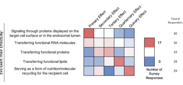
Survey participants were asked to rank several potential mechanisms of EV-cell interaction through which EVs exert their effects from the most (1) to
least (5) likely (Figure 8). Signalling through membrane
proteins was ranked as the most likely mechanism. The next two most likely mechanisms were considered to be the delivery of functional proteins and RNAs, respec-tively. Transfer of functional lipids and nutrition (molecular recycling) were the next most likely.
Certain conditions may need to be met before EV uptake can occur. For example, dendritic cell-derived EVs can only bind activated T-cells that express
high-affinity LFA1 [91]. Cellular uptake of EVs may also be
regulated by competitive mechanisms. For example, when working with unfractionated peripheral blood mononuclear cells (PBMC), MSC-EVs are mainly taken up by monocytes and only scarcely by T and NK cells; yet adding EVs to singularly cultured T or
NK cells increases uptake significantly [92].
Furthermore, small and large EVs may have different membrane compositions that could give rise to diver-gent uptake mechanisms.
EV uptake in vivo also shows selectivity. Red blood cells are the most abundant cells in the body and produce a high number of EVs that contain unique
miRNAs [93,94]. These EVs are only found in blood/
serum and are usually not observed in the surrounding tissue. EVs present in the circulation and interstitial fluid of zebrafish are taken up only by endothelial cells and macrophages, but not muscle cells (despite being
bathed in EVs) [95]. These observations support the
notion that there are likely differential uptake mechan-isms for EVs, depending on cell type and/or EV subpopulation.
Specific proteins or lipids that may prevent uptake in one cell type versus another by inhibiting docking, fusion, or internalization of EVs have not been identi-fied. An interesting consideration is how the extracel-lular matrix (ECM) may influence EV processing or uptake in neighbouring cells. Unlike cell culture mod-els, the in vivo ECM may contain proteins and pro-teases that contribute to or prevent EV uptake. As such, experiments conducted in vitro likely do not recapitulate in vivo uptake mechanisms accurately.
How do variables such as dose, time, pH, and temperature affect EV uptake?
“One dose does not fit all”. EV uptake by cells should be determined through specific, well-designed
dose-dependent uptake profiles of human serum small EVs by murine and human cell lines showed that EVs from the same source could be internalized by one, several
or all of the cell lines depending on the dose [96].
Therefore, considering a fixed EV dose for different target cell lines may entail misleading results.
With respect to time, EV uptake has been
reported in as little as 15 min [87], which is
consis-tent with endocytosis rates, while lysosomal
degrada-tion of EVs has been reported within hours [85,97].
If EV cargo is delivered to cells so rapidly, a rethink of days-long experiments after a single addition of EVs may, depending on the functional readout, be in order.
Considering pH, ILVs are formed in the acidic lumen of the MVB, so exosomal proteins should be
somewhat pH resistant. Nevertheless, pH can alter EV interactions with cells. For example, some viral mem-brane fusion proteins are inactive at pH 7, but undergo conformational changes at pH 5, leading to membrane
fusion [98]. An EV that has entered the endosomal
pathway will also undergo progressive acidification. As a fusion of EVs and cellular membranes would lead to the delivery of the EV content into the cell
cytoplasm [9], understanding how pH influences the
cell–EV interactions is an interesting avenue of research that requires further work.
Regarding temperature, it was previously shown that EVs are taken up by cells at 37°C, but EVs can bind to the
cell surface when endocytosis is blocked at 4°C [99,100].
Experimental manipulation of temperature might pro-vide valuable mechanistic clues into EV uptake.
1 2 1 1 2 3 3 9 3 3 2 3 6 4 5 1 1 1 1 3 6 4 4 3 2 1 1 2 1 2 9 1 2 6 1 1 7 4 6 3 2 3 2 1 1 1 0 1 2 3 4 5 6 7 8 9 10 Survey Question
(Bubble Size & Label =
# of Responders)
E
EV-Cell Fusion
Most cell types, sooner or later, internalize at least a proportion of stained EVs, seemingly regardless of the cells of origin.
EV-cell fusion is most likely to occur through endosomal uptake and acidification.
Proteins on the EV surface are required for most fusion events between EVs and cellular membranes.
EV-cell fusion events are actually quite rare in vivo, and may involve minority subpopulations of EVs and specific uptake pathways.
Current in vitro studies of uptake (anything involving 2D tissue culture plastic substrates) are not worthwhile as unrepresentative of in vivo biology.
Strongly Disagree Neutral Strongly Agree Total # Responders 28 23 24 26 30
Figure 7.EV-cell fusion. Five questions regarding EV-cell fusion were administered in the post-workshop survey. For each question,
participants’ answers are depicted horizontally on a Likert-scale from 0 to 10, with bubble size reflecting of the number of
responders at each point on the scale. Responders agree that recipient cells internalize EVs from different cell types through endosomal uptake and acidification, and that proteins on the EV surface are responsible for fusion events. Survey participants are
Membrane fusion and stability
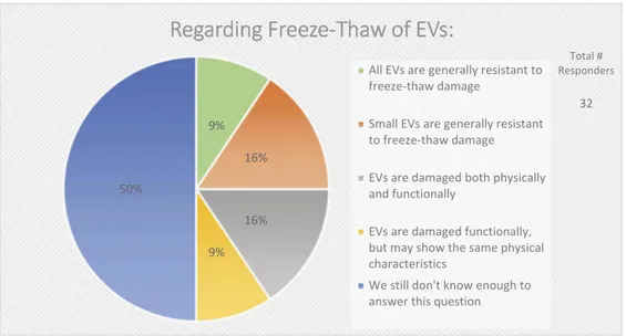
In the context of EV uptake and function, it is important to define EV structural and functional stability. Structural stability is the tendency of an EV to remain intact over time and under different conditions. Functional stability is the retention of a particular effect. On questions of EV stability, over half (67%) of survey respondents agreed that current technologies for studying EVs are not adequate to measure both functional and physical stability of EVs (Figure 9). When asked how freeze-thaw cycles may affect EV stability, 50% felt that more must be learned about
potentially detrimental effects (Figure 10).
What about the relationship between fusogenicity and stability? Is an EV that fuses with a cellular
mem-brane inherently“unstable”? Here, the molecular
com-position is likely to define membrane stability and tendency to fuse. Although membranes are coated with sugars and proteins, it is the lipid composition that most heavily dictates thermodynamic favourability for fusion. In principle, all vesicle-cell membrane fusions are thermodynamically favourable. The barriers to fusion are however kinetic, which may be lowered in relation to lipid compositions. For example, the inser-tion of cholesterol influences curvature and fusion
properties of small vesicles [101–103]. Cone-shaped
phospholipids induce high curvature, while those of cylindrical shape are found in more planar membranes. Fusion is thermodynamically favourable for more curved vesicles, as it brings the vesicles to a lower energy state. Small vesicles have a higher curvature and tighter membranes, lending to a very high-energy state and a possible predisposition for fusion. Supporting this likelihood, a portion of synthetic small unilamellar vesicles fuses with each other given
enough time [104,105]. Contrastingly, the recovery of
EVs as small as 30–50 nm in diameter from biological
and culture fluids suggests a certain stability of even small EVs or subsets thereof. EVs may be more likely to fuse after isolation than when in a complex mixture such as plasma. However, current techniques cannot easily distinguish between a large EV and a similarly-sized EV that formed by the fusion of several smaller vesicles. When asked about the relationship between EV size and stability, 50% of respondents believed that
size has a large impact on physical stability (Figure 9).
EV-cell fusion is thermodynamically favourable but high kinetic barriers prevent spontaneous membrane coalescence. Lowering kinetic barriers can alter lipid membranes, including increased bilayer curvature, and the presence of protein-based fusion catalysts.
Total # Responders 30 30 29 28 29
Figure 8.EV-cell interactions. In the post-workshop survey, participants were asked to rank order the most to least likely ways in which EVs interact with target cells. Answers are depicted in a heat map, with pink shades indicating a higher number of responders, and blue indicating a lower number of responders. Responders believe that EVs primarily interact with target cells by signalling through proteins displayed on the target-cell surface or endosomal lumen. Transferring functional RNA, proteins and lipids is seen as a secondary effect. Most believe that EVs are indirectly a form of nutrition or molecular recycling for recipient cells.
The functional capacity of an EV is likely dependent on membrane protein topology. Overall, membrane protein composition appears to be heavily influenced by EV size, the cell type of origin and cellular activation state. One technique to study EV membrane composi-tion is free radical incorporacomposi-tion or electron-dense lipid labelling. Incorporating free radicals in membrane samples can provide useful topological information on both the membrane and/or associated/integral proteins
based on the preferential localization of the radical
used [106,107]. On the other hand, using lipids
con-taining radicals at different positions in the tail could help provide information about the transmembrane
region itself [108]. Some new techniques to make
pseudo-membranes containing differently shaped
phospholipids can allow for further study of how com-position can influence membrane curvature and fusion
by using nuclear magnetic resonance (NMR) [109].
9% 16% 16% 9% 50%
R
Regarding Freeze-Thaw of EVs:
All EVs are generally resistant to freeze-thaw damage
Small EVs are generally resistant to freeze-thaw damage
EVs are damaged both physically and functionally
EVs are damaged functionally, but may show the same physical characteristics
We still don't know enough to answer this question
Total # Responders
32
Figure 10.Storage of EVs. In the post-workshop survey, participants were asked to choose from five options whether or not they believe freeze-thawing causes damage to EVs. Responders agree that we do not know enough about how freeze-thawing affects EV stability, uptake, and functionality.
3 2 4 7 4 5 1 2 1 1
3 1 2 7 4 5 1 2 1
0 1 2 3 4 5 6 7 8 9 10
Survey Question
(Bubble Size & Label =
# of Responders)
E
EV Stability
Current technologies are adequate to measure both functional and physical stability of EVs.
Physical stability of EVs (defined here as the tendency to maintain vesicular form) is related to size.
Strongly Disagree Neutral Strongly Agree Total # Responders 30 26
Figure 9.EV stability. Two questions regarding EV stability were administered in the post-workshop survey. For each question,
participants’ answers are depicted horizontally on a Likert-scale from 0 to 10, with bubble size reflecting of the number of
responders at each point on the scale. While EVs are physically stable, most survey participants believe that current technologies need to be improved to simultaneously measure the functional and physical stability of EVs.
Many questions remain to be answered about EV functional stability: Is the physical structure or the encapsulated content most important? If the structure of an EV has been compromised, does that necessarily affect its carrier/delivery function? Also, does the pre-sence or abpre-sence of cargo reciprocally influence EV stability?
Finally, it is worth noting that our knowledge on EV membrane interactions with inorganic nanostructures and surfaces is poor and fragmented, albeit of key importance for future EV processing and engineering technologies (e.g. colloidal gold nanoplasmonic assays
[110], microfluidics/lab-on-chip applications [111], EV
supported lipid bilayers [112]).
In vivo administration of EVs: how membrane components and associates affect distribution
One of the most alluring aspects of EVs is the potential for the delivery of therapeutic drugs/molecules, which requires an understanding of the biodistribution of EVs introduced to healthy and diseased organisms. When introducing EVs to in vivo models, many factors need to be considered, including dose, route of administra-tion, source of the administered EVs, and techniques for assessing biodistribution (i.e. labelling).
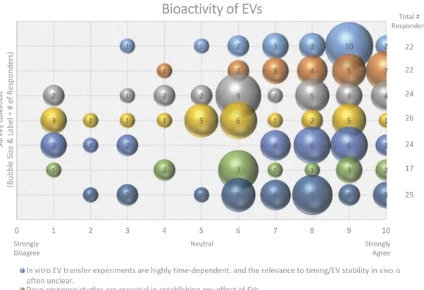
An overwhelming majority (87%) of workshop sur-vey respondents suggested that EV transfer experi-ments are highly time dependent, and that the in vivo
1 1 2 3 3 10 2 1 2 3 4 5 7 2 1 2 2 9 1 5 3 4 3 1 1 1 5 6 1 2 5 1 2 6 6 4 2 1 3 2 3 1 1 7 2 1 1 2 1 5 4 7 2 3 0 1 2 3 4 5 6 7 8 9 10 Survey Question
(Bubble Size & Label =
# of Responders)
B
Bioactivity of EVs
In vitro EV transfer experiments are highly time-dependent, and the relevance to timing/EV stability in vivo is often unclear.
Dose-response studies are essential in establishing any effect of EVs. Most EVs in vivo are bioactive.
EVs in circulation (blood) are less likely to be bioactive and are cleared rapidly. EVs are most likely to have a signaling function in tissue, i.e., locally.
Tumor-bearing mice accumulate more EVs in cancer tissue mostly because of vascular leakiness.
The apparently low rates of EV:cell fusion indicated by systems such as the Cre/lox stoplight system may reflect sensitivity or idiosyncrasies of the assay and not imply that fusion is really so rare.
Strongly Disagree Neutral Strongly Agree Total # Responders 22 22 24 26 24 17 25
Figure 11.Bioactivity of EVs. Seven questions regarding the bioactivity of EVs were administered in the post-workshop survey. For
each question, participants’ answers are depicted horizontally on a Likert-scale from 0 to 10, with bubble size reflecting of the
number of responders at each point on the scale. Responders believe that the use of EVs forin vitro transfer experiments is
time-dependent, that dose–response studies are important, and that EVs have a greater functional impact in the local tissue
environ-ment. Survey participants are undecided on how to determine and identify bioactive EVs. The survey reveals the need for improved technology for the study of EV-cell fusion.
![Figure 1 ). ISEV standardization efforts [ 32 , 33 ] suggest that multiple membrane-associated proteins should be measured to demonstrate the presence of the lipid bilayer and thus EVs](https://thumb-eu.123doks.com/thumbv2/123dokorg/5533157.64979/8.914.115.435.87.1098/standardization-multiple-membrane-associated-proteins-measured-demonstrate-presence.webp)