UNIVERSITÀ DI PISA
Scuola di Dottorato di Ricerca in
“Scienza del Farmaco e delle Sostanze Bioattive
”
Dottorato di Ricerca in
“Scienza del Farmaco e delle Sostanze Bioattive”
XIX CICLO (2004-2006)
ALZHEIMER DISEASE:
DESIGN AND SYNTHESIS OF NEW BACE
INHIBITORS
CANDIDATO: Dott.ssa Elisa Ghilardi TUTOR: Prof. Marco Macchia
DIRETTORE DELLA SCUOLA (Prof.ssa Claudia Martini)
INTRODUCTION ...1
ALZHEIMER DISEASE ...2
ALZHEIMER DISEASE GENETICS...4
PHARMACOLOGICAL TREATMENT OF AD TODAY ...5
ALZHEIMER DISEASE PATHOLOGY...8
Β-SECRETASE ...10
BACE INHIBITORS ...15
CHOLESTEROL AND AD……IS THERE A CONNECTION?..25
THE AIM OF THE THESIS...27
SYNTHESIS OF PATENTED COMPOUNDS ...35
3-AMINO-PIPERIDINE DERIVATIVES ...40
ANALOGUE OF TETRALIN DERIVATIVES ...50
CARBAZOLE DERIVATIVES ...58 EXPERIMENTAL SECTION ...81 GENERAL DETAILS ...82 BIOLOGICAL ASSAY ...83 SYNTHETIC PROCEDURES ...85 3-AMINOPIPERIDINE DERIVATIVES...89
ANALOGUE OF TETRALINE DERIVATIVES...95
CARBAZOLE DERIVATIVES ...97
ACKNOWLEDGMENTS ...111
ALZHEIMER DISEASE
Alzheimer disease (AD) is a progressive degenerative disease of the brain that is characterized by neocortical atrophy, neuron and synapse loss, and the presence of extracellular senile plaques and intracellular neurofibrillary tangles (NFTs) (Fig. 1). The primary clinical manifestation of AD is a profound global dementia that is marked by severe amnesia with additional deficits in language, “executive” functions, attention, and visiospatial and constructional abilities.
Fig. 1. Neurofibrillary Tangles and Amyloid Plaques
The neurodegenerative changes occur primarily in the hippocampus and entorhinal cortex and in the association cortices of the frontal, temporal, and parietal lobes (Fig. 2). Although the temporal progression of the neuropathological changes of AD is not fully known, recent studies suggest that the hippocampus and entorhinal cortex are involved in the earliest stage of the disease, and that frontal, temporal, and parietal association cortices develop pathology as the disease progresses. This “spreading” of the pathology from those regions of the brain in which hallmarks of the disease (amyloid plaques, reactive gliosis, NFTs) can be first detected, to other regions of the brain is notable, and
while generally accepted as being a genuine feature of the pathology, no explanation for it has yet emerged.
Fig. 2. Neurodegenerative changes in Alzheimer Disease brain patients
As the population ages, the projected number of individuals that will be affected by dementia, and AD in particular, indicates that a serious public health problem is looming. However, intense research over the past decade has begun to uncover some of the cell and molecular processes leading to neuronal loss with the discovery of possible targets for therapeutic intervention, raising the hope that we may be able at least to halt the progression of the disease.1
Diagnosis of AD is based on clinical features which are confirmed by brain hystopathological evidence. There are three clinical stages in AD progression, mild, moderate and severe. They are associated with progressive decline of cognitive and physical functionalities over 5 – 8 years. The initial stage usually lasts 2 – 3 years and is characterized by short-term memory impairment often with symptoms of anxiety and depression. In the moderate state these symptoms appear as visual hallucinations, false beliefs and reversal of the sleep cycle.2
ALZHEIMER DISEASE GENETICS
In AD, three genes have been clearly identified as causative and one gene as a risk factor. Major clues came from families suffering from autosomal dominant, early onset forms of AD (familial AD or FAD). Other than the fact that FAD is clearly hereditary and manifests itself at earlier ages (<60 years), it is indistinguishable from the sporadic form of AD (with respect to behavioral patterns, disease progression, plaque deposition, tangle formation).
The first identified FAD-causing mutations were found in the gene encoding the amyloid-‚ precursor protein (APP) on chromosome 21. At least five such mutations have been identified. These are found near the proteolytic cleavage sites in APP that result in Aβ, and all lead to increased production of Aβ in general or specifically a 42-residue form of Aβ (Aβ42) in transfected cells, in transgenic mice, and in the plasma of mutant carriers. Two other disease-causing mutations are found within the Aβ‚ region, near an alternate proteolytic processing site; however, the resultant mutant Aβ‚ aggregates in the cerebral vasculature and causes hereditary cerebral hemorrhage with amyloidosis, either with or without features of AD. A related genetic clue that Aβ is involved in the early molecular events leading to AD is the fact that all Down’s syndrome (trisomy 21) patients invariably develop AD by age 50. Down syndrome patients carry an extra copy of the APP gene, located on chromosome 21, and they produce more A‚ from birth and develop amyloid plaques as early as age 12. These early plaques are of the diffuse kind and contain Aβ42 almost exclusively. Because victims of Down syndrome are fated to develop AD at early ages, the observance of Aβ42-specific diffuse plaques suggest that these plaques could be the precursors of the dense, neuritic plaques found in the AD brain.
APP mutations, however, account for only about 10% of FAD cases and only 2% of all incidences of AD. Most FAD is caused by missense mutations in the genes for presenilin-1 (PS1) and presenilin-2 (PS2), located on chromosomes 14 and 1, respectively. These genes encode multi-transmembrane proteins, the normal biological roles of which were completely unknown upon discovery. More than 70 different mutations in the presenilins have been identified that lead to FAD. Virtually all are missense mutations, and these are located in various regions of the primary sequence
in Aβ42 production, implicating this particular Aβ species in the etiology of AD. Considerable effort has gone into identifying genes associated with the more common late-onset, sporadic form of AD. Such searching led to the pinpointing of the ApoE gene. ApoE is a lipid transport protein that comes in three allelic variants: E2, E3, and E4. The ApoE4 allele is a major risk factor for late-onset AD: those who carry one or two copies of this allele may not necessarily develop AD, but these carriers are at substantially increased risk. As a function of age, the risk increases with the number of ApoE4 alleles inherited, with the mean age of onset being some 15 years earlier and the incidence of AD 10 times more likely in individuals who inherit two E4 rather than two E3 alleles. Inheritance of ApoE2 decreases risk of the disease and increases age of onset (i.e., it appears to be a protective factor). Yet some individuals homozygous for E4 show no AD symptoms in their 90s, illustrating the principle that this allele is a risk factor, not a determinant of whether AD will develop. Apparently, the ApoE variants differentially affect Aβ deposition and resulting neurodegeneration. A dose-dependent increase in the density of neuritic Aβ plaques and vascular Aβ deposits are associated with ApoE4. More recently, studies with transgenic mice have demonstrated that apoE is required for amyloid formation and glial activation caused by FAD-mutant APP.3
PHARMACOLOGICAL TREATMENT OF AD TODAY
There is a prominent loss of cholinergic, noradrenergic, dopaminergic, and GABAergic neurons transmission in AD. Neurotransmitter-based treatments with cholinesterase inhibitors (ChEls) and N-methyl-D-Aspartate (NMDA) receptor antagonists are now in current use. Hence today the treatment of AD is based on a control of symptoms of the disease.
Cholinesterase inhibitors. The aim is to enhance the cholinergic neurotransmission.
This strategy is based on evidences found in AD patients: (1) brain biopsies and authopsy have shown that AD patiens have reduced cortical activity of ChAT which is the enzyme that synthesizes Ach from choline. (2) There is a loss of cholinergic neurons in the nucleus basalis of Meynert and other subcortical nuclei. These large neurons are mainly responsible of cerebrocortical cholinergic supply and play an important role in
revealed the presence of NFT in these neurons. (3) Furthermore cholinergic antagonists, such as scopolamine interfere with learning ability. While cholinergic agonists have been found to facilitate learning, which lends support to the important physiologic role of acetylcholine in attention and learning.
ChEls act by slowing the biochemical breakdown of acetylcholine; the result is that cholinergic neurotransmission is prolonged. Three ChEls are commonly used to treat patients AD: donezepil, rivastigmine, and galantamine (Fig. 3). ChEls are indicated in patients with mild to moderate AD, althought some studies suggested a small benefit also in patients suffering from more advanced stages of AD.
Fig. 3. Cholinesterase Inhibitors
Memantine. Overstimulation of NMDA receptors by glutamate is implicated in
neurodegenerative disorders. Glutamate is the principal excitatory neurotransmitter in the brain. Glutamatergic overstimulation may result in neuronal damage and this a
CH3O CH3O O N HCl . Donezepil (aricept) (1997) O H CH3 CH3 C H3 C H3 C H3 O COOH COOH OH H H O H (S) (R)
Rivastigmine tartrate (exelon) (2000)
N O CH3O H OH H CH3 HBr .
calcium overload and has been implicated in neurodegenerative disorders. Glutamate stimulates a number of postsynaptic receptors including the NMDA receptor, which has been particularly implicated in memory process, dementia, and the patogenesys of AD. Memantine (Fig. 4) is a non-competitive NMDA receptor antagonist and reduces glutamatergic exocitotoxicity. Memantine is used in moderate to severe dementia. This substance acts with a non competitive NMDA receptor antagonism with a voltage-dependance and a low-moderate affinity. Memantine have a fast kinetic of blocking/unblocking the receptor which is very important, infact other NMDA receptor antagonists (es. Ketamine, amantadine) are high affinity compounds with neuropsychiatric side effects. Furthermore the fast block/unblock kinetics means that memantine sits on the receptor just long enough to prevent pathological activation af the glutamate receptors and then quickly goes away when physiological activation of glutamate receptors is needed. Hence memantine preserve the normal-physiologic activation of NMDA receptors which is required for learning and memory.
New studies are necessary to establish whether association between memantine and cholinergic treatment may represent a complementary or even synergistic strategies in AD treatment.2
Fig. 4. NMDA Receptor Antagonist
CH3 NH2
CH3
ALZHEIMER DISEASE PATHOLOGY
AD is caracterized pathologically by the presence in the brain of beta-amyloid plaques (Aβ), neurofibrillary tangles (NFT), and neuronal loss.
NFT are aggregated filaments of a protein called τ, and the density of these filaments in the brain are related to the severity of dementia. It is unclear why tangles are formed and whether tangles are related to plaques formation (some evidences show that Aβ are present especially in the end stage of the disease and they are unique to AD). Besides a remarkable rate of NFT are the first sign for identification of a neurologic disorder which may not be AD, indeed we can also find these tangles in other neurodegenerative disease.2
The loss of neurons is probably triggered by inflammation around plaques.
Plaques are composed of Beta-amyloyd tissue (Aβ) which tends to aggregate in insoluble forms in the extra-cellular space. Aβ is generated by the proteolytic process of the Amyloid Precursor Protein (APP) which is a type 1 transmembrane protein encoded by a gene on chromosome 21. APP is expressed in neurons and glial cells in which APP is integrated in the ER. The biological functionality of APP is still unknown.
APP is metabolised by two different pathways: a non amyloidogenic pathway and an
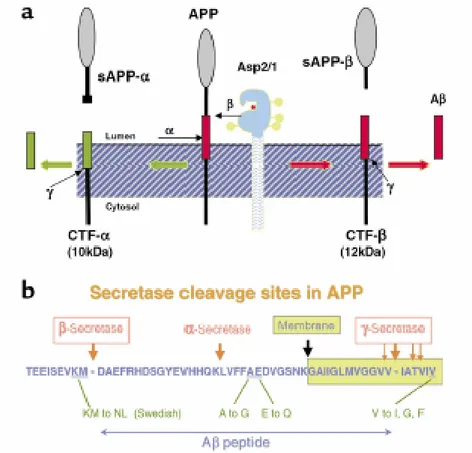
Fig. 5. APP Cleavage and Amyloid Domain
(a) A schematic representation of the sequential cleavages of APP at the β- and γ- sites to generate β-amyloid. (b) The amyloid domain of APP.
In the non amyloidogenic pathway, an α-secretase first cleaves APP between lys16 and leu17 (Fig. 5). Two fragments are originated: sAPPα (which is soluble) and αCTF (88aa). Then γ-secretase cleaves αCTF to generate P3 ( a protein of 3 Dalton weight) and γCTF. APP is processed in this pathway for 90% and the fragments are soluble so they are not implicated in the course of the disease. It is found that sAPPα has a neurotrophic and neuroprotective functionalities against deficiency of glucose and glutamate toxicity. Furthermore sAPPα stimulates neurons growth and regulates synaptogenesis.
The amyloidogenic pathway also involved two sequential cleavages (Fig. 5). APP is first cleaved by a β-secretase to generate a soluble fragment sAPPβ and a membrane bound , 99 amino acid residue, C-terminal fragment (C99). C99 is subsequently cleaved
Aβ42. Approximately 90% of Aβ produced is Aβ40, but Aβ42is more prone to aggregate as fibrils and is the main component of amyloid deposits.2
Hence pharmacological inhibition of β- and γ-secretase could provide a targeted therapy for the treatment of AD.
γ-Secretase would appear to be an aspartyl protease like BACE although γ-secretase is associated to presenilins 1 and 2 to exert its proteolytic and biological activity. Moreover a large number of substrates have been identified for this enzyme such as Notch, Delta and Jagged4 which are important for normal biological processes. Hence inhibiting γ-secretase may lead to a variety of unwanted side effects, especially if the inhibitor was unable to attain high blood-brain barrier permeability and was thus present at high concentrations in the periphery.
Β-SECRETASE
β- and γ- secretase are the enzymes involved in the amyloidogenic pathway for the generation of Aβ. Today there is a great interest in the development of inhibitor drugs against β-secretase. A number of factors have encouraged this research line: first of all the fact that the activity of β-secretase is an early step in the production of Aβ. Furthermore the absence of phenotype in mice devoid of a functional β-secretase gene suggests that the inhibition of this protease during clinical treatment is physiologically tolerated.
β-secretase also termed BACE (β-site App Cleaving Enzyme) is new member of the pepsin family of aspartyl proteinases. BACE is a type I transmembrane protein, the first member of the pepsin family that exist as an integral membrane protein. β-secretase is a novel aspartyl –protease of 501 amino acids containing a single transmembrane domain near the C-terminus as well as a signal sequence and pro-peptide region at the N-terminus.
β-secretase cuts APP at the sequence EVKM*DAEF (the asterisk is pointing out the cleavage site). Furthermore a Swedish family showed a double mutation in the
which causes a Familial AD in Swedish family3 has allowed to develop a first series of BACE-inhibitors which mime the mutated APP sequence.
BACE is highly expressed in the brain and the gene is located on chromosome 11. A homologue called BACE 2 exists in vascularized tissue such as heart, kidney and placenta and the gene has been identified on chromosome 21, but little is known about the significance of this homologue.2 It has been shown that BACE 1 and BACE 2 exhibit 52% amino acid sequence identity and 68% similarity, and BACE 2 cleaves APP and short peptides in a β-secretase-like manner. However, BACE 2 is not well expressed in the brain and this suggests that it may not contribute to the development of AD.5
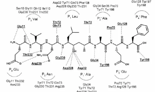
The first structure of β-secretase was reported by Tang et al. Tang group have discovered a potent peptidic BACE inhibitor (OM99-2) (Fig. 6) and have shown an 1.9Å X-ray crystal-structure of the enzyme with the inhibitor (Fig. 7).
Fig. 6. Schematic representation of memapsin 2 residues interacting with the inhibitor in each sub-sites
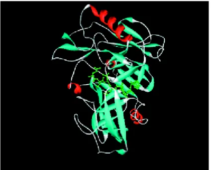
Fig. 7. X-ray crystal structure of β-secretase bound to inhibitor OM99-2
The inhibitor is located in the cleft of the bilobal BACE-structure.
This X-ray structure has given research groups vital information about the enzyme structure and the nature of protein-ligand interactions, and it has been essential for the development of new BACE inhibitors.
The bilobal structure of β-secretase has the conserved general folding of aspartyl proteases with an extensive β-sheet organization. The inhibitor is located in the substrate binding cleft between the amino- and carboxy-terminal lobes, and as expected the transition-state mimicking hydroxyethyl moiety is coordinated with the two active site aspartates (Asp32 and Asp228). As other aspartyl proteases, β-secretase possesses a “flap” that partially covers the cleft, and the backbone of the inhibitor is mostly in an extended conformation. However, β-secretase does display some structural differences, at least compared with pepsin. Four insertions are located on the molecular surface near the aminoterminus of the inhibitor, and together these insertions significantly enlarge the molecular boundary of β-secretase compared with pepsin. Two other insertions are located at the surface near the inhibitor C-terminus. In general, the β-secretase active site is more open and accessible than that of pepsin, and most of the hydrogen bond interactions between the enzyme and the backbone of the inhibitor are highly conserved among eukaryotic and HIV aspartyl proteases. However, the S2 and S4 subsites are relatively hydrophilic and open to solvent, and the hydrophilic character of these
proteases, such as pepsin, gastricsin, and cathepsins D and E, suggesting that these differences could be exploited for the design of selective inhibitors. In contrast, the P3’ and P4’ inhibitor side chains OM99-2 point toward the molecular surface and have little interaction with the protease. The backbone of residues P2’-P4’ deviates from the regular extended conformation, with a kink at P2’, another unusual feature for an aspartyl protease that might be turned to advantage in inhibitor design.5
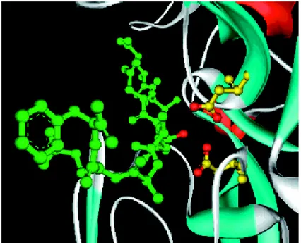
Fig. 8. X-ray crystal structure of β-secretase bound to inhibitor OM99-2
Close-up view showing the interaction of the inhibitor hydroxyl group with the two catalytic aspartates (yellow).
Asp 32 and Asp 228 lie in the centre of the active site and they seems to be critical for catalytic activity. Mutational studies on HIV-1 protease (an aspartyl protease) have shown a complete loss in activity with the removal of either carboxyl.6
A number of mechanism have been proposed for the proteolytic cleavage of peptides by aspartyl proteases. These mechanism are based on X-ray crystal structures, kinetics, and general physical organic principles. In general most of the mechanism that have been put forward are based on the general acid- general base theory, and suggest formation of a tetrahedral intermediate as a key step in the cleavage of the peptide bond. It has been proposed that the catalytic aspartates have an overall –1 charge, specifically one aspartate is protonated and the other one is deprotonated.7,8 Further, a bound water molecule in involved in the most proposed mechanism for proteolysis.9
Computational studies using X-ray structures of BACE revealed that the enzyme can adopt at least two conformations in binding ligands. An open conformation has been identified when BACE is bound with a non-peptidomimetic inhibitor, and a closed conformation has been found when BACE is bound with a peptidomimetic inhibitor.10 One critical issue in any proper description of the mechanism is the identification of the protonation states of the catalytic aspartates, and the location of the protons. Hence other computational studies tried to understand the role of the two aspartates around the enzymatic activity. These studies have shown that the monoprotonated form is preferred when an inhibitor is bound to the enzyme and specially the protonation of the inner oxygen of Asp 228 is preferred over all other proton location.11
On the other hand, the most stable protonation state is the di-deprotonated when the inhibitor is removed.11
BACE 1 is found in the endoplasmic reticulum (ER), Golgi network, cell surface and in endosomes.12,13
Furthermore BACE and APP are both endocytosed into endosomes for cleavage. Endosomes are likely to be the major site for β-secretase processing because of the acidic pH optimum for the enzyme activity. This is quite important in for the design of BACE inhibitors. It should be considered that a molecule have to penetrate across at least five biological barriers: (i) intestinal membrane when absorbed; (ii) Phase I and II metabolic processes, especially in the liver but also in a range of other tissues; (iii) the blood-brain barrier when entering the brain; (iv) the cell membrane when reaching the site of action, and; (iv) the endosomal membrane when binding to BACE.10
All of the above characteristics have to be considered to design and develop new BACE-inhibtors and goes a long way to explain the poor in vivo results obtained with the potent, but non drug-like peptidic and peptidomimetic inhibitors that have been studied by many groups.
BACE INHIBITORS
Many things have to be considered in designing BACE inihibitors: a) The fact that BACE is an aspartyl protease with a characteristic catalytic site, b) BACE contains subsites (termed exosites) that could regulate the catalytic activity giving more possibilities for the inhibitors design, c) the inhibitor have to penetrate the brain barrier, hence we need small molecule, d) the inhibitor have to penetrate the cell and especially endosomes. BACE inhibitors research is divided in two lines: one concerning peptidomimetic structures and one non-peptidomimetic structures.
Peptidomometic inhibitors
Statines. The statine isostere is derived from the natural product pepstatin, which is a
potent inhibitor for many aspartyl protease, although not BACE. Tang and Gosh proposed peptidominetic statine inhibitors based on the sequence of Swedish-mutant APP. OM99-1 and OM99-2 (Fig. 9) have been designed which have shown to be potent inhibitors.14
Fig. 9. Statine Isosteres; Gosh and Tang Inhibitors
OH H2N-Val-Val-Val Ala-Sta-OH O Pepstatine Ki=300 nM Ala-Glu-Phe-OH O CH3 OH H2N-Val-Asn OM99-1 Ki=36 nM
Ala-Glu-Phe-OH O CH3 OH H2N-Glu-Val-Asn OM99-2 Ki=1.6 nM
OM99-1 and OM99-2, peptidic inhibitors of seven and eight residues, contained a non-hydrolysable Leu-Ala based hydroxyethylene dipeptide isostere. Subsequently, detailed information on the subsite from X-ray crystal structure of BACE bound to OM99-2,15-17 lead to the design by Gosh and Tang a more potent inhibitor OM00-3 (Fig. 10) with a Ki value of 0.3 nM.18
Fig. 10. Gosh and Tang Inhibitor OM00-3
Val-Glu-Phe-OH O CH3 OH H2N-Glu-Val-Asn OM00-3 Ki=0.3 nM
Hydroxymethylcarbonyl isosteres. Hydroxymethylcarbonyl isosteres (norstatine) are
truncated versions of the common statine transition state mimic. Kiso et al reported a series of octapeptides with BACE inhibitory activity. KMI-008 (Fig. 11) have shown an IC50 of 413 nM,19 altought the chemical-fisycal characteristics were not suitable as a drug due to its large molecular weight, vulnerability to exopeptidases, and the presence of many natural peptide bonds.
Fig. 11. Hydroxymethylcarbonyl Isosteres by Kiso group
N H2 N H N H N H NH N H N H N H O OH O O O OH O O CH3 O O OH O Ph O OH OH O Ph KMI-008
Then Kiso reported two compounds (Fig. 12) KMI-358 (IC50=16nM) and KMI-370 (IC50= 3.4 nM) as BACE inhibitors with a smaller molecular weight than KMI-008.20
Fig. 12. KMI-358 and KMI-370 by Kiso group
N H2 N H N H N H O O O OH N H O O OH O X COOH Ph X= H; KMI-358:IC50=16 nM X= COOH; KMI-370: IC50= 3.4 nM
The β-N-oxalyl DAP residue in P4 plays an important role in enhancing the inhibitory activity on BACE, but these inhibitors isomerised in acqueous and organic solvent that results in lowering the inhibitory BACE activity. Hence, Kiso have tried to solve the problem modifying the β-N-oxalyl DAP moiety with a tetrazole carbonyl derivative, and two other potent inhibitors were synthesized: 420 (IC50= 8.2 nM) and KMI-429 (IC50= 3.9 nM) (Fig. 13).21
Fig. 13. KMI-420 and KMI-429 by Kiso group
N H2 N H N H N H O O O OH N H O O OH O X Ph N H N N N X= H; KMI-420:IC50=8.2 nM X= COOH; KMI-429: IC50= 3.9 nM
Hydroxyethylene (HE) dipeptide isosteres. In this class of compounds the amide bond
is replaced by an all-carbon framework and a Hydroxyl group in positioned to engage the catalytic aspartates. This type of derivative was used extensively for the development of rennin and HIV-protease inhibitors. Gosh and Tang also reported a series of compounds with a lower molecular weight than OM99-2. In the new molecules P3’, P2’ and P4 were deleted and the P2 side chain was replaced with designed ligand to allow hydrogen bonding in the S2 subsite (Fig. 14).22 Some such compounds have shown a Ki comparable to that for OM99-2.22
Fig. 14. Gosh and Tang. Hydroxyethylene Dipeptide Isosteres
N H N H N H OH CH3 O O Ph N H N H O O SO2Me boc Ki= 2.5 nM
The Elan research group reported their first generation of statine-based inhibitors with an hydroxyethylene dipeptide structure. Three compounds have shown interesting IC50 values in comparison with statine-based inhibitors (Fig.15).23,24
Fig.15. Elan; Hydroxyethylene Dipeptide Isosteres
Ac-Val-Met N H Ala-Glu-Phe-OH OH O IC50= 20 nM n-Pr2N O N H N H O OH CH3 O F OH O IC50= 26 nM
n-Pr2N O N H N H O OH CH3 O F F IC50= 30 nM
Hydroxyethylamine (HEA) isosteres. The hydroxyethylamine isosteres were widely
used as aspartyl protease inhibitors, especially they gave the basis for the Amprenavir (HIV-protease inhibitor).25
The Tamamura group have designed and synthesized a range of BACE inhibitors based on the hydroxyethylamine dipeptide isostere, and TK-3 have shown a μM inhibitory activity against BACE (Fig. 16).26
Fig. 16. Tamamura Group. Hydroxyethylamine Isosteres
H-Glu-Ile-Asp N H NH Met-Val-Leu-Asp-NH2 O OH TK-3 IC50= 0.049 microM
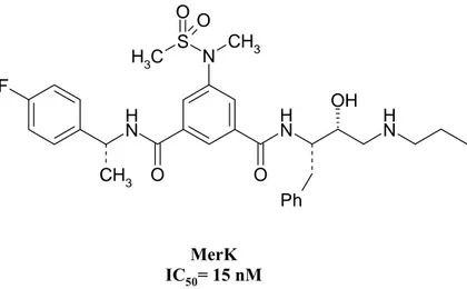
The Merck research group have recently reported cell permeable BACE inhibitors with a low molecular weight containing hydroxyethylamine dipeptide isosteres (fig. 17).27
Fig.17. Merck. Hydroxyethylamine Dipeptide Isosteres F CH3 N H O O N H OH N H Ph N CH3 S O C H3 O MerK IC50= 15 nM
Carbocyclic and heterocyclic peptidomimetic inhibitors. Recently Hanessian group
have reported a novel series of peptidomimetic inhibitors where the hydroxyethylene subunit of the original OM99-2 was replaced at the P1’ position with a cyclopentane ring moiety. In particular a compound (Fig. 18) have shown an IC50 of 25 nM.28
Fig. 18. Hanessian group. Carbocyclic Peptidommimetic Inhibitors
Further, replacement of cyclopentane ring by a cyclopentanone ring resulted in enhancement of inhibitory activity in compound reported below (IC50= 10 nM) (Fig. 19).28 C H3 NH N H N H O O SMe O OH H N H O O N H IC50= 0.025 μM
Fig.19. Hanessian group. Carbocyclic Peptidommimetic Inhibitors
Non peptidomimetic inhibitors
Piperidines. Piperidine derivatives have shown to be able to inhibit the aspartyl protease
renin.29-32 X-ray structure of renin bound to piperidine inhibitors showed that the protonated piperidine nitrogen was positioned between the two catalytic aspartates. Substituents on the 3-position seemed to fill into the S1-S3 subsites, and the 4-aryl ring take up a new binding pocket under the enzyme flap.33
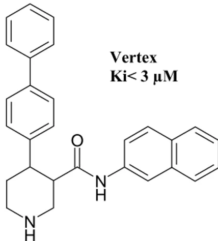
Several research groups have disclosed patent applications about piperidine as BACE inhibitors. A compound have been reported (Fig. 20) by a Vertex patent with a Ki < 3 μM.34 C H3 NH N H N H O O SMe O OH H N H O O N H O IC50= 0.010 μM
Fig. 20. Vertex. Piperidine Derivatives BACE Inhibitors N H N H O Vertex Ki< 3 µM
An Elan patent reported piperidine and other cyclic amines as BACE inhibitors.35,36 Especially a piperidine compound (Fig 21) have shown a modest BACE inhibitory activity (IC50=11 μM)
Fig. 21. Elan. Piperidine Derivatives BACE inhibitors
Furthermore from Actelion 3-/4- aminomethyl piperidines and 3-/4- aminopiperidines37 were disclosed (Fig. 22).38
N H O O Elan IC50= 11 μM
Fig. 22. Actelion. Piperidine BACE inhibitors N O N C5H11 C5H11 Actelion Ki < 3 µM
The De Novo group has described a series of piperazine inhibitors exemplified by compound in Fig. 23 IC50=3 μM.39
Fig.23. De Novo group. Piperazine Derivatives BACE inhibitors
Miscellaneous chemotypes. Natural substances and other small molecule have been
reported as BACE inhibitors.
(-) Epigallocatechin gallate (Fig. 24) has shown to be the most potent inhibitor of BACE with an IC50 of 1.6 μM. It is also non-competitive with substrates.40
OH N N O H Cl Cl Br Br De Novo IC50= 3 μM
Fig. 24. (-) Epigallogallocathechin gallate O OH O H O OH OH OH OH OH OH (-) epigallocathechin gallate
Genetics company performed a pharmaco-phore search starting from the crystal structure of OM99-2 bound to BACE and reported several new classes of inhibitors (Fig. 25).41
Fig. 25. Genetics Company BACE Inhibitors
S N N NH S Cl Cl Cl OH N N H IC50= 10 µM IC50= 14.4 µM
Takeda research group disclosed trisubstituted long chain-amines with more than one aromatic ring in the chain, in particular a tetralin compound (Fig.26) have shown an inhibitory activity of 250 nM on BACE.42
Fig. 26. Takeda group. Tetralin Derivatives BACE inhibitors
O
N
IC50= 250 nM
CHOLESTEROL AND AD……IS THERE A CONNECTION?
Research over the last 10 years has revealed some correlations between yholesterol and the Alzheimer’s disease. It is increasingly recognized that coronary artery disease,43 hypertension,44 atherosclerosis,45 diabetes,46 hypercolesterolemia47 increase the risk for the AD onset.
Cholesterol is a necessary component of all cell membranes and is important both as a structural component and as a modulator of cell fluidity. Cholesterol is synthesized in the body and it is also obtained through the diet from animal products. Cholesterol is carried in the blood on lipoproteins which are divided in different classes: chylomicrons, very-low density lipoproteins (VLDL), intermediate-density lipoproteins (IDL), low-density lipoproteins (LDL), and high density lipoproteins (HDL).
The association of cholesterol with dementia may vary depending upon when cholesterol is measured in the life-span and/or relative to the course of the disease. High cholesterol may be a risk factor if measured in mid-life many years before clinical onset, but as the disease pathology progresses cholesterol levels may fall such that it appears that high cholesterol is protective.
APOE has been identified as a major risk gene for AD, including sporadic AD. The APOE gene codes the apolipoprotein E, which has several important functions: it transport lipids between cells,48 is critical for the coordination of cholesterol in the
repair, growth, and maintenance of myelin and neuronal membranes during development or after injury.49
APOE gene exists in three isoforms: ε2, ε3, ε4. Normally, the ε3 allele is the most common followed by the ε4 allele.50 Several studies have shown that inheritance of the APOE ε4 allele is associated with elevated cholesterol levels and an increased risk of developing AD compared to individuals with only the APOE ε2 or APOE ε3 alleles.51-53 Although the mechanism behind the contribution pf APOE ε4 to an increased AD risk is currently unknown.
There is the idea that statines could have a role in decreasing the risk of AD. Statines inhibit HMG (hydroxyl-3-methyl-glutaryl-CoA)54 which is involved in cholesterol synthesis. The result of inhibiting this enzyme is a decrease of LDL levels and an increase of HDL levels. Recent studies have shown that reduction in cholesterol levels, due to the statines effect, increases the non amyloidogenic α-secretase activity55 and decreases BACE activity perhaps with an inhibiting mechanism.56
However the research in this field is in its infancy and further rigorous studies to reveal the real relationship between cholesterol and AD risk and precise regulation mechanisms are required.
This PhD thesis has been carried out in collaboration with Siena Biotech. The aim of this work concerns the design and synthesis of novel active molecules in Alzheimer Disease therapy.
Alzheimer’s Disease (AD) is characterized by the production in the brain of extracellular amiloyd plaques composed largely of the β-amyloid peptide (Aβ). Genetic evidence obtained from familial forms of AD suggests that up-regulation of the production of the 42 amino-acid form of Aβ has a primary role in the disease. The Aβ peptides are generated by successive cleavages of the amyloid precursor protein (APP) by β- and γ-secretases, both of which have emerged as strong therapeutic targets for AD intervention. Our work focused the attention on β-secretase (BACE) which is an aspartyl protease and it is responsible for the initial cleavage of APP.
This PhD work has been developed around the research of new non peptidic BACE inhibitors. Small molecules, which can penetrate blood brain barrier, have to be found in order to reach the action site and regulate Aβ production and aggregation.
In particular, the work reported in this thesis deals with the research of non peptidic compounds with low molecular weight, good stability and good bio-availability.
This PhD work started with the analysis of issued patent disclosing BACE inhibitors. Looking at the literature we found a considerable number of patents and other publications, brought out by well-known companies, disclosing structures that inhibit BACE. A large number of peptidic and non peptidic structures have been reported.
In particular, we focused our attention on Elan, Actelion and Takeda patents that disclosed non peptidic structures with BACE inhibitory activity in the μM range (Fig. 27).
Fig. 27. Patented BACE inhibitors
N H O O N N O O N N H2 S N O O O OH N H O O 1 ELAN 2 ACTELION 3 TAKEDA 4 ELAN
This research work has been divided in four parts:
Synthesis of patented compounds: Compounds 1 and 4 (Fig. 27) have been re-synthesised and then taken as model for the next studies.
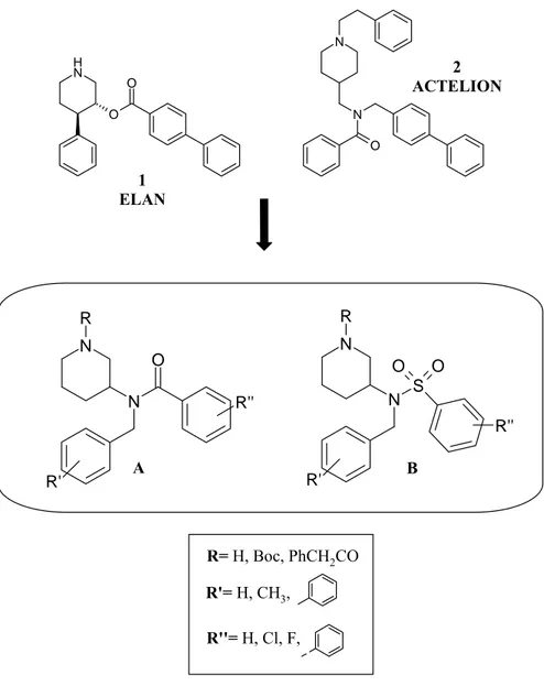
3-Aminopiperidine Derivatives: Looking at the Elan (1) and Actelion (2) structures (Fig. 27) we have designed a new piperidine scaffold, which is an hybrid of the two patented molecules, trying to blend the two patented structures with some modifications. Two series of compounds A and B have been carried out from this work (Fig. 28).
Fig. 28. 3-Amino piperidine derivatives N H O O N N O N N S R O O R' N N R O R' R'' R'' R'= H, CH3, R''= H, Cl, F, R= H, Boc, PhCH2CO 1 ELAN 2 ACTELION A B
In both series of compounds we have introduced a single substitution on piperidine ring in position 3 which is characterized by a nitrogen atom bearing aryl groups widely substituted (R’, R’’), and by a carboamido (A series) or a sulfonamido (B series) moiety. As concerning endocyclic nitrogen piperidine we have introduced a substituent R that may be an hydrogen atom, a Boc group, or a phenyl-acetyl group.
Analogue of Takeda Tetralin Derivatives: Based on structure 3 disclosed by Takeda group a new compound 5 (Fig. 29) has been designed and synthesised.
Fig. 29. Tetralin Derivative
O N O O N O 3 TAKEDA 5
The aim of this work was to create a new molecule with more polar characteristics than the referring Takeda disclosed compound 3. Hence in compound 5 tetralinic moiety has been replaced with benzodioxane, and piperidine group has been substituted with dimethylamino group.
Carbazole Derivatives: In order to find new classes of potential hits as BACE inhibitors
a random and focused screen of commercially available compounds was carried out at Siena Biotech. One of the hit classes identified was a series of carbazole derivatives. They found that some carbazole derivatives (fig. 30) showed inhibitory activities on BACE ranging between 1 and 10 μM.
Fig. 30. Screened carbazole derivatives N O H N SO2 R N O H N H Br Br 6 R= H 7 R= CH3 8
Inspired by screened compounds 6, 7 and 8 five series of molecules came out. Two first series C and D (Fig. 31) were based on a carbazolic structure 6 and 7, where R’ and R’’ are aryl group widely substituted and X may be a carboamidic (C series) or a sulfonamidic (D series) moiety.
Fig. 31. New carbazole derivatives: C, D series
N O H N SO2 R N O H N R' X R'' R'= Aryl, Alkyl R''= Aryl 6 R= H 7 R= CH3 C Series X=CO D Series X=SO2
Compound 6 and 7 also ispired two other series of new molecules: indole derivatives and tetrahydrocarbazole derivatives (Fig.32). We have proposed a sulfonamidic structure where carbazole group was substituted with an indole (E series) and a
Fig. 32. New carbazole derivatives: E, F series N O H N SO2 R N OH N R' SO2 R'' N OH N R' SO2
R'' R'= Aryl, AlkylR''= Aryl
R'= Aryl, Alkyl R''= Aryl E Series Indole derivatives F Series Tetrahydrocarbazole derivatives 6 R= H 7 R= CH3
Finally screened compound 8 gave the start for the last series of β-naphtylaminic derivatives (G series) where we wanted to explore the activity of naphtylaminic compounds making modification around 3,6-di-Bromo-carbazolic moiety (Fig. 33).
Fig. 33. β-naphtylaminic derivatives: G series
N O H N H Br Br N O H N H N O H N H N O H N H G Series ß-Naphtylaminic derivatives 8 G1 G2 G3
All the synthesised compounds have been tested on BACE at highest concentration of 20 μM with a time-resolved quenching assay developed at Siena Biotech laboratories in order to evaluate a possible BACE inhibitory activity.
SYNTHESIS OF PATENTED COMPOUNDS
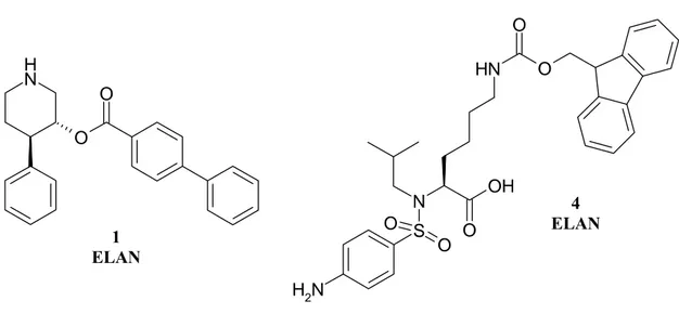
In the first part of this PhD work we have been interested in the synthesis of patented compounds 1 and 4 (Fig. 34) in order to take their strucuture as model for the next studies.
Fig. 34. Patented Elan and Actelion compounds
N H O O N H2 S N O O O OH N H O O 4 ELAN 1 ELAN
Patented synthesis and adapted strategies have been followed for the synthesis of these compounds.
Synthesis of compound 1 :
The original patent37 described a well-constructed synthesis which would have involved many steps showing disadvantages in term of yields. Moreover, it would have started from a non commercially available compound which would have to be synthesised from appropriate precursors without clear literature references reported in the patent (Fig 35).
Fig. 35 Original patented synthesis
N O O Si Ph Ph boc N H O O 6 steps
Therefore, we planned a novel synthetic pathway (scheme 1) referring to literature and well known procedures also improved in our laboratories.
A Grignard reaction with phenylmagnesium bromide has been made on the commercially available N-benzyl-4-piperidone 9. After one night the reaction afforded compound 10 with a good yield (99%). Compound 10 has been dehydrated with a 20% solution of H2SO4 in CH3COOH to obtain olefine 11. After purification by flash chromatography olefine 11 has been submitted to an oxidative hydroboration with a boron-DMS complex. The reaction afforded alcool 12 where the hydroxyl group has been introduced into the 3 piperidine position, trans with respect to the phenyl group. Alcool 12 has been purified by flash chromatography (yield 68%). A traditional method for esterification reaction has been done to obtain compound 13.
Scheme 1. Compound 1 Synthesis N O MgBr Et2O N OH N BH3-DMS complex THF NaOH/H2O2 N OH Cl O Et3N/DMAP CH3CN N O O H2/Pd/C EtOH/CH3COOH N H O O H2SO4 sol 20% CH3COOH 10 11 12 13 1 9
Hence alcool 12 has been treated with 4-biphenyl-carbonyl-chloride in the presence of Et3N as the base, and DMAP as the catalyst. Subsequently, final product 1 has been obtained by a catalytic hydrogenation. Compound 13 has been dissolved in EtOH and CH3COOH under a H2 atmosphere with 10% Pd on carbon. The desired compound 1 has been obtained with a 80% yield after purification by flash chromatography.
synthesis of compound 4:
Original patented reaction conditions57 have been mainly followed for compound 4. The synthesis has been shown in scheme 2.
Commercially available N-Boc-L-Lysine 14 has been treated with benzylchloro formiate in a 2N NaOH solution to protect the free amine group. After 2 hours the reaction afforded the desired product 15 in good yields. Hence, the carboxyl moiety has been protected with a benzyl group by reaction with benzyl-bromide in DMF as solvent. The desired product 16 has been purified by flash chromatography and a yellow oil has been obtained (87% yield). Reaction for 2 hours with a solution of trifluoroacetic acid has cleaved the Boc group and afforded compound 17.
The amine group has been functionalised, first with an isobutyl moiety by a reductive amination (compound 18) and then with a sulfonamidic moiety through reaction with the 4-nitrobenzensulfonyl chloride in classical sulfonylation method (compound 19). An overnight catalytic hydrogenation, with 0.1 equivalent of Pd, should have reduced the nitro group to an amino group, and subsequently released the carboxyl moiety and the amine protected with the benzyl group. These conditions, reported in the patent did not lead to the desired product 21, but to a compound where only the nitro group had been reduced with no removal of the Cbz and benzyl groups (compound 20).In this case we have optimised reaction conditions increasing Pd quantity to 0.4 eq and also increasing reaction time to 4 days. In this way the catalytic hydrogenation led to the desired product 21 with a 65% yield. The last step was the introduction of fluorenyl group by reaction with the Fmoc-chloride and a mixture 1:1:1 of 2N aq K2CO3, THF and CH3CN. Final product 4 has been obtained in 44% yield.
Scheme 2. Compound 4 Synthesis Boc-NH NH2 COOH O Cl O Boc-NH COOH N H O O Br DMF/K2CO3 Boc-NH N H O O O O CH2Cl2/TFA N H O O O O N H2 CHO MeOH/CH3COOH NaCNBH3 N H O O O O N H S Cl O O O2N DIEA/CH2Cl2 NH2 N COOH S NH2 O O N H O O O O N S O NO2 O H2 - Pd/C CH3COOH Fmoc-Cl K2CO3/THF/CH3CN N H N COOH S NH2 O O O O N H O O O O N S O NH2 O H2 - Pd/C CH3COOH NaOH 2N overnight 4 days 14 15 16 17 18 19 20 21 4
3-AMINO-PIPERIDINE DERIVATIVES
The second part of this thesis is based on the study of patented compounds from Elan 37 and Actelion39 patents (Fig.36). We started from the fact that compounds 1 and 4 (Fig. 36) showed some structural analogies: piperidine, bi-phenyl, and aryl groups are represented in both structures.
Fig. 36. Compounds from Elan and Actelion patent
N H O O N N O 1 ELAN 4 ACTELION
In order to analyse spatial disposition of these three common pharmacophoric groups, a theoretical study has been performed in Siena Biotech. Viewer Pro Program was used to create a superimposition of referring structures from Elan an Actelion patents (Fig. 37). Compound alignment was carried out using Accelrys DS Pro software. Putative common features were first tethered and a flexible overlay carried out. Subsequently the molecules were minimised prior to a second rigid overlay to give the alignment hypothesis shown in. The resulting 3D model obtained from the program allowed us to analyse in detail the spatial conformation of disclosed reference compounds and their structural analogies.
Fig. 37. Comparison of the Actelion (green) and Elan (grey) molecules
Superimposition was obtained by fitting the piperidine nitrogen of two reference compounds, and Heteroatoms are coloured by atom type (N blue, O red).
From the spatial superimposition of the two compounds we can observe that i) the two piperidine rings are aligned; ii) the two biphenylic groups of two referring compounds efficiently superimpose in spite of the fact that they are in different positions inside the original molecules; iii) the phenyl ring of compound from Elan patent resulted aligned to the benzoyl one of the compound from Actelion patent, albeit in the first one this scaffold is directly linked to the piperidine ring and in the second one is sensitively far away.
Fig. 38. Carbazole derivatives: C, D series N N S R O O R' N N R O R' R'' R'' N H O O N N O R'= H, CH3, R''= H, Cl, F, R= H, Boc, PhCH2CO 1 ELAN 4 ACTELION A B
Looking at the Elan and Actelion structures (1 and 4) two new series of hybrid piperidine scaffold A and B (Fig.38) have been designed trying to blend the two patented structures with some modifications. We have introduced a single substitution on piperidine ring in 3 position which is characterized by a nitrogen atom bearing variously substituted (R’, R’’) aryl groups; in A series a carboamido moiety and in B series a sulfonamido moiety were synthesised. As concerning nitrogen piperidine it may be unsubstituted, or we have introduced a substituent R that may be a Boc group, or a phenylacetyl group.
with Siena Biotech it has been possible to learn this new methodologies and, a large number of compounds have been easily synthesised.
Scheme 3. A Series synthesis:
N NH2 boc N NH boc N N boc O NaCNBH3/CH3COOH DCM DCM PS-DIEA/DMAP CHO R' COCl R'' R' R' R'' 23-26 A1-A7 23 R'=H 24 R'=4-Ph 25 R'=2-Ph 26 R'=3-Ph 22
Scheme 3a. A Series Synthesis
TFA/CH2Cl2 N N boc O R' R'' N N H O R' R'' A1-A2 A8-A9
Scheme 3b. A Series Synthesis
N N H O PhCH2COCl PS-DIEA/DMAP DCM N N O O R' R' R'' R'' A9 A10
Commercially available 3-Amino piperidine (Scheme 3) has been treated with the respective aldehydes to obtain amines 23-26. The reaction gave good yields for the aldehydes (> 90%) and the products have been purified by ionic exchanged pre-packed silica columns, which are specific for purification of molecules which have basic moieties. The second step for the synthesis of 3-amino piperidines (A series) was the acylation of amines 23-26 with the R’’ aryl-acyl-chlorides. In this reaction PS-DIEA was used to neutralise the reaction mixture. PS-DIEA is dimethyl-isopropyl-ethyl amine which is supported on poly-styrene. This resin captured HCl that was released during the reaction and remained in a solid state. Hence we had got rid of it by filtration, avoiding problems of purification. Purification of compounds A1-A7 has been carried out with pre-packed silica gel columns. Boc-clevage (Scheme 3a) has been done by reaction of compounds A1-A2 with a solution of TFA in CH2Cl2. The reaction was completed in 2 hours and led to the desired compounds (A8-A9) generally in good yields. Both compounds A8 and A9 have been purified with ionic exchanged pre-packed columns. The last step was the acylation of compound A9 at piperidinic nitrogen with a phenyl-acetyl group (Scheme 3b). Reaction conditions have been the same of the acylation in step two with good yields for compound A10.
Synthesis for B series (Scheme 4 and 4a) was similar to that of the same of A series, and it started from amines 23-26 obtained as described in scheme 3.
Scheme 4. B Series synthesis
N NH boc N N S boc O O DCM PS-DIEA/DMAP SO2Cl R'' R' R' R'' 23-26 B1-B7 23 R'=H 24 R'=4-Ph 25 R'=2-Ph 26 R'=3-Ph
Scheme 4a. B series Synthesis
TFA/CH2Cl2 N N S H O O PhCH2COCl PS-DIEA/DMAP DCM N N S O O O R' R' R'' R'' N N S boc O O R' R'' B8 B9 B1
Amines 23-26 was submitted to sulfonylation with the R’’ aryl-sulfonyl-chlorides (compounds B1-7). The outcome of step two was not the same for acylation or sulfonylation in series A and B, and generally we obtained the best result and yields with sulfonyl reagents (TLC have shown a clearer reaction mixture in sulfonyl reactions than acyl chlorides ones). The purification for compounds B1-B7 has been carried out with pre-packed silica column.
Boc-clevage (Scheme 4a) has been done by reaction of compound B1 with a solution of TFA in CH2Cl2. The resulting compound B8 has been purified with ionic exchanged pre-packed silica columns.
The last step (Scheme 4a) was the acylation of compound B8 at piperidinic nitrogen with a phenyl-acetyl group. Reaction conditions have been the same of the acylation, with PS-DIEA DMAP and CH2Cl2, with good yields for compound B9.
RESULTS and DISCUSSION
Series A and B compounds have been shown in table 1 and 2 and percentage of BACE inhibition have been reported. All synthesised compounds have been tested on BACE with a time-resolved fluorescence quenching assay at the highest concentration of 20 μM.
Table 1. Type A Compounds N N R O R' R'' A Compound R R’ R’’ % inhibition A1 Boc H 4-Ph No activity A2 Boc 4-Ph H 27 A3 Boc 4-Ph 4-Ph No activity A4 Boc 4-Ph 2,4-Cl 32 A5 Boc 4-Ph 4-F 29 A6 Boc 2-Ph H No activity A7 Boc 3-Ph H No activity A8 H H 4-Ph No activity A9 H 4-Ph H No activity A10 O 4-Ph H No activity
Type A Compounds: In A series, as concerns R group on nitrogen piperidine atom, Boc
cleavage led to a loss of activity (A8, A9), moreover the substitution with a phenylacetyl group (A10) showed again no inhibitory activity on BACE. Results in Table 1 also showed that when benzylamine group was unsubstituted (R’=H) no BACE inhibitory activity have been reported (A1, A7). A percentage of inhibition on BACE
moiety (A2); whereas, if a phenyl group is simultaneously introduced in position 4- of the two aromatic portions (R’= R’’= 4-Ph) we found a loss of activity (A3). Halogen atom R’’ introduction into the benzenecarbonyl position is tolerated (A4, A5). A loss of activity is also reported shifting phenyl group R’ in position 2- or 3- on benzylaminic moiety (A6, A7).
Table 2. Type B Compounds
N N S R O O R' R'' B Compound R R’ R’’ % inhibition B1 Boc H 4-Ph No activity B2 Boc 4-Ph H 30 B3 Boc 4-Ph 4-Ph 40 B4 Boc 4-Ph 2,4-Cl No activity B5 Boc 4-Ph 4-F No activity B6 Boc 2-Ph H No activity B7 Boc 3-Ph H No activity B8 H H 4-Ph No activity B9 O H 4-Ph No activity
Type B compounds: In B series, just like in A series, no BACE inhibitory activity was
found when piperidine nitrogen atom was not substituted (B8). The introduction of a phenyl-acetyl group (R) on piperidine ring (B9), led to a loss of BACE inhibitory activity. Furthermore, we found again that BACE inhibition is preserved when a phenyl substitution was on position 4- benzylaminic moiety (B2). Whereas, in this case a simultaneously introduction of 4-phenyl on both aromatic substituents is tolerated (B3). Contrairly to A series, in B series we found a loss of activity when halogen atom was introduced as R’’ on phenyl sulfonamidic group (B4, B5). Once again, shifting phenyl group R’ from 4- to 2- or 3- position on benzylaminic moiety, a loss of activity was reported (B6, B7).
ANALOGUE OF TETRALIN DERIVATIVES
Research of tetralin derivatives has been based on a detailed study of Takeda patent 42 where the key compound 3 has been reported to inhibit BACE with an IC50 of 0.35 μM (Fig. 39).
Fig. 39. Patented compound by Takeda group
O
N
3 TAKEDA
In this study, the patent has been analysed in detail to examine all disclosed structures reported. This analysis showed that lateral moieties of the the general formula (Fig. 40), Ar, X, Y and amine group were mainly explored, whereas central moiety AB (circled in red) did not cover many disclosed structures. In fact, we found that central core could be only a tetralin, a 1,2 dihydronaphthalene, or a tetrahydroquinoline. (Fig. 40).
Fig. 40. Central cores disclosed in Takeda patent N R Y N R' R'' X Ar A B
Hence, we planned to work on possibile replacements of the central cores reported in the patent. The idea was to insert in this central core heterocyclic moieties in order to increase the Takeda molecules polarity. Infact, this compound has shown an interesting BACE inhibitory activity, but his high lipophilicity could be a problem in his development because of a predicted poor oral bioavailability.
Looking for new heterocicles a large number of structures have been designed with new central cores.
Furthermore another patented structure (27) reported by takeda has been considered (Fig. 41). This molecule was less active on BACE (IC50= 2.93 μM) but it was a little more polar than compound 3 because of the presence of dimethylamine instead of piperidine.
Fig. 41. Tetralin structure reported by Takeda group
O
N
27 TAKEDA
Approximately a hundred structures, unprecedented in the literature, have been designed related to the referred takeda compounds. All new molecules contain different central cores from compounds disclosed by Takeda group. Lateral chain was reduced to a methylene group instead of an ethylene, as reported in patented compounds 3 and 27. This modification should help in reducing polarity of the new molecule, and besides, in Takeda patent structures with shorter lateral chain were also reported. Furthermore, looking across literature references, we have seen that a shorter lateral chain , with only a methylene, was synthetically more accessible to introduce than the ethylene.
New designed structures, hence, have been submitted to a theorical study based on Lipinsky, Veber and Norinder’s rules. This study, based on predicted theoric calculus, allowed us to select 7 structures (Fig. 42) with the best physico-chemical characteristics to penetrate the Blood-Brain Barrier.
In this study, all the designed molecules have been evaluated on the base of restrictions of the three most important parameters for a good bio-availability for CNS: RBN (number of rotatable bonds), MlogP (parameterization of the hydrophobicity) and BBB (blood-brain barrier permeability) were made to their optimum value in order to obtain compounds with better predicted physicochemical properties.
Fig. 42. New structures related to Takeda disclosed compounds O O N O O O N O O N O O N N O O N O N O O N O O O N
Based on this results a further selection has been done considering the synthetic accessibility of these new compounds. Therefore, a 1,4-benzodioxanic compound (5) has been sorted out as the best candidate for our synthetic work (Fig. 43).
Fig. 43. 1,4-benzodioxanic structure O O N O 5
Central core for compound 5 was a 1,4-benzodioxane moiety that should give to the molecule a higher polar characteristics than referring Takeda compounds (3, 27) due to the presence of two oxygen atoms in the central core and a dimethylamine group instead of piperidine group on the lateral chain.
Synthetic pathway for compound 5 has been carried out according to literature and Takeda patent (Scheme 5).
Commercially available 4-benzyloxy-2-hydroxy-benzaldehyde 28 was treated with epichloridrine to obtain aldehyde 29 58. Aldehyde 29 was reduced submitted to a Bayer-Villiger oxidation to an instable formiate intermediate that spontaneously produced phenol 30 (59). Phenol 30 was converted to the 1,4-benzodioxane by cyclization in KOH 2N (compound 31) (58). A catalytic reduction with Pd/C 10% afforded compound 32 with free phenol group. Hence, we have introduced a bi-phenyl group by reaction with 4-phenylbenzyl chloride in CH3CN (compound 33). The subsequent transformation of primary alcol into a iodide by reaction with imidazole, iodine and triphenyl phoshine (compound 34) 42, allowed us to introduce subsequently a di-methyalmine group to obtaine final desired product 5 42. Compound 5 has been treated with Et2O·HCl to achieve its hydrochloride salt form 5·HCl (17% yield).
Scheme 5. Compound 5 Synthesis O Cl N O O BnO OH OH CHO OBn O CHO Cl OH OBn O OH Cl OH OBn O O O H OH O O OH O O O N O MeOH/H2O2 KOH H2/Pd Cl N H K2CO3/CH3CN O O I O K2CO3/THF I2/Ph3P Imidazol/THF KHSO4 Et2O/HCl O O N O HCl . [1] [3] [1] [2] [2] 29 30 31 32 33 34 5 28 5 HCl
RESULTS and DISCUSSION
Compound 5 has been tested on BACE with a time-resolved fuorescence quenching assay. In spite of the general structure which closely resembled patented compounds 3 and 27, with the exception of the introduction of oxygen atoms in the central core and of the lateral chain cut, unfortunately, compound 5 did not shown any significant inhibitory activity on BACE.
Table 4. Tetralin Derivatives Results
Compound Structure IC50 (μM)
3 O N 0.35 27 O N 2.93 5 O O N O > 10 μM
CARBAZOLE DERIVATIVES
In order to find new classes of potential hits as BACE inhibitors a random and focussed screening of commercially available compounds was carried out at Siena Biotech. One of the hit classes identified was a series of carbazole derivatives, some of which showed inhibitory activities on BACE ranging between 1 and 10 μM (Fig 44).
Fig. 44. Screened carbazole derivatives
N O H N SO2 R N O H N H Br Br 8 6 R= H 7 R= CH3
We then envisaged the possibility of a new series of BACE-inhibitor compounds related to these carbazolic structures.
Some computational studies on compound 6 have been carried out in Siena Biotech to giustify the activity with a possible interaction between carbazole derivatives and BACE on the basis of β-secretase catalytic site.
In the protease family the binding site is usually mapped using a sub-pocket organization. The binding site (Fig. 45) is considered to be divided in two main regions defined by the cutting point of the substrate peptide. The binding site part that recognises the C-terminus region of the peptide is called prime region (S’). The side at the N-terminus of the peptide is termed non-prime region (S). The two regions are mapped in sub-pockets starting from the cutting point towards the periphery of the substrate peptide. The S2’, S1, and S3 pockets were mainly involved in lipophilic
interactions. On the contrary, S1’, S2, S4 pockets were mainly involved in hydrophilic interactions
Fig. 45. BACE Binding Site Map.
O O O O NH2 N H2 HN+ NH O NH O NH O NH2 N H2 HN+ NH3+ OH NH2 O NH3+ OH N H2 NH2 HN+ S1 S3 S2 S1' S2' ASP32 ASP228 ARG235 S4 ARG307 LYS321 ASN233 SER325 GLN74 LYS107 PHE108 THR329 LYS224 ARG128 Cutting Point Prime Region Non-Prime Region [S'] [S]
The main pockets of the BACE1 binding site are shown in this schema. For each pocket hydrophilic interactions are also shown. In red: the catalytic aminoacids Asp32 and Asp228 activate a water molecule that functions as the nucleophile responsible for amide cleavage
Carbazole binding mode hypotheses
With the aim of guiding the exploration of the carbazole class, two putative binding modes based on docking calculation were hypothesised. Docking studies were performed using the X-ray structure of BACE from the Protein Data Bank: 1FKN (Fig. 46).60
Fig. 46. 3D Binding Site Map of BACE on 1FKN co-crystal structure
Due the high pseudo-symmetry of this class of molecules two opposite possible binding modes were found (a and b) (Fig. 47 and 48).
Fig. 47. Carbazole binding mode a
O O O O N H2 NH2 HN N S O OH N O S2 S1 S3 S1' ASP32 ASP228 ARG235 S2'
Fig. 48. Carbazole binding mode b
Carbazole group could occupy the subsites S1, S2, and S3 (fig. 47), but it could place itself also in S1’ and S2’ side (fig. 48).
Inspired by screened compounds 6 and 7 two series of analogous molecules were designed (C and D) (Fig. 49). C and D series of carbazole derivatives were based on a
O O O O N H2 NH2 HN N S O OH N O S2 S1 S3 S1' ASP32 ASP228 ARG235 S2'
series) or a sulfonamidic moiety (D series). In both series R’ may be an aryl or alkyl group such as phenyl, benzyl , phenylethyl, or cyclohexyl, and R’’ is an hydrogen, halogen, methyl, or methoxy group.
Fig. 49. C,D Series N O H N SO2 R N O H N R' O R'' N O H N R' S O O R'' 6 R= H 7 R= CH3 R'=
R''= H, 4-Cl, 4-Me, 3,4-di-Cl, 4-OMe
C D
Compound 6 and 7 also inspired two other series of analogous strucutures: indole derivatives (E series) and tetrahydrocarbazole derivatives (F series) (Fig. 50). We have
indole and a tetrahydrocarbazole and where R’ may be an aryl or alkyl group such as benzyl, or phenylethyl, and R’’ is an hydrogen, halogen, methyl, or methoxy group.
Fig. 50. E, F Series N O H N SO2 R N OH N R' SO2 N OH N R' SO2 R'' R''
E series - Indole derivatives F series - Tetrahydrocarbazole derivatives 6 R= H
7 R= CH3
R'=
Screened compound 8 gave the idea for the last series of β-naphtylaminic derivatives (Fig. 51) where we wanted to explore the activity of naphtylaminic compounds making modification around carbazolic moiety.
Fig. 51. G Series N O H N H Br Br N O H N H N O H N H N O H N H G series ß-Naphtylaminic derivatives
8
G1 G2 G3
As concerns the synthesis of carbazole derivatives, some reaction conditions, already used at Siena Biotech laboratories, have been optimised for our series of compounds (Scheme 6).
Scheme 6. C Series synthesis N H Cl O N O KOH/DMF R'NH2 EtOH N OH NH R' N OH N R' O PS-DIEA/DMAP DCM R'' COCl R'' 37-40 C1-9 37 R'= 38 R'= 36 39 R'= 40 R'= 35
The commercially available carbazole 35 was alkylated by a reaction with Epichlorohydrin in DMF and KOH as base. The reaction led to the desired epoxide 36 with a 50% yield after crystallization from Et2O. This reaction could have two different type of mechanism: 1) SN2: epychloridrine may exchange a chloro atom with the carbazole nitrogen atom (Scheme 7); 2) Epoxide ring opening: the carbazole nitrogen atom may be alkylated by the primary oxirane carbon atom, then epoxide may re-form on the other side by elimination of chloridric acid (scheme 8).
Scheme 7. SN2 N H Cl O N O KOH DMF
Scheme 8. Epoxide ring opening
N H Cl O N Cl O H DMF KOH N O
The resulting epoxide was treated with different amines to obtain amino-alcol 37-40. After one night at 65°C, then we could generally see the product that precipitated in the reaction mixture as a white solid, and the yield ranged between 70 to 80%. Amino-alcol 37-40 were treated with acyl-chlorides in the presence of PS-DIEA as base, and catalytic DMAP in CH2Cl2 and the reaction afforded compounds C1-9 with yields from 30 to 75%.
Scheme 9. D series synthesis N OH NH R' N OH N R' S O O PS-DIEA/DMAP DCM R'' SO2Cl R'' 37 R'= 38 R'= 37-40 D1-14 39 R'= 40 R'=
The D series synthesis (Fig. 9) started from amino alcol 37-40 prepared as in scheme 8. Amino alcol 37-40 were treated with sulfonyl-chlorides in the presence of PS-DIEA as base, and catalytic DMAP in CH2Cl2 and the reaction afforded compounds D1-14 with yields from 30 to 75%.
Synthesis for series E compounds is reported below (Scheme 10).
Scheme 10. E series synthesis
N OH NH R' N OH N R S O O PS-DIEA/DMAP DCM N H Cl O N O KOH/DMF R'NH2 EtOH R'' SO2Cl R'' 43-46 E1-14 43 R'= 44 R'= 45 R'= 46 R'= 42 41
Commercially available indole 41 was alkylated by a reaction with epichlorohydrin in DMF and KOH as base. The reaction leaded to the desired epoxide 42 with a yield of 50% after purification by flash cromtography. Epoxide 42 was treated with different amines to obtain the amino-alcol 43-46. Also in this case after one night at 65°C, we could generally see the product precipitated in the reaction mixture as a white solid and the yield ranged between 70 to 80%. Amino-alcol 43-46 were treated with sulfonyl chlorides in the presence of PS-DIEA as base, and catalytic DMAP in CH2Cl2 and the reaction afforded compounds E1-14 with yields from 30 to 75%.
Scheme 11. F series synthesis N OH N R' S O O PS-DIEA/DMAP DCM Cl O N O KOH/DMF R'NH2 EtOH N H N OH NH R' R'' SO2Cl R'' 49 R'= 50 R'= 49-50 F1-7 48 47
Commercially available 1,2,3,4-tetrahydrocarbazole 47 was alkylated by a reaction with epichlorohydrin in DMF and KOH as base. The reaction leaded to the desired epoxide 48 with a 50% yield after purification by flash cromtography. Hence epoxide was treated with different amines to obtain the amino-alcol 49-50. Also in this case after one night at 65°C, we could generally see the product precipitated in the reaction mixture as a white solid and the yield ranged between 70 to 80%. Amino-alcol 49-50 was treated with sulfonyl-chlorides in the presence of PS-DIEA as base, and catalytic DMAP in CH2Cl2 and the reaction afforded compounds F1-7 with yields from 30 to 75%.