ORIGINAL ARTICLE
Cranial structure and condylar asymmetry of patients with juvenile
idiopathic arthritis: a risky growth pattern
Maria Grazia Piancino1,2&Rosangela Cannavale1&Paola Dalmasso3&Ingrid Tonni4&Umberto Garagiola5& Letizia Perillo6&Alma Nunzia Olivieri7
Received: 5 May 2018 / Revised: 10 June 2018 / Accepted: 12 June 2018 # International League of Associations for Rheumatology (ILAR) 2018
Abstract
The aim of the study was to evaluate the cephalometric differences and condylar asymmetry between patients with juvenile idiopathic arthritis (JIA) and normal control group. Sixty-two JIA patients with a latero-lateral cephalogram and orthopantomography, seeking for orthodontic therapy, and 62 normal matched subjects were comprised in the study. Cephalometric analysis was used for the evaluation of facial morphology while the method of Habets et al. (J Oral Rehabil 15(5): 465–471,1988) was used to compare the condyles in orthopantomography. The significance of between-group differences was assessed using the Mann–Whitney test, as appropriate. The results showed a prevalence of the upper maxilla with hypomandibulia (class II), hyperdivergency with short vertical ramus posterior and posterior rotation of the mandible in JIA children (SNB, ANB, NSL/ML, Fh/ML, NL/ML, ArGo, MLP < 0.0001, ML/Oc P < 0.004, ArGo/GoGn P = 0.02, no difference for SNA). The condyles of the JIA group resulted highly asymmetric (P < 0.0001). The growth pattern of JIA patients resulted clearly different from normal subjects. This serious impairment of the cranial growth may be considered as an indicator of the need for early and continuous orthognatodonthic therapy during the entire period of development for all JIA patients, indepen-dently from temporomandibular joint signs or symptoms. To this end, it is important that rheumatologists and orthognathodontists set up a multidisciplinary treatment planned to control the side effects of a deranged growing pattern, to strictly avoid any orthodontic therapies that may worsen function and growth, and to promote treatments improving the phys-iology and bphys-iology of the cranial development.
Keywords Cephalometry . Condylar asymmetry index . Juvenile idiopathic arthritis (JIA) . Orthopantomography (OPT)
Introduction
Juvenile idiopathic arthritis (JIA) is an autoimmune chronic inflammation of one or more joints, characterized by an onset before the age of 16 years, during childhood and/or adolescent development [1]. One of the affected joints is the temporo-mandibular joint (TMJ) that is a bilateral synovial joint with a high degree of movement, bilaterally coordinated, and influ-enced by cranial structure and occlusion especially during growing.
TMJ involvement by JIA is recognized only in the most severe JIA subtype classification. However, in their everyday clinical practice, rheumatologists know that the TMJ might be involved also in less severe JIA and likely in all affected chil-dren, even when symptoms or signs are not detectable with routine clinical examination nor diagnostic imaging. The TMJ involvement may become clear at a later stage of develop-ment, when the physiological morphology is now lost
* Maria Grazia Piancino
1
Department of Surgical Sciences-Orthodontic division, Dental School, University of Turin, Turin, Italy
2
Turin, Italy
3
Department of Public Health and Paediatrics, University of Turin, Turin, Italy
4 Orthodontic Division, Dental School, University of Brescia,
Brescia, Italy
5
Department of Biomedical Sciences, University of Milan, Milan, Italy
6
Multidisciplinary Department of Medical-Surgical and Dental Specialties, University of Campania, Luigi Vanvitelli, Italy
7 Department of Woman and Child and General and Specialized
Surgery, University of Campania , Luigi Vanvitelli, Italy
resulting in facial asymmetries and functional impairment, even in the absence of previous or actual pain or clinical signs. The TMJ shows three special features: first, it is completely immature at birth. The functional reason for this characteristic is due to the fact that sucking is a vital reflex at birth and it is characterized by antero-posterior, linear shifts occurring at the occlusal plane level. For this reason, the joint is completely flat and immature. The entire develop-ment of the condyle and TMJ will occur during growing from 0 through 21st year of age. Second, as showed in pre-vious studies, the condylar cartilage is not a primary growth center, as the growth plates of the long bones, but it is char-acterized by an adaptive type of growth as showed with histological studies [2–4]. The condylar cartilage acts as a secondary cartilage, with the aim to resist intermittent pres-sure and movements (chewing, swallowing, speaking) and to produce rapid growth during early life [3,5]. The capa-bility to adapt to functions, loads, cranial development, and vectors makes this a unique joint of its kind. Third, it is a bilateral joint with a high degree of movement and the con-dylar components (right and left) are part of a rigid struc-ture, which is the mandible, highly influenced by functional movements. It results that the two sides are simultaneously involved during any type of displacement, both in a sym-metrical or asymsym-metrical way depending on the considered movement.
In our previous study [1], it has been clearly shown that the two condyles of children with JIA grow in a severely asym-metrical way with respect to the control group. This means that one of the two condyles, in these children, is growing faster than the contralateral. The inflammatory status of JIA that involves the TMJs might be responsible of the altered growth pattern of the condyles and mandible resulting in a highly unstable and asymmetrical cranial structure. The asym-metry might be due to the altered cranial growth pattern re-sponsible of functional instability and to the fact that the ill-ness mainly involves the joint subjected to the higher and unbalanced load.
It is known from previous cephalometric studies in normal children that the long face hyperdivergent cranial type with prevalent vertical growth and posterior rotation of the mandi-ble is also frequently associated with asymmetrical cranial growth [6,7]. More specifically, previous cephalometric stud-ies in patients with JIA have claimed that these children are characterized by prevalence of superior maxilla (skeletal class II malocclusion) and long face with posterior rotation of the mandible [8–19]. Cephalometric analysis showed a well-positioned maxilla (SNA near to normal values) and a retruded little mandible (SNB less than normal values). This combination leads to a hyperdivergent skeletal class II with hypomandibulia, posterior rotation of the mandible and aug-mentation of the anterior facial height (NSL/ML and Fh/ML more than normal).
Based on these considerations, we hypothesized that this type of cranial structure (hyperdivergent class II) might be the consequence of an insufficient growth in length of the vertical ramus of the mandible likely related to the inflammatory status of the TMJ responsible of the hyperdivergent cranial structure and, consequently, of the condylar asymmetry between sides. The aim of this study was the evaluation of the impaired cra-nial structure growth together with the condylar asymmetry of JIA patients compared to normal subjects.
Material and methods
Subjects
Sixty-two patients with JIA diagnosed according to the ILAR 2003 criteria (58 females 93.5%; 4 males (6.5%), mean age, 11.1 ± 3.8 years; Table 1), referred at the Department of Orthodontics of the University of Turin, University of Naples, University of Brescia and University of Milan, were included in this retrospective study. The treatment was assigned according to American College Rheumatology (ACR) recommendations [20]. Our patients were treated with non-steroidal anti-inflammatory drugs (NSAIDs) or cortico-steroids according to juvenile idiopathic arthritis (JIA) sub-type at the beginning of the disease. Patients who did not achieve clinical remission [21] were started treatment with methotrexate. A biological drug was associated with metho-trexate in non-responders patients.
Inclusion criteria were (1) diagnosed JIA and rheumatolog-ic treatment and (2) orthopantomography and lateral teleradiography.
Exclusion criteria were (1) any previous orthodontic treat-ment, (2) any maxillofacial surgery, (3) TMJ involvetreat-ment, (4) history of maxillofacial trauma, and (5) genetic diseases, syn-dromes, or other congenital deformities.
Patients were referred to the Orthodontics divisions for routine orthodontic and dental screening.
Controls
Sixty-two subjects, age and sex matched with patients with class I occlusion without crossbite, were selected for the study. The inclusion criteria were (1) normal growth and develop-ment with no significant medical history, (2) bilateral molar class I with minor or no crowding. Exclusion criteria were (1) crossbite, (2) asymmetrical malocclusion, (3) functional devi-ation of the mandible, (4) history of maxillofacial trauma, (5) TMJ pathology, and (6) prosthodontic treatment and orthognathic surgery. The age and sex distributions in the two groups are shown in the Table1.
Cephalometric examination
The craniofacial structure was evaluated by cephalometric analysis of the latero-lateral teleradiography.
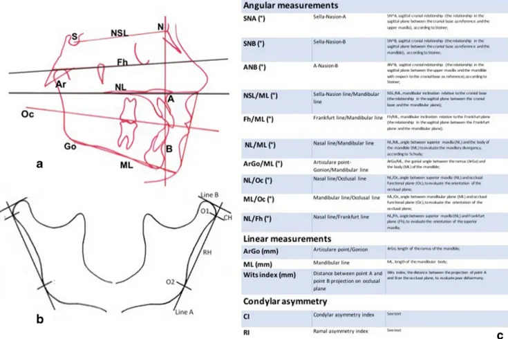
Tracings of craniofacial structures with angular and linear measurements were performed manually on acetate films on profile radiographs. Landmarks are reported in Fig.1.
Orthopantomographic examination
The method introduced by Habets et al. [22] was used to quantify asymmetries between the mandibular condyles and the rami. The outlines of the condyle and the ascending ramus of both sides were traced on acetate paper. On the tracing paper, a line (A, the ramus tangent) was drawn between the most lateral points of the condylar image (O1) and of the ascending ramus image (O2). A line perpendicular (B) to the ramus tangent was drawn from the most superior point of the condylar image. The vertical distance on the ramus tangent from B line to the most lateral point of the condyle (O1) was measured. This distance was called condylar height (CH). The distance on the ramus tangent between the two originally marked most lateral points of the image (O1 and O2) was called ramus height (RH) and measured (Fig.1). To express the symmetry between the condyles and the rami on the OPT image, the following formula |(R-L) /(R + L)|× 100% was used. The absolute value of the difference between the mea-sured condylar or rami sizes of the right (R) and left (L) were divided by the sum of the same condylar or rami sizes and respectively expressed in percentages. This calculation allows individual differences in sizes and provides a value for (a)
symmetry of each individual. The result of this ratio-formula gives a range of asymmetry from 0% (complete symmetry) to100%. According to the study by Habets et al. [22], a 6% difference between the condylar vertical sizes in an OPT is an acceptable limit for diagnosing a condylar asymmetry.
Statistical analysis
Data were expressed as mean ± SD and interquartile range. The statistical distribution of the quantitative measures was found to be non-Gaussian (tested by the Shapiro–Wilk test) and the significance of between-group differences were assessed using the Mann–Whitney test. All the tests were two tailed and statistical significant level was set at 5%.
Results
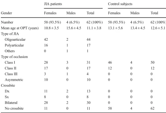
Demographic characteristics, type of JIA, and dental occlu-sion are presented in Table1. As regards the type of JIA, 44 patients (70.96%) were diagnosed with oligoarticular type, 17 (27.41%) with polyarticular type, and 1 (1.61%) with other type of JIA. In the JIA group, class I dental malocclu-sion was observed in 28 females and in 3 males; class II dental malocclusion was observed in 17 females; class III dental malocclusion was observed in 3 females and 1 male while asymmetric dental malocclusion (Class II and Class III) was observed in 10 females. Furthermore, posterior crossbite was present in 51 subjects (79.03%)while no crossbite was present in 11 females.
Table 1 Demographic and occlusal features of JIA patients and control subjects
JIA patients Control subjects
Gender Females Males Total Females Males Total Number 58 (93.5%) 4 (6.5%) 62 (100%) 58 (93.5%) 4 (6.5%) 62 (100%) Mean age at OPT (years) 10.8 ± 3.5 15.6 ± 4.5 11.1 ± 3.8 13.1 ± 5.6 13.4 ± 4.5 12.6 ± 5.1 Type of JIA Oligoarticular 42 2 44 Polyarticular 16 1 17 Others 0 1 1 Type of occlusion Class I 28 3 31 46 4 50 Class II 17 0 17 12 0 12 Class III 3 1 4 0 0 0 Asymmetric 10 0 10 0 0 0 Crossbite Dx 11 2 13 0 0 0 Sx 8 0 8 0 0 0 Bilateral 28 2 30 0 0 0 No crossbite 11 0 11 58 4 62
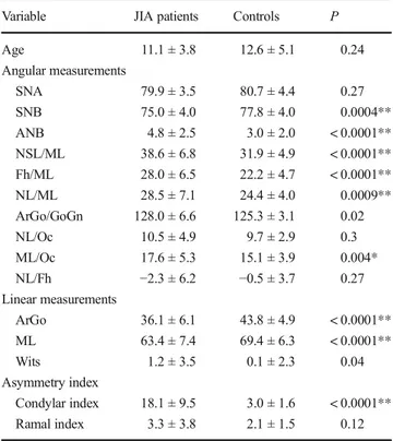
The lateral cephalometric values of the two groups and the statistical analysis are shown in Table2.
The following angular measurements, ANB, SNB, NSL/ ML, Fh/ML, and NL/ML, showed a high significant differ-ence (P < 0.0001) as well as ML/Oc (P = 0.004) and ArGo/ GoGn(P = 0.02) comparing JIA patients with controls. As regards linear measurements, ArGo and ML also showed a high significant difference comparing the JIA group with con-trols (P < 0.0001). Any difference was found for SNA, NL/ Oc, NL/Fh.
As regards condylar asymmetry, the results showed a high significant difference in the range of asymmetry of the con-dyle, being the patient group highly asymmetrical (P < 0.0001). No differences were found in the range of asymmetry of the ramus between groups (P = 0.47).
Discussion
This retrospective study evaluated the cranial structure togeth-er with the condylar asymmetry of patients with JIA, com-pared with normal subjects. The results showed an important
difference between JIA patients and control group for cepha-lometric angular and linear measurements (SNB, ANB, NSL/ ML, Fh/ML, NL/Oc, ArGo, ML, ArGo/GoGn, ML/Oc, and ArGo and ML (P < 0.0001). These findings are in agreement with the literature but the originality of this study is the eval-uation in the same sample of the condylar asymmetry that resulted highly asymmetric in JIA patients (P < 0.0001). With respect to normal subjects, JIA patients present different cranial features that, together with the condylar asymmetry, lead us to consider that their growth pattern is clearly different from that of normal subjects.
Regarding the cranial structure, the most important result is the reduced value of ArGo (P < 0.0001). ArGo is a linear measurement that refers to the height of the vertical ramus of the mandible. Its shortness due to insufficient growth is in-volved in the hyperdivergent pattern growth of the skull, to-gether with the backward rotation of the mandible (NSL/ML, FH/ML P < 0.0001). In fact, the value related to the hyperdivergency of the skull that is represented by the angle between the upper maxilla and the mandible seriously in-creased (hyperdivergent) in children with JIA (NL/MLP < 0.0001). Furthermore, the horizontal body of the mandible
Fig. 1 a Cephalometric landmarks and planes. b Method of Habets/Hansson used to evaluate condylar and mandibular ramal asymmetry. c Definition of cephalometric angular and linear measurements considered in the study to compare craniofacial features of JIA patients with the control group
(MLP < 0.0001) resulted significantly shorter as well, likely due to the alteration of growth mainly of vectorial origin.
The values of ANB resulted higher (skeletal class II) in JIA patients versus the control group (P < 0.0001), in agreement with the literature. ANB is an angular measurement that refers
to the skeletal classification on the sagittal plane; the results showed that the upper maxilla is prevalent with respect to the mandible in JIA patients. Interestingly, the increase of ANB is not due to a bigger growth of the upper maxilla (SNA not significant), but to a decrease of the mandibular growth (SNB P < 0.0001). The skeletal class II of JIA patients is due to a severe lack of growth of the mandible likely related to the inflammatory status of the joints and, consequently, to the vectorial unbalance of the mandibular functions and im-paired muscular activation. (Fig.2).
Regarding the condyle, the percent difference between the right and left condylar height in orthopantomography resulted highly different from the control group, being the condyles of JIA children were much more asymmetric than those of nor-mal children [1].
The results of this study showed that the growth pattern of JIA patients is clearly different from normal subjects and that the cephalometric and condylar values are related to the illness and to each other. The growth of the skull is a complex phe-nomenon due to genetic and environmental factors. Any path-ological event will not act on one bone or district only but it will determine the reaction and compensation of all the neigh-boring structures with the aim to preserve the best function. To this end, the dentoalveolar compensation happens in JIA chil-dren to preserve occlusion and function despite the illness effects. Cephalometric characteristics of JIA patients could be explained considering that the condylar contribution to the growth process is hindered by the inflammatory status and that the growth of both the vertical ramus and the hori-zontal body of the mandible is decreased, requiring a dentoalveolar compensation during dental eruption. This leads
Table 2 Comparison of cranial structure and percent difference of right and left condyles and mandibular rami of JIA patients with control group Variable JIA patients Controls P Age 11.1 ± 3.8 12.6 ± 5.1 0.24 Angular measurements SNA 79.9 ± 3.5 80.7 ± 4.4 0.27 SNB 75.0 ± 4.0 77.8 ± 4.0 0.0004** ANB 4.8 ± 2.5 3.0 ± 2.0 < 0.0001** NSL/ML 38.6 ± 6.8 31.9 ± 4.9 < 0.0001** Fh/ML 28.0 ± 6.5 22.2 ± 4.7 < 0.0001** NL/ML 28.5 ± 7.1 24.4 ± 4.0 0.0009** ArGo/GoGn 128.0 ± 6.6 125.3 ± 3.1 0.02 NL/Oc 10.5 ± 4.9 9.7 ± 2.9 0.3 ML/Oc 17.6 ± 5.3 15.1 ± 3.9 0.004* NL/Fh −2.3 ± 6.2 −0.5 ± 3.7 0.27 Linear measurements ArGo 36.1 ± 6.1 43.8 ± 4.9 < 0.0001** ML 63.4 ± 7.4 69.4 ± 6.3 < 0.0001** Wits 1.2 ± 3.5 0.1 ± 2.3 0.04 Asymmetry index Condylar index 18.1 ± 9.5 3.0 ± 1.6 < 0.0001** Ramal index 3.3 ± 3.8 2.1 ± 1.5 0.12 *P < 0.01; **P < 0.001
Fig. 2 a Image of the right profile of the face of a JIA patient. b Latero-lateral teleradiography of the same patient.c Cephalometry of the same patient showing in red the altered vectors working on the mandible and on the temporomandibular joint. The forces are carried out during swallowing and chewing. The altered vectors are due to the hyperdivergent cranial structure due to the insufficient growth of the vertical ramus of the mandible, as showed in this study. This is a critical cranial structure in non-JIA patients also; in JIA patients, the altered loads
on the temporomandibular joint are worsen by the inflammatory status of the systemic illness. The aim of the multidisciplinary treatment in JIA patients should be the control of the TMJ loads and mandibular vectors (orthognathodontists) together with the control of the inflammatory status (rheumatologists). Teeth are not the final aim, but a means to improve the functional balance. Any traumatic and anti-physiologic dental therapy, in those patients during growing, must be avoided to prevent a worsening of functional vectors and TMJ loads
to a typical hyperdivergent facial development with a serious backward rotation of the mandible, steep mandibular plane, decrease of the posterior facial height, and increase of the anterior one. The mandibular plane steepness together with the posterior mandibular rotation is due to the insufficient growth of the vertical ramus (ArGo) that resulted shorter with respect to the control group. This type of hyperdivergent cra-niofacial pattern of growth of JIA patients is related to the effects of the autoimmune disease leading to a hyperdivergent cranial structure that is highly unstable from a functional point of view and that is responsible for a progressive alteration of the neuromuscular coordination between sides. The force vec-tor’s alteration during chewing and swallowing changes and worsens the physiological development of facial bones.
But the results of this study also showed, in the same pa-tients, a serious asymmetry of the height of the condyles be-tween sides. It is intriguing to underline that the asymmetry of the condyles has been related to the hyperdivergency of the skull in previous studies of non-JIA subjects [6, 7], as de-scribed in the introduction of this article. This can explain part of the condylar asymmetry of JIA patients which severity is likely due to the fact that the effects of the inflammatory pro-cesses are more evident on the side where the skeletal insta-bility and the muscular imbalance of the hyperdivergent skull cause the hyperloading.
These serious and undeniable features may be considered as an indicator of the need of early and continuous orthognatodonthic therapy during the entire period of devel-opment of all JIA patients, independently of the presence of TMJ signs or symptoms. Identifying and diagnosing affected children at an early stage of development and at an early stage of the disease make it possible for an early orthognathodonthic therapy aimed not to the repositioning of teeth within the dental arches, but to improving the balance of masticatory function and growth of the condyles during the developmental stages. Teeth alone are not the final aim, but a means to im-prove the functional balance that is the basic aim of any ther-apy, especially important for JIA patients.
The limitation of this study could be related to the limited number of patients and matched controls. However, the high significance obtained for all the homogeneous measurements let us clearly conclude that JIA patients undergo a special type of cranial growth that should be known by both the rheuma-tologists and orthognathodontists in order to set up a multidis-ciplinary treatment plan from the onset of the illness to reduce and control the side effects of a deranged growing pattern. To this end, it is important to strictly avoid in these patients any orthodontic therapy worsening function and growth; any trau-matic, mechanical, and anti-physiologic dental therapy, espe-cially during growing, risks much more than in normal sub-jects, the worsening of functional vectors; and the enhance-ment and unbalance of TMJ loads which, in turn, will be further aggravated by the inflammatory status of JIA. The
improvement of physiology and biology of the cranial devel-opment during orthognathodonthic therapy can readily be ob-tained when the treatment choice is based on gnathological, non-mechanical concepts as well as the appliances used [23]. The simultaneous control of the unlucky restarts of the illness by rheumatologists is, of course, of utmost importance to avoid irreversible bone defects.
The conclusion of this study is due to the significant differ-ences between children with and without JIA. These results are consistent with the special adaptive type of growth of the temporomandibular joint that is very sensible to the inflam-matory environment during growing [24,25]. This favors a craniofacial morphology with functional vectors at risk for temporomandibular joint derangement. The simultaneous rheumatological and gnathological intervention is important to plan the best multidisciplinary treatment aimed to protect and improve both growth and function of the temporomandib-ular joint.
Compliance with ethical standards
Diclosures None.
References
1. Piancino MG, Cannavale R, Dalmasso P, Tonni I, Filipello F, Perillo L, Cattalini M, Meini A (2015) Condylar asymmetry in patients with juvenile idiopathic arthritis: could it be a sign of a possible temporomandibular joints involvement? Semin Arthritis Rheum 45(2):208–213.https://doi.org/10.1016/j.semarthrit.2015. 04.012
2. Kjellberg H, Fasth A, Kiliaridis S, Wenneberg B, Thilander B (1995) Craniofacial structure in children with juvenile chronic ar-thritis (JCA) compared with healthy children with ideal or postnormal occlusion. Am J Orthod Dentofac Orthop 107(1):67–78 3. Thilander B, Carlsson GE, Ingervall B (1976) Postnatal develop-ment of the human temporomandibular joint. I. A histological study. Acta Odontol Scand 34(2):117–126
4. Merida Velasco JR, Rodriguez Vazquez JF, De la Cuadra Blanco C, Campos Lopez R, Sanchez M, Merida Velasco JA (2009) Development of the mandibular condylar cartilage in human spec-imens of 10-15 weeks’ gestation. J Anat 214(1):56–64.https://doi. org/10.1111/j.1469-7580.2008.01009.x
5. Durkin JF, Heeley JD, Irving JT (1973) The cartilage of the man-dibular condyle. Oral Sci Rev 2(0):29–99
6. Chen F, Terada K, Wu L, Saito I (2007) Dental arch widths and mandibular-maxillary base width in Class III malocclusions with low, average and high MP-SN angles. Angle Orthod 77(1):36–41.
https://doi.org/10.2319/011006-15R.1
7. Chen F, Wu L, Terada K, Saito I (2007) Longitudinal intermaxillary relationships in class III malocclusions with low and high mandib-ular plane angles. Angle Orthod 77(3):397–403. https://doi.org/ 10.2319/0003-3219(2007)077[0397:LIRICI]2.0.CO;2
8. Fjeld M, Arvidsson L, Smith HJ, Flato B, Ogaard B, Larheim T (2010) Relationship between disease course in the temporoman-dibular joints and mantemporoman-dibular growth rotation in patients with juvenile idiopathic arthritis followed from childhood to
adulthood. Pediatr Rheumatol Online J 8(13):13.https://doi.org/ 10.1186/1546-0096-8-13
9. Farronato G, Carletti V, Maspero C, Farronato D, Giannini L, Bellintani C (2009) Craniofacial growth in children affected by juvenile idiopathic arthritis involving the temporomandibular joint: functional therapy management. J Clin Pediatr Dent 33(4):351–357 10. Hsieh YJ, Darvann TA, Hermann NV, Larsen P, Liao YF, Bjoern-Joergensen J, Kreiborg S (2016) Facial morphology in children and adolescents with juvenile idiopathic arthritis and moderate to severe temporomandibular joint involvement. Am J Orthod Dentofac Orthop 149(2):182–191. https://doi.org/10.1016/j. ajodo.2015.07.033
11. Hu Y, Billiau AD, Verdonck A, Wouters C, Carels C (2009) Variation in dentofacial morphology and occlusion in juvenile idi-opathic arthritis subjects: a case-control study. Eur J Orthod 31(1): 51–58.https://doi.org/10.1093/ejo/cjn085
12. Kjellberg H, Kiliaridis S, Thilander B (1995) Dentofacial growth in orthodontically treated and untreated children with juvenile chronic arthritis (JCA). A comparison with Angle Class II division 1 sub-jects. Eur J Orthod 17(5):357–373
13. Kjellberg H (1995) Juvenile chronic arthritis. Dentofacial morphol-ogy, growth, mandibular function and orthodontic treatment. Swed Dent J Suppl 109:1–56
14. Kjellberg H (1998) Craniofacial growth in juvenile chronic arthritis. Acta Odontol Scand 56(6):360–365
15. Mericle PM, Wilson VK, Moore TL, Hanna VE, Osborn TG, Rotskoff KS, Johnston LE Jr (1996) Effects of polyarticular and pauciarticular onset juvenile rheumatoid arthritis on facial and man-dibular growth. J Rheumatol 23(1):159–165
16. Ronchezel MV, Hilario MO, Goldenberg J, Lederman HM, Faltin K Jr, de Azevedo MF, Naspitz CK (1995) Temporomandibular joint and mandibular growth alterations in patients with juvenile rheu-matoid arthritis. J Rheumatol 22(10):1956–1961
17. Ronning O, Barnes SA, Pearson MH, Pledger DM (1994) Juvenile chronic arthritis: a cephalometric analysis of the facial skeleton. Eur J Orthod 16(1):53–62
18. Sidiropoulou-Chatzigianni S, Papadopoulos MA, Kolokithas G (2001) Dentoskeletal morphology in children with juvenile idio-pathic arthritis compared with healthy children. J Orthod 28(1): 53–58.https://doi.org/10.1093/ortho/28.1.53
19. Stabrun AE (1991) Impaired mandibular growth and micrognathic development in children with juvenile rheumatoid arthritis. A lon-gitudinal study of lateral cephalographs. Eur J Orthod 13(6):423– 434
20. Beukelman T, Patkar NM, Saag KG, Tolleson-Rinehart S, Cron RQ, DeWitt EM, Ilowite NT, Kimura Y, Laxer RM, Lovell DJ, Martini A, Rabinovich CE, Ruperto N (2011) 2011 American College of Rheumatology recommendations for the treatment of juvenile idiopathic arthritis: initiation and safety monitoring of ther-apeutic agents for the treatment of arthritis and systemic features. Arthritis Care Res (Hoboken) 63(4):465–482.https://doi.org/10. 1002/acr.20460
21. Wallace CA, Ruperto N, Giannini E, Childhood A, Rheumatology Research A, Pediatric Rheumatology International Trials O, Pediatric Rheumatology Collaborative Study G (2004) Preliminary criteria for clinical remission for select categories of juvenile idiopathic arthritis. J Rheumatol 31(11):2290–2294 22. Habets LL, Bezuur JN, Naeiji M, Hansson TL (1988) The
Orthopantomogram, an aid in diagnosis of temporomandibular joint problems. II The vertical symmetry. J Oral Rehabil 15(5): 465–471
23. Piancino MG, Kyrkanides S (2016) Understanding masticatory function in unilateral crossbites. John Wiley & Sons Inc, Ames, Iowa USA.
24. Isola G, Anastasi GP, Matarese G, Williams RC, Cutroneo G, Bracco P, Piancino MG (2017) Functional and molecular outcomes of the human masticatory muscles. Oral Dis. https://doi.org/10. 1111/odi.12806
25. Lux CJ, Raeth O, Burden D, Conradt C, Komposch G (2004) Sagittal and vertical growth of the jaws in Class II, Division 1 and Class II, Division 2 malocclusions during prepubertal and pubertal development. J Orofac Orthop 65(4):290–311.https://doi.org/10. 1007/s00056-004-0336-9