Research Doctorate School in Biological and
Molecular Sciences
Cadmium uptake, cellular localization and structural and
physiological effects in Pteris vittata.
Supervisors: PhD student: Prof. Laura Maria Costantina Forino, Mirko Balestri
Dott. Monica Ruffini Castiglione,
2
INDEX
INTRODUCTION
51.4. Heavy metals. 5
1.2. Classification of heavy metals.
71.3. Cadmium (Cd).
71.4. Cadmium toxicity.
111.5. Cadmium uptake, transport and distribution in plants.
121.6. Cadmium toxicity in plants.
161.7. How do plants tolerate high metal (Cd) concentration in soil?
191.8. Plant mechanisms for Cd detoxification.
211.9. Remediation of cadmium contaminated soils.
251.10. Phytoremediation.
261.11. Methods of phytoremediation.
28 1.11. a. Phytostabilisation. 28 1.11. b. Rhizofiltration. 29 1.11. c. Phytovolatilization. 30 1.11. d. Phytoextraction. 301.12. Cd hyperaccumulators.
311.13. Pteris vitatta L.
333
MATERIALS AND METHODS
382.1. Plant material.
382.2 Cadmium treatments.
382.3. Anatomical observations.
392.3.a. Light microscopy. 39
2.3.b. Fluorescencemicroscopy. 40
2.3.c. Scanning electron microscope (SEM). 40
2.4. Cadmium content determination.
412.5. Translocation factor and bioconcentration factor.
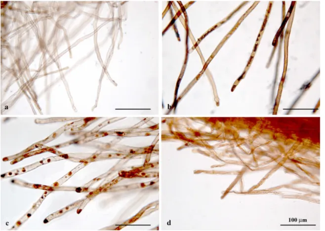
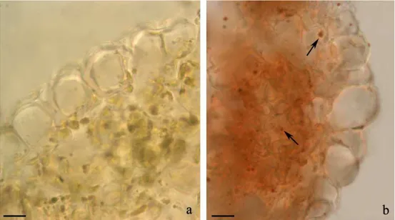
412.6. Histological staining of cadmium.
422.7. Physiological traits.
422.7.a. Determination of relative water content
.
422.7. b. Chlorophyll and carotenoids determination. 43
2.7.c. Electrolytic conductivity method for membrane damage estimation. 43
2.7.d. Extraction and determination of hydrogen peroxide. 44
2.7.e. Thiobarbituric acid reactive substances (TBARS) determination. 44
2.7.f. Extraction and determination of ascorbate and glutathione. 45
2.7.g. Extraction and determination of proline. 46
2.7.h. Enzyme extraction and assays. 46
4
RESULTS
493.1. Root morphology and anatomy.
493.2. Cadmium content determination.
583.3. Cadmium bioconcentration and transfer factors.
603.4. Cadmium localization in tissues.
613.5. Physiological traits.
653.5. a. Relative water content and photosynthetic pigment estimation. 65
3.5.b. Oxidative stress evaluation. 67
3.5.c. Proline and non enzymatic antioxidants. 69
3.5.f. Antioxidant enzymes. 72
DISCUSSION
744.1. Root morphology and anatomy.
744.1.a. Border like cells. 77
4.2. Cadmium content determination.
784.3. Cadmium bioconcentration and transfer factors.
804.4. Cadmium cellular localization.
804.5. Physiological traits.
81CONCLUSIONS
855
INTRODUCTION
1.1. Heavy metals.
Heavy metals constitute a very heterogeneous group of elements widely varied in their chemical properties and biological functions. Typically the term “heavy metals” refers to elements with atomic number greater than 20 that exhibit metallic properties, and includes transition metals lanthanides actinides as well as the metalloids arsenic and antimony. Heavy metals are natural constituents of the earth crust, that have a largest availability in soil and in aquatic ecosystems and a relatively smaller proportion in the atmosphere.
Some heavy metals such as zinc, iron, copper, chromium and cobalt are essential micronutrients necessary for metabolic function of animals, plants and many micro-organisms; but depending on the dose, all heavy metals can have toxic effects on living organisms via metabolic interference and mutagenesis.
Heavy metals pollution can originate from natural and anthropogenic sources. The principal natural source of heavy metals in the environment is from crustal material that is either weathered on (dissolved) and eroded from (particulate) the Earth’s surface or injected into the Earth’s atmosphere by volcanic activity. These two sources account for 80% of all the natural sources; forest fires and biogenic sources, account for 10% each (Nriagu, 1980). In many instances, the inputs of these metals from anthropogenic sources exceed the contributions from natural sources by several times (Adriano, 1986).
6 Currently large areas of the world are contaminated by heavy metals, caused by anthropogenic activities that alter their biochemical balance and geochemical cycles. The heavy metal pollution of anthropogenic origin, comes from mining, smelting, burning of fossil fuels, application of fertilizers, pesticides, as well as from sewage and municipal wastes.
The land and water pollution by heavy metals represents a great potential threat to both the environment and human health and it is a widespread worldwide issue.
All countries have been affected: in Europe, 1400000 sites were affected by heavy metals (McGrath et al., 2001), in USA 600000 brown fields were contaminated with heavy metals and needed reclamation (McKeehan, 2000), in China one-sixth of total arable land has been polluted by heavy metals, and more than 40% has been degraded to varying degree due to erosion and desertification (Liu, 2006). Soil and water pollution is also severe in India, Pakistan and Bangladesh, where small industrial units are pouring their untreated effluents in the surface drains, which spread over near agricultural fields (Lone, 2008).
Some of the heavy metals, i.e. arsenic (As), cadmium (Cd), lead (Pb), mercury (Hg), are cumulative poison. They cannot be biologically destroyed but are only transformed from one oxidation state or organic complex to another (Gisbert et al., 2003). These heavy metals are persistent, they can be accumulated without being metabolized in other intermediate compounds, thus they cannot be easily broken down in the environment.
Accumulation of heavy metals in the environment and particularly in aquifers and soil, from which they can be transferred into living organisms either via the food chain, is a matter of continuously growing environmental concern.
7
1.2. Classification of heavy metals.
Heavy metals can be classified into four major groups based on their health importance and/or their toxicity. Essential: Cu, Zn, Cr, Mn and Fe. These metals, also called micronutrients, are toxic when taken in excess of requirements; Non essential: Ba, Al, Li and Zr; Low toxicity: Ni and Al; High toxicity: As, Pb, Hg, Cd (Raikwar et al., 2008) .
One of the most toxic heavy metal is cadmium (Cd). Cadmium is considered among the top ten on the ATRSD (Agency for Toxic Substances and Disease Registry) priority list of hazardous substances which is based on the combination of their frequency, toxicity and potential for human exposure.
1.3. Cadmium (Cd).
Cadmium (Cd, atomic number 48, atomic weight 112.411) is a soft, silver-white
metal, widely distributed in the earth's crust (EPA 1985) with concentrations reported
between 0.1 and 0.5 ppm and higher levels in sedimentary rocks (Morrow, 1997). It is commonly associated with zinc, lead and copper ores. Cadmium is also a natural constituent of ocean water at average levels ranging from <5 to 110 ng/L with higher levels reported near coastal areas. It has a melting point of 321.8 °C and a boiling point of 765.8 °C. It belongs to group IIb of elements in the periodic table and in aqueous solution has the stable 2+ oxidation state (Callender, 2004)
8 Natural emissions of cadmium in the environment can result from volcanic eruptions, forest fires, generation of sea salt aerosols, or other natural phenomena (Morrow, 1997).
Cadmium is refined and consumed for uses in batteries (83%), pigments (8%), coatings and platings (7%), stabilizers for plastics (1.2%), and nonferrous alloys, photovoltaic devices, and other uses (0.8%) (USGS, 2008). The main anthropogenic sources of cadmium in the environment are nonferrous metal mining and refining, manufacture and application of phosphate fertilizers, fossil fuel combustion, and waste incineration and disposal.
The main cadmium compounds found in air are cadmium oxide, chloride, and sulfate, and these compounds are expected to undergo minimal transformation in the atmosphere (EPA, 1980). Atmospheric concentrations of cadmium are generally highest in the vicinity of cadmium-emitting industries (Pirrone et al., 1996). Cadmium emitted in the atmosphere from combustion processes condenses onto very small particulates that are in the respirable range (<10 µm) and are subject to long-range transport (Wilber et al., 1992). These cadmium pollutants may be transported from a hundred to a few thousand kilometers and have a typical atmospheric residence time of about 1–10 days before deposition occurs (EPA, 1980). Larger cadmium-containing particles from smelters and other pollutant sources are removed from the atmosphere by gravitational settling, with substantial deposition in areas downwind of the pollutant source. Cadmium-containing particulates may dissolve in atmospheric water droplets and be removed from air by wet deposition.
Atmospheric fallout of cadmium to aquatic systems is another major source of cadmium to the environment (IARC, 1993). Cadmium may be released to water by natural weathering process, by discharge from industrial facilities, by leaching from landfills or
9 soil, or phosphate fertilizers (Morrow, 1997). Cadmium is more mobile in aquatic environments than most other heavy metals. In surface water and groundwater, cadmium can exist as hydrated ion or as ionic complexes with other inorganic or organic substances.
The speciation of Cd is generally considered to be dominated by dissolved forms, except in cases where the concentration of suspended particulate matter is high such as “muddy” rivers and reservoirs and near-bottom benthic boundary layers, and underlying bottom sediments in rivers and lakes (Li et al., 1984).
In polluted or organic-rich waters, adsorption of cadmium by humic substances and other organic complexing agents plays a dominant role in transport, partitioning, and remobilization of cadmium (EPA, 1979).
While soluble forms may migrate in water, cadmium is relatively nonmobile in insoluble complexes or absorbed to sediments. Precipitation and sorption to mineral surfaces, hydrous metal oxides, and organic materials are the most important processes for removal of cadmium to bed sediments (EPA, 1979). The mode of sorption of cadmium to sediments is important in determining its disposition to remobilize. Cadmium associated with carbonate minerals, precipitated as stable solid compounds or co-precipitated with hydrous iron oxides, is less likely to be mobilized by resuspension of sediments or biological activity. Cadmium that is adsorbed to mineral surfaces such as clay, or to organic materials, is more easily bioaccumulated or released in the dissolved state when the sediment is disturbed (EPA, 1979). Cadmium may re-dissolve from sediments under varying ambient conditions of pH, salinity, and redox potential (EPA, 1979; Muntau and Baudo 1992). However, cadmium is not known to form volatile compounds in the aquatic environment, so partitioning from water to the atmosphere does not occur (EPA 1979).
Wet and dry deposition of cadmium from the atmosphere may also contribute sizable amounts of cadmium to soil in the areas surrounding sources of atmospheric
10 emissions, such as incinerators and vehicular traffic, which may release cadmium from burned fuel and tire wear (EPA, 1985).
High-temperature sources, such as smelters and incinerators, release small particles that are ideal for long-range atmospheric transport. Also, vapours emitted from high temperature processes will preferentially condense onto smaller particles, thus making vapour emissions available for transport (Steinnes and Friedland, 2006). Aerosols containing cadmium can be carried very long distances in the atmosphere before being deposited to soils. Long-range atmospheric deposition is more evident in organic-rich soils as they have a tendency to concentrate heavy metals (Steinnes and Friedland, 2006).
Other important sources of cadmium to soil include direct application methods and
accidental or fugitive contaminations. Direct application emissions refer to phosphate
fertilizers, sewage sludges and residual ashes from wood, coal, or other types of combustion. Contamination sources include industrial site contamination, mine waste dumps, and corrosion of metal structures (Alloway and Steinnes, 1999).
The levels of Cd found in the soil cover a wide range of concentrations. As estimated by Wagner (1993), non-polluted soil solutions contain Cd concentrations ranging from 0.04 to 0.32 µM. Soil solutions which have a Cd concentration varying from 0.32 to about 1 µM can be regarded as polluted to a moderate level. In polluted soils Cd concentration can be as high as 35 µM.
Generally, in soils, cadmium will bind strongly to organic matter and this will, for the most part, immobilize cadmium (Autier and White, 2004). For Cd to be bioavailable, a shift from the solid-phase form to that of soil solution is essential. This shift is mediated by the presence of organic matter, pH, redox potential, temperature and concentration of other elements. A low soil pH, which is becoming prevalent in many areas of the world due to acid rain, increases the bioavailability of Cd (Elinder, 1992). Contamination of soil by
11 cadmium is of concern because this heavy metal is considered toxic to life with no known function in any biological organism.
1.4. Cadmium toxicity.
Accumulation of Cd in soils can become dangerous to all kinds of organisms. Regarding its potential toxicity for soil organisms and soil microbial processes, Cd was classified by Duxbury (1985) as an element of ‘intermediate toxicity’.
High concentration of cadmium in the soil can be mutagenic and carcinogenic in many animal species (Degraeve, 1981). Recent studies have indicated that a single high dose of cadmium can give rise to necrosis of the testicles. Long-term, low-dose exposure to cadmium did not give rise to this effect, but may cause changes in male sex hormone levels in animals (Deckert, 2005).
Cadmium contamination represents a great threat to both environment and human health since Cd, compared with other non-essential elements, is extracted by plants from the soil, and transferred to food chain (Xiao et al., 2007). The largest source of cadmium exposure for non smoking adults and children is through dietary intake; this heavy metal
can be accumulated in the human body with a half-life exceeding 10 years. Daily dietary
intake of Cd ranges from 40-50 µg/ day (WHO, 2000). Cadmium levels in food can vary greatly depending on the type of food, agricultural and cultivating practices, and amount atmospheric deposition and other anthropogenic contamination. In general, leafy vegetables, such as lettuce and spinach, and staples, such as potatoes and grains, contain relatively high values of cadmium. Peanuts, soybeans, and sunflower seeds have naturally high levels of cadmium. Meat and fish contain lower amounts of cadmium, with the
12
exception of animal organ meats, such as kidney and liver, as these organs concentrate
cadmium (Morrow, 1997).
It has been shown that a high fiber diet and a diet rich in shellfish increase the dietary cadmium intake substantially. Cadmium accumulates, prevalently, in the kidney, the absorption of cadmium in the lung is 10-50%, while the absorption in the gastrointestinal tract is only a few percent. There is evidence that exposure to Cd, derived
from the diet, is associated with renal dysfunction (Salt et al., 1995). According to current
knowledge, renal tubular damage is probably the critical health effect of cadmium exposure.
Cd exposure has also been linked with pulmonary emphysema and possibly bone
demineralization (Salt et al., 1995). The International Agency for Research on Cancer
concluded in 1993 that there was sufficient evidence to classify cadmium as a human carcinogen (Järup et al., 1998).
Recent data indicate that adverse health effects from cadmium exposure may develop in about 1% of the adult general population at an average daily intake of 30 µg over a life-span. This intake is already exceeded by some population groups in Europe, and the margin is very narrow for large groups. Therefore, measures should be taken to reduce cadmium exposure in the general population to minimize the risk of adverse health effects.
1.5. Cadmium uptake, transport and distribution in plants.
Plants are continually exposed to potentially toxic chemicals, including cadmium. Higher plants can uptake Cd, depending on its availability and concentration, from soil or water; rather little is taken up directly from the atmosphere (Clemens, 2006).
13 The root system is the main interface of ion exchange between plants and their environment, thus in each process roots play a central role. Low Cd+2 concentrations in the soil solution in combination with low diffusion coefficient for Cd+2 in aqueous solution suggest that transpiration-driven mass-flow of the soil solution dominate in the delivery of Cd+2 to plant roots (Gallego et al., 2012).
Before the metal can move from the environment into the plant, it must pass the surface of the root. The first barrier blocking heavy metals entrance into cells is the cell wall. This structure is identified as a pivotal site of heavy metal storage in plants (Gallego
et al., 2012). Analysis of Cd localization under electron microscope showed that root cell
walls contain the majority of the metal, as compared to cytoplasm content. Indeed, because of their negative charge, cell walls have significant capacity for heavy metal binding and retention (Schtzendbel and Polle, 2002).
However, cadmium can enter plant cells and this can either be a passive process, with metal ions moving through the porous cell wall of the root cells, or an active process by which metal ions move symplastically through the cells of the root (Lux et al., 2010)
Cell walls in root epidermis and apoplast have apparent free spaces (AFS) of macro- and micropores, where passive ion uptake takes place. The macropores, called water free spaces (WFS), are freely accessible to ions as well as uncharged molecules. The micropores, called Donnan free spaces (DFS), have carboxylic groups (R-COO–) that attract cations and serve as cation exchangers, while anions are repelled. The number of cation exchange sites, (CEC), varies among species (Marschner, 1995).
Passive uptake of ions is affected by the valence and radius of the ion (Marschner, 1995). Uptake increases with decreasing valence of the ion; therefore the cell may more easily take up ions of low electric valence and uncharged molecules.
14 Moreover, Cd ions can move symplastically through the cells of the root. This process requires that the metal ions traverse the plasmalemma, a selectively permeable barrier that surrounds cells (Pilon-Smits, 2005). Special plant membrane proteins recognize the chemical structure of metals; these proteins bind the metals, which are then ready for uptake and transport.
Cadmium can enter root cells as Cd+2 through transmembrane carriers engaged in the uptake of Ca +2, Fe +2, Mg +2, Cu +2 and Zn +2 (Clemens, 2006; Roth et al., 2006).
The ZIP-IRT1transporters (zinc regulated transporter/iron regulated transporter-like protein) are the best-studied nonspecific transporters responsible for high-affinity iron uptake from the soil. Cadmium ions can enter root cells through ZIP transporters, such as orthologues of AtIRT1 and TcZNT1/TcZIP4, through orthologues of the wear TaLCT1 transporter, or via cation channels, such as depolarization-activated calcium channels (DACC), hyperpolarization activated calcium channels (HACC), and voltage-insensitive cation channels (VICC), all of which are relatively non-selective between cations (Lux et
al., 2010). In addition, Cd is thought to enter root plant cells as Cd-chelates through YSL (yellow stripe-like) proteins, which belong to the OPT superfamily of oligopeptide transporters(Curie et al., 2009).
The rate of both active and passive nutrient transport through the cell membranes can be described by the Michaelis-Menten equation (v = (Vmax Cs)/(Km + Cs)), and depends on the two factors Vmax and Km (Maschner, 1995). Vmax is the capacity factor, which denotes the maximum rate of transport when all available binding sites (carriers) are loaded. The second factor, Km, is the Michaelis-Menten constant, equal to the ion concentration given when the transport rate is half of Vmax. The rate of transport (v) also depends on the ion concentration of the substrate (Cs). The uptake rate is independent of the source of the element, as long as the concentration remains constant.
15 It is hypothesised that the transport of Cd within the plant occurs in the xylem as it follows water transport upwards in the xylem (Greger and Landberg, 1996).
Due to its high mobility and water solubility, Cd readily enters the roots through the cortical tissue and can reach the xylem via an apoplastic and/or a symplastic pathway (Salt
et al., 1995). The symplasmic pathway is formed by the cytoplasm of individual root cells
connected by plasmodesmata. The Cd species transported through the symplams are unknown, but could include Cd+2 or Cd-chelates. (Verbruggen et al., 2009). Cadmium is loaded from the symplasm into the xylem possibly by YSL proteins, (Mills et al., 2005;
Dal Corso et al., 2008; Curie et al., 2009), and also by heavy metal P1B-ATPases, such as
orthologues of AtHMA2 and AtHMA4 (Dal Corso et al., 2008; Verbruggen et al., 2009). These proteins play an important role in transporting transition metal ions against their electrochemical gradient using the energy provided by ATP hydrolysis.
It is also possible for Cd+2 and Cd-chelates to reach the xylem solely via an extracellular, apoplasmic pathway in regions of the root lacking a Casparian band (Lux et
al., 2004). The uptake of cationic elements reaching the xylem via an apoplasmic pathway
is generally restricted to the extreme root tip and to regions in which lateral roots are being initiated (White, 2001). Supporting the presence of an apoplasmic pathway, are observations that the root tip is the most active region of the root for Cd influx (Piñeros et
al., 1998). Root tips lack the Casparian band, and Cd is therefore transported apoplastically
through cell walls directly to the xylem.
Generally Cd is mainly retained in the root tissues, adsorbed to negative charges on cell walls and macromolecules in cells, or is taken up by the root cell and accumulates in the cytoplasm and vacuoles. Only small amounts (10-30%) are transported to the above-ground tissues moving upwards in the negative charged cell walls of the xylem (Cataldo et
16 Different chelators may be involved in the translocation of Cd cations through the xylem, such as organic acids. Since the metal is complexed within a chelate it can be translocated upwards in the xylem without being absorbed by the high cation exchange capacity of the xylem (Von Wiren et al., 1999).
In light of what has been said cadmium concentrations are often (but not always) greater in roots than in shoots, suggesting that Cd transport to the xylem is restricted in most plants, and it is lower in seeds, fruits, and tubers, for which it is supposed that Cd is not readily translocated in the phloem (Lux et al., 2010). However, it has also been hypothesized that Cd accumulation in developing fruits could occur via phloem-mediated transport, implicating a systemic diffusion of heavy metals into the plant body (Hart et al., 1998).
Shoot Cd concentrations are determined largely by Cd entry to the root, sequestration within root vacuoles, translocation into the xylem and phloem, and dilution within the shoot through growth (Lux et al., 2010).
This heavy metal is toxic to plant cells, even at low concentrations; leaf concentrations greater than 5–10 µg Cd g−1 DM are toxic to most plants (White and Brown, 2010).
1.6. Cadmium toxicity in plants.
Cadmium is not an essential element for plant metabolism and can be strongly phytotoxic, causing rapid death (Kuzovkina et al., 2004). The main visible symptoms of Cd toxicity in plants are chlorosis and leaf rolls.
17 Toxicity symptoms observed in plants growing in the presence of excessive amounts of cadmium may result from a wide range of interactions at cellular level (Hall, 2002).
The plant plasma membrane is considered the first living structure affected by Cd toxicity. Ion leakage (Iannone et al., 2010), protein oxidation, cross-linking of protein thiols (Meharg, 1993; Romero-Puertas et al., 2002), inhibition of membrane proteins like H-ATPase (Fodor et al., 1995) and changes in lipid composition linked to peroxidation of unsaturated fatty acids induced by free radicals (Groppa et al., 2001; Quartacci et al., 2001; Cuypers et al., 2011) are some of the mechanisms probably involved.
In most environmental conditions, Cd enters first the roots, and consequentely they are likely to experience Cd damage first (Sanità di Toppi and Gabrielli, 1999). In root tip cells of Allium cepa, Cd damages nucleoli (Sanità di Toppi and Gabrielli, 1999), in rice, it altered the synthesis of RNA and inhibited ribonuclease activity (Shah and Dubey, 1995) and in Pisum sativum cadmium concentrations caused a reduction of root growth, which is directly related to reduction of apex length, mitotic activity and percentage of DNA-synthetizing cells (Fusconi et al., 2007). It is known to interfere in the symbiosis between roots and microbes, as well as to increase plant predisposition to fungal invasion (Kabata-Pendias and (Kabata-Pendias, 2001).
It also induces biochemical change in roots and leaves, such as lignification of cell walls in root tissues and leaf main vein (Sanità di Toppi and Gabrielli, 1999).
Being highly mobile in phloem, Cd can accumulate in any part of the plant causing stunted growth and leaf epinasty (Chaffei et al., 2004).
In addition, cadmium interacts with water balance (Barcelò and Poschenrieder, 1990; Costa and Morel, 1994) and reduces net photosynthetic rate, damaging the photosynthetic apparatus, in particular the light harvesting complex II (Krupa, 1988), and
18 the photosystems II and I (Siedlecka and Baszynsky, 1993; Siedlecka and Krupa, 1996). Besides, Cd inhibits photosynthesis by decreasing the transcription of the photosynthesis-related genes psbA, psaB and rbcL and inactivates enzymes involved in CO2 fixation.
Moreover, this heavy metal alters chloroplast ultrastructure, stomatal conductance and leaf transpiration (Perfus-Barbeoch et al., 2002).
Cd actively inhibits the stomatal opening, but how it must yet be established. Probably the stomatal movements are not directly affected by Cd, but rather are due to the strong interference of Cd with movements of K+, Ca2+ and abscisic acid in the guard cells (Barcelò et al., 1986; Barcelò and Poschenrieder, 1990).
It was also shown that Cd induces peroxisome-senescence in leaves by activating the glyoxylate cycle enzymes malate synthase and isocitrate lyase, as well as peroxisomal peptidases, being the latter well-known leaf senescence-associated factors (Chaffei et al., 2004). Another important reason for Cd toxicity is its high chemical similarity with functionally active ions situated in active sites of enzymes and signalling components, in particular Zn, but also Ca and Fe, deregulating the homeostasis of the latter elements or causing their displacement from proteins (Roth et al., 2006).
Protein carbonylation under cadmium stress is able to alter the redox cell status mainly by modifying the antioxidant defence system and thus increasing reactive oxygen species (ROS) at cell level (Romero-Puertas et al., 2004). Cadmium was found to produce oxidative stress (Hendry et al., 1992; Somashekaraiah et al., 1992), but, in contrast with other heavy metals such as Cu, it does not seem to act directly on the production of reactive oxygen species (Salin, 1988). On the other hand, Cd ions can alter the activity of several antioxidative enzymes. This results in oxidative injuries and converges into a general redox homeostasis impairment.
19 Oxidative stress is a disturbance of the cellular redox balance in favour of the pro-oxidants, and can lead to disruption of cellular macromolecules (e.g., degradation of proteins, cross-links in DNA, membrane fatty acids peroxidation).
One of the major targets of Cd toxicity in plant cells is the mitochondrial electron transfer chain, that represents the site of most rapid Cd-induced ROS generation (Wang et
al., 2004; Heyno et al., 2008). As a result, these ROS might lead to membrane lipid
peroxidation, increasing the passive permeability to H+ of the mitochondrial inner membrane (Kessler and Brand, 1995) and impaired ATP generation, resulting in mitochondrial damage and induction of apoptosis during stress situations.
Apart from the damage they cause, Cd-induced ROS also exert a positive role, as ROS influence cell growth and induce biological repair mechanisms for Cd- and ROS-induced damage. Reactive oxygen species play crucial roles in normal physiological processes and are ideal signalling molecules, as they are small and able to diffuse over short distances (Halliwell, 2006;Thevenod, 2009).
Cadmium induced oxidative damage in a variety of plant species, in different organs or in cell suspensions, at different metal concentrations and exposure times, leading to a vast number of responses.In order to cope with high concentration of Cd, few plants species have evolved complex mechanisms that serve to control the uptake, accumulation and detoxification of this metal (Verbruggen et al., 2009).
1.7. How do plants tolerate high metal (Cd) concentration in soil?
The sensitivity of plants to heavy metals depends on an interrelated network of physiological and molecular mechanisms that include uptake and accumulation of metals
20 through binding extracellular exudates and cell wall, complexation of ions inside the cell by various substances, for example, organic acids and phytochelatins; general biochemical stress defence responses such as the induction of antioxidative enzymes and activation or modification of plant metabolism to allow adequate functioning of metabolic pathways and rapid repair of damaged cell structures (Benavides et al., 2005)
Plants evolved several effective mechanisms for tolerating high concentrations of Cd in soil. In some species, tolerance is achieved by preventing toxic metals uptake into root cells. A first barrier against Cd stress, operating mainly at the root level, can be the immobilization of Cd by means of the cell wall (Nishizono et al., 1989) and extracellular carbohydrates (mucilage, callose) (Verkleij and Schat, 1990; Wagner, 1993). Preventing Cd ions from entering the cytosol through the action of the plasma membrane could theoretically represent the best defence mechanism (Sanità di Toppi and Gabbrielli, 1999). These plants, coined excluders, have little potential for metal extraction.
A second group of plants, termed indicators, shows poor control over metal uptake and transport processes. In these plants, the extent of metal accumulation reflects metal concentration in the rhizospheric soil. Indicator species have been used for mine prospecting to find new ore bodies (Raskin et al., 1994).
In addition, a third group of plants, termed accumulators, does not prevent metals from entering the root. Accumulator species have evolved specific mechanisms for detoxifying high metal levels accumulated in the cells. These mechanisms allow bioaccumulation of extremely high concentration of metals.
21
1.8. Plant mechanisms for Cd detoxification.
Plants employ various strategies to counteract the inhibitory effect of Cd, and it is thought that nutrient management is a possible way to overcome Cd toxicity. The importance of mineral nutrition on Cd stress tolerance has been scarcely studied (Hall, 2002). Apart from nitrogen (N), phosphorus (P) and potassium (K), sulphur (S) plays an important role in plant nutrition. This essential macronutrient not only is involved in growth and development, but it also affects stress tolerance (Marschner, 1995). Sulphur is known for its role in the formation of the sulphur-containing amino acids cysteine (Cys) and in the synthesis of proteins, vitamins, chlorophyll and glutathione (GSH, γ -glutamylcysteinyl-glycine), which is involved in stress tolerance.
Glutathione is the most important sulphur-containing antioxidant and redox buffer in plants, playing an essential role in plant metabolism and stress tolerance (Foyer et al., 2006). Glutathione is composed of the amino acids, glutamate, cysteine and glycine. Its significance lies mostly in its role as a reductant, as well as in its ability to detoxify harmful components within a cell. Compounds able to oxidize GSH at high rates include ROS such as superoxide or hydroxyl radical. Glutathione forms glutathione disulfide (GSSG) after oxidation as a result of its antioxidant activity. Glutathione is maintained in a predominantly reduced state by specific enzymes named glutathione reductases (GR) located in the cytosol, plastids, mitochondria, and peroxisomes.
Glutathione reductase is the enzyme that, in conjunction with NADPH, catalyzes the reduction of GSSG to GSH (Carlberg and Mannervik, 1985).
22 Glutathione reductase has been detected in bacteria, yeast, plants and animals. It is essentially responsible for maintaining the GSH levels in the cell. Glutathione reductase activity has been shown to increase in plants exposed to environmental stress.
Glutathione is also a precursor for phytochelatins (PC) (Kneer and Zenk, 1992). Therefore, GSH has been implicated in aiding plants to cope with various environmental stresses, either directly by binding and detoxifying, or indirectly through conversion into phytochelatins.
Phytochelatins (PC) are a family of peptides with the general structure (γ -Glu-Cys)n-Gly, where n = 2–11. Phytochelatins form various complexes with Cd (with molecular masses of about 3600 or 2500 D), due to the presence of the thiolic groups of Cys, which chelate Cd and, as a result, prevent it from circulating as free Cd2+ inside the cytosol (Grill et al., 1989).
Phytochelatins synthesis from glutathione is catalysed by a transpeptidase, named phytochelatin synthase, which has been shown to be activated only in the presence of heavy metal ions, in particular Cd and As (Benavides et al., 2005).
Plants have several metal-sensitive enzymes, such as alcohol dehydrogenase, glyceraldehyde-3-phosphate dehydrogenase and ribulose-1,5-diphosphate carboxylase. Kneer and Zenk (1992) found that these enzymes were able to tolerate cadmium (Cd) at 10 to 1000 times greater concentration when it was complexed with phytochelatins.However, Leopold et al. (1999) found that when Silene vulgaris, a heavy metal tolerant plant, was exposed to copper and cadmium there was no detectable heavy metal-phytochelatin complexation. They concluded that not all plants are able to form stable heavy-metal-PC complexes.
In Thlaspi caerulescens, Cd has been found in the apoplast and in the vacuole (Vàzquez et al., 1992). It has been demonstrated that the physiological mechanism of Cd
23 tolerance is not based on an enhanced synthesis of phytochelatins (Ebbs et al., 2002; Schat
et al., 2002) but on a preferential compartmentalization of the metal in the plant. The vacuolar compartmentalization plays a very significant role in Cd detoxification and tolerance, preventing the free circulation of Cd ions in the cytosol and forcing them into a limited area.
It is also possible for plants to bind oxygen free radicals and to detoxify organic contaminants using other enzymatic and non-enzymatic antioxidants. Specifically, ascorbic acid is an important and major plant antioxidant due to its high abundance (Noctor et al., 1998).
As an antioxidant, ascorbic acid can manage ROS through the ascorbate-glutathione cycle. In this cycle, ascorbate and ascorbate-glutathione are not consumed. They transfer reducing power from NADPH and reduce hydrogen peroxide to H
2O. Hydrogen peroxide
is reduced to H
2O by ascorbate peroxidase using ascorbate, which generates
monodehydroascorbate. Monodehydroascorbate is a radical. It can be reduced to ascorbate by monodehydroascorbate reductase. If not reduced rapidly, monodehydroascorbate is disproportionated into ascorbate and dehydroascorbate. Dehydroascorbate is reduced to ascorbate by dehydroascorbate reductase using reduced glutathione as the reducing agent. Oxidized glutathione is in-turn reduced by glutathione reductase using NADPH. The ascorbate-glutathione cycle presents in at least four different subcellular locations including the cytosol, chloroplast, mitochondrion and peroxisome.
Apart from the enzymes of the ascorbate-glutathione cycle the protection against ROS may be mediated by other hydrogen peroxide scavenging enzymes such as catalase (CAT) and peroxidase (POD).
Catalase, which can be found in primarily in the peroxisomes and root nodules, converts hydrogen peroxide (H
24 O
2 + H2O2 =>H2O + ½ O2
Peroxidase catalyzes the reduction of H
2O2 to water using an external electron
donor as substrate. Peroxidase localization is cytoplasmic or associated with cell walls. H
2O2 + Sub H2 => 2 H2O + Sub
Cadmium treatment affects the activities of antioxidative enzymes, but contrasting results have been reported. For example, in leaves of Cd-exposed Helianthus annuus plants, the activities of ascorbate-glutathione related defence enzymes were decreased (Gallego et al., 1996). Roots and leaves of Phaseolus vulgaris as well as suspension cultures of tobacco (Nicotiana tabacum) cells contained elevated APX activities after Cd exposure (Chaoui et al., 1997). In Phaseolus aureus seedlings, Cd induced elevated guaiacol peroxidase (POD) but decreased CAT activities (Shaw, 1995).
Presently, the involvement of antioxidative enzymes in plant responses against Cd toxicity is unclear because Cd is not contained in the group of transition metals like copper and zinc, and it is not involved directly on the production of ROS via Fenton-type reactions or Haber-Weiss reactions (Schtzendbel and Polle, 2002).
Finally, compatible solutes, such as proline, may protect plants from stress through contribution to cellular osmotic adjustment, membrane integrity protection and stabilization of enzymes/proteins, playing multiple functions in stress protection and signalling (Szabados and Savouré, 2009). Proline might have a role: (a) in providing protection to the enzymes against Cd damage (Shah and Dubey, 1997); (b) in protecting macromolecules against denaturation (Schobert and Tschesche, 1978); (c) in conferring rigidity to the cell wall (Muñoz et al., 1998), and (d) in scavenging hydroxyl radicals (Smirnoff and Cumbes, 1989).
25
1.9. Remediation of cadmium contaminated soils.
Soils contaminated with Cd arean environmental problem that requires an effective and affordablesolution. Soil remediation is defined by Allen (1988) as the return of soil to a condition of ecological stability together with the establishment of plant communities it supports or supported to conditions prior to disturbance. The current remediation technique of heavy metal from contaminated soil-water are expensive, time consuming and environmentally destructive (Chhotu and Fulekar, 2009).
Conventional technologies involve the removal of metals from polluted soils by transportation to laboratories, soil washing with chemicals to remove metals, and finally replacing the soil at its original location or disposing of it as hazardous waste (Francis et
al., 1999). Excavation is a commonly used remediation method that produces rapid
remediation results. However, this decontamination strategy is an ex situ approach and can be often expensive because of the operation, transport and special landfill requirements. Moreover, this method can damage the soil structure and ecology (USEPA, 1997).
The capping method consists in covering the contaminated soil with a hard cover to reduce exposure. Stabilization and solidification are in situ physical treatments where soil is mixed with cement or stabilizer to create a hardened mixture. These remediation technologies have the advantage of immediately reducing the risk factors arising from metal contamination, but may only be considered temporary alternatives because the metals have not been removed from the soil environment.
Immobilization of heavy metals through the addition of lime (Krebs et al., 1999), phosphate (Ebbs et al., 1998) and biosolid compost (Bolan et al., 2003) have been suggested as remediation techniques. The addition of biosolid compost decreased the concentration of the soluble and exchangeable Cd fraction but increased the concentration
26 of organic-bound Cd fraction in soil. Although the formation of soluble metal–organic complex reduces the phytoavailability of metals, the mobility of the metal may be facilitated greatly in soils receiving alkaline-stabilized biosolid compost because of the reduction of metal adsorption and increased concentration of soluble metal–organic complex in solution (Brown et al., 1997; Dinel et al., 2000; Gove et al., 2001).
No single soil remediation is suitable for all situations. A careful investigation of the contaminated site characteristics, contaminant problem, treatment options and treatment timeframe must be considered in order to achieve a successful clean up of a site. Biological, physical, and chemical technologies may be used in conjunction with one another to reduce the contamination to a safe and acceptable level. Conventional methods to remediate metal-contaminated soils can be used at highly contaminated sites but are not applicable to large areas. These remediation methods require high energy input and expensive machinery (Schnoor, 1997). At the same time they destroy soil structure and decrease soil productivity (Leumann et al., 1995). In this context, cost-effective methods such as phytoremediation have been proposed (Vieira da Cunha et al., 2008).
1.10. Phytoremediation.
In recent years, extensive research has been conducted to generate cost effective technologies that include use of microorganisms/biomass or live plants to clean polluted areas.
Some micro-organism-based remediation techniques, such as bioremediation, show potential for their ability to degrade and detoxify certain contaminants. Although these biological systems are less amenable to environmental extremes than other traditional
27 methods, they have the perceived advantage of being more cost-effective (Cunningham et
al., 1997). However, bioremediation is most applicable for sites that have been
contaminated with organic pollutants because heavy metals are not subject to degradation. Over the past decade there has been increasing interest for the development of plant-based remediation technologies for cleaning up contaminated sites, which are cost effective, have aesthetic advantages and long term applicability, a concept called phytoremediation.
Phytoremediation can be defined as “the efficient use of plants to remove, detoxify or immobilise environmental contaminants in a growth matrix (soil, water or sediments) through the natural biological, chemical or physical activities and processes of the plants”.
Plants are unique organisms equipped with remarkable metabolic and absorption capabilities, as well as transport systems that can take up nutrients or contaminants selectively from the soil. Phytoremediation involves growing plants in a contaminated soil, for a required growth period, to remove contaminants from the soil, or facilitate immobilisation or degradation of the pollutants. Certain species of plants can be used successfully to clean up heavy metal polluted soils if their biomass and metal content are large enough to complete remediation within a reasonable period (Ebbs and Kochian, 1998). The plants can be subsequently harvested, processed and disposed. The harvested plants are much lighter to transport than soil, this reduces costs of remediation.
Phytoremediation is an alternative or complimentary technology that can be used along with or, in some cases in place of mechanical conventional clean-up technologies that often require high capital inputs and are laborious and energy intensive. Phytoremediation is an in situ remediation technology that utilizes the inherent abilities of living plants and can be used to clean up heavy metals, pesticides, explosives, crude oil, solvents and polyaromatic hydrocarbons.
28 Phytoremediation is also considered to be more aesthetically appealing than some traditional remediation technologies. The plants may be relatively easier for the public to accept, as they are more attractive compared to bare soils or caps. Moreover, it is an ecologically friendly, solar-energy driven clean-up technology, based on the concept of using nature to cleanse nature.
1.11. Methods of phytoremediation.
Plants have evolved a great diversity of genetic adaptations to handle the accumulated pollutants that occur in the environment and there are several ways in which plants can be used to clean up or remediate sites contaminated with heavy metals. Plants can stabilize or remove metal contaminants through one of or a combination of different methods.
1.11. a. Phytostabilisation.
Phytostabilisation is the use of certain plants to immobilize soil and water contaminants, it is the use of plant roots to limit contaminant mobility and bioavailability in the soil. The plants are used to (1) decrease the amount of water percolating through the soil matrix, which may result in the formation of a hazardous leachate, (2) act as a barrier to prevent direct contact with the contaminated soil and (3) prevent soil erosion and the distribution of the toxic metal to other areas (Raskin and Ensley, 2000).
29 Phytostabilization can occur through the sorption, precipitation, complexation, or metal valence reduction. It is useful for the treatment of cadmium, lead, arsenic, chromium, copper and zinc. Some of the advantages associated with this technology are that the disposal of hazardous material/biomass is not required (USPA, 2000) and it is very effective when rapid immobilization is needed to preserve ground and surface waters.
This technique can also be used to re-establish a plant community on sites that have been denuded due to the high levels of metal contamination. Once a community of tolerant species has been established both the potential for wind erosion and the amount of water available in the system are reduced, and thus also the spread of the pollutants and the leaching of the soil contaminants are reduced.
1.11. b. Rhizofiltration.
Rhizofiltration is primarily used to remediate extracted groundwater, surface water, and wastewater with low contaminant concentrations (Raskin and Ensley, 2000). It is defined as the use of plants, both terrestrial and aquatic, to absorb, concentrate, and precipitate contaminants from polluted aqueous sources in their roots. Rhizofiltration can be used for cadmium, lead, copper, nickel, zinc, and chromium which are primarily retained within the roots (USPA, 2000).
The contaminants are either adsorbed onto the root surface or are absorbed by the plant roots. Plants used for rhizofiltration are not planted directly in situ but are acclimated to the pollutant first. Plants are hydroponically grown in clean water rather than soil, until a large root system has developed. Once a large root system is in place the water supply is substituted for a polluted water supply to acclimatize the plant. After the plants become
30 acclimatized they are planted in the polluted area where the roots uptake the polluted water and the contaminants along with it. As the roots become saturated they are harvested and disposed of safely.
1.11. c. Phytovolatilization.
Phytovolatilization involves the use of plants to take up contaminants from the soil, transforming them into volatile forms and transpiring them into the atmosphere (USPA, 2000). Mercuric mercury is the primary metal contaminant that this process has been used for. The advantage of this method is that the contaminant, mercuric ion, may be transformed into a less toxic substance (that is, elemental Hg). The disadvantage to this is that the mercury released into the atmosphere is likely to be recycled by precipitation and then redeposited back into lakes and oceans, repeating the production of methyl-mercury by anaerobic bacteria (Jadia and Fulekar, 2008).
1.11. d. Phytoextraction.
Phytoextraction is the name given to the process where plant roots uptake metal contaminants from the soil and translocate them to their above soil tissues. The roots and shoots are subsequently harvested to remove the contaminants from the soil. Salt et al. (1995) reported that the costs involved in phytoextraction would be more than ten times less per hectare compared to conventional soil remediation techniques. Phytoextraction also has environmental benefits because it is considered a low impact technology.
31 Furthermore, during the phytoextraction procedure, plants cover the soil and erosion and leaching will thus be reduced. With successive cropping and harvesting, the levels of contaminants in the soil can be reduced (Vandenhove et al., 2001).
In some cases it is possible to recycle the metals through a process known as phytomining, though this is usually reserved for use with precious metals. Metal compounds that have been successfully phytoextracted include cadmium, zinc, copper, and nickel. Ideally, the plants used for phytoextraction are hyperaccumulators of the contaminant in question.
1.12. Cd hyperaccumulators.
Plants often contain trace concentrations of many contaminants of concern. At low levels, plants can usually metabolize or dispose of these compounds without any significant injury. Generally, at high contaminant concentrations in soil or water, plants often suffer and/or die because of their inability to metabolize these harmful elements. However, some plants can survive and/or thrive when they accumulate high concentrations of toxic elements.
A class of plants called hyperaccumulators combines extremely high tolerance to, and foliar accumulation of trace elements (Verbruggen et al., 2009). Hyperaccumulation is an active process that depends on an internal hypertolerance mechanism to resist cytotoxic levels of the accumulated metals and on a powerful scavenging mechanism for the efficient uptake of the pollutants (Salt et al., 1995).
Hyperaccumulators are conventionally defined as species capable of accumulating metals at levels 100-fold greater than those typically measured in common
non-32 accumulator plants. Brooks et al. (1977) first coined the term “hyperaccumulator” to define plants with nickel concentrations higher than 1,000 µg.g -1 (0.1%) in their dried leaves. This criterion was also considered appropriate to specify hyperaccumulation of copper and lead, while for zinc a threshold of 10,000 µg.g -1 (1.0%) in dried plant material as suggested because of greater background concentrations of this metal. A foliar cadmium concentration above 100 µg.g -1 DW (0.01%) is considered exceptional and is used as a threshold value for Cd hyperaccumulation (Baker et al., 2000).
Hyperaccumulators have recently gained considerable interest because of their potential use in phytoremediation strategies, phytomining, and food crop biofortification (Pilon-Smits, 2005).
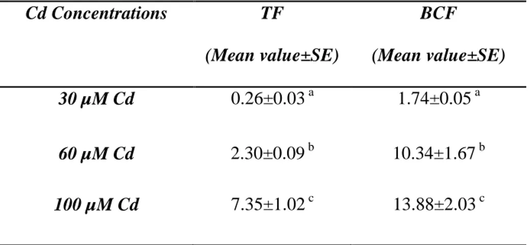
The metal hyperaccumulation characteristic is not common in terrestrial plants; to date, approximately 450 plant species from 45 plant families have been reported to hyperaccumulate metals. Many of the known hyperaccumulators are biennial or short-lived perennial herbs, or are shrubs or small trees. Ideally, hyperaccumulators should have a high rate of accumulation, be fast growing, and have a high production of biomass (Watanbe, 1997). The concentration of the contaminant is generally very high in these plants when grown in contaminated media. They must have both a bioconcentration factor (BCF) and transfer factor (TF) greater than one. The BCF is the plant to soil ratio for a particular contaminant, while the TF is defined as the ratio of concentration of metals in the leaves compared to those in the roots (MacFarlane et al., 2007).
Most hyperaccumulators bioconcentrate Ni, about 30 species absorb either Co, Cu, and/or Zn, just few species accumulate Mn and Cd. Still, minimal information is available on cadmium accumulation in plants, as hyperaccumulation of Cd is a very rare phenomenon due to its nonessential nature and high phytotoxicity to plants.
33 Cd hyperaccumulation is present only in some populations of Thlaspi caerulescens,
Arabidopsis halleri, and Thlaspi praecox, all belonging to the Brassicaceae family, and Sedum alfredii (Crassulaceae) (Verbruggen et al., 2009). Recently, Tagetes patula has
been identified as a novel Cd accumulator plant; this species is able to tolerate Cd induced toxicity by activation of its antioxidant defence system (Liu et al., 2011).
Most of the hyperaccumulating plants are dicots or monocots, Pteris vittata L. is a fern that removes arsenic from soil and/or water, and it can be defined as an arsenic hyperaccumulator (Ma et al., 2001).
1.13. Pteris vitatta L.
The perennial fern Pteris vittata (Fig. 1) is considered a suitable plant for phytoremediation of As-contaminated sites (Chen et al., 2002; Zi-Zhuang et al., 2006), having characteristics of fast growing, high biomass production and wide geographic distribution.
34
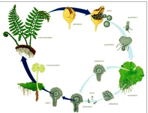
P. vittata belongs to the Pteridaceae family. Like others ferns, the life history of P. vittata consists of a cycle between a diploid sporophyte generation and a gametophyte
generation (Fig. 2). During the sporophyte generation, cluster of sporangia called sori are formed on the fronds. Sporangia contain spore mother cells, each of which produces four haploid spores via meiosis. After maturity, the spores scatter to locate favourable conditions (humidity). A spore germinates and grows into a young green plant known as a prothallus, which is the gametophyte of the fern. This produces the archegonia and antheridia, which produce a single egg and swimming sperm by mitosis, respectively. As the male and female sex organs have differing maturation times, the sperm seeks a separate individual on which the archegonia are already established so fertilization can occur. This restores the haploid gametophyte into a diploid sporophyte, producing a new independent generation.
35
Pteris vittata is very efficient at removing arsenic from soil, it is the first known
arsenic hyperaccumulator fern. It cannot only take up high amounts of arsenic from soil and water, but it can transport arsenic very efficiently from its roots to its fronds (Ma et al., 2001). The amounts of As accumulated in fronds can be up to 93% of the total As content in the plants. Recent results showed that trichome on the fronds contained the highest levels of As compared with the other frond tissues, including epidermal and mesophyllous cells (Li et al., 2005).
Because of its fast-growth and arsenic hyperaccumulation, this fern exhibits potential for use in the phytoremediation of arsenic-contaminated soils. In reality, soil or groundwater contaminated with a single heavy metal is rare; in sites contaminated with As often high levels of other heavy metals, such as Cd, Cu, Pb, Cr and Zn, co-exist (Groudev
et al., 2001; Kim et al., 2003) and therefore multipollutant removal is the goal of
phytoremediation .
There is an increasing awareness of soils contaminated with As or co-contaminated with As and other metals (Liao et al., 2005), as cadmium, and the requirement for developing approaches for some remediation of the co-existing contaminants. Recently, it has been noted that P. vittata may have potential for phytoremediation of multiple toxic metals (Xie et al., 2009).
While there are several studies regarding the effect of As on P. vittata (Lombi et
al., 2002; Wang et al., 2002; Li et al., 2005; Forino et al., 2012), little is known about the
tolerance and accumulation of Cd in this fern (Xiao et al., 2007; Drava et al., 2012). The few studies that have taken in account the impact of Cd on P. vittata biomass (Fayga et al., 2004; Drava et al., 2012) and its accumulation in the fern organs (Gupta and Devi 1994; Drava et al., 2012), have shown contradictory results, probably related to the different
36 experimental conditions that lead to roots with different Cd uptake rate (Redjala et al., 2011).
1.14. Aim of the work.
Looking at phytoextraction, not only quantitative parameters, as biomass, should be considered but qualitative root characteristics may also be looked into, because altered root morphology could directly influence the absorption of water and thus affect plant growth and efficiency of phytoremediation (Li et al., 2009).
In some angiosperm species exposure to Cd has been found to result in changes in the relative proportion and size of root tissues and cell types, and in the formation of Casparian band and suberine lamellae closer to the root apex (Martinka and Lux, 2004; Zelko and Lux, 2004;Vaculík et al., 2009).
Little information is available about the root morphological changes induced by cadmium in ferns. Our previous study showed in P. vittata roots, treated with different As concentrations, the occurrence and the modulation of stress induced morphogenic response (SIMR) and of border-like cells released from the root cap; traits that were suggested to be involved in adjusting the rate of root uptake and its metabolic activity (Forino et. al, 2012). We have hypothesized that cadmium, as arsenic, could determine the occurrence of SIMR in P. vittata roots, which might influence Cd tolerance in this fern.
Given that the study of Cd absorption, uptake, tissue localization and the analysis of structural changes at cellular level are essential to understand Cd tolerance in plant, we analyzed plants of P. vittata exposed to different concentrations of Cd. Moreover, because it is not known whether Cd induces common plant defence pathways, we investigated the
37 sequence of physiological reactions, including H
2O2 production, chlorophylls and
carotenoids content, production of proline, changes in ascorbate-glutathione-related antioxidant systems and other antioxidant enzymes occurring in fronds after Cd exposure.
In particular the aim of this study was:
1. to verify the presence of Cd induced morpho-anatomical changes in the root system;
2. to assess the ability of P. vittata to take up and translocate Cd, comparing Cd content in both roots and fronds;
3. to localize cadmium at cellular level in both roots and fronds;
4. to analyze the oxidative stress induced by Cd and the plant antioxidant response; 5. to know the highest Cd concentration that P. vittata can tolerate in specific
experimental conditions.
Information obtained from this study can enhance the understanding of cadmium tolerance in ferns.
38
MATERIALS AND METHODS
2.1. Plant material.
Spores of Pteris vittata, collected from mature fertile fronds, germinated within two weeks period by sowing on a mixture of potting soil (75%), and sand (25%). After sowing, the substrate was kept moist by watering at frequent intervals. After four weeks, most of the germinated spores developed into a gametophyte stage constituted by several young prothalli. Fourteen weeks were needed for the development of tiny sporophytes. When the plants attained a height of 3-4 cm with 2 or 3 fronds, they were transferred to 10 cm plastic pots filled with the same growth media as the seedbed and allowed to grow up until they reached a height of 10 cm.
The plants were watered on a daily basis or as necessary.During fern growth, the temperature in the greenhouse was 23 °Cwith 16/8 h light/dark photoperiod.
Healthy and uniform Pteris vittata plants with 6 - 7 fronds were selected for the experiments.
2.2 Cadmium treatments.
The plants were acclimatized in a hydroponic system to promote root growth. After acclimatization in 0.2 strength Hoagland nutrient solution for two weeks, 5 plants of P.
39 µM, 60 µM, 100 µMof CdCl2 and maintained in a greenhouse for five and fifteen days.
Air supply during hydroponic cultivation was provided by an aquarium air pump in the medium solution.
2.3. Anatomical observations.
2.3.a. Light microscopy.
After 5 days of treatment with Cd, 5 roots from control plants and 5 roots per sample from treated plants were isolated and stained with an aqueous solution of Giemsa (Lillie, 1965) and observed at the optical microscope to evaluate the meristematic region size and the length and position of root hairs. Analyses were performed on the apical 1 cm segment of the root using a Leiz DMRB microscope equipped with a Leica DFC420 digital camera. The distance of root hair appearance from the apex and the hair length were evaluated by captured images and imported into Image J software for image analysis. The mean value of the hair length was achieved by measuring 50 hairs randomly selected from five different samples for each treatment; the measurements were performed at a distance of 2.5-3 mm from the apex.
Other roots from the same plants were isolated and fixed in FAA (10% formaldehyde, 5% acetic acid and 45% ethanol; Sass, 1958) for 24 h, dehydrated in an ethanol series and subsequently embedded in LR White acrylic resin (SIGMA). Serial cross sections of the root apex were cut at 3 µm with an Ultratome Nova microtome, than stained with Toluidine Blue (Feder and O’Brien, 1968) and analyzed by optical microscope. Histological analysis was performed to assess the differentiation pattern of
40 root hairs and their number in the first 3 mm from the apex on at least 100 transverse sections randomly selected and belonging to three different samples for each Cd treatment.
2.3.b. Fluorescencemicroscopy.
After 5 days of treatment, 5 roots from control plants and 5 roots per sample from treated plants, previously fixed in methanol, were stained with Toloudine blue, embedded in agarose (6%) and cut using razor blades as described by Zelko et al. (2012).
Series of hand sections of roots were prepared at 1 mm intervals from the root apex to the base. For suberin visualization in fluorescence microscopy, the free hand sections were stained by 0.01% Fluorol yellow 088 dissolved in lactic acid for 30 min and washed
in distilled water according to Lux et al. (2005). The samples were placed into a drop of
0.1% FeCl3 dissolved in 50% glycerin prior to observation. The sections for bright-field
observations were stained with Toluidine Blue. The sections were observed under a Zeiss Axioskop 2 plus epifluorescence microscope (Jena, Germany) and documented by an Olympus DP 72 digital camera.
2.3.c. Scanning electron microscope (SEM).
Regarding SEM observation, roots from control and treated plants were fixed in glutaraldehyde (2% in phosphate buffer solution at pH 7.4), dehydrated in a gradient of an alcohol and acetone mixture, critical point dried, mounted with a double adhesive tape on
41 stubs, and sputter-coated with gold. Samples were examined at 10 KV with a JEOL SSM-5410 scanning electron microscope.
2.4. Cadmium content determination.
Five control plants and 5 plants treated with Cd for 15 days were separated into fronds (aboveground biomass) and roots (belowground biomass), oven-dried at 60°C for 5 days, then were digested with nitric-perchloric acid (3:1, v/v) at 100°C. A microwave oven MLS-1200 Mega (Milestone-FKV, USA) equipped with a UV generation device was used to eliminate the interfering organic substances. Then, the measurements of Cd content were made by a graphite furnace atomic absorption spectrophotometer. Three samples of every plant part were used for cadmium detection.
2.5. Translocation factor and bioconcentration factor.
To evaluate the phytoextraction potential of Pteris vittata two parameters were calculated: the translocation factor (TF) and the bioconcentration factor (BCF). The TF, defined as the cadmium concentration ratio between frond and root biomass, was used to measure effectiveness of a plant in translocating cadmium from root to frond. The BCF represents the ratio between the Cd concentration in the frond and that in the culture medium. It is an useful parameter for evaluating the ability of plants in accumulating heavy metals with respect to the initial concentration of heavy metals in the substrates (Tu and Ma, 2002).