Alma Mater Studiorum – Università di Bologna
DOTTORATO DI RICERCA IN
BIOLOGIA CELLULARE E MOLECOLARE
Ciclo XXVI
Settore Concorsuale di afferenza:
05/E2
Settore Scientifico disciplinare:BIO/11
Titolo Tesi
Staphylococcus aureus bones and joints infections:
in vivo studies and host immune response
Presentata da: Alessia Corrado
Coordinatore Dottorato
Relatori
Ch.mo Prof. Vincenzo Scarlato Dott. Giuliano Bensi
Dott. Emiliano Chiarot
Ch.mo Prof. Vincenzo Scarlato
Il mio progetto di dottorato, presentato in questo lavoro di tesi, è stato focalizzato sul settaggio di un modello animale di artrite/osteomielite mediate da Staphylococcus aureus, che nell’uomo è il batterio maggiormente responsabile di queste gravi e debilitanti patologie. Queste malattie sono difficili da trattare anche per la costante crescita di Stafilococchi multi resistenti agli antibiotici (MRSA) e attualmente nessun vaccino contro questo batterio è disponibile sul mercato.
Il modello settato è riproducibile e duraturo e mima ciò che avviene nell’uomo, con le due classiche fasi di infezione: la fase acuta e quella cronica. Nel presente lavoro è stata studiata la risposta immunitaria dell’ospite durante tutte le fasi della patologia, sia considerando ciò che avviene nel sito di interesse (articolazioni del ginocchio) sia la situazione sistemica.
Questo modello è stato inoltre utilizzato per testare l’efficacia di un vaccino contro S. aureus proposto da Novartis.
4 TABLE OF CONTENTS
1 Introduction 7
1.1 Staphylococcus aureus 7
1.2 Staphylococcus aureus joints and bones infections 9
1.3 Novartis proposed Combo Vaccine 16
1.4 Aim of the project 18
2 Results 20
2.1 S. aureus shows a particular tropism for bones and joints causing both arthrosynovitis and osteomyelitis in mice 20
2.2
Setting up of an arthrosynovitis mouse model of infection suitable for long lasting in vivo studies 28
2.2_1CFU counts and histological analysis 28
2.2_2Serological analysis 33
2.2_3Cellular analysis 41
2.3
The acute arthrosynovitis model can be used to test therapeutic and preventive treatments against S. aureus infections 47
5 2.3_1 Ampicillin treatment reduced bacterial load in kidneys but was
not efficacious in killing S. aureus in knee joints 47
2.3_2 The Novartis vaccine proposed against S. aureus infections reduced bacterial burden in joints of infected mice 50
3 Discussion 54
4 Material and Methods 66
4.1 Bacterial strains 66
4.2 Preparation of Bacteria and infection 67
4.3 In vivo imaging 67 4.4 Histopathology examination 68 4.5 Immunohistochemistry 68 4.6 Confocal microscopy 69 4.7 Immunization protocol 70 4.8 Antibiotic treatment 70
4.9 Mice and infection model 70
4.10Antibodies measurements 71
4.11 Cytokine analysis 72
4.12 Flow Cytometry 72
6 5 Bibliography 75 6 Acknowledgments 88
7
1 Introduction
1.1 Staphylococcus aureus
Staphylococcus was first isolated in 1880 in United Kingdom, by the surgeon Sir Alexander Ogston in pus from a surgical abscess in a knee joint (1). Staphylococcus aureus is a facultative anaerobic Gram-positive coccus, member of the Firmicutes, also known as "golden staph" and “Oro staphira”. S. aureus appears as grape-like clusters when observed through a microscope, and it has large, round, golden-yellow colonies, often surrounded by a hemolytic halo, when grown on blood agar plates (2).
S. aureus is the most prevalent specie of staphylococci that leads
Staph-mediated infections. It frequently colonizes different body districts such as nares, axilla, vagina, pharynx and skin. It has been reported that 20% of the human population are long-term carriers of S. aureus and worldwide approximately 30-50% of the healthy adults
are colonized at any time point during their life (3).
Although S. aureus is not always pathogenic, S. aureus disease-associated strains are often able to promote infections by, for example, producing potent protein toxins, and expressing cell-surface proteins that bind and inactivate antibodies (4, 5).
Methicillin-resistant Staphylococcus aureus (MRSA) resistant strains have become resistant to most widely used β-lactam antibiotics.
8
MRSA strains are most often found associated with institutions such as hospitals, but are also becoming very common in community-acquired infections (6).
There are many clinical manifestations of staphylococcal infections, ranging from skin and soft tissues diseases to the most invasive pathologies represented by the metastatic infections and endocarditis (7, 8, 9, 10, 11).
In most cases infections occur after breaches of the skin or mucosal barriers through wounds, trauma or surgical intervention that allow the organism to access the tissues or the bloodstream; once the skin or mucosa have been penetrated, the infection may spread to the blood causing invasive diseases. Figure 1 reports the different staphylococcal diseases, arranged on the basis of their frequency and severity (12).
9
Figure 1: S. aureus diseases
The pyramid reports the broad spectrum of Staphylococcus aureus diseases. The severity of them decreases from top to bottom, on the other end the frequency decreases from bottom to top.
Adapted from “Strategies for and advances in the development of
Staphylococcus aureus prophylactic vaccines”; Jane Broughan et al. 2011
1.2 Staphylococcus aureus joints and bones infections
Staphylococci are the principal causative agents of two major types of bones infections, septic arthritis and osteomyelitis, which involve the inflammatory destruction of joints and bones. These diseases lead serious morbidity and are often difficult to treat (13). The most common routes of infection for both septic arthritis and osteomyelitis are either haematogenous, resulting from bacteremia, contiguous, when the infection is transmitted from local tissue, or direct, resulting from infiltration of bone, often following surgeries, injuries, or implantations of a foreign body, for example joint replacement (13,
10
14, 15, 16, 17). Among the possible routes of infection most septic joints develop as a result of hematogenous seeding of the vascular synovial membrane due to a bacteremic episode (18,19). Infections may result in acute or chronic forms and affect native joints, especially knee and hip, or prosthetic joints, long bones, vertebrae and almost any other bone. The incidence of septic arthritis is between 2 and 10 in 100,000 in the general population but it may be as high as 30–70 per 100,000 in rheumatoid arthritis sufferers or recipients of prosthetic joints (15, 20, 21) and it is more common in children than adults, and in males than in females (22). Haematogenous osteomyelitis usually effects children and elderly people (17) and in children the incidence is between 1 in 5000 and 1 in 10,000 (23). Local spread of infection from contiguous tissue to bone or direct infection can occur at any age, with foreign body implants representing an important risk factor (17). The presence of an implant is frequently associated with chronic osteomyelitis, where antibiotic treatment is often ineffective, and removal of the implant and debridement are required (14). Some cases of relapsing osteomyelitis with several decades between subsequent episodes have been documented, and there are records of reactivation fifty or even eighty years after the first infection (14, 24, 25). Staphylococcus, principally S. aureus, accounts for between 37%
and 67% of septic arthritis isolates as resulting from studies carried out in a range of nations (15, 26, 27, 28).
11
Figure 2: Knee joint anatomy
From:http://www.practical-wellness-guide.com/physical/total-personal-health-care-and-wellness
Septic arthritis is a joint pathology characterized by bacterial colonization and fast articular destruction (22). Inflammation with infiltration of leukocytes into the joint fluid is the result of bacterial growth within the synovium (15, 20). The production of bacterial toxins, host matrix metalloproteinases (MMPs) and reactive oxygen species and lysosomal enzymes, promote the destruction of cartilage. This latter process, mediated by polymorphonuclear leukocytes, begins with degradation of host proteoglycans followed by collagen breakdown within a few hours after the infection. The containment of the inflammation within the joint results in increasing pressure which prevents blood and nutrient supply to the joint increasing joint damage (15, 20, 29, 30).
12
Osteomyelitis includes a range of infections in which bacteria colonize bone with associated inflammation and bone destruction. Acute osteomyelitic foci are characterized by pus-forming inflammation at the site of microbial colonization. Damage to bone matrix, compression and destruction of vasculature is also observed as the infection spreads to surrounding soft tissues, which can further exacerbate bone necrosis (16, 17). Sections of dead bone, known as sequestra, may then detach resulting in separate infectious foci
which, due to the lack of vasculature, are protected from immune cells and antibiotics (16,17). Such areas of dead, infected tissues that are inaccessible to the immune response or antimicrobials can lead to chronicization of the infection (16).
The infection by S. aureus is accompanied by a rapid recruitment of polymorphonuclear granulocytes and activated macrophages that are then followed by T cells. Although monocytes and macrophages are important to clear bacteria, they also play a pivotal role in the destructive inflammation within the joint (13). Moreover, a number of host cytokines play a significant role in the pathogenesis of osteomyelitis and arthritis, and there is strong evidence that production of these cytokines is induced by staphylococcal infection of bone and joints, and that they directly contribute to bone destruction. In particular, the inflammatory cytokines tumor necrosis factor α (TNFα), interleukin 1 (IL-1) and IL-6 seem to be especially important in bone physiology and pathology (32). In patients with
13
acute osteomyelitis, plasma levels of TNFα, IL-1β (the secreted form of IL-1) and IL-6 are all elevated (32, 33). High levels of IL-1β, IL-6 and TNFα are also found in the synovial fluid of patients with septic arthritis (34, 35). The local source of these cytokines is not fully understood. Production of IL-1β can be induced in human osteoblast-like cell lines by a variety of stimuli, including TNFα (36). Infiltrating immune cells may therefore be a more likely source of IL-1β and TNFα in bone in response to infection (37, 38, 39, 40). Moreover IL-6 is produced by osteoblasts in response to a variety of signals, including infection with S. aureus (37, 38). In addition to staphylococcal induction of inflammatory mediators that modulate the actions of osteoblasts and osteoclasts, bacteria of this genus are involved in more direct interactions with bone cells. Invasion and persistence of S. aureus in ‘non-professional phagocytic’ host cells in vitro has been described for many different cell types, including
epithelial cells, endothelial cells and keratinocytes (41, 42). Electron microscopy has demonstrated the presence of bacteria within osteoblasts and osteocytes of embryonic chicks following injection with S. aureus, indicating that internalization by bone cells could also occur in vivo (43). The internalization of S. aureus by bone cells in vivo provides a protective niche for the bacterium, where it is
shielded from immune effector mechanisms and antibiotics may help to explain persistent cases of osteomyelitis. Increasing evidence supports the importance of staphylococcal surface components as
14
virulence determinants by enabling initial colonization. In a number of studies, mutations in these surface receptors strongly reduced the ability of staphylococci to produce infection. In a murine septic arthritis model, inoculation of mice with a strain mutated in the collagen adhesin gene showed that septic arthritis occurred 43% less often than in the corresponding wild type (44). Also vaccination with a recombinant fragment of the S. aureus collagen adhesin was able to reduce the sepsis-induced mortality rate to 13%, compared to 87% of the control group (45). Nevertheless, the role of collagen adhesion of S. aureus as a major virulence factor has recently been questioned
since approximately 30 to 60% of clinical isolates do not display collagen binding in vitro or the cna-encoded collagen adhesin (46).
Staphyloccal fibronectin binding proteins (FbpA and FbpB) may als o play a major role in the colonization and virulence of septic arthritis. In a recent study all the tested clinical isolates (n = 163) contained one or both of the coding regions for these binding proteins and 95% of these strains had a comparable fibronectin binding capacity to that seen in a staphylococcal reference strain known to efficiently bind fibronectin (47
).
These receptors may play an additional role in an intracellular immune-avoidance strategy. S. aureus survives intracellularly after internalization by cultured osteoblasts (48). Initial adherence to cells is mediated by S. aureus fibronectin receptors (49) possibly using fibronectin as a bridge between the host cell and the bacterial receptors. Following adherence, bacteria may be15
internalized by host mechanisms involving membrane pseudopod formation (seen in established bovine mammary epithelial cell lines) or through receptor-mediated endocytosis via clathrin-coated pits (seen in mouse osteoblasts and epithelial cells, 49, 50).
Following internalization, staphylococci may induce apoptosis via a host caspase-dependent mechanism or survive intracellularly (49, 51, 52, 53). Induced apoptosis may exacerbate the host cell damage seen in septic arthritis. Moreover, staphylococci may escape clearance by the immune system and antimicrobial therapy by persisting within these host cells. This survival was recently demonstrated in vivo when S. aureus cells were found in the cytoplasm of embryonic chicken osteoblasts and osteocytes in mineralized bone matrix (43). In another study, S. aureus was found within polymorphonuclear neutrophils in an infection model, and these infected host cells were able to establish infection in naïve animals (54). Therefore, this pathogen may utilize invasion as an immune avoidance technique during the host inflammatory response. Following the down regulation of the adaptive immune response through T-cell apoptosis (mediated by superantigens, other toxins, and invasion), fulminant and/or persistent infection may result.
Antibiotic therapy during septic arthritis is efficacious only when rapidly applied and, often, in combination with joint drainage (29). The antibiotic therapy is based on appropriate culture and antibiotic sensitivity results (55).
16
Osteomyelitis, on the contrary, results often refractory to antibiotic treatment, a problem exacerbated by the increasing levels of antibiotic resistance among Staphylococcus spp. This is further complicated by the emergence of particularly persistent, antibiotic-resistant ‘small colony variant’ forms that may be selected for by certain current treatment regimens (14, 56, 57). Staphylococcal bone infections are thus likely to be a continuing and probably increasing problem, and the understanding of host-pathogen interactions in bones and joints will become crucial for the development of novel therapeutic and preventing strategies.
1.3 Novartis proposed Combo vaccine
The treatment of staphylococcal infections in the last years has been complicated also by the emergence of multidrug resistant strains, which can cause invasive diseases that frequently lead to death. Presently,no vaccines are available to prevent this kind of infections. Novartis Vaccines & Diagnostic (NVD) has engaged in the research and development of an effective vaccine against S. aureus infections. Using different approaches, including reverse vaccinology, proteomic and comparative genomics analyses (58), Novartis selected a combination of four highly conserved antigens, which were proposed as components of a potential innovative multiprotein-based vaccine. The vaccine formulation is composed of two surface proteins FhuD2, and Sur 2, and three secreted proteins, a toxoid derivative of
-17
Hemolysin (Hla) (H35L) and EsxA-B, a fusion protein of two secreted toxins.
Hla is a pore-forming toxin whose expression is associated with staphylococcal pneumonia in mice and humans (59). Its expression is up-regulated in exponential phase and consistently expressed by clinically relevant strains. The protein included in the vaccine carries a substitution of histidine 35 with leucine that produced a mutant toxin (H35L) without hemolytic or lethal activity (60). EsxA-B derives from the fusion of the proteins EsxA and EsxB, both of them being expressed by clinically relevant strains and involved in abscess formation (61). FhuD2 is a lipoprotein involved in iron uptake, up regulated in iron deplete-conditions (58). Finally Sur 2 is a protein of still unknown function. This antigen combination has been tested using different animal models of S. aureus infections (pneumonia, abscess and sepsis model), resulting protective in all the cases and effective in preventing the infections carried out by different S. aureus strains.
18
1.4 Aim of the project
Since the relative importance of S.aureus mediated joints and bones infections both in terms of frequency and severity (fig. 1), the aim of this work was the development of an animal model of septic arthrosynovitis and osteomyelitis resembling the natural disease in humans and suitable for testing preventive and therapeutic tools. The hematogenous source of infection (intravenous inoculation) was preferred among the others due to its higher frequency of occurrence in humans. Our attention was focused to track in the long term bacterial infiltration in joints and bones using different microbiological and histopathological tools, which could allow us to have a complete overview of the situation and to understand the mechanisms set up by the host to contain or eradicate bacterial infection. Antibodies response, cytokines profiles during the time and cellular recruitment were therefore monitored for a period that allowed us studying both acute and chronic phases of these diseases in situ. The systemic infection and the host immune response were also evaluated. Finally the Novartis proposed vaccine against S. aureus diseases was tested in the model of acute infection, and bacterial reduction in situ and systemically was evaluated and compared to a standard antibiotic prophylaxis.
Having a powerful tool to study specific bacterial mediated diseases is nowadays an important requirement for the scientific community to shed light on the complex interactions between host and pathogens
19
and to test treatments for preventing or contrasting infections. We believe that our work could improve the knowledge in the field of S. aureus dependent pathologies, opening the possibility for further
20
2 Results
2.1 S. aureus shows a particular tropism for bones and
joints causing both arthrosynovitis and osteomyelitis
in mice
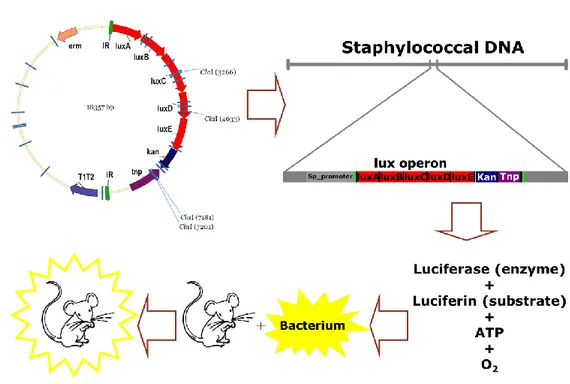
Since S. aureus was demonstrated to have a particular tropism for bones and joints in humans during natural infections (13), we started analyzing the interaction between pathogen and host infecting mice with bioluminescent bacteria. Bioluminescence (BLI) has been used as a powerful tool for studying host-pathogen interactions for years (62) and nowadays many different S. aureus bioluminescent strains are available on the market (Perkin Elmer). Many of them were generated using a plasmid (pXEN5, Perkin Elmer) carrying the Photorhabdus luminescens lux operon (figure 3), which allows
random integration of lux genes into either the bacterial chromosome or natural plasmids carried by the pathogen. Once integrated in bacterial DNA downstream of a functional promoter, this operon is able to make bacteria bioluminescent without the need of extra sources of substrates (fig.3). The lux operon is a 9-kilobase fragment of the Photorhabdus luminescens genome that controls bioluminescence through the catalyzation of the enzyme luciferase (Meighen, 1991). The lux operon has a known gene sequence of luxCDAB(F)E, where lux A and lux B code for the components of luciferase, and the lux CDE codes for a fatty acid reductase complex
21
that makes the fatty acids necessary for the luciferase mechanism (63). Lux C codes for the enzyme acyl-reductase, lux D codes for acyl-transferase, and lux E makes the proteins needed for the enzyme acyl-protein synthetase. Luciferase produces blue/green light through the oxidation of reduced flavin mononucleotide and a long-chain aldehyde by diatomic oxygen. The reaction is summarized below (Silverman et al., 1984): FMNH2+O2+CHO → FMN + R-COOH + H2O + Light.
Figure 3. Generation of bioluminescent bacteria using the pXEN5 random integration system. pXEN5 plasmid used for random integration
of Photorhabdus luminescens lux operon in DNA of recipient bacteria and mechanism of action of the inserted lux operon. luxA-E are lux genes, Kan is the resistance for the antibiotic kanamycin.
22
The light produced by this enzymatic reactions is captured by special imaging systems, in our case IVIS100 and IVIS Spectrum-CT, which exploit a special cooled camera enclosed in a light-proof box to capture all the photons emitted by the live mouse. Bioluminescent spots from the mouse body can be related to specific body areas and the intensity of the light emitted can be quantified.
The bioluminescent strains we used and their features in term of integration are reported here below in figure 4.
Figure 4. List of bioluminescent S. aureus used for in vivo imaging studies. Parenteral strains are reported in the brackets and whether the lux
operon was inserted inside the chromosome or into a stable native plasmid is reported on the right.
All the strains used were highly bioluminescent and suitable for in vivo studies, as reported in the site
http://www.perkinelmer.com/Catalog/Family/ID/Gram%20Positive.
S. aureus bioluminescent strains were therefore used to infect mice
in the lateral tail vein (i.v.) and infection progression was followed for at least 1 week after the injection using an IVIS 100® machine
23
(Perkin Elmer). The i.v. route of infection was chosen trying to mimic a hematogenous source of infection, often occurring in humans. As reported in figure 5A, 1 day after injection S. aureus reached the knee joints being able to establish a local infection that persisted for at least 7 days. Bioluminescence values, expressed as photon/second, (p/s) were calculated considering only our region of interest (ROI) reported as red circle in figure 5A, and a very good correlation could be observed when BLI was compared to colony forming units (CFUs) counted after knee joint washes the last day of the experiment (fig. 5B). Similarly to what obtained for Xen 36 strain, when other S. aureus strains were used to infect animals (fig. 5C) bacteria rapidly and lastingly localized in the joints, underlying that all the different S. aureus strains we tested had the ability to reach this body district. Finally we confirmed the presence of bacteria in knee joints after i.v. inoculation using confocal microscopy, as reported in figure 5D.
24
Figure 5. Staphylococcus aureus showed a particular tropism for joints in a mouse model of intravenous infection. A) Bioluminescence
observed during the time from day 0 through day 7 in mice intravenously infected with S. aureus Xen 36 strain (1x10⁷ CFU/mouse). In red, Region Of Interest (ROI) used to quantify the signal. B) A good correlation expressed as Spearman R value was measured when bioluminescence in the selected ROIs was compared to CFU recovered in knee joint washes of animals infected with Xen 36 strain. C) Signals observed at different time points using 3 S. aureus bioluminescent strains commercially available. S. aureus tended to localize in joints. D) Confocal microscopy analysis of a mouse knee from an infected animal. In yellow: bone (osteocalcin), in red: muscle (phalloidin), in blue: nuclei (dapi), in green: S. aureus
Since in humans S. aureus is able to cause osteomyelitis, we used the in vivo imaging approach also to better understand whether
25
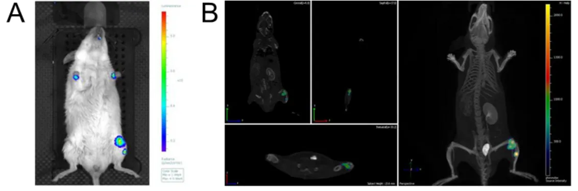
bacteria which had reached the joint could invade the bone tissue. Mice were again i.v. infected with the highly bioluminescent strain Xen 36 and then followed for 1 week post infection. Using the IVIS spectrum-CT system® which combines a camera able to acquire bioluminescent signals and a tomography system that can scan sections of the animal body, we found that bioluminescent bacteria were not only present in the knee joint area but also in the tibia. This is shown in figure 6 where the 2D (A) and the 3D (B) analysis of the same infected animal are reported.
Figure 6.Staphylococcus aureus was localized both in knee joints and bones. A) 2D bioluminescent acquisition of a CD1 mouse i.v. infected with
Xen 36 S. aureus strain. Two bioluminescent signals were detected in the left leg. B) 3D analysis of the same animal. The 2 bioluminescent signals turned to be one in the knee joint and the other in the left tibia. In both the cases an IVIS spectrum-CT system® was used to acquire images.
These findings allowed us to confirm that S. aureus-mediated hematogenous infection could cause arthrosynovitis and osteomyelitis, nevertheless, to better characterize bacterial
26
localization and local damages we carried out histopathological analysis of tissues obtained from infected mice. One week after infection with S. aureus Newman strain, mice were sacrificed, knees were removed, fixed and tissue slices were prepared. Hematoxylin and Eosin (H&E) staining were performed and histopathological analysis of these preparations was compared to slices derived from joints and bones of not infected mice. As shown in figure 7A on the right (red-framed image), when animals were infected with S. aureus the normal architecture of bones and joints was effaced by the presence of mixed inflammatory cells (with prevalence of neutrophils and macrophages) as compared with a control animal on the left (black-framed image). In particular, the tibia was destroyed and granulomas/abscesses were observed. Periostal inflammation was severe, causing thickening of the periostal tissue. Inflammation was evident in the synovia, which appeared severely thickened. In the enlargement, elongated cells can be seen which formed a wall around foci of inflammation composed by neutrophils and macrophages. Within the inflammatory focus, clusters of bright pick eosinophylic slightly hyaline material consistent with the presence of bacteria were observed. Indeed, immune-histochemical staining demonstrated that S. aureus was present in bone abscesses (figure 7B_1) and a wall of neutrophils was built up all around these structures (fig. 7B_3).
27
Figure 7.Staphylococcus aureus was responsible for causing arthritis and osteomyelitis in mice. A) Hematoxylin and Eosin (H&E)
staining of mouse legs i.v. infected with S. aureus Newman strain. On the left (surrounded with a black frame) a slice from a not infected animal is reported as negative control, while on the right (red frame) a slice from a mouse infected and sacrificed 1 week after injection is reported. F= Femur T= Tibia. The smallest panel is an enlargement of a specific area of the red-frame image. B) Immune-histochemistry staining of knees from animals treated intravenously with S. aureus. In B1) S. aureus was stained in red (this antibody recognizes soluble and structural antigens of Staphylococcus aureus whole bacterium) in B3) neutrophils were marked in violet (the antibody recognizes a currently undetermined structure on the neutrophil membrane). B2) is the isotype control of B3.
28
2.2 Setting up of an arthrosynovitis mouse model
of infection suitable for long lasting in vivo studies
2.2_1 CFU counts and histological analysis
For the setting up of a long lasting and reproducible mouse model of hematogenous S. aureus-mediated arthrosynovitis Xen 36 did not appear to be a suitable strain. In fact, the infectious dose required to be able to follow in vivo a detectable bioluminescent signal was too high and mice showed several signs of disease after the first week from the infection. The S. aureus Newman strain was therefore chosen for these experiments since it has been largely used in research worldwide being well adapted for animal studies. The first objective was to find a dose high enough to homogeneously infect a group of mice both systemically and in the knee joints, but reducing the risk of causing severe disease and/or death.
Immediately after the infection, S. aureus disappeared from the blood and rapidly disseminated in all the organs (64). In particular, the most infected organs were the kidneys, as illustrated in figure 8, which were therefore chosen as markers for systemic infection.
29
Figure 8. Staphylococcus aureus intravenously injected in CD1 mice reached all organs. CFU counts in organs 4 days after intravenous
administration of about 1x10⁷CFU/mouse of S. aureus Newman strain. CFU were normalized among different organs reporting the total colonies counted in that organ.
Mice were intravenously infected with doses ranging from 1x10⁷ to 1x10⁴ CFU/mouse and followed up to 3 weeks post infection. Kidneys of infected animals were collected and knee joints washes were performed. In figure 9 we reported the results that we obtained. Mice treated with the dose of 1x10⁷ showed evident signs of illness and, from four days after the infection, they started dying, while doses as low 1x10⁵-1x10⁴ resulted in poor or even absent infection. For these reasons a dose of 1x10⁶ was chosen for our purposes since no animal died during this observation period and all of them resulted significantly infected both systemically and locally.
30
Figure 9. The dose of 1X10⁶ CFU/mouse was used to establish a long lasting and reproducible model of arthrosynovitis in mice. A) CFU
counts in knee joints washes of CD1 mice intravenously infected with S. aureus Newman strain. Animals were infected with different doses from 1x10⁷ to 1x10⁴ CFU/mouse and then sacrificed at day 4, and 1, 2 and 3 weeks after injection. Single dots represented single animals and different colors were used for the different doses. B) CFU counts in kidneys of mice infected i.v. with Newman strain. Doses ranging from 1x10⁷ to 1x10⁴ CFU/mouse were used and animals were sacrifice 4 days, 1, 2 and 3 weeks after injection. Single dots represented single animals and different colors were used for the different doses.
After this preliminary experiment, we inoculated animals with 106 CFUs and we followed the progression of S. aureus dissemination up to 90 days after the infection. Also in this case CFU counts in kidneys were used as marker of systemic infection and CFU counts in joint washes as marker of local infection in the knees joints. We additionally measured CFUs in the blood as a possible second
31
marker of systemic infection since it had been reported that during the chronicization of the pathology S. aureus could escape from the organs ad exploit the bloodstream for further spreading (64)
As reported in figure 10, the peak of the infection seemed to be reached between 1 and 2 weeks after the inoculum when CFUs recovered in joints and kidneys were maximal (fig.10 A-B). Starting from 1 month after infection, the CFUs started to decrease both in kidneys and in knee joints as the infection was being controlled by the host to a certain extent. On the other hand, CFUs in the blood seemed to increase during time reaching the highest level at the latest time point 90 days after infection (fig.10 C). Some animals actually died during the experiments not always showing evident signs of disease. In particular, 2 out of 24 died between day 15 and day 30 and 4 out of 18 died between day 30 and day 90.
32
Figure 10. Bacteria could be detected up to 90 days after infection in knee joints, kidneys and blood. A-B-C) Logarithmic scale of bacterial
titers in knee joint (A) kidneys (B) and blood (C) were determined at indicated time points. Each point represents the median and the bars reported the interquartile range.
To better understand whether the infection was being controlled or was becoming chronic, we performed H&E staining of slices from knee joints of infected mice up to 90 days post intravenous infection. In figure 11 we reported the results of our observations expressed in terms of severity and chronicization (age-grade) of all samples analyzed. These analyses demonstrated that after intravenous infection with S. aureus, mice developed arthrosynovitis and osteomyelitis and that both an acute (7-14 days) and a sub-acute/chronic (90 days) phase could be clearly detected.
33
Figure 11. Intravenous infection with S. aureus resulted in an initial acute phase that finally chronicized. A-B) Severity (A) and age grade (B)
of arthrosynovitis and osteomyelitis during the time. A specific score (indicated in the tables) was assigned for severity and age grade of the disease for each knee slides analyzed at each time point. Six mice for time point were analyzed and medians plus interquartile range were reported.
2.2_2 Serological analysis
The humoral response during the time both in situ and systemically was also analyzed. IgM and IgG against alpha-hemolysin (Hla), one of the most important and highly immunogenic virulence factors of S. aureus, were titrated both in sera and in the joint washes of infected
mice at each time point as marker of infection progression.
Anti-Hla IgM peaked seven days after the bacterial injection both in the knee joints and in the serum, while IgG level reached the maximum level at 14 and 30 days after inocula in joint and serum respectively, decreasing then in knee washes along with CFU
34
decrease, while remaining almost stable during the time in the sera of infected animals as expected (fig.12).
Figure 12. IgM and IgG could be detected both in the sera and in the knee lavages of infected animals. A) Hla IgG (black line) and IgM (grey
line) curves in knee joint washes of infected animals from day 0, in red and pink, respectively through day 90. Medians and interquartile ranges were reported. B) Hla IgG (black line) and IgM (grey line) curves in sera of infected animals from day 0 (pre-infection time) through day 90. Medians and interquartile ranges were reported.
As cytokine secretion can be considered a reliable indicator of immune activation, we also assessed cytokine levels at different time points after infection. In order to understand if the local inflammatory response was specific and if differences existed with the systemic response, we measured cytokine levels both in the serum and directly in situ. The cytokines analyzed were IL-1α, IL-1β, IL2, IL-3, IL-4, IL-5, IL-6, IL-9, IL-10, IL-12 (p40), IL-12 (p70), IL-13, IL-17,
35
eotaxin, G-CSF, GM-CSF, IFN, KC, MCP-1, MIP-1α, MIP-1β, RANTES and TNF-α. In the knee joints (fig.13) all the cytokines that increased (IL-1α, IL-1β, IL-6, IL-10, IL-12 (p40), IL-17, eotaxin, G-CSF, GM-G-CSF, IFN, KC, MCP-1, MIP-1α, MIP-1β, RANTES) in comparison with time 0 (not infected animals) showed a profile of expression that somehow correlated with the observed trend of CFU number variation during infection (fig. 10A). Interestingly, 9 and IL-13 were the only 2 cytokines that displayed an opposite trend, decreasing during the time as compared to controls. Finally some of them did not significantly change during the time (2, 3, 4, IL-5, IL 10, IL12p70 and TNF-α).
36
Figure 13. Cytokines profile in knee joint washes of infected animals during the time. A panel of 23 cytokines was analyzed in knee joint
washes. In red data obtained with negative controls (not infected animals) are reported. In each graph the median and interquartile ranges of one cytokine is reported in pg/ml. The name of the cytokine analyzed is reported above each graph.
37
The situation in the serum (fig. 14) was different and more complicated: some cytokines did not change during the time (IL-1α, IL-2, IL-3, IL-4, IL-9 and RANTES); others (IL-6, eotaxin and KC) were secreted immediately after the infection (3 days) and then decreased during the time, or showed a kind of bimodal behavior (IL-1β, IFNothers increased rapidly remaining then stable (IL-5, IL-10, IL-12(p40), IL-12(p70), IL-13, IL-17, G-CSF, GM-CSF, MCP-1, MIP-1β, TNF-α). MIP-1 seemed to be secreted only at the latest time point.
38
Figure 14. Cytokines profile in sera of infected animals during the time. A panel of 23 cytokines was analyzed in sera of infected mice. In red
data obtained with negative controls (not infected animals) are reported. In each graph the median and interquartile ranges of one cytokine is reported in pg/ml. The name of the cytokine analyzed is reported above the single graphs.
39
Some of the cytokines found in the knee joint washes well correlated with the CFU recovered in that site at different time points. In particular, as reported in figure 15, for most of them this correlation could be detected 14 days after infection (6, 12(p40), 13, IL-17, G-CSF, KC, MCP-1, MIP-1β, RANTES), while for IL-12(p40) and MIP-1α a good correlation was found at days 30 and 90 post infection respectively.
40
Figure 15. Cytokines which correlate with CFU number in knee joints.
A correlation analysis between cytokines levels and CFUs recovered in the knee joint of single animals was performed. In the panel only significant correlations are reported together with Pearson R index and statistical analysis for each situation. Single dots represent single animals. Each graph reports the name of the respective cytokine and, on the right, the different time points are reported.
41
2.2_3 Cellular analysis
We also analyzed at each time point the immune cell populations recruited in the knee joint space and present in the blood of infected animals. First we wanted to understand if and how intravenous infection with S. aureus could affect the immune cell composition in knee joint washes. In figure 16 we reported the number of live cells recruited in joint washes 1 week after infection with S. aureus Newman strain and we compared this number with that obtained from cells recovered in mice mock-infected with PBS alone. As shown in the picture, as early as 1 week post infection the immune cells counted in the knee joints of infected animals were almost 100 fold higher than those recovered in uninfected mice.
42 Figure 16. The number of live cells in knee joint washes increased after S. aureus infection. Live cells recovered in knee joint washes of
control mice injected with 100 µl of PBS (blue dots) and infected mice injected with the same volume of 2x10⁶ S. aureus Newman strain (red dots). Logarithmic scale was used to graph these data and medians line are aso reported.
The next step was staining those cells with specific antibodies directed against different cellular markers to better characterize the multiple cell populations. Through the gating strategy reported below, we were able to identify neutrophils, macrophages (only in knee joint washes), monocytes, dendritic cells, eosinophils (only in blood), B cells, CD4+ and CD8+ T cells, NK cells.
43
Figure 17. Gating strategy used for the identification of immune cells in knee joint washes and blood. Cellular composition of knee joint
washes and blood was analyzed using the gating strategy reported above. Gate edges were underlined in pink and the name of the single cell subsets identified using that strategy was reported above the panel or above the single gate. X and Y axes reported the physical or immunological parameter used for preparing each specific panel. Black arrows indicated the analysis sequence.
Locally, in the knee joints (fig. 18), cells belonging to the myeloid lineage increased starting from three days after the infection with an evident peak at 1 - 2 weeks post inoculum, which recalls the observed bacterial profile described in figure 10A. Neutrophils were the most abundant population increasing up to 1000 fold as
44
compared to the basal situation, but also monocytes and macrophages showed a sharp increase (10-50 fold). A similar situation, although less evident, was observed when myeloid cells were stained and counted in the blood. In this case a decrement in eosinophils, especially immediately after infection, was observed. Different the behavior of lymphoid cells in situ and systemically. Indeed, while lymphoid cells decreased in the blood starting 3 days after the injection with S. aureus, they increased in the joints. Noteworthy, this was true for all the lineages but not for B cells that significantly decreased in number also in the joints. During the chronic phase the cellular composition both in blood and in the knee joint came back to the baseline level.
45
Figure 18.Immune cell analysis of mouse knee joint washes. Absolute
cell numbers of immune cells identified in knee joint washes of S. aureus infected animals and control mice. Starting from time 0 (control animals in red) and through all the observation period (90 days, black line and dots) animals were sacrificed at established time points and immune cells recruited in knee joints were collected and stained. In the graphs, single subset populations are shown as indicted in the titles.
46
Figure 19. Immune cell analysis of mouse blood. Absolute cell number
of immune cells per ml of blood identified in S. aureus infected animals and control mice. Starting from time 0 (control animals in red) throughout all the observation period (90 days, black line and dots) animals were sacrificed at established time points and immune cells in the blood were collected and stained. In the graphs, single subset populations are shown as indicted in each title.
47
2.3 The acute arthrosynovitis model can be used to
test therapeutic and preventive treatments against S.
aureus infections
2.3_1 Ampicillin treatment reduced bacterial load in
kidneys but was not efficacious in killing S. aureus in
knee joints
In humans antibiotics are the first and most used treatment for S. aureus septic arthritis and osteomyelitis (65). Nevertheless,
sometimes they are not able to eradicate bacteria from these sites because of the low vascularization of bones and joints and of the increase of S. aureus antibiotic resistant forms. In order to investigate whether a classical antibiotic treatment would be able to reduce bacterial burden in our model, mice infected i.v. with about 2X10⁶ bacteria of the S. aureus Newman strain were treated i.p. with 100µg/g weight of ampicillin 24 and 48 hours after infection, as reported in literature (66). Five days later they were sacrificed, kidneys were collected and knee joint washes were performed. Interestingly, a significant reduction in CFU counts was observed in kidneys (fig. 20A), indicating that the antibiotic treatment had some positive effect on the containment of the systemic infection. On the contrary, a CFUs reduction in knee joints (fig. 20B) was not
48
observed, which could be expected on the basis of the reported difficulties to successfully treat with antibiotics such localized infections.
When the cell composition in knee joint washes was analyzed no substantial differences were observed, with the exception of B cells, which appeared to be more abundant in the animals treated with ampicillin (fig. 20C).
49
Figure 20. Mice treated with ampicillin after infection with S. aureus Newman strain showed reduced bacterial load in kidneys but not in knee joints. A) CFU counts in knee joint washes of mice intravenously infected with S. aureus Newman strain and treated intraperitoneally twice with ampicillin 100 µg/g of body weight 7 days after inoculum. Each dot represented data from one single animal and black lines showed median values. The dotted grey line stated the lower detectable value. B) CFU counts in kidneys of mice intraperitoneally treated with ampicillin after being infected with S. aureus Newman at the dose of 2X10⁶ CFU/mouse. Each dot represented one single animal and black lines showed median values. The dotted grey line stated the lower detectable value. C) Immune cell recruitment in knee joint washes of mice infected with S. aureus, treated twice with ampicillin and sacrificed 7 days after bacterial inoculum. Fold change in respect to the normal situation (median of naïve mice for each cellular subset) was plotted, medians and interquartile range were shown. N= Neutrophils, Mo= Monocytes, Ma= Macrophages, DC= Dendritic cells, B= B cells, CD4+= CD4+ T cells, CD8+ =CD8+ T cells. Statistical analysis was performed with a Mann-Whitney t test in all the cases and only statistically significant differences were reported.
50
2.3_2 The Novartis vaccine proposed against S. aureus
infections reduced bacterial burden in joints of infected
mice
The described model of acute arthrosynovitis in mice was then used to test the Novartis proposed vaccine against S. aureus mediated infections. Novartis vaccine is a combination (called “combo”) of 4 protective antigens, a detoxified form of alfa-hemolysin (HlaH35L) where histidine 35 was mutated with a leucine, the fusion protein EsxA-B that consisting of the 2 extracellular secretion system proteins A and B both involved in infection persistence (61), the highly conserved ferric-hydroxamate uptake D2 (FhuD2) (58) protein, important in the early stages of infection and Sur 2, a surface lipoprotein highly conserved and still poorly characterized. Different groups of mice received two immunizations with a two-week interval either with the combo vaccine adjuvanted with aluminum hydroxide (alum) or the adjuvant alone as negative control. Ten days after, the animals were infected with about 2X10⁶ CFU/mouse intravenously, they were sacrificed 7 days after and kidneys, blood, serum were collected together with knee joint washes to perform microbiological and immunological analysis.
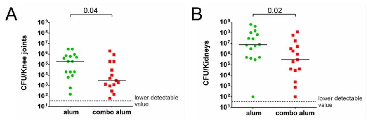
As shown in figures 21A and 21B, immunization with the protein combination resulted in a significantly lower bacterial burden both in
51
the knee joints (A) and in the kidneys (B), suggesting that, in this model, the efficacy of a preventive vaccine treatment may be more efficacious than a classical therapeutic approach in reducing S. aureus mediated infection in joints.
Figure 21. Mice immunized with the S. aureus combo vaccine resulted protected against a subsequent systemic infection with S. aureus Newman strain. A) CFU counts in knee joint washes of mice immunized
and intravenously infected with S. aureus Newman strain 7 days after inoculum. Each dot reports the data from a single animal and black lines indicate the median values. The dotted grey line states the lower detectable value. B) CFU counts in kidneys of mice infected with S. aureus sacrificed 7 days later on. Each dot is referred to a single animal and black lines show median values. The dotted grey line states the lower detectable value. In both the cases, statistical analyses were performed with a Mann-Whitney t test.
Further analyses were then performed in the attempt to understand the possible mechanism(s) of action of the vaccine. To this purpose, we analyzed both the humoral and the cellular immune components.
52
First we measured antibody titres against each single antigen of the vaccine in the knee joint washes. In figure 22A IgG titres against all the antigens are reported, demonstrating that all of them induced seroconversion in immunized animals and that measurable antibody levels could be detected in the knee joints.
Moreover, immune cells were analyzed comparing the results obtained for the vaccinated animals to those obtained for the negative controls. Fewer dead cells were found in knee joint washes of combo-immunized mice and, even if the number of total cells recovered was more or less the same in both the samples, the number of neutrophils recruited was lower in the immunized group indicating the lower state of general inflammation (fig.22B). Interestingly, B cells were preserved in knee joints after immunization with protein combo as if the vaccination could increase the number of cells recruited and/or shield them from S. aureus mediated toxicity (fig. 22B).
Cytokine levels were also measured in this site. Even if none of the analyzed mediators significantly changed between the 2 groups, it was interesting to note that almost all the pro-inflammatory cytokines (like IL-1, IL-1, and G-CSF) were a little less in knee joint washes of combo immunized animals in respect to mocked immunized mice (fig. 22C), confirming the observation already done of a general lower state of inflammation of treated animals.
53
Figure 22. IgG titres, immune cells recruitment and cytokines analysis of immunized mice. A) IgG titres of combo antigens from knee joint
washes of mice immunized with alum or combo/alum and infected with S. aureus Newman strain. Mean Fluorescence Intensity (MFI) was used as titer and box-whiskers analysis (median plus 25 and 75 percentile) of row data was reported. Statistical analysis was performed with a Mann-Whitney t test. B) Immune cell recruitment in knee joint washes of mice immunized and infected with S. aureus 7 days after infection. Fold change in respect to the basal situation (median of naïve mice for each cellular subset) was plotted, medians and interquartile range were reported. N= Neutrophils, Mo= Monocytes, Ma= Macrophages, DC= Dendritic cells, B= B cells, CD4+= CD4+ T cells, CD8+ =CD8+ T cells. Statistical analysis was performed with a Mann-Whitney t test. Only statistically significant differences were reported. C) Cytokine analysis of knee joint washes from mice immunized with alum or combo/alum and infected with S. aureus Newman strain. Data were reported in pg/ml and only the cytokines from a panel of 23 mediators analyzed which were measurable were shown. Medians and interquartile range were reported and statistical analysis was performed with a Mann-Whitney t test. Only statistically significant differences were shown.
54
3 Discussion
In the present work we set a new, long lasting and reproducible mouse model of S. aureus mediated arthrosynovitis-osteomyelitis that allowed us to unravel the host immune response during these pathologies. Using this model we were also able to demonstrate that the Novartis proposed Vaccine against staphylococcal infections overcame the activity of a commercial antibiotic in reducing bacterial load in knee joints. High IgG titres against all the vaccine components were found in knee joint washes of vaccinated animals one week after infection with S. aureus Newman strain. Moreover, vaccination seemed to preserve the immune cells recruited in joints after infection by bacteria-mediated killing and this was particularly clear for B cells. Finally, a lower degree of general inflammation was observed in knee joints of vaccinated animals as compared to the controls that reflected a lower recruitment of neutrophils in situ. We can conclude that this vaccine could become a powerful tool for preventing S. aureus-mediated arthrosynovitis and osteomyelitis in humans, pathologies that still have often poor outcomes.
Septic arthrosynovitis and osteomyelitis are severe and painful joints and bones diseases often associated with treatment failure and poor prognosis. Most of these pathologies are due to Staphylococcus aureus that is also responsible for the ones with the highest
55
Since the pathophysiology of these diseases is not completely understood, the use of animal models has proven invaluable for studying them as well as for testing the efficacy of experimental preventive and therapeutic treatments (68). To mimic the human disease as closely as possible, several features of the host bacterium relationship should be clarified. During the past decades, the use of experimental models of staphylococcal infections was useful to understand the involvement of several bacterial virulence factors as well as the host response to the bacterial infection. Dogs, rabbits, rats, chickens, and mice (69,70,71,72,73) have been used to develop Staphylococcus aureus model of arthrosynovitis and osteomyelitis,
but it is believed that the mouse model is optimal because of the resemblances between the murine and the human immune and inflammatory systems.
Nevertheless, the existing animal models generally study osteomyelitis and arthritis like two different diseases but since these pathologies often coexist in humans, they do not completely reflect the full scenario of the natural disease. Moreover, most of them focus only on the acute phase of the disease while the chronic phase, which occurs later on during the infection, is often the most debilitating one (74,75). Recently Horst et al. developed a novel hematogenous murine model of acute and chronic osteomyelitis, but the immune response induced has not been analyzed neither locally nor systemically (72).
56
Based on this background, we thought it would be important to have a consistent model of osteomyelitis and arthritis established in mice after infection with S. aureus, which would allow to study both the acute and chronic phases, dissecting the humoral and cellular host immune response locally and systemically, and to evaluate the efficacy of therapeutic and preventive tools.
An important issue was how the staphylococci spread through the body to reach the joints. The most common routes of infection for both septic arthritis and osteomyelitis in humans are the haematogenous, the contiguous, or the direct ones (13, 14, 15, 16, 17), but it was clearly shown that most of bacterial joint infections in humans are caused by bacteria which spread hematogenously (18). Thus we decided that intravenous (i.v.) inoculation of bacteria mimicking the hematogenous route of infection would be the optimal delivery route for the animal model that we wanted to set up. Indeed after inoculum, bacteria are required to adapt to the environment within the host, to survive bactericidal components in the blood, disseminate to synovial tissue and penetrate various structures to reach the joint space (69) being in this way really similar to what happens during natural infections in humans.
We reported in figure 5 using the In Vivo Imaging System (IVIS®) that S. aureus showed a particular tropism for bones and joints
57
considering its ability to quickly and long lastingly colonize these tissues. This was somehow expected since S. aureus expresses several receptors (adhesins) specific for bone matrix components such as fibronectin (76), collagen (77) and bone sialoprotein (78). Once reached the joints S. aureus was able to cause both arthrosynovitis and osteomyelitis and the infection lasted for months progressing to a chronic phase (figures 7, 10A and 11). Interestingly, only combining histopathological and microbiological analysis of both the in situ and the systemic situation we were able to clearly identify an acute and a chronic phase of arthrosynovitis and osteomyelitis. On the one hand CFU decrease in the knee joints and kidneys seemed to push toward the resolution of the pathology, but on the other hand the rise of CFU in the blood and, in particular, the increase in age grade and severity of histopathological analysis of knee joints showed a completely different scenario. The acute phase was therefore characterized by a high bacterial burden in joints and kidneys, but medium level of severity of the lesions in the knee joints, while when the infection became chronic, CFU numbers decreased, as the host immune system was able to control bacteria, but the severity of the lesions dramatically increased with destruction and deformation of the bone architecture, and bacteria used again the blood as vehicle for further dissemination. Taken together, these data allowed us in defining the two distinct phases of infections, an acute
58
phase that picks from 7 to 14 days after inoculum, and a chronic phase, that could be clearly seen at the latest time point.
Like in humans, the acute phase was shown to be highly symptomatic and was characterized by a particularly active immune response.
Interestingly, the host immune response, both humoral and cellular, seemed to be quite different between blood and knee joints indicating that a specific immune response is built up against this pathogen in situ. While in the blood the situation was quite complicated probably
representing a multi-organ infection, during this phase all the pro-inflammatory cytokines (like IL-1β, IL-6 and IL12p40) peaked in the knee joints between 7 and 14 days after infection, reflecting the profile observed for CFU counts and allowing the recruitment of the cellular components of the host immune response. Moreover, a good correlation between some of the cytokines we analyzed and CFU counts in knee joints was observed (figure 15). This important finding could pave the way for using new potential biomarkers for humans, that could be not only useful in the specific diagnosis of the diseases, but above all to define the degree of severity and the maturation of disease, helping to figure out the possible prognosis and to give the most proper and accurate care for a patient.
As expected, IgM and IgG levels rose significantly early during the acute phase indicating that a strong adaptive immune response was mounted against the pathogen really soon and that the inflammation
59
state of the joints was useful in allowing the diffusion of antibodies and the recruitment of plasmacells in situ. But inflammation means also cellular recruitment. We therefore approached the cellular analysis both in the knee joints and in the blood using, in our knowledge for the first time in these models, a flow cytometer analysis with a specific staining for different immune cells. This approach allowed us analyzing almost all the possible immune cells recruited in this site at the same time.
Both myeloid and lymphoid cells were rapidly and strongly recruited in situ. Monocytes and macrophages increased between 10 and 20
fold starting immediately after infection. These cells play an important role in controlling initial stages of infection not only directly through the phagocytosis of the bacteria, but also producing chemoattractant molecules for neutrophils recruitment. Their role in the early stages of infection is in any case controversial. Verdrengh et al. (79) indeed studied the role of these cells in staphylococcal arthritis using monocytopenic mice. During the time these mice exhibited a significantly less severe arthritis than the control animals and this was accompanied by decreased serum levels of the proinflammatory cytokines TNFα and IL-6. In contrast, infection-triggered mortality was increased in the monocytopenic mice as compared with the control animals. Notably, the monocytopenic mice exhibited elevated bacterial burden in the blood and kidneys. This study indicated a dual role of mononuclear phagocytes in the pathogenesis of S.
aureus-60
induced infection. On the one hand, absence of macrophages led to a favorable outcome concerning the severity of arthritic lesions, but on the other hand the direct and indirect clearance of bacteria mediated by monocytes/macrophages was decreased, resulting in poor survival. Neutrophils were actually the population which showed the highest increment among all the cells changing almost 1000 fold as compared to not infected mice. This increment was evident in the blood too, even if less impressive, probably suggesting a greater production due to an increased demand from other tissues and organs. It is not strange to see that neutrophils were strongly recruited in the knee joints immediately after infection peaking at 14 days, since it had been already demonstrated that they played an essential role in controlling bacterial spreading in these kinds of infections. Indeed mice depleted of these cells died in a few days after infection with S. aureus and showed more severe signs of arthritis, increased bacterial burden in blood and kidneys and exhibited increased levels of the proinflammatory cytokines TNFα, IL-6, and INFγ, reflecting the severity of their disease (80,81).
The role of T cells in arthrosynovitis and osteomyelitis was already reported by Abdelnour et al. (82) who showed using a immune-histochemical approach an increase of these cell populations in the joints between 3 and 14 days after the bacterial inoculum. We confirmed their findings but our approach was certainly more suitable for studies where a large number of animals and multiple analyses
61
were required. During the first days after infection circulating lymphoid cells left the blood spreading into tissues where they were recruited by the presence of infective agents. Interestingly, while CD4+ and CD8+ cells incremented in the knee joints from day 3 to day 14 confirming our hypothesis, this was not true for B cells whose number suddenly decreased coming back to basal level only between day 14 and day 30.
One possible explanation for this behavior is the production by S. aureus of protein A (Spa). In recent studies, it was shown that SpA
has the properties of a B cell superantigen by virtue of interactions with a large supraclonal B cell set via high affinity framework-mediated interactions with soluble and cell-associated B cell receptors (BCR, 83). Moreover, other studies demonstrated that a domain of SpA forms a complex with human Fab via a conformational surface on BCR (84) and that this target is conserved among amphibian, avian, and mammalian species (85).
SpA toxic activity in vivo was already demonstrated by Goodyear et al. who injected in mice purified protein A and then followed the fate of peripheral B cells expressing BCRs with VH regions capable of binding SpA. They observed a rapid down-regulation of BCRs and the coreceptors CD19 and CD21, the induction of an activation phenotype, and limited rounds of proliferation. Apoptosis followed through a process heralded by the dissipation of mitochondrial