A
A
l
l
m
m
a
a
M
M
a
a
t
t
e
e
r
r
S
S
t
t
u
u
d
d
i
i
o
o
r
r
u
u
m
m
–
–
U
U
n
n
i
i
v
v
e
e
r
r
s
s
i
i
t
t
à
à
d
d
i
i
B
B
o
o
l
l
o
o
g
g
n
n
a
a
DOTTORATO DI RICERCA IN
Biologia Cellulare e Molecolare
Ciclo XXIX
Settore Concorsuale di afferenza: 05/D1
Settore Scientifico disciplinare: BIO/09 FISIOLOGIA
TITOLO TESI
PHYSIOPATHOLOGICAL PROTEIN RELEASE BY GLIAL
CELLS: FOCUS ON PURINE NUCLEOSIDE PHOSPHORYLASE
(PNP), SUPEROXIDE DISMUTASE 1 (SOD1), AND α-SYNUCLEIN.
Presentata da: Francesca Massenzio
Coordinatore Dottorato
Relatore
Chiar.mo Prof. Giovanni Capranico Prof.ssa Barbara Monti
1
INDEX
ABSTRACT ... 4
1. INTRODUCTION ... 8
1.1 THECENTRALNERVOUS SYSTEM ... 8
1.1.1 MICROGLIAL CELLS ... 9
1.1.1.1 Origin of microglia ...10
1.1.1.2 Microglial phenotypes ...11
1.1.1.3 Physiological functions of microglia ...11
1.1.1.4 Pathological functions of microglia ...12
1.1.2 ASTROCYTES ... 16
1.1.2.1 Physiological role of astrocytes ...17
1.1.2.2 Pathological role of astrocytes ...19
1.1.3 NEURON-GLIA INTERACTIONS ... 20
1.1.4 GLIAL CELL SECRETION ... 22
1.1.4.1 Glial vescicular release mediated by physiopathologycal stimuli ...22
1.1.4.2 Vescicule-mediated release of proteins ...24
1.2PURINENUCLEOSIDEPHOSPHORYLASE(PNP) ...26
1.2.1 PNP DEFICIENCY ... 27
1.3 SUPEROXIDEDISMUTASE1(SOD1) ...28
1.3.1 AMYOTROPHIC LATERAL SCLEROSIS (ALS) ... 30
1.3.2 MUTANT SOD1 IN ALS ... 33
1.3.3 GLIAL CELL INVOLVEMENT IN ALS ... 34
1.4ALPHA-SYNUCLEIN(Α-SYN) ...36
1.4.1 PARKINSON’S DISEASE ... 38
1.4.2 MUTANT α-syn IN PARKINSON’S DISEASE ... 40
1.4.3 GLIAL CELL INVOLVEMENT IN PARKINSON’S DISEASE ... 42
2. AIMS ...44
3. MATERIAL AND METHODS ...48
3.1PRIMARYRATCULTUREOFMICROGLIAANDASTROCYTES ...48
3.2PRIMARYRATCULTUREOFCEREBELLARGRANULENEURONS(CGNS) ...50
3.3IMA2.1ANDN9IMMORTALIZEDCELLSLINES ...52
3.4CULTURETREATMENTS ...52
3.4.1 TREATMENT WITH LPS ... 52
2
3.4.3 TREATMENT WITH ADENOSINE TRIPHOSPHATE (ATP) AND
2'-3'-O-(4-BENZOYLBENZOYL)-ATP (BZ-ATP) ... 53
3.4.5 L-SERINE TREATMENT ... 54
3.4.6 TREATMENT WITH THREALOSE ... 55
3.5GLIALCELLTRANSFECTION ...55
3.5.1 PURIFICATION OF PLASMID DNA ... 55
3.5.2 TRANSFECTION OF GLIAL PRIMARY CULTURES ... 56
3.5.3 TRANSFECTION OF GLIAL CELL LINES ... 57
3.6CELLSANDCONDITIONEDMEDIA ...58
3.7PURIFICATIONOFEXTRACELLULARVESICLESFROMCONDITIONEDMEDIUM ...58
3.8CO-CULTUREOFCEREBELLARGRANULENEURONS(CGNS) WITH PRIMARYMICROGLIAOR ASTROCYTESOVEREXPRESSING WT/ MUTSOD1 ...59
3.9IMMUNOCYTOCHEMISTRY ...59
3.10NITRICOXIDEDETECTIONASSAY ...61
3.11HOECHSTSTAININGANDNUCLEICOUNTING ...61
3.12WESTERNBLOT ...62
3.13HIGHPERFORMANCELIQUIDCHROMATOGRAPHY(HPLC) ...64
3.14QUANTITATIVEREALTIMEPCR ...65
3.15STATISTICALANALISYS ...66
4. RESULTS ...67
4.1PNPRELEASEBYGLIALCELLS ...67
4.1.1 PNP EXPRESSION AND RELEASE BY RAT PRIMARY CULTURES OF MICROGLIA, ASTROCYTES AND DIFFERENTIATED CGNS ... 67
4.1.2 INFLAMMATORY STIMULI DO NOT MODIFY CONSTITUTIVE PNP SECRETION FROM CULTURED MICROGLIA, ASTROCYTES AND DIFFERENTIATED CGNS ... 69
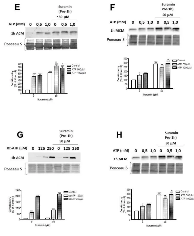
4.1.3 ATP ENHANCES CONSTITUTIVE PNP SECRETION FROM CULTURED MICROGLIA AND ASTROCYTES ... 72
4.1.4 PNP RELEASE IS MEDIATED VIA THE LYSOSOMAL SECRETORY PATHWAY ... 73
4.1.5 INVOLVEMENT OF P2X7 RECEPTORS IN PNP RELEASE ... 76
4.2SOD1RELEASEFROMRATPRIMARYCULTURESOFMICROGLIAANDASTROCYTESIN PHYSIOPATHOLOGICALCONDITIONS ...80
4.2.1 MICROGLIAL SOD1 RELEASE THROUGH LYSOSOMES ... 80
4.2.2 INTRACELLULAR MUTANT SOD1 ACCUMMULATION IN PRIMARY CULTURES OF MICROGLIA AND ASTROCYTES ... 83
4.2.3 AUTOPHAGIC PATHWAY ALTERATION IN MICROGLIAL CELLS OVEREXPRESSING MUTANT SOD1 ... 86
3
4.2.4 EFFECT OF WILD-TYPE AND MUTANT SOD1 OVEREXPRESSION ON MICROGLIA AND
ASTROCYTE ACTIVATION AS WELL AS ON MICROGLIAL PHENOTYPES ... 89
4.2.5 EFFECT OF GLIAL CELLS OVEREXPRESSING WILD-TYPE OR MUTANT SOD-1 ON NEURONAL SURVIVAL/DEATH IN CONTACT CO-CULTURES ... 92
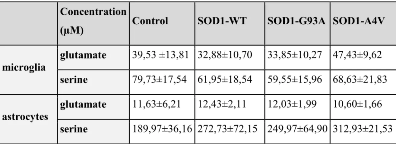
4.2.6 GLUTAMATE AND SERINE RELEASE BY MICROGLIA AND ASTROCYTES OVEREXPRESSING WT AND MUTANT SOD1 ... 95
4.2.7 TREHALOSE REDUCES THE INTRACELLULAR ACCUMMULATION OF SOD1... 96
4.2.8 TREHALOSE REDUCES THE ACTIVATION STATE OF MICROGLIAL CELLS OVEREXPRESSING MUTANT SOD1 ... 99
4.2.9 NEUROPROTECTIVE FUNCTION OF TREHALOSE IN GLIA-NEURON CO-CULTURE MODEL ... 100
4.3RELEASEOFALPHA-SYNUCLEINBYGLIALCELLLINESINPHYSIOPATHOLOGICAL CONDITIONS. ... 103
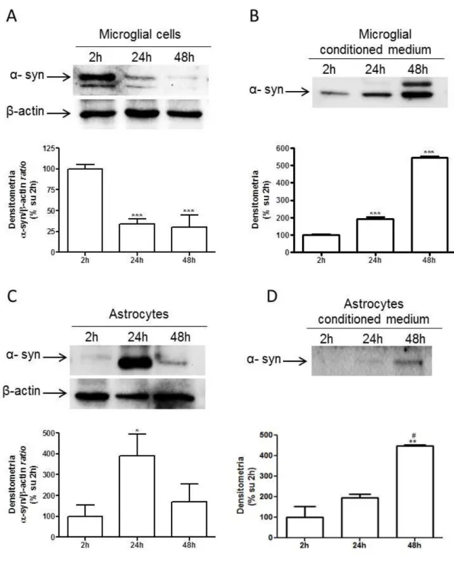
4.3.1 α-SYNUCLEIN EXPRESSION AND RELEASE FROM RAT PRIMARY CULTURES OF MICROGLIA, ASTROCYTES AND DIFFERENTIATED CGNS ... 103
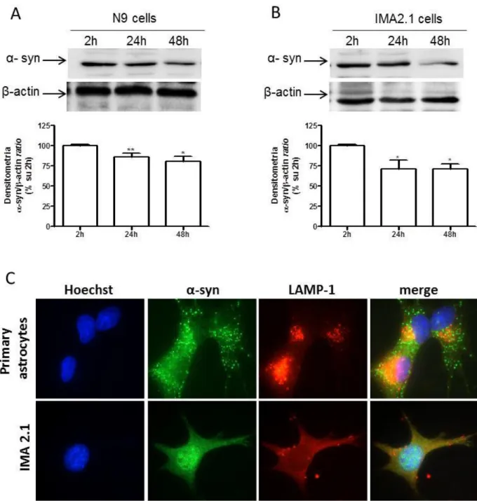
4.3.2 α-SYNUCLEIN EXPRESSION IN MURINE IMMORTALIZED CELL LINE OF MICROGLIA (N9) AND ASTROCYTES (IMA2.1) ... 107
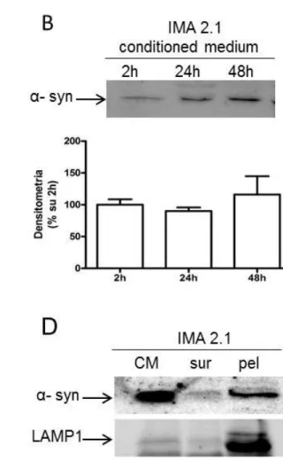
4.3.3 α-SYNUCLEIN RELEASE IS MEDIATED BY LYSOSOMAL SECRETORY PATHWAY ... 109
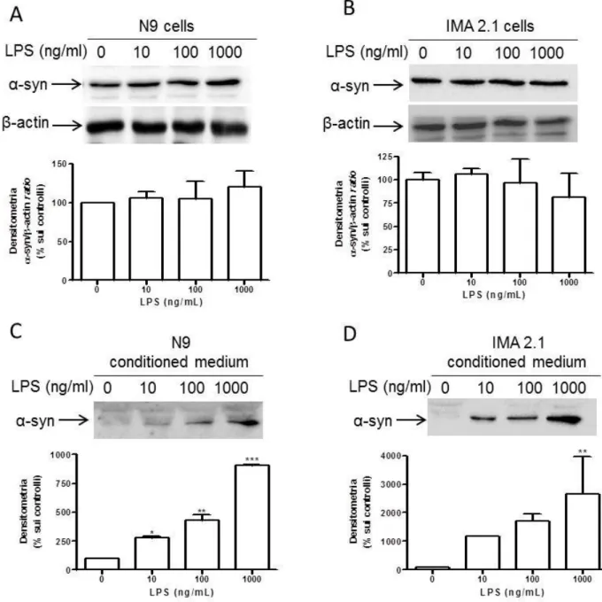
4.3.4 EFFECT OF INFLAMMATORY STIMULI ON α-SYNUCLEIN RELEASE ... 111
4.3.5 GLIAL ACTIVATION THROUGH ATP DOES NOT CHANGE α-SYNUCLEIN RELEASE ... 114
4.3.6 ACTIVATION OF NMDA RECEPTOR MEDIATED BY SERINE DOES NOT INTERFERE WITH PHYSIOLOGICAL GLIAL RELEASE OF α-SYNUCLEIN ... 117
4.3.7 INTRACELLULAR EXPRESSION AND EXTRACELLULAR RELEASE OF α-SYNUCLEIN FROM N9 AND IMA 2.1 CELL LINES OVEREXPRESSING WT AND MUTANT α-SYNUCLEIN ... 118
4.3.8 EFFECT OF WILD-TYPE AND MUTANT α-SYNUCLEIN OVEREXPRESSION ON MICROGLIA AND ASTROCYTE ACTIVATION ... 121
4.3.9 EFFECT OF WILD-TYPE AND MUTANT α-SYNUCLEIN OVEREXPRESSION ON MICROGLIA AND ASTROCYTE VIABILITY... 123
4.3.10 GLUTAMATE AND SERINE RELEASE BY MICROGLIA AND ASTOCYTES OVEREXPRESSING WT AND MUTANT α-SYNUCLEIN ... 124
5. DISCUSSION ... 126
5.1PNPRELEASEBYPRIMARYGLIALCELLS... 127
5.2SOD1RELEASEBYPRIMARYGLIALCELLSINPHYSIOPATHOLOGICALCONDITIONS ... 129
5.3ALPHA-SYNUCLEINRELEASEBYGLIALCELLLINES... 134
6. CONCLUSIONS ... 139
4
ABSTRACT
Previous studies from our laboratorory highlighted the neuroprotective role of glial cells against neuronal damage especially through protein release (Polazzi and Monti, 2010). Given the involvement of glial cells not only in the surveillance and homeostasis of the Central Nervous System, but also in pathological conditions, in this thesis we focused our study on the glial release of three different proteins, Purine Nucleoside Phosphorylase (PNP), Superoxide Dismutase 1 (SOD1) and α-synuclein related to different neuropathological conditions, i.e. PNP-deficiency, Amyotrophic Lateral Sclerosis (ALS) and Parkinson’s disease (PD).
Purine Nucleoside Phosphorylase (PNP) protein is a ubiquitous enzyme, which catalyzes the intracellular cleavage of ribonucleosides to generate purine bases. Physiologically, PNP plays a crucial role in the metabolism of purines and PNP deficiency causes a rare inherited disease determining severe combined immunodeficiency and mental retardation (SCID). PNP histochemical localization is restricted to glial cells, whereas its expression in neurons is still debated. Similarly to human disease, PNP deficiency in mice, due to missense mutations in the gene codifying for this enzyme, showed a reduction in number of immature and peripheral T-cells and in T-cell proliferation. Neurological symptoms have been described in 50% of patients; neurological involvement seems to be related with the severity of enzyme deficiency because GTP is necessary for neurotransmission. Notwithstanding the relevant role of glial cells in PNP-deficiency, the role of PNP in glial cell physiology has never been studied before.
Superoxide Dismutase 1 (SOD1) catalyzes the dismutation mechanism, which is the transformation of superoxide to H2O2, which in turn is converted
to H2O by catalase, peroxiredoxin (Prx) or glutathione peroxidase. Catalytic
activity is required to maintain low levels of intra and extracellular ROS, superoxide and H2O2, indispensable in signaling or in defense against
pathogens. If the level of ROS is too high, it may be cause of oxidative stress. A previous published study from our laboratory demonstrated constitutive SOD1 production and release by microglia and the consequent accumulation in
5
conditioned medium (MCM) with a neuroprotective role of microglia in case of neuronal damage; SOD1 was found to be one of the neuroprotective released molecules. On the other hand, mutations in SOD1 are a key player in the onset of Amyotrophic Lateral Sclerosis (ALS); approximately 20% of familial cases (FALS) are due to mutations in the gene coding for SOD1. Neurodegenerative diseases, stroke, trauma and hypoxia affect neuronal survival indirectly by triggering neuroinflammation. Microglia are then activated (or become reactive) in response to insults acquiring a phagocytic phenotype and releasing inflammatory mediators, cytokines and chemokines. The acute neuroinflammatory response seems to be beneficial to the CNS, since it is an attempt to reduce injury and restore damaged tissues. Because of the dual role of SOD1 and microglial cells, we decided to study the effect of wild-type and mutant SOD1 (G93A and A4V) in glial cell pathophysiology.
Alpha-synuclein is a ubiquitous protein belonging to a family that also includes β- and γ –synucleins. In the brain, α-synuclein represents 0.5-1.0% of all cytosolic proteins and is primarily localized at presynaptic terminals. The protective or toxic effect of α-synuclein depends on its expression levels in the cell: α-synuclein in physiological conditions would have a protective function, while its accumulation and its aggregation into oligomers or fibrils would have a detrimental effect on cells. Some point mutations (A53T, A30P and E46K) in the α-synuclein gene are related to the onset of an autosomal dominant juvenile form of Parkinson’s disease. Taking these evidences together and because most studies on α-synuclein were performed on neurons, we decided to analyze as a first step whether α-synuclein could be secreted by glial cells and then we tried to define the conditions of this secretion and its role.
Therefore, the first aim of this thesis was to analyze the release of PNP, SOD1 and α-synuclein by glial cells in physiopathological conditions in in
vitro models: primary cultures of rat astrocytes and microglia for PNP and
SOD1, as well as immortalized mouse astrocytes and microglia cell lines for α-synuclein (respectively IMA2.1 and N9). In particular, for all three proteins the effect of different glial activating stimuli (LPS, IFNγ and ATP) on their release, as well as their vesicular secretion through exosomes were analysed. Considering the role of mutations in the genes codifying for SOD1 and
α-6
synuclein in familial forms of neurodegenerative diseases, respectively ALS and PD, a second aim was to investigate whether and how overexpression of wild-type SOD1 or mutations linked to ALS (G93A and A4V) could alter the functioning of microglia and astrocytes, determining activation and thereby contribute to inflammation, and whether this condition could affect neuronal survival and/or death. In parallel, a preliminary study on α-synuclein release was carried on by a preliminary investigation on overexpression of wild-type or PD-linked mutant α-synuclein (A53T and A30P) in glial cells.
By using the above-mentioned in vitro approaches, we observed that all brain cells, but mainly microglia and astrocytes, constitutively release PNP. Glial stimulation with potent inflammatory agents, like lipopolysaccharide and interferon-γ, did not modify PNP release, while ATP and Bz-ATP led to a dose-dependent increase of release mediated by P2X7 receptors and occurred
through the secretion of lysosomal vesicles in the extracellular medium. The mechanisms activated by ATP seem to be common in pathological conditions such as ischemia, traumatic injury and seizure activity and might suggest a pathological role of extracellular PNP in the mechanisms of PNP-deficiency.
Regarding SOD1, this enzyme is constitutively released by microglial cells and astrocytes, even through vesicles and mutant SOD1 (fALS murations) appears to be less released than wild-type SOD1. Intracellular accumulation of mutant SOD1 in both microglia and astrocytes correlates to dysfunction of autophagy mechanisms and promotes the activation of microglial cells. In addition, we developed a primary co-culture in vitro model in order to evaluate the effect of glial cell SOD1 overexpression on neuronal survival and death. We show that microglia exert neuroprotection in cerebellar granule cells exposed to the neurotoxic stimulus glutamate, but microglial mutant SOD1 overexpression appears to be toxic to neurons. Astrocytes, on the other hand, seem to be less susceptible to mutant SOD1overexpression and do not display a neuroprotective function against glutamate excitotoxicity, even if overexpression of wild-type SOD1 seems to provide neuroprotection. Trehalose, a disaccharide known to be able to restore the physiological autophagic flux, showed significant ability in restoring the physiological pathway of SOD1 expression in glial cells and significantly reduced the death
7
of cerebellar granule neurons due to glutamate excitotoxicity, giving promising expectations for future studies.
Moreover, α-synuclein is constitutively released by microglial cells and astrocytes, even through vesicles. The increased release after LPS but not ATP treatment underlies the different response of glial cells to different stimuli. Since activation of glial cells is strictly related to the progression of neurodegenerative diseases, inflammatory pathways might be responsible for the impairment of glial cell function. According to this hypothesis, we observed that overexpression of α-synuclein mutations linked to Parkinson’s disease promoted an increased release of synuclein compared to wild-type α-synuclein. Thus, α-synuclein overexpression, even wild-type α-synuclein, promoted the activation of microglia, though not of astrocytes. We could speculate that the increased release, together with increased activation, might be responsible for reduced neuroprotection and for diffusion of potential toxic mutant α-synuclein.
8
1. INTRODUCTION
The central nervous system is composed by two different cells type with a ratio of 1 to 10-50; neurons and glial cells or neuroglia (Rezaie et al., 2002). While neurons are specialized in the conduction of electric signals, glial cells do not participate directly in signal transmission, though they play an important role during neuronal development and are also involved in the trophic and protective support of neurons throughout life. In the Central Nervous System (CNS) there are four types of glial cells: ependymal cells, oligodendrocytes, astrocytes and microglial cells.
Ependymal cells, whose functions are not clear, are columnar cells located close to the brain ventricles and the central canal of the spinal cord.
Oligodendrocytes are responsible for the formation of the myelin sheet in the CNS. The myelin sheet acts as an insulator of axonal segments and is indispensable for nerve conduction.
Astrocytes are the most numerous cells in the CNS; they have a high number of ramifications that allow them to contact blood vessels and create the blood-brain barrier.
Microglia are the resident macrophages in the central nervous system. They are involved in the surveillance of the environment; in case of injury or neuronal damage, glial cells are the most responsive cells helping neurons restore physiological conditions (Hartline 2011). Microglial cells represent 5-20% of the glial population in the central nervous system.
9
The major brain areas rich in microglia are the hippocampus, telencephalic region and basal ganglia; the cerebellum and brain stem are much less populated (Lawson et al., 1990).
Since glial cells establish numerous interactions with neurons regulating their function, homeostasis and protection from stress and damage, the involvement of glial cells in the most known neurodegenerative diseases is well-established, though it remains to be characterized.
1.1.1 MICROGLIAL CELLS
Microglia are the resident mononuclear phagocytes of the CNS (Lawson et al., 1990). Defining the origin of microglia has not been easy and there have been several schools of thought.
The concept of neuroglia was introduced in 1856 by Rudolf Virchow; (Verkhratsk, 2006); he defined “nervenkitt” cells with the ability to form a kind of nervous adhesive tissue, able to keep connected adjacent cells. At the beginning of the nineteenth century, Robertson defined “mesoglia”, phagocytic cells with mesodermal origin present in the CNS. Mesoglia mainly correspond to oligodendrocytes, renamed by Santiago Ramon y Cajal “third element of the nervous system” to further differentiate them from neurons (first element) and neuroglia (second element) (Florent et al., 2012). The concept of the third element was clarified by Del Rio-Hortega; he called “microglial cells” the neuronal, non-astrocytic third element as distinct from neuroectodermal oligodendroglia or oligodendrocytes (Rezaie et al., 2002).
10
1.1.1.1 Origin of microglia
Regarding the origin of microglia, there are two well-known hypotheses: the neuroectodermal hypothesis and the myeloid-monocytic hypothesis.
1) The neuroectodermal hypothesis has been supported by studies on the identification of microglial markers during the development of the brain, germinal matrix, central nervous system or in the neuroectoderm.
2) The hypothesis that microglia derives from blood monocytes, originating in the bone marrow and circulating in the CNS between birth and the postnatal period, is the most-approved hypothesis. According to this myeloid-monocytic hypothesis, resident microglia and in general resident brain macrophages, are derived from circulating blood monocytes during embryonic life and in the postnatal period (Polazzi et al., 2010; reviewed by Kettenmann H. et al., 2011). However, data related to microglia during brain development, before vascularization and the formation of macrophages in the neural epithelium from precursors different from the ones originating monocytes, together with the phenotypic differences between mononuclear phagocytes in the embryonic-fetal stage and neonatal-adult period (Takahashi, 2001), suggest that prenatal microglia might arise from mesodermal progenitors.
There is evidence supporting the existence of two different microglial populations during fetal and embryonic development; a population with myeloid/mesenchymal origin, not necessarily monocytes; and a second population, which represents a transient form of fetal macrophages derived from blood precursors, probably monocytes, that are related to amoeboid microglia observed in the post-natal period in rodents (Rezaie et al., 2005b). This second microglial population lays close to blood vessels and can be replaced by precursors that originate in the bone marrow and express the histocompatibility complex II, indicating their monocytic origin (Eglitis et al., 1997; Hickey et al., 1998). The phagocytic ability of the two cell populations is different, in fact a reduced phagocytic activity is observed in unstimulated cells
11
with branched phenotype, which however is not observed in the amoeboid phenotype (Szabo et al., 2013).
1.1.1.2 Microglial phenotypes
Microglial cells are known for their plastic ability and functional characteristics depending on their activation state; indeed, it is not difficult to observe morphological microglial changes depending on their function (Szabo et al., 2013). In physiological conditions, microglial cells are in a state of non-activation, also call "resting" or “quiescent” state, in which microglial morphology is characterized by numerous processes that originate from the soma with distal arborization (Streit et al., 1999). In conditions of stress, inflammation, injury or particular signals, microglia change their morphology, shifting to an “activated” state (Stence et al., 2001). Cells undergo an expansion of the cell body, ramifications become shorter, less arborized and located around the soma. This morphological change can induce phagocytic functions in microglial cells and an amoeboid ability that allows cells to move towards lesions (Thored et al., 2005).
1.1.1.3 Physiological functions of microglia
It is well-known that microglia, even when in a “resting” state, monitor the brain microenvironment and synapses. Microglial cells, in fact, are essential for the proper functioning of the CNS, since they are a sensor of pathological conditions (Polazzi et al., 2010). Their highly-branched morphology and plasticity ability, allow microglia to supervise the extracellular parenchyma of the CNS and rapidly activate in response to pathological conditions, exerting typical macrophage functions such as phagocytosis (Neumann et al., 2006, Napoli et al., 2009), secretion of pro-inflammatory cytokines (Banati et al., 1993, Gehrmann et al., 1995, Minghetti et al., 1998) and the ablity to behave as antigen presenting cells (Beauvillain et al., 2008, Aloisi, 2011).
12
The neuroprotective role of microglia is carried out not only as a response to neuropathological threats, but also under physiological conditions. During brain development, microglia support synaptogenesis through the local synthesis of neurotrophic factors, regulating also synaptic transmission and remodeling; the interactions between microglia and neurons contribute to the development of synapses and neuronal homeostasis, also as a result of injury, which arrange for the recovery of neuronal circuits (Polazzi et al., 2013).
During development, microglia have a role in synaptic maturation, while in the adult brain they have a fundamental role in synaptic plasticity. Microglial cells are able to exert neurotoxic and neurotrophic effects because of their activation state. One of the most peculiar features of microglia is the ability to modify their behavior in response to different signals originating from other cells located in the brain parenchyma. This function suggests the ability to promote neuronal cell survival or death depending on the nature of the neural signals that regulate the activation of microglia. Damaged neurons may release or express molecules able to modulate the activation of microglia. This suggests that different types of neuronal death, apoptosis or necrosis, can induce different microglial responses, which can in turn be responsible for different neuronal fate (Polazzi et al., 2001).
Apoptosis or programmed cell death is crucial in CNS development, since it allows the removal of neuronal subpopulations. Apoptotic death is a common mechanism of neuronal removal during development of the nervous system, which occurs only in pathological conditions in the adult brain, such as in neurodegenerative diseases. Microglia are involved in this process in response to various signals derived from neurons triggering the process of programmed cell death through the release of neurotoxic molecules (De la Rosa et al., 2000).
1.1.1.4 Pathological functions of microglia
Loss of physiological functions is associated with the onset of psychiatric disorders or neurodegenerative diseases. Theoretically, every neurological disorder involves inflammation with the consequent activation of
13
resident microglia, accompanied by an increase in cell number and in phenotypic change, a phenomenon known as "gliosis".
Neurodegenerative diseases, stroke, trauma and hypoxia, affect neuronal survival indirectly by triggering the state of neuroinflammation. Microglia are then activated (or become reactive) in response to insults adopting a phagocytic phenotype and releasing inflammatory mediators, cytokines and chemokines. The acute neuroinflammatory response seems to be beneficial to the CNS, since it is an attempt to reduce injury and restore damaged tissues (Wake et al., 2013, Kiefer et al., 1995, Streit et al., 1999, Imai et al., 1997, Lalancette et al., 2007, Yanagisawa et al., 2008, Madinier et al., 2009).
Microglia and monocyte-derived macrophage functions –cascade of events. London et al., Front. Cell. Neurosci., 2013.
On the other hand, chronic neurodegenerative diseases including amyotrophic lateral sclerosis (ALS), Alzheimer's disease (AD), Parkinson's disease (PD), tauopathies and multiple sclerosis (MS), are known to be associated with chronic neuroinflammation. Although several differences have been found between these chronic diseases, neuroinflammation is a response to long-term injury or initial insult.
A typical feature of neuropathological conditions is the activation of microglia and the subsequent release of inflammatory mediators leading to an increased oxidative and nitrosative stress. This condition promotes the inflammatory cycle; the duration of the inflammatory response and the activation of microglia in this case are harmful to the nervous tissue (Frank-Cannon et al., 2009, Polazzi et al., 2010).
14
Activated microglia have two different phenotypes M1 and M2.
1) Classically activated microglia, M1 (LPS or IFNγ) have pro-inflammatory, neurotoxic properties, inhibiting the proliferation of lymphocytes; M1 activated macrophages secrete pro-inflammatory cytokines such as interleukin IL-1α, IL-1β, tumor necrosis factors (TNF) and nitric oxide (NO).
2) Alternatively activated microglia, M2 (IL-4, IL-13), are able to repair small damage and show an anti-inflammatory phenotype, contributing to the trophic support of neurons and have the ability to degrade aggregates and increase neuroprotective functions due to the production of anti-inflammatory interleukins (Geissmann et al., 2008, Hu et al., 2014).
In vitro studies in cell cultures have shown the ambivalent role of
microglial cells on neurons; neuroprotective but also neurotoxic, while in vivo studies mainly support the neuroprotective potential of activated microglia (Streit et al., 2008). It has not been clearly defined whether the activation state of microglia is beneficial or detrimental to neuronal survival, though the most accredited hypothesis today is that microglia may have a dual role depending on the nature, duration and extent of injury (Ransohoff et al., 2009).
In case of acute injury, microglia are able to remove damaged neurons through its phagocytic activity. This ability is attenuated by pro-inflammatory cytokines, suggesting that microglia involved in the inflammatory response could have reduced phagocytic activity (Koenigsknecht-Talboo et al., 2005). In neurodegenerative diseases, microglial activation occurs before neuroinflammation. A possible explanation may be that microglial activation induces neuroprotection through the elimination of toxic molecules (Polazzi et al., 2010). Furthermore, because neurodegenerative diseases cause alterations in many other cell besides neurons, not only are neuronal functions altered, but also the cross-talk between different cell types (Appel et al., 2010). In neurodegenerative diseases, the role of microglia in the early stages, seems to be linked to an increase of inflammatory functions resulting in neurodegeneration (Polazzi et al., 2010). However this does not exclude the hypothesis of microglial dysfunction; the loss of neurotrophic/neuroprotective microglial functions induces neurodegeneration (Streit 2014). During aging
15
and the onset of neurodegenerative diseases, there is a reduction in the phagocytic ability of microglia and in the trophic support to neurons. Therefore, if on one side the primary activation of microglia reflects a physiological mechanism focused on neuroprotection mediated by the secretion of cytokines and neurotrophic factors, the removal of damaged cells (Napoli et al., 2009) and phagocytosis of polymorphonuclear neutrophils (Neumann et al., 2006), on the other side the overactivation or the loss of microglial functions increases preexisting neuropathology (Streit et al., 2009). Alteration of microglial phenotype and as consequence the impairment of physiological functions in glial cells, might promote neuronal damage through a “non-cell autonomous” mechanism (Kathrin et al., 2014). According to this disease mechanism, neurodegeneration is strongly influenced by toxicity or mutant protein expression in non-neuronal cells which could be spread to neighbouring neurons.
In the literature, one of the main methods used to activate microglia both in vitro and in vivo is treatment with the bacterial endotoxin (LPS) (Hetier et al., 1988). In response to LPS microglia release cytokines, chemokines and NO. This effect is mediated by TLR4, belonging to the Toll-like receptor family (Takeda et al., 2003). Microglial activation, however, is reduced by treatment with anti-inflammatory compounds, followed by the reduction of inducible nitric oxide synthase (iNOS) expression. Microglial activation promotes on one hand the decreased production of reactive oxygen species, while on the other hand it increases the neuroprotective functions of microglia (Nathan et al., 2002). Microglia-mediated neuronal homeostasis has been also found altered in serious neuronal damage, which could be mediated by glutamate excitotoxicity and is involved in the majority of neurodegenerative processes. Glutamate acts via N-Methyl-D-Aspartate (NMDA) receptors and in physiological conditions its excess derived from synaptic activity is employed by astrocytes. In pathological conditions, glutamate uptake by astrocytes is compromised inducing an increase of the intracellular calcium concentration and neuronal cell death. LPS-activated microglia express the GLT-1 protein involved in glutamate uptake (Hanisch et al., 2007). LPS-treatment of hippocampal slices induces the stimulation of microglia and a fast and transient
16
increase in post-synaptic AMPA-mediated currents in the hippocampal neurons of the CA1 region. This effect was not observed in animals lacking microglia, indicating that LPS stimulation requires microglia and confirming its neuroprotective role. Following stimulation, microglia produce adenosine triphosphate (ATP), which acts as transmitter and recruits astrocytes that release glutamate allowing an increase of excitatory synaptic transmission through the glutamate metabotropic receptors (Iadecola et al., 2007).
1.1.2 ASTROCYTES
Astrocytes are the most numerous cells of the CNS (Parpura et al., 2012). In the human brain, the number of astrocytes was estimated to be more than five times the number of neurons (Hartline et al., 2006). During CNS development, the generation of astrocytes is subsequent to the initial production of neurons.
Astrocytes exert several important functions during the development of gray and white matter. They are also responsible for the formation and function of synapses during development, thanks to the release of molecular signals, such as thrombospondin. At the end of the nineteenth century, astrocytes were distinguished in protoplasmic and fibrous astrocytes based on morphological and anatomical differences. Protoplasmic astrocytes have been found in the gray matter and wrap around synapses through numerous branched processes, fibrous astrocytes have been found in the white matter and contact the nodes of Ranvier through long thin fiber-like processes. Both types of astrocytes establish direct contact with blood vessels and form gap junctions between the distal processes of
17
neighbouring astrocytes. At the end of the nineteenth century, Camillo Golgi theorized that astrocytes could be connected to blood vessels (Golgi 1871). However, only recent studies have highlighted the dynamic processes supporting these structural interactions, such as the dialogue between astrocytes and other vascular elements. Astrocytes have an ordered arrangement and a minimal overlapping, and each one interfaces with blood vessels and includes hundreds of synapses (Bushong et al., 2004). The cell surface is covered by lamellar extensions and protrusions which make astrocytes able to interact with surrounding synapses. Most astrocytes possesses the so called "end-feet", contact blood vessels and contribute to the formation of the blood-brain barrier (BBB), a diffusion barrier that prevents the influx of foreign molecules into the brain parenchyma (Simard et al., 2001).
1.1.2.1 Physiological role of astrocytes
Astrocytes play essential functions in the CNS in physiological conditions thanks to the processes able to contact synapses, axons at the nodes of Ranvier, neuronal cell bodies and blood vessels at the endothelial cell layer. The connections with blood vessels allow astrocytes to collect glucose for energy supply, adjust blood flow and the cerebral homeostasis of ions and transmitters (Iadecola et al., 2007). Astrocytes play a synaptic role during development, but also in the adult brain, especially in remodeling and synaptic pruning: they are involved at GABAergic and glutamatergic synapses in neurotrasmitter uptake to prevent excitotoxicity by glutamate and play a direct role in synaptic transmission by releasing molecules synaptically active such as purines (ATP and adenosine), γ-aminobutyric acid (GABA) and D-serine, modulating cell excitability and increasing the concentration of the intracellular calcium ion (Ca2 +) (Shimizu et al., 2007, Bittner et al., 2010). D-serine binds to post-synaptic NMDA receptors (NMDARs), which also bind glutamate released from the pre-synaptic terminal. These events facilitate NAMDAR activation and induction of synaptic plasticity (Verkhratsky et al., 2010). Astrocytes modulate synaptic function through cross talk and exchanging gliotransmitters, ions, second messengers and nutrients through gap junctions
18
(Parpura et al., 2009, Ullian et al., 2004). These observations support the concept of "tripartite synapses", according to which astrocytes represent the third unit element of signal transmission together with presynaptic and postsynaptic terminations (Volterra, 2005). The main role of astrocytes in this “model" is to isolate and support the function of individual synapses, ensuring spatial specificity in synaptic transmission (Hewett et al., 2009).
Astrocytes play also a metabolic role, since they are responsible for the storage of glycogen granules in the CNS, especially in high-density synaptic areas. In conditions of hypoglycemia and high neuronal activity, glycogen is able to support cell activity. Glycogen stored in astrocytes may provide lactate, which once transferred to adjacent neural elements can be used as aerobical fuel by neuronal mitochondria (Bélanger et al., 2011). The involvement of astrocyte glycogen stores is required for the formation of long-term memory in rats (Pellerin et al., 2007).
Astrocyte functions. (Maragakis et Rothstein, 2006)
Furthermore, although not presenting action potentials along their processes, astrocytes express sodium and potassium channels which may be involved in evoked currents and show forms of excitability, for example in the case of Ca2+ concentration increases. These increases are necessary for the intracellular communication that is established between astrocytes and between astrocytes and neurons (Molofsky et al., 2012).
19
1.1.2.2 Pathological role of astrocytes
Astrocytes have role in every aspect of neuronal function, through the regulation of blood flow, supply of energy substrates and modulation of synaptic function and plasticity, therefore damaged astrocytes are the primary cause of serious neural dysfunction (Maragakis et al., 2006). Following specific acute injuries, inflammation, chronic neurodegenerative diseases or stress, astrocytes respond through the reactive astrogliosis process and the expression of glial fibrillary acidic protein (GFAP), a sensitive and reliable marker for the immunohistochemical identification of reactive astrocytes. In the same way as microglial activation, reactive astrogliosis is not an on-off phenomenon, but rather a continuum event modulated by changes in the surrounding microenvironment and is regulated by specific inter and intracellular signaling (Yamanaka et al., 2008). Depending on the severity of the insult, reactive astrogliosis leads to a series of changes in astrocytes at the molecular, cellular, and functional (gain or loss of function) level, affecting adjacent cells in a beneficial or harmful way. Reactive astroglial cells play several essential beneficial functions in response to CNS insults, such as repairing the BBB, neuroprotection and limiting the spread of inflammatory cells. Loss or alteration of these functions in the astrogliosis process produces neuronal dysfunction and could be a primary cause of disease in the CNS such as neuropathology, psychiatric disorders, trauma and stroke. Animal models have shown that loss of astrocyte function leads to altered CNS function such as excitotoxicity-induced neurodegeneration, caused by an altered uptake of glutamate or increased inflammation and infection due to the loss of astrocyte barrier functions. Following specific inflammation signals, such as LPS exposure, a gain of harmful functions may also occur in reactive astrocytes, such as the inhibition of axonal regeneration, production of reactive oxygen species (ROS), glutamate release and the impairment of the BBB functionality. Moreover, the involvement of other cell types, such as microglia, macrophages and lymphocytes, stimulates astrocytes to produce cytotoxic levels of molecules which are normally non-toxic and contribute to inflammation and chronic neuropathic pain (Verkhratsky et al., 2010, Molofsky et al., 2010,
20
Rappold et al., 2010). Understanding these processes could simplify treatment strategies to preserve the beneficial role of astrocytes. Recent studies have shown that astrocytes are the main players in many developmental diseases, such as autism and schizophrenia, characterised by defects in synaptic formation (Parpura et al., 2012, Sofroniew et al., 2009). In fact, in the development of white matter, dysfunction or malfunction of astrocyte connections or gap junctions leads to demyelination. Given their strong involvement in developmental diseases, astroglial cells may be a useful target in the development of new therapies. The functionality of astrocytes is altered not only in neurodegenerative diseases, such as ALS, Alzheimer's and Parkinson's disease, but also in neurodevelopmental diseases such as Rett syndrome, X-fragile, Alexander’s disease, epilepsy, autism, psychiatric disorders and in brain tumors.
1.1.3 NEURON-GLIA INTERACTIONS
Glial cells represent about 90% of all cells in the human brain. The initial role as brain “glue” has been replaced with a more important role of key players in brain physiology, metabolism, development, and even neurological diseases (Barres, 2008). An essential aspect of normal brain function is the bidirectional interaction and communication between neurons and neighbouring glial cells; dysregulation of this interaction may contribute to the onset of neurodegenerative diseases. Together with astrocytes, microglia can release neuromodulatorymolecules involved in intracellular signalling and the communication with neurons.
21
The first evidence of interaction between microglia and neurons is given by the complementary expression of the microglial receptor for chemokines (CX3CR1) and its neuronal ligand (CX3CL1), thus suggesting the ability of microglia to modulate neurotransmission (Béchade et al., 2013). The CX3CL1/R1 pathway seems to be involved in synaptic maturation and an alteration of this pathway can lead to delayed glutamatergic synaptic maturation and alteration of hippocampal development (Paolicelli et al., 2011). Another example of neuron-microglia interaction is related to the CD200R membrane protein. CD200R is a membrane receptor exclusively expressed by microglia while its ligand, CD200 is expressed by neurons, oligodendrocytes and astrocytes. CD200-deficient mice display LTP inhibition, further underlining the role of microglia in neuronal homeostasis and synaptic plasticity (Costello et al., 2011). In fact, microglia is a sensor of neuronal activity modulating most contacts between neurons and synaptic elements (Tremblay et al., 2011, Wake et al., 2013). The signaling modulating this function is not known, though it seems to be mediated by the secretion of signaling molecules by microglia, including cytokines (Elkabes et al., 1996, Hanisch, 2002) and neurotransmitters (Hanisch, 2002, Flierl et al., 2007), which regulate synaptic functions. In vivo studies suggest a role of microglia in the regulation of hippocampal (Battista et al., 2006, Ziv et al., 2006) and sub ventricular area neurogenesis (Thored et al., 2009)after stroke. In an in vitro co-culture model of neural stem cells with microglia or conditioned medium from microglia, the secretion of factors by microglia needed for neurogenesis was oberved (Walton et al., 2006). Microglia-neuron interaction and the consequent regulation of neuronal homeostasis by microglia were also found in severe neuronal damage, such as glutamate excitotoxicity. Glutamate acts via N-methyl-D-aspartate (NMDA) receptors inducing an increase of neuronal intracellular calcium concentration, and consequent neuronal death. Neurotoxicity due to glutamate is involved in numerous neurodegenerative processes, such as ischemia and brain injury. In physiological conditions, the excess of glutamate resulting from synaptic activity is uptaken by astrocytes, while in pathological conditions, glutamate uptake by astrocytes is compromised. LPS-activated microglia express the GLT-1 protein implicated
22
in glutamate uptake (Hanisch and Kettenmann, 2007). Microglia stimulation with lipopolysaccharide (LPS) treatment of hippocampal slices, induces a rapid and transient increase in the spontaneous post-synaptic AMPAergic current in hippocampal CA1 neurons. This effect has not been found in animal models with microglia depletion, indicating that LPS stimulation requires microglia and confirming the neuroprotective role of microglial cells. Following this stimulation, microglia recruit astrocytes through the production of ATP. Astrocytes release glutamate allowing an increase of excitatory synaptic transmission through metabotropic glutamate receptors (Pascual et al., 2012).
Astrocytes express neurotransmitter receptors, but at the same time, thanks to the increase in calcium concentration, they release neurotransmitters (ATP, Glutamate, D-serine, and GABA) essential for the modulation of neuronal activity (Haydon, 2001, Volterra, 2005) Glutamate release from astrocytes seems to be a key modulator of synaptic transmission depending on the kinetic of calcium elevations. Slow elevation in astrocytes induces slow inward currents in CA1 neurons by acting on post synaptic NMDA receptors (Fellin et al., 2004, Angulo et al., 2004). Faster calcium elevation induces presynaptic modulation of synaptic release, by acting on presynaptic mGluR5 receptors (Shigetomi et al., 2008). Activation of glutamatergic receptors on astrocytes, induces the release of D-serine, which acts as a co-agonist of NMDA receptors (Mothet et al., 2005). D-serine is synthetized by astrocytes from L-serine by serine racemase (SR), mainly expressed in glial cells and overexpressed in activated glial cells increasing the production and release of D-serine (Wu et al., 2004). Released D-serine promotes the production of reactive oxygen species (ROS) and the overactivation of NMDA receptors exacerbating glutamate effects and neuronal damage (Mustafa et al., 2010).
1.1.4 GLIAL CELL SECRETION
1.1.4.1 Glial vescicular release mediated by physiopathologycal stimuli
The correlation between neuroinflammation and neurodegeneration has not been yet clarified. Many physiological CNS functions involving glia,
23
including neurodevelopment, memory maintenance, neuroprotection, homeostatic and metabolic mechanisms, may be mediated by direct cell-cell contact or by the secretion of molecules in a paracrine way both through the release of soluble factors and through extracellular vesicles (EVs), able to transfer potential toxic, aberrant, non-folded or neuroprotective molecules to neighbouring cells.
In 1909, Hans Held observed granular inclusions in glial processes (Held, 1909). A year later, Jean Nageotte observed secretory granules in glial cells of the grey matter indicating an active secretion from these cells (Nageotte, 1910). Alois Alzheimer called these granules, gliosomes. Glial cell secretion includes neuotransmitters, neuromodulators, hormones, peptides, metabolic substrates, ROS, growth factors, plastic factors, inflammatory factors and neuroprotective molecules. The release of these molecules is mediated by several pathways; vesicle-based exocytosis of D-serine (Martineau et al., 2013) or glutamate (Montana et al., 2009), diffusion through plasmalemmal pores/channels, release of ATP and/or glutamate through anion channels, P2X7
receptors (Cotrina et a., 1998, Suadicani et al., 2006) and extrusion through plasmalemmal transporters (Unichenko et al., 2013).
Microglia and astrocytes exert their immune function mainly in the inflammatory process following acute and chronic neurodegenerative diseases, among which psychiatric disorders are included (Shie et al., 2011). In these pathological conditions, glial cells become activated (Skaper et al., 2012; Collins et al., 2012; Eikelenboom et al., 2012) and exert their function by releasing several factors.
ATP represents one of the most important neuro/glio-transmitters; during pathology, ATP is released from damaged cells and acts both as a cytotoxic factor and a proinflammatory mediator, being a universal "danger" signal (Franke et al., 2012).
D-serine upregulates glial activation (Panatier et al., 2006). Glutamate excitotoxicity is one of the main events responsible of neuronal degeneration and motor neuron death in Amyotrophic Lateral Sclerosis (ALS) and the impairment in astrocyte uptake of glutamate could be responsible for this event (Ilieva et al., 2009). Release from neural cells could be altered by glial cell
24
activation through neuroinflammatory stimuli; bacterial LPS and interferon- γ (IFN-γ), which are able to induce a wide range of inflammatory activities, increase phagocytosis, chemotaxis, cytokine secretion, activation of the respiratory burst and induction of nitric oxide synthase (Zielasek, Hartung, 1996), while the neuro/glio-transmitters ATP, which is released by injured and dying neural cells, leads to activation of P2X7 receptors in glial cells (Franke et
al., 2006; Weisman et al., 2012; Skaper et al., 2010).
Polazzi et al. showed that neuroprotection is due partly to molecules with a molecular weight lower than 30kDa, such as TGF-β2, SOD1 and ApoE. Transforming growth factor-beta 2 (TGF-β2), as well as SOD1 are constitutively released by microglia and neuroprotection could derive partly from the interaction between microglial released TGFβ-2 or SOD1 with neuronal receptors (Polazzi, 2013).
1.1.4.2 Vescicule-mediated release of proteins
It is known that glial cells signal to neighbouring cells by releasing molecules via regulated exocytosis through lysosomes (Verderio et al., 2012; Bräuer et al., 2004) and glial cell activation, commonly occurring in most neurodegenerative diseases and affecting protein release from these cells by influencing their secretory pathways (Bianco et al., 2005; Hanamsagar et al., 2011). Glia-cell release of molecules may be mediated by extracellular vesicles that can be classified according to size, cargo and origin.
Extracellular vesicles (EVs) comprise shedding microvesicles (MVs), exosomes, and apoptotic bodies. Apoptotic bodies are released during apoptosis, whereas the other types of vesicles are derived from healthy cells (Cocucci et al., 2009, Thery, 2011). Exosomes contain proteins arising from the plasma membrane, endocytic pathway and cytosol. They contain limited amounts of proteins from other intracellular compartments (nucleus, endoplasmic reticulum, Golgi apparatus). RNAs are transported by EVs from cell to cell and can modulate gene expression in the recipient cell. In the immune system, MHC-peptide complexes are secreted by exosomes from
25
antigen presenting cells (APCs) suggesting a role for exosomes in the adaptive immune response.
Communication within the nervous system could be mediated by EVs; exosomes are secreted by neurons, Schwann cells, astrocytes and microglia (Potolicchio et al., 2005) and they may be responsible of the transfer of pathogenic proteins such as prions, beta-amyloid peptides, superoxide dismutase 1, tau and α-synuclein between glial cells and neurons promoting neuronal damage.
Activation of microglia, mediated by ATP (activation of P2X7 receptors), seems to promote the release of microvesicles with irregular shape and size (0,1-1µm), carrying pro-inflammatory cytokines to recipient neurons increasing inflammation (Verderio et al., 2012). Similar to microglia, astrocyte-derived MVs are increased by activation of P2X7 receptors though these vesicles can be up to 8 μm in size and carry intact mitochondria and lipid droplets (Falchi, 2013).
While microglia appear to be mainly involved in the spread of inflammatory molecules, astrocytes seem to be mainly implicated in the propagation of pathogenic proteins in neurodegenerative disorders. Astrocytes expressing mutant SOD1 (copper-zinc superoxide dismutase 1) secrete
26
increased amounts of exosomes, carrying mutant SOD1. These vesicles can transfer mutant SOD1 to neurons and induce motor neuron death suggesting a role of EVs in the pathogenesis of ALS (Basso et al., 2013). Exposure of astrocytes to amyloid peptide triggers the release of pro-apoptotic exosomes that contain ceramide and PAR4 (prostate apoptosis response 4). These exosomes are taken up by astrocytes and promote their apoptosis suggesting that exosome-mediated astrocyte death may contribute to neurodegeneration in Alzheimer’s disease (Wang et al., 2012).
Purine Nucleoside Phosphorylase (PNP) is a ubiquitous enzyme, which catalyzes the intracellular cleavage of ribonucleosides to generate purine bases. Physiologically, PNP plays a crucial role in the metabolism of purines (Bzowska et al., 2000) and its deficiency causes a rare inherited disease determining severe combined immunodeficiency (SCID) (Markert, 1991).
PNP protein, a trimer of approximately 90,000 daltons, has been found in most body tissues, showing the highest levels in lymphoid tissue, thus explaining why immune cells are predominantly impaired in PNP deficiency. Accordingly, PNP inhibitors have been developed as T-cell selective immune-suppressants and to treat many forms of leukemia (Balakrishnan et al., 2010; Robak et al., 2006; Bantia, Kilpatrick, 2004). PNP knock-out mice recapitulate most pathological features of PNP-deficiency (Arpaia, et al., 2000). In this animal model, TAT-mediated intracellular delivery of PNP corrects the enzyme deficiency (Toro, Grunebaum, 2006). Concerning neurological defects in brains from PNP-KO mice, morphological abnormalities and apoptosis were detected in the cerebellum, which was smaller than normal with reduced Purkinje cell number (Mansouri et al., 2012).
Despite all data concerning PNP involvement in brain physiopathology, little is known regarding its specific brain cell distribution. PNP histochemical localization is restricted to glial cells, whereas its expression in neurons is still debated (Castellano et al., 1990). Additionally, Dalmau and collaborators
1.2 PURINE NUCLEOSIDE PHOSPHORYLASE
(PNP)
27
(1998) suggested that microglial cells could be crucial for the development of normal central nervous system (CNS) by controlling purine metabolism. They showed different PNP staining depending on microglial morphology and observed PNP labeling in blood vessels, some nerve fibers and astrocytes, suggesting a complex mechanism of cell interactions in the regulation of extracellular purine levels.
1.2.1 PNP DEFICIENCY
Severe Combined Immunodeficiency (SCID) is a heterogeneous disease characterized by a prenatal disorder in the development of T lymphocytes leading to profound abnormalities of cell-mediated immunity, antibody deficiency, severe viral, bacterial and fungal infections, chronic diarrhoea, failure to thrive and skin rashes as typically seen in Omenn syndrome (OS). This pathology was first described in a five-year old child with T-cell immunodeficiency (Giblett et al., 1975). SCID is caused by mutations in different genes mainly involved in the development and function of both T and
28
B cells; natural killer cells are present in 50% of patients with SCID and are able to protect against viral, bacterial and fungal infections.
PNP deficiency is a rare form of SCID (Markert, 1991). As in the human disease, PNP deficiency in mice, due to missense mutations in the gene codifying for this enzyme, showed a reduction in numbers of immature and peripheral T cells and in T cell proliferation (Snyder et al., 1997). Furthermore, mutations in PNP caused loss of deoxyguanosine kinase activity. PNP- deficiency could be due to block of purine recovery as a consequence of GTP depletion or to the accumulation of PNP substrates inosine, guanosine, deoxyinosine, and deoxyguanosine.
The abnormal accumulation of dGTP preferentially in mitochondria, inhibits mitochondrial DNA synthesis as a first step of T- lymphocyte apoptosis (Young, 1994).
Pathways of purine metabolism, Archives of Disease in Childhood, 1987.
Neurological symptoms have been described in 50% of patients; neurological involvement seems to be related with the severity of PNP deficiency because GTP is necessary for neurotransmission. These evidences support a direct relation between neurological dysfunction and PNP deficiency.
Superoxide dismutase (SOD) is an enzyme that exists in three isoforms: SOD1 (CuZnSOD); SOD2 (MnSOD); SOD3 (extracellular SOD); each one is produced from a different gene with distinct localization, but share the same
29
function. This distinct localization is important for the compartmentalization of redox reactions.
This enzyme catalyzes the dismutation mechanism, which is the transformation of superoxide by SOD to H2O2, in turn converted in H2O by catalase, peroxiredoxin (Prx) or glutathione peroxidase (GPx) (Go, Jones D, 2011). The conversion involves alternative reduction and oxidation of a transition metal, such as copper (Cu) and manganese (Mn) that are located in the active site of the enzyme (Miao, St Clair, 2009; Zelko et al., 2002; Yamakura, Kawasaki H, 2010; Ushio-Fukai, 2011). Catalytic activity is required to maintain low levels of intra and extracellular ROS, superoxide and H2O2,, indispensable in signaling or in defense mechanisms against pathogens.
If the level of ROS is too high, it may be cause of oxidative stress.
SOD1 is a 153 amino acid homodimer ubiquitously expressed, mainly in the cytoplasm. Each SOD1 subunit binds to a zinc (Zn) and a copper (Cu) atom (Pasinelli, Bown R, 2006; Bruijn et al., 2004). The genomic sequence coding for SOD1 has high similarity among rat, mouse and human, thus indicating a considerable conservation for this gene.
Philbert Ip., 2011
Polazzi et al. demonstrated a constitutive SOD1 production and release by microglia and the consequent accumulation in microglia conditioned medium (MCM). SOD1 was also found among the low molecular weight neuroprotective molecules released by microglia. Treatment of primary cerebellar granule neurons with 6-hydroxydopamine (6-OHDA) induced death of 50% of cells in 24h, however, co-exposure to MCM was able to restore cell
30
viability. The neuroprotective ability of SOD1 was confirmed by exposing primary cerebellar granule neurons to exogenous SOD1 in presence or absence of SOD1 inhibitors; neuroprotection was abrogated by SOD1 inhibitors. Polazzi et al. also demonstrated an ATP-mediated increase of SOD1 release by microglia, confirming the involvement of the lysosomal secretory pathway (Polazzi et al., 2013).
Mutations in SOD1 are a key player in the onset of Amyotrophic Lateral Sclerosis (ALS); approximately 20% of familial cases (FALS) are due to mutations in the gene coding for SOD1. More than 125 mutations have been identified, 114 of them cause the disease, six are silent and five are intronic variants that do not lead to ALS. Although most of the 114 mutations are missense, twelve mutations are deletions or nonsense mutations that produce truncated proteins; all these mutations are specific to FALS and they have been found only in 1% of sporadic ALS (SALS). The majority of mutations cause reduced dismutation activity while few maintain full catalytic function. There is no clear correlation between enzyme activity and clinical disease progression, therefore it is still unclear how mutant SOD1 leads to motor neuron degeneration (Pasinelli, Bown, 2006). However, it is known that SOD1-mediated toxicity in ALS is not due to a loss of function rather to gain of one or more toxic properties that are independent from SOD1 activity (Lewis et al., 2014).
1.3.1
AMYOTROPHIC
LATERAL
SCLEROSIS
(ALS)
Amyotrophic lateral sclerosis (ALS), also known as motor neuron disease, Charcot's disease (from the French neurologist who first described it in 1860) or Lou Gehrig's disease (Pasinelli and Brown R.H, 2006), is a progressive, lethal, neurodegenerative disease, leading to paralysis of the voluntary muscles. ALS affects adults of both sexes around 55 years; the incidence and prevalence of ALS are respectively 1-2 and 4-6 per 100,000 cases every year (Pasinelli P, and Brown, 2006). Mutations in superoxide dismutase 1 (SOD1), valosin containing protein (VCP), ubiquilin 2
31
(UBQLN2), charged multivesicular body protein 2b (CHMP2B), optineurin (OPTN), TAR DNA-binding protein 43 (TDP43), fused in sarcoma (FUS), chromosome 9 open reading frame 72 (C9ORF72) have been identified and promote the definition of proteinopathy for ALS (Takeuchi et al., 2015). A hallmark of neurodegenerative proteinopathies is the formation of misfolded protein aggregates that cause cellular toxicity. A key feature of misfolded protein diseases is the ability of the pathogenic protein species to propagate in a prion-like manner to neighbouring cells and to initiate or promote damage.
Approximately 90-95% of ALS cases are sporadic (SALS), the remaining 5-10% are hereditary, familial ALS (FALS), 20% caused by dominant mutations in the gene coding for Cu/Zn superoxide dismutase 1 (SOD1, chromosome 21q22.11). In ALS patients, more than 150 pathogenic mutations in SOD1 have been identified. Despite the involvement of several genes in FALS and SALS, there are similarities between all ALS forms: progressive degeneration of lower motor neurons (atrophy, cramps, twitching) and death of cortical neurons, suggesting a common pathogenic pathway (Grada et al., 2014). Motor neuron degeneration leads to impairment of neuromuscular function, paralysis of skeletal muscles, paralysis of breathing muscles (diaphragm and intercostal muscles) that culminate in respiratory failure, which causes death (Pasinelli and Brown, 2006). The main pathological hallmark of ALS is the atrophy of motor neurons but accumulation of phosphorylated neurofilament inclusions and the deposition of inclusions (spheroids) in axons, known as Lewy bodies, as well as activation and proliferation of astrocytes and microglia are observed (Hall et al., 1998; Wilms et al., 2001). Unfortunately, there is currently no therapy available for this neuropathology; the only approved drug, riluzole, only prolongs the life of patients by a few months (Pasinelli and Brown, 2006; Lewis et al., 2014). Therefore, the characterization of the cellular and molecular processes involved in the onset and progression of the disease is necessary to identify new potential targets useful for therapeutic strategies, as well as for the identification of biomarkers for early diagnosis and therapeutic follow-up. When the disease affects upper motor neurons, it is defined as primary lateral sclerosis (PLS). The majority of patients affected by PLS develop consequent
32
problems in lower motor neurons (MND), satisfying the criteria for the diagnosis of ALS. Impairment in lower motor neurons (MND) classified the pathology as progressive muscular atrophy (PMA), though disease progression is not different from ALS. Frontotemporal lobar degeneration (FTLD) affects 3 or 4 persons/100,000 per year and is characterized by abnormal behaviour and language. The main hallmark of FTLD are inclusions containing TAR DNA-binding protein 43 (TDP-43), similar to the ones found in the motor neurons of ALS patients.
In physiological conditions, TDP-43 is located in the nucleus while in the pathology the phosphorylated protein and C-fragment aggregates are located in the cytoplasm. The presence of both TDP-43 and mutant SOD1 aggregates in FALS is controversial; there are groups supporting the absence of TDP-43 aggregation (Al-Chalabi and Leigh, 2005; Chio et al., 2009) and groups supporting the presence of these aggregates (Wills et al., 2009). These evidences may classify ALS and FTLD as a different manifestation of the same disease spectrum.
Interaction between ALS and FTD. Robberecht & Philips. Nature Reviews Neuroscience 14, 248-264.
The mechanism involved in the onset of ALS is not clearly defined; cellular degeneration, protein misfolding, presence of aggregates in the cytoplasm, ROS, excitotoxicity, impairment of axonal transport and abnormal calcium metabolism could be due to the loss of physiological function or gain of toxic function in the proteins responsible of the disease.
Recent studies suggest a correlation between exposure to solvents, pesticides, metals, chemical compounds and the onset of ALS regarding the neurotoxic properties of these substances. (Bonvicini et al., 2010).
33
ALS Neurodegeneration mechanisms. Robberecht & Philips, Nature Reviews Neuroscience 14, 248-264.
1.3.2 MUTANT SOD1 IN ALS
As previously described, microglia may show a direct neuroprotective effect against neuronal damage, as well as through the involvement of small neuroprotective molecules released such as SOD1.
The function of SOD1 is controversial; neuroprotective in physiological conditions, but responsible for the onset of ALS, if mutated.
Many hypotheses have been considered about the role of SOD1 mutations in ALS. SOD1 mutations could i) alter the enzymatic activity of SOD1 through the copper aberrant catalysis or the binding of a wrong metal due to an alteration in the configuration of the active channel, or ii) cause a rapid reduction in the bond between SOD1 and copper resulting in the formation of superoxide anion (Bruijn et al., 2004), or iii) give SOD1 an unstable conformation promoting the aggregation of the protein. Protein aggregates increase with disease progression (Johnston et al., 2000; Wang et al., 2002).
34
The G93A mutation is the most common SOD1 mutation in Europe and is also the most studied one, especially in mouse models. Substitution of G93 is predicted to destabilize the β -barrel (Hart et al., 1998).
Transgenic mice overexpressing human SOD1 G93A mutation show the first symptoms at 3 months of age but the severity of the disease depends on the expression levels of the mutant protein. The progressive weakness characteristic of these mice coincides with microglial activation, astrogliosis, loss of motor neurons in the spinal cord and intracellular aggregates both in motor neurons and glia, thus well-representing the human disease. The inhibition of the proteasome ability to remove aberrant proteins seems a sufficient condition to induce the formation of aggregates in glial cells expressing mutant SOD1 (Bruijn et al., 2004).
These evidences led to a "non-cell autonomous" hypothesis for neurodegeneration in ALS, in which non-neuronal cells of the brain play a central role in inducing neuronal death (Wang et al., 2009).
Mutant SOD1 causes astrogliosis, activation, infiltration and the inflammatory response of microglia, (increased production of nitric oxide and toxic cytokines), leading to further damage to motor neurons and accelerated disease progression (Polazzi and Monti, 2010).
1.3.3 GLIAL CELL INVOLVEMENT IN ALS
Involvement of microglia in ALS was postulated in the early 90s in in
vivo models of the disease, by studying microglial activation in the brain and
spinal cord of patients. Activation of microglia correlates with disease progression, leading to the idea that immuno-inflammatory mechanisms may contribute to ALS. Neuro-inflammation in ALS is also indicated by the fact that anti-inflammatory drugs could exert protective effects in mouse models of ALS. However, the role of microglia in neuro-inflammation and whether microglial activation is a cause or a consequence of the degeneration of motor neurons has not been fully clarified. Metabolism of nitric oxide (NO) may provide a link between microglial activation and oxidative damage to motor neurons. Mutant SOD1 is able to catalyze the production of reactive oxygen species such as peroxynitrite, which promote the expression of inducible nitric
35
oxide synthase (iNOS) (Li et al., 2005). iNOS converts arginine into citrulline and produces NO. Production of NO may play an important role in the transition from M2 to M1 phenotype and consequently in neuronal toxicity (Lewis et al., 2014).
Astrocytes are the most abundant cells in the adult nervous system. In mouse models of ALS, astrocyte degeneration is due to the interaction of glutamate with one of its receptors, GluR2, which is less expressed than in control conditions. The GluR2 subunit is impermeable to Ca2+ and thus protects motor neurons from excitotoxicity. Mutant SOD1 expressed in
astrocytes alters the impermeable property of the receptor, making motor neurons vulnerable to excitotoxicity (Ilieva et al., 2009). Pharmacological blockade of the glutamate receptor slows the degeneration of astrocytes, delays the onset of symptoms and improves life expectancy. Thus the GluR2 glutamate receptor may be investigated as a therapeutic target. Although the expression of mutant SOD1 confined to astrocytes is not sufficient for disease development, selective reduction of mutant SOD1 in astrocytes slows down disease progression and the consequent activation of microglia, demonstrating the functional cross-talk between astrocytes and microglia. (Nagai et al., 2007).