Alma Mater Studiorum – Università di Bologna
DOTTORATO DI RICERCA IN
Scienze Veterinarie
Ciclo XXX
Settore Concorsuale: 07H5
Settore Scientifico Disciplinare: VET/09 Clinica chirurgica veterinaria
NEW APPLICATIONS OF ALPHA
2-AGONISTS IN VETERINARY MEDICINE
Presentata da:
Dott.ssa Carlotta Lambertini
Coordinatore Dottorato
Supervisore
Prof. Arcangelo Gentile
Dott.ssa Noemi Romagnoli
Sommario
SOMMARIO ... 2
ABSTRACT... 4
RIASSUNTO ... 6
ALPHA2-ADRENOCEPTOR AGONISTS IN VETERINARY MEDICINE ... 8
INTRODUCTION ... 9
MECHANISM OF ACTION OF ALPHA2-ADRENOCEPTOR AGONISTS ... 11
SEDATIVE EFFECTS ... 18 ANALGESIC EFFECTS ... 27 CARDIOVASCULAR EFFECTS ... 32 RESPIRATORY EFFECTS ... 37 OTHER EFFECTS ... 39 ALPHA2-ANTAGONISTS ... 44
NEW APPLICATIONS OF ALPHA2-AGONISTS ... 46
IN VETERINARY MEDICINE ... 46
ALPHA2-AGONISTS FOR SEMEN COLLECTION ... 47
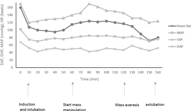
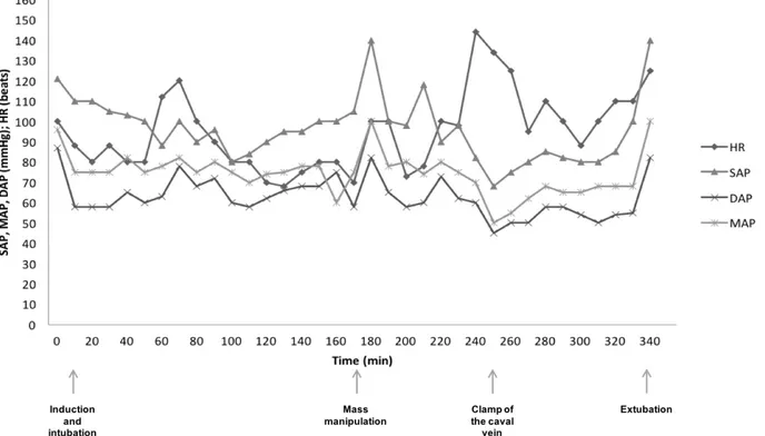
DEXMEDETOMIDINE INFUSION DURING SURGERY IN TWO DOGS UNDERGOING ADRENALECTOMY FOR A SUSPICION OF A PHEOCHROMOCYTOMA ... 69
ALPHA2-AGONISTS FOR THE PERIOPERATIVE MANAGEMENT OF ANESTHESIA IN NON-HUMAN PRIMATES UNDERGOING CRANIOTOMY ... 82
DISCUSSION AND CONCLUSION ... 90
Abstract
Alpha2-agonists are a class of drugs widely used in veterinary anaesthesia; moreover by means
of their action on adrenoceptors that are widespread distributed in several tissues, they can be beneficial for different clinical applications. The aim of this work was to describe new applications of alpha2-agonists in veterinary medicine.
In cats, high dose medetomidine is administered to perform semen collection by urethral catheterization. We have investigated the haemodynamic effects of high dose medetomidine (0.13 mg kg-1) administered to healthy male cats. Haemodynamic evaluations were performed before and
after medetomidine administration and consisted of: clinical examination, blood pressure evaluation and transthoracic echocardiography. Significant hemodynamic alterations were observed, even if they were similar to that provided by lower dosages. The cats recovered without clinical alterations.
Despite their cardiovascular side effects, low doses of alpha2-agonists can be beneficial for the
maintenance of a good cardiovascular stability for specific conditions.
In humans, dexmedetomidine helps in maintaining a good hemodynamic stability if administered for pheochromocytoma ablation. We have described the administration of dexmedetomidine for the anesthetic management of two dogs with a suspicion of pheochromocytoma undergoing
adrenalectomy. Dogs received dexmedetomidine intramuscularly (0.001 mg kg-1) and
dexmedetomidine and remifentanil were administered (0.0005 mg kg-1h-1 and 0.0003 mg kg-1min-1,
respectively) throughout the surgery. In this study dexmedetomidine infusion together with remifentanil provided satisfactory intraoperative anesthetic and hemodynamic control in two dogs with a suspicion of pheochromocytoma.
In patients undergoing craniotomy, dexmedetomidine, increasing the cerebral vascular resistance, prevents alteration of the cerebral blood flow. We have described the administration of dexmedetomidine in five Macaca fascicularis undergoing craniotomy for physiologic research. The
Macaca were sedated with ketamine (8 mg kg-1) and dexmedetomidine (0.02 mg kg-1)
intramuscularly. Dexmedetomidine was administered by infusion (0.012 mg kg-1h-1) throughout the
Riassunto
I farmaci alfa2-agonisti sono largamente utilizzati in anestesia veterinaria; inoltre, grazie alla loro
azione sui recettori alpha-adrenergici, distribuiti in diversi tessuti, sono utilizzati per diverse applicazioni cliniche. L’obiettivo del presente studio è stato quello di descrivere nuove applicazioni degli alfa2-agonisti in medicina veterinaria.
Nel gatto, la medetomidina somministrata ad alte dosi consente la raccolta del seme mediante cateterismo uretrale. Abbiamo valutato gli effetti emodinamici della medetomidina somministrata al dosaggio di 0.13 mg kg-1 in gatti sani. Le valutazione emodinamiche sono state eseguite prima e dopo
la somministrazione di medetomidina mediante visita clinica, misurazione della pressione sistemica ed ecocardiografia transtoracica. Dallo studio sono state evidenziate alterazioni emodinamiche significative, ma simili a quelle riportate dopo somministrazione di dosi più basse.
I farmaci alpha2-agonisti, nonostante le alterazioni cardiovascolari che inducono, se
somministrati a basse dosi, possono contribuire al mantenimento di una buona stabilità emodinamica in condizioni cliniche specifiche.
Nell’uomo, la somministrazione di dexmedetomidina in pazienti sottoposti a rimozione di un feocromocitoma contribuisce a mantenere parametri emodinamici intraoperatori stabili. Abbiamo descritto la somministrazione perioperatoria di dexmedetomidina in due cani sottoposti a surrenalectomia per un sospetto di feocromocitoma. Entrambi hanno ricevuto dexmedetomidina intramuscolo (0.001 mg kg-1) e dexmedetomidina e remifentanil sono stati somministrati in infusione
(0.0005 mg kg-1h-1 e 0.0003 mg kg-1min-1, rispettivamente) per tutta la chirurgia. Il protocollo
utilizzata ha permesso di mantenere un piano anestesiologico e condizioni emodinamiche stabili in due cani con sospetto di feocromocitoma.
Nei pazienti sottoposti a neurochirurgia, la dexmedetomidina previene alterazioni significative del flusso cerebrale. Abbiamo descritto la somministrazione di dexmedetomidina in esemplari di
Macaca fascicularis sottoposti a craniotomia. I macachi sono stati sedati con ketamina e
dexmedetomidina. La dexmedetomidina è stata somministrata in infusione continua (0.012 mg kg-1h -1) per tutta la procedura e ha permesso di mantenere un’analgesia adeguata e parametri emodinamici
Introduction
Alpha2-adrenoceptor agonists are a class of drugs widely used in veterinary anesthesia to obtain
sedation, analgesia and muscle relaxation. Their application has been described in small companion animals (Sinclair et al. 2003; Lemke 2004), in large animals (Daunt and Steffey, 2002; Valverde 2010; Gozalo-Marcilla et al. 2015) and in laboratory animals (Lee et al. 2010; Lugo-roman et al. 2010) among others.
The alpha2-adrenoceptor drugs most commonly used in clinical practice are xylazine,
detomidine, romifidine, medetomidine and dexmedetomidine.
Xylazine was first synthesized in Germany in 1962 as an antihypertensive drug but its sedative properties were soon discovered in animals, becoming the first alpha2-agonist used in veterinary
anesthesia. It was first used in ruminants, then in horses and cats (since 1970s), and finally in dogs (Green and Thurmon 1988; Paddleford and Harvey 1999).
The administration of alpha2-agonists also produces side effects which must be taken into
consideration. A decrease in heart rate and arrhythmias, and a biphasic pressure response are the most problematic side effects described after alpha2-agonist administration at therapeutic doses. In
addition, several endocrine effects have been recognized: decreased insulin release, glycogenolytic effects and decreased antidiuretic hormone release.
Since alpha2 adrenoceptors are involved in the regulation of several physiological mechanisms,
their administration may provide multiple effects other than those sedative and analgesic.
Among other applications, studies in veterinary medicine have reported their administration in male animals of several species for semen collection. In cats, high doses of medetomidine (0.13 mg kg-1) are needed to obtain semen release into the urethra. Since the effects of high dose medetomidine
in cats have never been described, the aim of this study was to investigate the hemodynamic effects induced by medetomidine administered intramuscularly in healthy cats at a dose of 0.13 mg kg-1.
In addition, dexmedetomidine administration has been reported to improve cardiovascular stability in human patients with pheochromocytoma and in those undergoing pheochromocytoma ablation. In dogs, adrenalectomy is the treatment of choice for patients with pheochromocytomas; however, mortality is high due to catecholamine release during mass manipulation. Alpha2-agonists
have been described to reduce the catecholamine release; therefore, one of the following studies described the use of dexmedetomidine infusion as an adjunct to a balanced anesthetic protocol in two dogs undergoing pheochromocytoma ablation.
Finally, dexmedetomidine has been described as a potential supplemental anesthetic drug in human patients undergoing intracranial surgery. To the best of our knowledge, there are no studies evaluating the use of dexmedetomidine as an adjunct for the anesthetic management of veterinary patients undergoing craniotomy. Therefore, we aimed to describe the use of dexmedetomidine administered by continuous rate infusion (CRI) for the anesthetic management of Cynomolgus
Mechanism of action of alpha
2-adrenoceptor agonists
Alpha2-agonists exert their action by means of agonism on the alpha2-adrenoceptors. In addition,
the majority of them, except for xylazine, have an imidazoline ring and can be combined with imidazoline receptors (Khan et al. 1999; Clarke et al. 2014).
Alpha- adrenoceptors
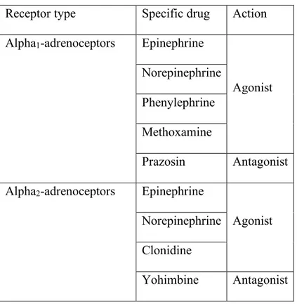
Alpha-adrenoceptors are differentiated on the basis of their sensitivity to agonists and antagonists (Table 1). Alpha2-adrenoceptors are located presynaptically and postsynaptically in both central and
peripheral sites while alpha1-adrenoceptors are only postsynaptic. Norepinephrine (NA) is the natural
ligand for both alpha1- and alpha2-adrenoceptors; when NA is released from a sympathetic neuron, it
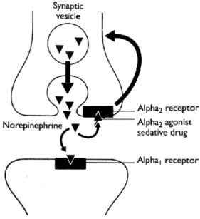
acts on both receptors inducing sympathetic stimulation. When activated, presynaptic alpha2
-adrenoceptors exert a negative feedback and prevent the additional release of NA, decreasing the sympathetic outflow from the central nervous system (CNS) (Figure 1). Postsynaptic alpha1 and
alpha2-adrenoceptors usually exert a stimulating effect.
Alpha2-adrenoceptors are transmembrane receptors formed by a long chain of amino acids with
hydrophilic and hydrophobic areas which cross the cell membrane seven times (Khan et al. 1999). Alpha2-adrenoceptors belong to the family of guanine nucleotide-binding proteins
(G-proteins)-coupled receptors. G-proteins are formed by alpha, beta and gamma subunits, and have been identified on the intracellular side of the cell membrane. The alpha subunit at rest binds to guanosine diphosphate (GDP). When the receptor is activated by its agonist, it changes its structure binding with the G-protein which reduces its affinity for GDP and, in the presence of magnesium, it becomes guanosine triphosphate (GTP). The alpha subunit leaves the G-protein, reaching the effector and, concurrently, the agonist leaves the receptor (Figure 2). The GTPase, which is now activated, hydrolyzes GTP to GDP with the release of an inorganic phosphate, and the receptor becomes inactive (Taylor 1990; Khan et al. 1999).
Alpha2-adrenoceptors are involved in more than one effector mechanism. Their activation
through the inhibitory G-protein (Gi-protein) inhibits the adenylyl cyclase, resulting in the decreased formation of 3',5'-cyclic adenosine monophosphate (cAMP), the second messenger of many biological mechanisms (Khan et al. 1999). The activation of the stimulatory G-protein (Gs- protein) causes hyperpolarization of the neuronal cells. However, other studies have suggested that the effect resulting from the activation of alpha2-adrenoceptors depends only on the agonist concentration; a
low alpha2-agonist concentration results in the inhibition of cAMP while high agonist concentration
induces an increase in cAMP (Eason et al. 1992; Gyires et al. 2009).
An alternative mechanism involves voltage-gated calcium ion channels coupled to a G-protein; this theory states that the decrease in calcium ion conductance results in the inhibition of the neurotransmitter release (Khan et al. 1999).
The presynaptic alpha2-adrenoceptors involved in the inhibition of the NA release are mainly
located in sympathetic nerve endings and in non-adrenergic neurons in the CNS. Postsynaptic alpha2-adrenoceptors have been identified in several tissues, such as the liver, pancreas, platelets, kidney, adipose tissue and the eye, where they are involved in several physiologic functions (Khan et al. 1999).
Four alpha2-adrenoceptor subtypes have been identified on the basis of pharmacological and
molecular studies: alpha2A, alpha2B, and alpha2C. The fourth type, alpha2D, identified in the rat brain
is the homologue of human alpha2A.
In the CNS, the alpha2A-subtype has been identified in the brain and mainly in the locus ceruleus,
the alpha2B-subtype has been found only in the thalamus and the alpha2C-subtype is widely distributed
but is mainly present in the basal ganglia (MacDonald and Scheinin 1995). Radioligand binding assays and real time polymerase chain reaction (PCR) have confirmed the presence of the alpha2A
-adrenoceptor subtype in the dog brainstem (Schwartz et al. 1999).
The most commonly used alpha2-agonists are characterized by a different selectivity towards the
alpha-adrenoceptors explains the differences in the sedative and physiologic effects obtained with their administrtaion. Schwartz and Clark (1998) evaluated the affinity of xylazine, detomidine and medetomidine for the different adrenoceptor subtypes. They found that detomidine and medetomidine have a higher affinity for all receptors when compared to xylazine.
Receptor type Specific drug Action
Alpha1-adrenoceptors Epinephrine
Agonist Norepinephrine
Phenylephrine Methoxamine
Prazosin Antagonist
Alpha2-adrenoceptors Epinephrine
Agonist Norepinephrine
Clonidine
Yohimbine Antagonist
Table 1-Classification of alpha-adrenoceptors based on their sensitivity for agonists and antagonists (Clarke et al. 2014)
Drug Selectivity alpha2: alpha1 receptors
Xylazine 160:1
Detomidine 260:1
Romifidine 340:1
Medetomidine 1620:1
Dexmedetomidine 1620:1
Figure 1-Mechanism of action of the alpha1 and alpha2 adrenoceptors (Clarke et al. 2014)
Imidazole receptors
On the cell membranes, imidazoline receptors are always associated with alpha2-adrenoceptors
with the unique exception of chromaffin cells (Farsang and Kaposci 1999). Three subtypes of imidazoline receptors have been identified:
• I1 receptors have been identified on the cell membranes in the ventrolateral medulla, in the cortex and in other regions of the brain. They have also been described outside the brain on chromaffin cells, on platelets and in the glomus caroticum (Farsang and Kaposci 1999). I1 receptors are involved in blood pressure regulations; their activation induces hypotension.
• I2 receptors have been localized on the outer side of the mitochondrial membrane. I2 receptors have been identified mainly in the brain (in the cortex and in the nucleus striatum) but also in several viscera: the pancreas, prostate, urethra, placenta, kidney, colon, endothelial cells, glomus caroticum, adrenal medulla, platelets and adipocytes (Farsang and Kaposci 1999; Khan et al. 1999).
• Not I1-Not I2 receptors have been identified on the axon terminal of the sympathetic neurons. In rabbits, these receptors have been described in the aorta, pulmonary artery, heart and iris; in rats they have been identified in the lungs and in the kidneis (Farsang and Kaposci 1999). The endogenous ligands of imidazoline receptors are: clonidine displacing substance (CDS), immunoreactive CDS and agmatine, involved in the regulation of several biological effects (Farsang and Kaposci 1999).
The mechanism of action of imidazoline receptors has not yet been clarified. Authors have hypothesized that the I1 receptor is coupled with a G-protein and activates choline phospholipid hydrolysis with the subsequent formation of products, such as diacylglyceride, arachidonic acid, prostaglandin and eicosanoids (Ernsberger et al. 1995; Ernsberger 1999; Farsang and Kapocsi 1999). Moreover, the activation of this receptor inhibits the Na+/H+ exchange and is involved in the activation of enzymes for catecholamines (Ernsberger 1999). The I2 receptor does not seem to be linked to a G-protein but, instead, to be coupled with a potassium channel (Okumara et al. 1992).
The imidazoline receptors are not involved in sedation; however, due to their widespread distribution, they regulate several mechanisms. They control catecholamine synthesis in the adrenal medulla, they modulate blood pressure and intraocular pressure, and they are involved in the modulation of glucose metabolism and body temperature (Ernsberger et al. 1995; Farsang and Kapocsi 1999). Central imidazole receptors have been demonstrated to be involved in the hypotensive and anti-arrhythmic effects mediated by clonidine and dexmedetomidine, respectively (Kamibayashi et al. 1995).
Sedative effects
The alpha2-adrenoceptor agonists are widely used in veterinary medicine to obtain sedation and
anxiolysis. Alpha2-agonist binding with alpha2A-adrenoceptors located in the locus coeruleus
prevents additional NA release with subsequent sedation (Sinclair 2003).
A study in rats has demonstrated that dexmedetomidine administration into the locus coeruleus induces sedation in a dose-dependent manner which is mediated by alpha2-adrenoceptors. In fact, this
hypnotic effect was reversed by the systemic administration of an alpha2-adrenoceptor antagonists,
such as atipamezole, which crossed the blood brain barrier (Correa-Sales et al. 1992). Instead, the administration of alpha1- or beta- adrenoceptor antagonists did not reverse the sedation (Doze et al.
1989).
The alpha2-agonists most commonly used for sedation in dogs, cats and horses are xylazine,
detomidine, romifidine, medetomidine and dexmedetomidine. They can be used alone or in combination with an opioid to improve the quality of sedation.
Alpha2-agonists are also commonly administered by constant rate infusion (CRI) as part of
balanced anesthetic protocols in several species (Bettschart-Wolfensberger et al. 2001; Carter et al. 2010; Pypendop et al. 2011; Pascoe 2015). Their intraoperative administration has been proven to reduce the mean alveolar concentration (MAC) of inhalant anesthetics, or the dose of propofol or alfaxalone required for the maintenance of a stable anesthetic plane (Ewing et al. 1993; Neges et al. 2003; Ringer et al. 2007). In equine anesthesia, the adjuncts of alpha2-agonists by CRI to a balanced
anesthetic protocol assure a better quality of recovery as well as a stable anesthetic plane (Ringer et al. 2007; Gozalo-Marcilla et al. 2015). In horses, the sedative effects observed after alpha2-agonist administration are decreased awareness, drop of the head, ptosis of the eyelids and of the lower lip, and ataxia (Valverde 2010).
Xylazine has been used in clinical practice since the 1960s. It does not have any imidazole ring and, therefore, it does not act on imidazole receptors (Clarke et al. 2014).
Xylazine provides a rapid onset of sedation after intramuscular (IM) and intravenous (IV) administration, while subcutaneous administration (SC) is less reliable and provides only poor sedation (Clarke et al. 2014; Rankin 2015).
In horses, xylazine is used to obtain sedation for standing procedures or as a premedicant drug before the induction of general anesthesia (Green and Thurmon 1988; Ringer et al. 2013; Rankin 2015). Suggested doses for this species are 0.5-1.0 mg kg-1 and 1.0-2.0 mg kg-1 for the IV and IM
routes, respectively. Xylazine administered to horses at 1.1 mg kg-1 provides deep sedation and
analgesia within 5-10 minutes, lasting 30-60 minutes and with an elimination half-life of approximately 50 minutes (Garcia-Villar et al. 1981; Jochle et al. 1991; Rankin 2015). The combination of xylazine, ketamine and guaifenesin (also known as triple drip) has been described for the maintenance of intravenous general anesthesia in horses. This protocol has been recommended only for procedures lasting less than 1 hour (Davidson 2008).
Cattle are particularly sensitive to xylazine, especially the Brahman breed; lower initial doses should be used in these cattle for sedation (Green and Thurmon 1988). In this species, deaths have been reported from hypoxemia after xylazine administration (Clarke et al. 2014). In cattle, xylazine 0.2 mg kg-1 reached its peak plasma concentration 15 minutes after IM administration and had an
elimination half-life of 30 minutes (Garcia-Villar et al. 1981).
In dogs and cats, xylazine has been used for short-term sedation to perform diagnostic procedures or as a premedicant agent prior to general anesthesia (Green and Thurmon 1988; Paddlerford and Harvey 1999). The recommended dose of xylazine in dogs and cats ranges from 0.25 to 0.5 mg kg-1
IV and 0.5 to 1.0 mg kg-1 IM. In dogs, xylazine 1.4 mg kg-1 administered IM reached its peak plasma
concentration after 15 minutes and had an elimination half-life of 30 minutes (Garcia-Villar et al. 1981). Xylazine has recently been described in association with ketamine in dogs to provide smooth, safe and effective total intravenous anesthesia (TIVA) for minor procedures (Ibrahim 2017). The
combination of ketamine (2 mg kg-1 IV for induction followed by an infusion of 10 mg kg-1 h-1) in
combination with xylazine (1 mg kg-1 IV followed by 1 mg kg-1 h-1) provided a stable anesthetic plane
for 60 minutes characterized by smooth induction and recovery, and good muscle relaxation without any paddling or tonic-clonic movement. The animals were able to stand 36 minutes after the disconnection of 60 minutes of anesthesia infusion (Ibrahim 2017).
Detomidine
Detomidine is more commonly used in horses and cattles than in small animals, and its potency has been shown to be similar among several species (Clarke et al. 2014). Detomidine is more potent than xylazine, and effective sedation can be achieved with lower doses; however, it has a wide therapeutic index (Rankin 2015).
In horses, detomidine can be used as a premedicant drug before general anesthesia but is more commonly used as a sedative agent for standing procedures. Conversely when either detomidine (0.005 mg kg-1 h-1) or saline solution were administered by CRI in isoflurane-anesthetized horses, the
authors did not find any differences in isoflurane requirement and quality of recovery between the two groups (Schauvliege et al. 2011). For standing procedures, even after high doses of detomidine, horses maintain the standing position with lower ataxia as compared with sedation provided by other alpha2-agonists (Clarke et al. 2014). In horses, detomidine given IV at 0.04 mg kg-1 has a median
half-life of 26 minutes (Hubbell et al. 2009; Rankin 2015). If the same dose is given to horses soon after exercise, the median half-life and the median volume of distribution increase significantly. This difference is mainly due to a different distribution of the cardiac output observed after exercise (Hubbell et al. 2009). These results might explain why higher dosages are necessary to obtain effective sedation in horses after exercise.
For more invasive standing procedures, such as laparoscopy or dental procedures, detomidine administered by CRI alone or in association with opioids has been reported to provide effective sedation and analgesia (Cruz et al. 2004; Virgin et al. 2010; Potter et al. 2016). Potter and colleagues
have described a protocol for standing dental procedures using acepromazine (0.02 mg kg-1) and
detomidine 0.01 mg kg-1 followed by detomidine CRI 0.0006 mg kg-1 min-1 in combination with either
buprenorphine (0.01 mg kg-1) or morphine (0.1 mg kg-1). Both protocols provided effective sedation
with the horses receiving buprenorphine having a higher sedation score and a higher incidence of side effects in the postoperative period (box walking, abdominal pain and shivering) (Potter et al. 2016). The concurrent administration of methadone (0.2 mg kg-1) as an adjunct to low doses of detomidine
(0.0025 mg kg-1) did not provide more sedation than that observed after the administration of
detomidine alone (0.005 mg kg-1) (Gozalo-Marcilla et al. 2017). A higher degree of sedation was
observed when methadone was administered with higher doses of detomidine (0.01 mg kg-1), with
the peak of the sedative effect observed 15 minutes after administration.
In ruminants, detomidine provided a more potent sedation when compared to xylazine but has been used to a lesser extent. In this species, it provides sedation similar to that provided in horses (Riebold 2015).
In both calves and horses, the sublingual administration of an oro-mucosal gel formulation of detomidine has been described (Kaukinen et al. 2011; Hokkanen et al. 2014). In horses, detomidine gel administered at a dose of 0.04 mg kg-1 induced sedation within 40 min. In 54% of the horses
receiving oral detomidine, the sedation was scored moderate to heavy while in 41% of the horses, only mild sedation was obtained (Gardner et al. 2010). In calves, detomidine gel has a bioavailability of 34%, and a sublingual dose of 0.08 mg kg-1 has provided effective sedation similar to that obtained
with the IV administration of detomidine 0.01 mg kg-1 (Hokkanen et al. 2014).
Romifidine
Romifidine is widely used in horses, and it is labelled for use in this species in several countries. However, its use has also been described in small animals (Muir and Gadawski 2002; Selmi et al. 2002).
In horses, romifidine (0.08-0.12 mg kg-1) produces profound sedation for standing procedures
with less ataxia as compared with other alpha2-agonists (Clarke et al. 2014; Rankin 2015). A study
has reported that only one out of ten horses stumbled or fell forward 5 minutes after the administration of romifidine 0.08 or 0.12 mg kg-1 (Freeman and England 2000). In this species, romifidine is
commonly used as a premedicant agent but it is also administered by CRI during general anesthesia. Wojtasiak-Wypart and colleagues (2012) have reported that, when romifidine was administered IV to horses at 0.08 mg kg-1, peak sedation occurred within 15 minutes, and the sedative effect lasted
up to 2 hours, with an elimination half-life of 138 minutes.
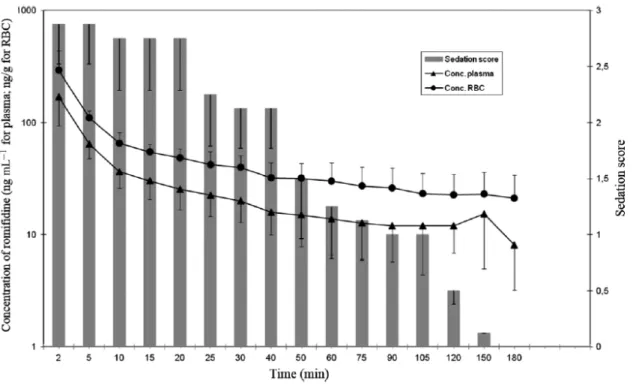
More recently we have described the partitioning of romifidine in red blood cells after IV administration at a dose of 0.1 mg kg-1 (Romagnoli et al. 2017). The Authors found that, after IV
administration, romifidine concentration in the red blood cells was two times higher than that in the plasma, and that the ratio between the two concentrations was constant over time (Figure 3). Romifidine has been detected in red blood cells up to 180 minutes after IV injection. This effect is explained by the lipophilicity of this drug; it can pass the membrane of the red blood cells binding to the membrane or to the intracellular molecules; as soon as the plasma concentration decreases, the drug is slowly released from the cells. The plasma concentration of romifidine has been found to be higher than 10 mg ml-1 in 75% of the animals included in the study from 30 to 180 minutes after IV
administration. The Authors have reported that romifidine (0.1 mg kg-1) provided good sedation in
all horses from 2 minutes after administration, lasting up to 105 minutes; however, half of the horses included were still sedated 120 minutes after injection and one horse up to 150 minutes. The Authors have hypothesized that romifidine partitioning in the red blood cells could explain the long-lasting sedative effect observed in horses after its administration (Romagnoli et al. 2017). In the same study, the mean clearance was 22.16 ± 6.67 ml min-1 kg-1, and the mean half-life of distribution and
Figure 3- Plasma and red blood cells concentration of romifidine and sedation score versus time observed in 8 horses after intravenous administration of romifidine 0.1 mg kg-1 (from
Romagnoli N, Al-Qudah KM, Armorini S, Lambertini C, Zaghini A, Spadari A, Roncada P (2017) Pharmacokinetic profile and partitioning in red blood cells of romifidine after single intravenous administration in the horse. Vet Med Sci 3, 187-197)
There is some discrepancy among studies concerning romifidine sparing effects on inhalant anesthetics. In isoflurane-anesthetized horses, romifidine, administered by CRI at 0.04 mg kg-1 h-1
(following a bolus of romifidine 0.08 mg kg-1 IV), has been shown to provide an isoflurane sparing
effect of 15% as compared to horses premedicated with xylazine (1 mg kg-1) and receiving saline
solution by CRI during surgery (Niimura Del Barrio et al. 2017). In the same study, romifidine infusion did not significantly alter the duration or the quality of recovery as compared with the control group. However, a previous study, in which an equal dose of romifidine was administered to isoflurane-anesthetized horses, had failed to find any sparing effects (Devisscher et al. 2010). The differences among the studies may have been due to the fact that different surgical procedures were performed on the horses involved.
In dogs, romifidine (0.01-0.08 mg kg-1) has been reported to produce effective sedation and a
sparing effect on the dose of propofol necessary for the induction of general anesthesia (Lemke 1999, England et al. 1996; Gomez-Villamandos et al. 2005)
Medetomidine
Medetomidine is a racemic mixture, widely used in both small and large animals (Bryant et al. 1991; Sinclair 2003; Ringer et al. 2007).
In horses, medetomidine (0.007 mg kg-1) produced more ataxia with respect to other alpha 2
-agonists up to recumbency (Bryant et al. 1991; Bettschart-Wolfensberger et al. 1999). For this reason, and due to its short duration of action, it is not commonly used for standing procedures but more often for premedication followed by CRI during general anesthesia (Ringer et al. 2007; Clarke et al. 2014). Its intraoperative administration, in horses anesthetized with isoflurane, helps in maintaining a stable anesthetic plane, reducing the inhalant anesthetic requirement as well as having a good cardiovascular stability (Bettschart-Wolfensberger et al. 1999, 2001). Of the other alpha2-agonists, medetomidine
has the shortest half-life and the most rapid clearance rate. In horses, when medetomidine was administered IV as a single bolus (0.007 mg kg-1), its mean elimination half-life was 51.3 min and its
total body clearance was 4.0 ml kg-1 h-1 (Bettschart-Wolfensberger et al. 1999). In the same species,
medetomidine (0.01 mg kg-1,) reaches peak plasma concentration 6.4 minutes after IV administration,
with an elimination half-life of 29 minutes (Grimsrud et al. 2012).
In small animals, medetomidine has been used as a single bolus for sedation or as CRI during general anesthesia. In dogs, doses of 0.03 mg kg-1 have been reported to provide similar sedation to
xylazine 2.2 mg kg-1(Cullen 1996; Vainio et al. 1989). In the same species, medetomidine 0.04 mg
kg-1 administered IV provided sedation in 15-20 minutes corresponding to a plasma concentration of
18.5 ng ml-1 (Kuusela et al. 2000). The administration of a low dose CRI of medetomidine (0.1 mg
kg-1 h-1) as part of a balanced anesthetic protocol contributed to the maintenance of a stable anesthetic
The more commonly used dosages suggested in clinical practice for obtaining effective sedation in dogs range from 0.02 to 0.15 mg kg-1; the duration of the sedative effect is dose dependent (Stenberg
et al. 1987).
In cats, medetomidine is commonly combined with ketamine or opioids to achieve effective sedation (Slingsby et al. 2015). The combination of medetomidine (0.03 mg kg-1) with buprenorphine
(0.02 mg kg-1) has been reported to significantly reduce the amount of inhalant anesthetic required
for maintenance of general anesthesia as compared with medetomidine alone (Grint et al. 2009). The addition of medetomidine (0.02 mg kg-1) to butorphanol (0.2 mg kg-1) has been reported to be more
effective than the addition of acepromazine (0.2 mg kg-1) in reducing the dose of alfaxalone necessary
for the induction and maintenance of general anesthesia (Schwarz et al. 2014).
Dexmedetomidine
Dexmedetomidine is the D-isomer of medetomidine and has a selectivity alpha2:alpha1 ratio of
1300:1 and an alpha2:imidazoline selectivity ratio of 32:1 (Khan et al. 1999). Dexmedetomidine, administered at half the dose of medetomidine, has been reported to provide similar effects (Kuusela et al. 2000; Bettschart-Wolfensberger et al. 2005).
In horses, dexmedetomidine has a rapid distribution and a short duration of action. For this reason, in this species, it is administered by CRI as part of a balanced anesthetic protocol in order to provide a constant level of sedation and to improve the recovery quality (Bettschart-Wolfensberger et al. 2005; Marcilla et al. 2012; Marly-Voquer et al. 2016). In equine species, its half-life of elimination, after a single bolus (0.0035 mg kg-1), is 28.96 ± 7.61 minutes (Bettschart-Wolfensberger
et al. 2005).
In dogs, dexmedetomidine (0.02 mg kg-1) provides similar sedation to that obtained with the
administration of medetomidine (0.04 mg kg-1) but with a longer lasting effect (Kuusela et al. 2000).
The concurrent administration of levomedetomidine (0.01-0.02 mg kg-1) did not provide any
levomedetomidine (0.08 mg kg-1) reduced the sedative and analgesic effects of dexmedetomidine
when administered together (Kuusela et al. 2000, 2001). In dogs sedated with dexmedetomidine (0.01 and 0.02 mg kg-1), the peak sedative effect was obtained 10 to 20 minutes after IV
administration, corresponding to a plasma concentration of 5.5± 3.0 and 14.0 ± 4.5 ng ml-1,
respectively (Kuusela et al. 2000). Dexmedetomidine, administered as a CRI (0.0015 mg kg-1 h-1 and
0.0045 mg kg-1 h-1) in sevoflurane-anesthetized dogs produced a dose-dependent decrease in the
MAC of the inhalant anesthetic (Hector et al. 2017).
In cats, dexmedetomidine (0.01 mg kg-1) has been used alone to obtain effective sedation, even
if the addition of opioids or ketamine provided more adequate sedation without increasing the incidence of adverse cardiovascular effects. In particular, its combination with butorphanol (0.2 mg kg-1) decreased the incidence of vomiting (Bhalla et al. 2017). When dexmedetomidine, at doses of
0.02-0.04 mg kg-1, was combined with alfaxalone (5 mg kg-1), general anesthesia was obtained (Selmi
et al. 2003; Rodrigo-Mocholí et al. 2016). Dexmedetomidine administered to cats as a premedicant drug before general anesthesia significantly reduced the dosage of the induction agent and the MAC of isoflurane in a plasma concentration related manner (from 0.006 to 11.46 ng ml-1) (Escobar et al.
2012; McSweeney et al. 2012). Dexmedetomidine administered at a dose of 0.025 mg kg-1 reached
its peak plasmatic concentration (10.2 ng ml-1) 17.8 minutes (range 2.6- 44.9) after IM administration
(Pypendop et al. 2017).
The oral administration (PO) of dexmedetomidine has been described in humans and small animal practice. In dogs dexmedetomidine is well absorbed systemically through the oral mucous membrane and this method of administration was effective for the sedation of fractious and fearful dogs (Cohen and Bennett 2015). In cats, the oral or the IM administration of dexmedetomidine has similar systemic bioavailability with comparable sedative and antinociceptive effects (Slingsby et al. 2009).
Analgesic effects
The analgesic effect of alpha2-agonists is the result of their action at the central and spinal levels.
At the spinal level, they bind to alpha2A, alpha2B and alpha2C receptors in the dorsal horn of the spinal
cord. The activation of noradrenergic receptors inhibits the NA and the substance P release from Ad and C fibers, thereby inhibiting the central transmission of the afferent nociceptive stimuli (Valverde 2010). In both the brain and the spinal cord, the alpha2-adrenoceptors interact with the opioid
receptors and share the same signal transduction system, the G protein. Opioid inhibition of nociceptive transmission at the spinal level might be mediated by the activation of the alpha2
-adrenoceptors. Among opioid receptors, the d-subtype has been found to be the main subtype involved in interaction with the adrenergic receptors (Omote et al. 1991). Studies support the interaction between alpha2-adrenergic receptors and opioid receptors. For example, Ossipov and
colleagues (1989) have demonstrated that systemic yohimbine attenuates the nociceptive effect of intrathecally-administered morphine and clonidine.
Studies have suggested that the analgesic effect of alpha2-agonists is dose dependent but shorter
lasting when compared to the sedative effect (Kramer et al. 1996). The analgesia provided by these drugs, even when locally administered, is reversed by the administration of the specific antagonist (Sabbe et al. 1994; Sinclair 2003).
Systemic administration
The effect of the systemic administration of alpha2-agonists has been evaluated on both somatic
and visceral nociception. Their application has been described in clinical trials and in experimental models applying thermal and mechanical thresholds, or duodenal and colorectal distension for somatic and visceral evaluation respectively. The results change with species and the drugs used, and also depend on the stimulus applied.
In horses, xylazine has been used to provide sedation and analgesia since it was first introduced in veterinary practice (Daunt and Steffey 2002). The analgesic effect of xylazine has been compared to opioids in a model of visceral analgesia in which colic was experimentally induced by inflating a balloon in the cecum (Muir and Robertson 1985). In that study xylazine (1.1 mg kg-1) provided a
more effective and longer lasting analgesia as compared with that provided by butorphanol (0.2 mg kg-1), meperidine (1.0 mg kg-1) and penthazocine (0.99 mg kg-1). In another study regarding the same
species, the combination of xylazine with opioids (morphine, butorphanol or nalbuphine) did not provide any adjunctive analgesia for dental dolorimetry as compared to xylazine administered alone (Brunson and Majors 1987). When xylazine was combined with butorphanol it potentiated its analgesic effect up to 30 to 60 minute after administration (Brunson and Majors 1987).
In a clinical study on horses with signs of colic, detomidine (0.020 and 0.040 mg kg-1) has been
used to relieve abdominal pain and was more effective in decreasing signs of pain than xylazine (0.5 mg kg-1), but also compared to butorphanol (0.1 mg kg-1) and flunixin meglumine (1.0 mg kg-1)
(Jochle et al. 1989). In the same species, an experimental study has been reported that detomidine (0.010 and 0.020 mg kg-1) significantly increased the colorectal and duodenal distension threshold
without affecting the thermal threshold (Elfenbein et al. 2009).
Alpha2-agonists have also been used to relieve pain in patients undergoing standing surgeries. In
mares, undergoing a standing flank laparotomy for ovariectomy and oophorectomy, detomidine (0.02 mg kg-1) provided better analgesia than the combination of xylazine and morphine (Jochle et al. 1991).
The efficacy of alpha2-agonists in managing surgical pain has also been described in small
animals (Sinclair 2003; Beckman 2013). In dogs, medetomidine alone (0.01, 0.03, 0.09, 0.18 mg kg -1) has been described to provide similar analgesic effect to xylazine (2.2 mg kg-1) in response to the
application of superficial pain stimuli (Vainio et al. 1989). Another study has demonstrated that, in this species, medetomidine (0.04 and 0.08 mg kg-1) alone provided analgesia for only a minimally
Dexmedetomidine has been compared to morphine for the management of postoperative analgesia in dogs undergoing abdominal or thoracic surgery, or spinal neurosurgery (Valtolina et al. 2009). In that study, dogs receiving dexmedetomidine (0.001 mg kg-1 followed by an infusion of
0.001 mg kg-1 h-1) or morphine (0.1 mg kg-1followed by 0.1 mg kg-1h-1) had similar pain scores during
the first 12 hours after surgery even if more dogs receiving dexmedetomidine required rescue analgesia (11/20) as compared with those treated with morphine (8/20) (Valtolina et al. 2009).
In dogs, the combination of dexmedetomidine with morphine or methadone has been demonstrated to be more effective than dexmedetomidine alone (0.01 mg kg-1) in suppressing the
withdrawal reflex induced by the application of toe pinches (Cardoso et al. 2014).
Combined alpha2-agonists and opioids also potentiate their analgesic effect in cats. In a study on
cats, dexmedetomidine (0.020 mg kg-1) combined with buprenorphine (0.010 mg kg-1) significantly
increased the thermal nociceptive threshold from 0.25 to 1.25 hours after administration (Slingsby et al. 2015). This effect was greater than the same doses of buprenorphine or dexmedetomidine administered alone.
Local administration
The local administration of alpha2-agonists has been described for epidural analgesia, local
infiltration, peripheral nerve block and intraarticular infiltration (Campoy and Read 2013).
Alpha2-agonists, by means of their action on alpha2A-adrenoceptors, enhance the action of local
anesthetic drugs (Yoshitomi et al. 2008). In an experimental study on guinea pigs, dexmedetomidine has been proven to increase the degree and the duration of the analgesic effects of lidocaine in a dose-dependent manner when injected intracutaneously. In the same study the improved anesthetic effect was reversed by yohimbine (alpha2A, -2B, and -2C adrenoceptor antagonists) but not by prozosin
In an experimental study on rats, dexmedetomdine (0.0005, 0.002, 0.006 and 0.02 mg kg-1) was
added to ropivacaine 0.75% to achieve a sciatic nerve block, and the sensory blockade to the application of a heat stimulus was evaluated (Brummet et al. 2009). The addition of dexemdetomdine increased the duration of the sensory block in a dose-dependent manner; however, when dexmedetomidine was used alone, no significant motor or sensory block was obtained (Brummet et al. 2008). At histopathological evaluation, a mild perineural inflammation was recorded 24 hours after the experiment; however, no signs of perinaural inflammation were found 14 days later. Moreover, the highest dose of dexmedetomidine (0.20 mg kg-1) did not provoke any histological
nerve damage (Brummet et al. 2009).
In cats, the combination of dexmedetomidine (0.001 mg kg-1) with bupivacaine (0.46 mg kg-1)
in achieving a sciatic and femoral nerve block did not decrease the response to toe pinching and did not increase the paw withdrawal threshold as compared to bupivacaine administered alone (Evangelista et al. 2017). Furthermore, in a previous study on dogs, dexmedetomidine (0.0001 mg kg-1) combined with ropivacaine (0.74 mg kg-1) to achieve a sciatic and femoral nerve block did not
increase the duration of the sensory block (Trein et al. 2017). These results differ from human studies which used dexmedetomidine in combination with local anesthetic drugs to perform local anesthetic techniques. Marhofer and colleagues (2013) have reported that in human volunteers, dexmedetomidine (0.02 mg kg-1) administered with ropivacaine (0.75%) to achieve an ulnar nerve
block, increased the duration of the sensory block as compared with ropivacaine alone when the pinprick test was applied.
Epidurally administered alpha2-agonists exerts their analgesic effect by means of their action at
the spinal level; however, systemic absorption cannot be excluded (Sabbe et al. 1994; Steagall et al. 2017). In dogs, epidurally or intrathecally administered dexmedetomidine provided comparable antinociceptive effects to systemically administered dexmedetomidine but without any signs of sedation (Sabbe et al. 1994). A study has compared the effects of the epidural administration of
lidocaine 2% alone or in combination with alpha2-agonists (xylazine 0.25 mg kg-1, romifidine 0.01
mg kg-1, detomidine 0.03 mg kg-1, dexmedetomidine 0.002 mg kg-1 or clonidine 0.005 mg kg-1) in
dogs undergoing ovariohysterectomy (Pohl et al. 2012) Among the other alpha2-agonists, xylazine
has been shown to determine a longer lasting analgesic effect lasting up to 4 hours (Pohl et al. 2012). In the same study, 3 out of the forty-two dogs involved which received detomidine, 4 which received xylazine and 5 which received clonidine required inhalation anesthesia in addition to the epidural; all bitches receiving epidural dexmedetomidine required isoflurane anesthesia.
In dogs undergoing orthopedic surgery, epidural medetomidine (0.015 mg kg-1) has provided
analgesia comparable to epidurally administered oxymorphone, but a higher incidence of cardiovascular side effects has been recorded. The epidural administration of medetomidine (0.005 mg kg-1) combined with morphine (0.1 mg kg-1) did not provide any benefit as compared to the use
of medetomidine alone (Vesal et al. 1996; Pacharinsak et al. 2003).
In horses, epidural detomidine has frequently been used to provide effective analgesia for standing procedures. Epidurally administered detomidine induced analgesia extending from the coccyx up to T15 and, as compared to xylazine, induced a higher degree of cardiovascular side effects and a more frequent change in hind limb position (Skarda and Muir 1996). When continuous IV or epidural detomidine infusions have been compared in mares undergoing standing ovariectomy, no significant differences in hormonal responses have been detected. However, mares which had not been received the local analgesia had a higher visual analogue scale (VAS) score when more painful stimuli were applied (Virgin et al. 2010).
The intraarticular administration of alpha2-agonists has also been described (Soto et al. 2014). In
dogs undergoing stifle joint surgery, intraarticular administered dexmedetomidine (0.0025 mg kg-1)
has provided postoperative analgesia comparable to intraarticular administered morphine (0.1 mg kg -1), lasting up to 6 hours (range 2-10 hours). Dogs treated with the combination of the two drugs did
Cardiovascular effects
Alpha2-adrenoceptor agonists, by means of their action on alpha2-adrenoeceptors, can
significantly impair the cardiovascular system. Bradycardia, arrhythmia, decreased cardiac output (CO) and increased systemic vascular resistance (SVR) are the most commonly reported alterations after alpha2-adrenoceptor agonist administration in dogs, cats and horses among others (England and
Clarke 1996; Pypendop and Verstegen 1998; Ko et al. 2001; Lamont et al. 2001; Carter et al. 2010). In an experimental study, dexmedetomidine (0.0005 mg kg-1min-1) has been demonstrated to
prevent arrhythmia induced by epinephrine in halothane-anesthetized dogs (Kamibahyashi et al. 1995). In that study, the authors hypothesized that this anti-arrhythmogenic effect was mediated by the action of dexmedetomidine on central imidazole receptors. In fact, the administration of two imidazole- alpha2-antagonists inhibited or reversed the dexmedetomidine action while the effect of
non-imidazole alpha2-antagonists was not significant.
Heart rate
In several species, the administration of alpha2-agonists has usually been associated with a
decrease in heart rate (HR) with respect to baseline values (Golden et al. 1998; Pypendop and Verstegen 1998; Ko et al. 2001; Lamont et al. 2001; Ilbäck and Stålhandske 2003; Carter et al. 2010). In small animals, medetomidine has been reported to induce a significant decrease in HR, and some authors have reported that this effect is not dose dependent; at low dosages, the decrease in HR is less pronounced and, at higher dosages, it is longer lasting (Pypendop and Verstegen 1998). Medetomidine administered by CRI in dogs (0.001-0.003 mg kg-1 min-1), has also been reported to
decrease the HR (Carter et al. 2010). A decrease in HR has also been reported in cats after medetomidine and romifidine administration, with medetomidine decreasing the HR up to 68% from baseline 15 minutes after IM administration (Lamont et al. 2001; Muir and Gadawski 2002).
Clonidine, xylazine, detomidine, medetomidine and romifidine are reported to produce profound bradycardia and atrioventricular (AV) blocks when administered to horses (Wagner et al. 1991; England and Clarke 1996). In equine species, xylazine (1.1 mg kg-1) has been found to induce a less
profound and a shorter-lasting alteration on HR than that of equipotent doses of detomidine (0.02 mg kg-1) and romifidine (0.08 mg kg-1) (Wagner et al. 1991; England et al. 1992).
There are two main mechanisms involved in the reduction of the HR observed after the administration of alpha2-adrenoceptor agonists. The interaction of these drugs with the central alpha2
-adrenoceptors reduces the sympathetic outflow, thereby reducing the HR. In addition, the interaction with peripheral alpha2-adrenceptors results in increased systemic vascular resistance (SVR) and
subsequent reflex bradycardia (Sinclair et al. 2003). The direct effects of alpha2-agonists on intrinsic
myocardial contractility have been excluded in a study carried out regarding the isolated ventricular myocardium of ferrets (Housman 1990).
Preemptive atropine administration has been hypothesized to reduce the incidence of bradycardia mediated by alpha2-agonists. However, the increase in HR induced by anticholinergic drugs in the
presence of a peripheral vasoconstriction due to the alpha2-agonists would increase the risk of
arrhythmia (Ko et al. 2001).
Arterial blood pressure
Alpha2-agonist administration is usually characterized by a biphasic pressure response; soon after
the drug administration, an initial increase in blood pressure is followed by a hypotensive phase (Pypendop and Verstegen 1998; Ilbäck and Stålhandske 2003).
This biphasic pattern has mainly been observed after the administration of higher doses of medetomidine (Pypendop and Verstegen 1998). When low doses of medetomidine are administered, central effects predominate, and hypotension is more frequently observed. Higher doses of medetomidine exert a more pronounced action on peripheral adrenoceptors with subsequent
vasoconstriction and a significant increase in arterial blood pressure (Pypendop and Verstegen 1998; Ko et al. 2001). However, as the peripheral action ceases, the central effect persists, and the decreased CO contributes to the normalization or the decrease of the systemic arterial pressure (SAP) (Carter et al. 2010). The hypertensive effect is seen especially after the IV administration of medetomidine; in fact, after IM administration, the peak blood concentration is delayed.
Higher doses of alpha2-agonists can cause a significant increase in blood pressure which can be
detrimental in patients affected by heart or cardiovascular disease. In a study in dogs, the administration of a high dose of medetomidine (0.02 mg kg-1) induced an increase in the SAP up to
200 mmHg even if of short duration (Pypendop and Verstegen 1998). Medetomidine 0.01 mg kg-1
given intravenously has been reported to cause sudden cardiac arrest due to the rupture of an aortic aneurysm in a dog affected by Spirocerca lupi (Joubert et al. 2005)
The biphasic response has been described in horses after the administration of clonidine, xylazine, detomidine, medetomidine or romifidine (England and Clarke 1996). Detomidine 0.02 mg kg-1 IV induced initial hypertension soon after its administration which lasted 15 minutes. This initial
increase was then followed by a significant decrease in the mean arterial pressure (MAP) with respect to the baseline values 1 to 2 hours after administration (Wagner et al. 1991). On the other hand, Wagner and colleagues (1991) have observed that xylazine 1.1 mg kg-1 IV and 2.2 mg kg-1 IM
administered in healthy horses induced a decrease in MAP 5 minutes after administration which became statistically significant 15 minutes later and lasted up to 120 minutes.
Studies in mice have demonstrated that the central hypotensive response after alpha2-agonists
administration is mediated by the alpha2A-adrenoceptor subtype while the increase in systemic blood
pressure is mediated by alpha2B-adrenoceptors (Link et al. 1996; MacMillan et al. 1996). In fact, in
alpha2B-deficient mice, the hypertensive response has not been observed after dexmedetomidine
administration. On the contrary, the disruption of the alpha2C-subtype did not produce any
of the alpha2A-subtype, the hypotensive response was lost after the administration of an alpha2-agonist
(MacMillan et al. 1996).
Data concerning the effect of alpha2-adrenoceptor agonists on pulmonary circulation are
controversial, and vary among drugs and species. In the pulmonary vessels, there is a low density of alpha2-agonists when compared to the systemic circulation, and neuronal regulation plays a minor
role in their regulation (Pypendop and Verstegen 1998; Lamont et al. 2001). In small animals, medetomidine did not affect the pulmonary vascular resistance index (PVRI) or the mean pulmonary arterial pressure (Pypendop and Verstegen 1998; Lamont et al.2001). In horses, detomidine and xylazine induced a significant increase in the PVRI (Wagner et al. 1991). Moreover, while xylazine did not affect the mean pulmonary arterial pressure, low and high dose detomidine induced a significant decrease in the mean pulmonary arterial pressure.
Cardiac output
The administration of alpha2-adrenoceptor agonists has been reported to induce a transient
decrease in both cardiac output (CO) and the cardiac index (CI) (England and Clarke 1996; Pypendop and Verstegen 1998; Carter et al. 2010).
The CO is determined by complex and coupled variables, but primarily depends on HR and stroke volume (SV). The SV, that is the volume of blood pumped by the heart in each cycle, is influenced by preload, afterload, contractility and lusitropic properties (Muir 2015). A study on the isolated ventricular papillary muscles of ferrets has demonstrated that dexmedetomidine has no intrinsic contractile effects on the myocardium (Housman 1990). There is a discrepancy among authors concerning the effect of alpha2-adrenoceptor agonists on SV and the stroke index (SI) in small
and large animals (Pypendop and Verstegen 1998; Yamashita et al. 2000; Carter et al. 2010). However, it is likely that the commonly observed decrease in CO is mainly due to bradycardia or to decreased contractility related to reduced sympathetic tone.
Stroke Volume decreased in automatically blocked dogs after medetomidine administration (de Morais and Muir 1995). In healthy dogs, medetomidine 0.001 mg kg-1induced a significant decrease
in CO and the CI without significant alterations in the SI. A lesser decrease in the CI index has been reported after the administration of low dose medetomidine (Pypendop and Verstegen 1998). In cats, medetomidine 0.02 mg kg-1 induced a significant decrease in the CI up to 37% of baseline 15 minutes
after administration; the value remained at 50% of baseline 30 minutes later (Lamont et al. 2001). In horses treated with xylazine or detomidine, the CO was significantly reduced by both drugs administered at different dosages; however, detomidine administration (0.02 mg kg-1 IV) was
associated with a lower CO and CI lasting up to 60 minutes (Wagner et al. 1991). In this latter study, the decrease in CO lasted longer than the reduction of the HR. However, in the absence of significant alterations in SV or the SI the authors recognized the decrease in HR as the main cause of the decreased CO.
In another study on horses treated with different dosages of medetomidine, detomidine or xylazine, the SV decreased from baseline, even if the variation was not statistically significant (Yamashita et al. 2000).
Respiratory effects
The administration of alpha2-agonists alone has been reported to decrease in fR. due to CNS
depression (Sinclair 2003). Respiratory depression has not always been associated with a significant alteration in blood gas tension (Lamont et al. 2001). In healthy dogs, low dose medetomidine (0.005 and 0.01 mg kg-1) reduced the sensitivity of the response to increased fractional concentration of
inspired carbon dioxide (FiCO2). This affected the fR, the tidal volume and the minute volume (Lerche
and Muir 2004). In horses, xylazine (0.01 mg kg-1) induced a significant decrease in the partial arterial
pressure of oxygen (PaO2) without altering the partial pressure of carbon dioxide in arterial blood
(PaCO2) (Lavoie et al. 1992a).
In horses, the sedation provided by alpha2-agonists is commonly associated with head and neck
dropping. The alteration in head carriage causes an increase in hydrostatic pressure which results in nasal mucosa congestion which contributes to increased airway resistance. In addition, these drugs provide a relaxation of the nostrils and head dropping which changes the conformation of the thorax and of the lungs. Taken together, these factors contribute to alterations in ventilator mechanics (Lavoie et al. 1992b).
In dogs, peripheral cyanosis has commonly been observed after medetomidine administration, also in healthy dogs. Sinclair (2003) has suggested that low blood flow through the peripheral capillary bed and high oxygen extraction were the main mechanisms inducing a cyanotic mucous appearance in alpha2-treated animals.
Ruminants, and especially sheep among others, are more sensitive to the respiratory depression induced by these drugs, even at low doses. Studies have described profound hypoxemia in sheep after the administration of xylazine, romifidine, detomidine, medetomidine or dexmedetomidine, despite their alpha receptor selectivity, and without a significant increase in PaCO2 (Celly et al. 1997; Kästner
et al. 2007). On the basis of their results, some authors have hypothesized that the hypoxia was not due to hypoventilation, to changes in body position or to the degree of sedation. Celly and colleagues
(1997) concluded that the increase in shunt fraction and the alteration of pulmonary mechanics, with a subsequent increase in P(A-a)O2 and a decrease in transpulmonary pressure recorded after the
administration of all the alpha2-agonists tested, were the main causes of hypoxemia (Celly et al.
1997). The hydrostatic stress, characterized by increased capillary pressure, and protein and erythrocyte extravasation was considered to be the main cause of the pulmonary edema and capillary congestion observed in dexmedetomidine-treated sheeps (Kastner et al. 2007)
The degree of respiratory depression induced by alpha2-agonists increases when they are
combined with other drugs, such as opioids. With these combinations, oxygenation of the patient is strongly recommended, especially in critically ill animals.
Other effects
Glycemic effect
Alpha2-agonists have been reported to increase serum glucose concentration and to decrease
insulin levels in several species (Feldberg and Symonds 1980; Ambrisko and Hikasa 2002; Restitutti et al. 2012). This hyperglycemic effect induced by alpha2-agonists has been reversed with the
administration of alpha2-antagonists (atipamezole, yohimbine and MK-467) (Maroto et al. 1992;
Ambrisko and Hikasa 2002; Restitutti et al. 2012).
Xylazine has been described to induce hyperglycemia in dogs and cats (Feldberg and Symonds 1980; Ambrisko and Hikasa 2002). In dogs xylazine (1-8 mg kg-1) increased blood glucose dose
dependently from two hours after IM administration and to a greater extent than medetomidine administered at equipotent sedative doses. In healthy dogs, the effect of medetomidine on glucose concentration depends on the dose administered (Ambrisko and Hikasa 2002). Medetomidine, administered at 0.01 mg kg-1, did not significantly alter blood glucose levels (Maroto et al. 1992;
Burton et al. 1997) while higher dosages (0.02-0.08 mg kg-1) induced a significant increase in blood
glucose (Maroto et al. 1992).
In another study, low dose medetomidine (0.005 mg kg-1 IM) also induced a significant increase
in plasma glucose concentration in healthy dogs and in dogs with insulinomas. In the latter, blood glucose increased by 20 mg dL-1 after medetomidine administration (Guedes and Rude 2013).
The mechanisms of the hyperglycaemic effects of alpha2-agonists has not yet been clarified and
the receptors involved vary according to species (Maroto et al. 1992; Ambrisko and Hikasa 2002). In the pancreas, alpha2-agonists decrease the insulin secretion by their action on alpha2
-adrenoceptors in the beta pancreatic cells. A study on the isolated rat pancreas has shown that alpha2
-agonists decrease insulin release by 80% while alpha1-agonists induce only a slight decrease in insulin
secretion, up to 25% (Hillaire-Buys et al. 1985; Ambrisko and Hikasa 2002). In healthy dogs, medetomidine (0.01-0.08 mg kg-1) and xylazine (1-8 mg kg-1) reduced insulin plasma levels dose
independently without significantly altering glucagone levels (Burton et al. 1997; Ambrisko and Hikasa 2002). In dogs with insulinomas, medetomidine (0,005 mg kg-1) also decreased plasmatic
insulin (by 78%) (Guedes and Rude 2013).
However, the glycemic effect of alpha2-agonists may depend on different mechanisms other than
the inhibition of insulin secretion and may also involve other receptors. In dogs, the alpha1-antagonist
prazosin was more effective than yohimbine in antagonizing the glycemic effect of clonidine (Maroto et al. 1992). In cattle, clonidine has been reported to increase glucose release from the liver in vitro (Gorewit 1980). These studies support the glycogenolytic effect on the liver induced by alpha2
-agonists by means of their affinity for alpha1-adrenoceptors. This explains the higher increase in
blood glucose observed after xylazine administration as compared to medetomidine (Ambrisko and Hikasa 2002). Dexmedetomidine in dogs (0.001 mg kg-1) significantly increased glucose levels only
120 minutes after IM administration. This delay of the hyperglycemic effect can be explained by the lower affinity of dexmedetomidine for the alpha1-adrenoceptors which are involved in the stimulation
of glycogenosis in the liver (Restitutti et al. 2012).
Imidazoline receptors may also be involved in the regulation of the glycemic effects of this class of drugs. Imidazoline I1 agonists have been reported to regulate the human glucose metabolism (Farsang and Kapocsi 1999; Ambrisko and Hikasa 2002).
Diuretic effect
In several species, alpha2-agonist administration is associated with an increase in diuresis, and
changes in urine specific gravity, pH, osmolality, creatinine concentration and electrolytic concentrations (Thurmon et al. 1978; Trim and Hanson 1986; Burton et al. 1998; Villela et al. 2005). The mechanisms of action primarily include a centrally mediated decreased secretion of the antidiuretic hormone (ADH) (Reid et al. 1979; Talukder and Hikasa 2009). In addition, the antagonism of the renal tubular effect of ADH and the alpha2-agonist mediated cardiocirculatory
alterations may contribute to their diuretic effect (Sinclair 2003). The magnitude of the diuretic effect is drug dependent and dose dependent.
In equine species and in cattle, xylazine induced a dose-dependent increase in urinary output (Thurmon et al. 1978; Thurmon et al. 1984; Trim and Hanson 1986). In ponies, xylazine 1.1 mg kg-1
induced a significant increase in urinary output for up to 2 hours with a peak effect between 30 and 60 minutes after administration. The treatment also induced a significant increase in potassium and sodium excretion without affecting their plasma concentrations (Trim and Hanson 1986). In mares, xylazine 0.5, 1.0, and 1.5 mg k-1 induced an increase in urinary output up to 1.82, 3.93, and 5.68 ml
kg-1 h-1, respectively (Thurmon et al. 1984). In cattle, the diuretic effect of xylazine 0.22 mg kg-1 or
0.44 mg kg-1 lasted up to 5 hours (Thurmon et al. 1978).
In dogs, both xylazine (0.24; 0.5; 1; 2; 4 mg kg-1) and medetomidine (0.005; 0.01; 0.02; 0.04;
0.08 mg kg-1) decreased ADH levels and increased urinary production dose dependently, with
xylazine inducing a greater increase as compared to medetomidine (Talukder and Hikasa 2009). In this study, the diuretic effect lasted up to 4 hours after administration. Both drugs also induced a significant decrease in creatinine concentration, in osmolality and in the pH of the urine samples. The urinary specific gravity decreased in a dose dependent manner with the lowest value recorded one to three hours after administration (Talukder and Hikasa 2009).
The hypertensive response observed after the administration of alpha2-agonists may be involved
in the diuretic effects; however, studies have highlighted that increased blood pressure alone cannot explain the diuretic effects. In dogs, clonidine (0.03 mg kg-1) induced an increase in blood pressure
and a significant decrease in plasma ADH from 10.9 ± 1.5 to 5.0 ± 1.1 ng ml-1. However, the
administration of two alpha2-antagonists (piperoxane and phentolamine) resulted in a reversal of the
pressure response without significant alteration in ADH concentration (Reid et al. 1979). On the contrary, atipamezole and yohimbine were effective in reversing the medetomidine-induced inhibition of the ADH release and the diuretic effect of medetomidine in dogs (Talukder et al. 2009). The effect of atipamezole in reversing the diuretic effect has been shown to be dose dependent and
greater as compared to that of yohimbine, probably because of its affinity for imidazole receptors (Talukder et al. 2009).
In dogs, xylazine and medetomidine administration also induced a significant increase in the atrial natriuretic peptide (ANP) (Talukder and Hikasa 2009; Talukder et al. 2009). However, in their study, Talukder and colleagues (2009) found exceptionally that atipamezole administration resulted in an additional increase in the ANP in a dose-dependent manner as compared to a reversal of the diuretic effect induced by medetomidine. Therefore, the atrial natriuretic peptide, released in response to atrial distension which, in this study, may have been associated with increased blood pressure, seems to be minimally involved in the diuretic effect induced by alpha2-agonists (Talukder et al.
2009).
Some authors have suggested not administering alpha2-agonists to patients with urinary
obstructions and to take into consideration fluid loss when managing critical patients (Daunt and Steffey 2002; Sinclair 2003).
Stress response
Alpha2-agonist administration has been described to reduce the perioperative release of
catecholamine. In dogs, medetomidine (0.01, 0.02, 0.04, 0.08 mg kg-1) and xylazine (1, 2, 4, 8 mg kg -1) suppressed NA release in a dose-dependent manner. Medetomidine also reduced epinephrine
release dose dependently and with a greater potency than xylazine (Ambrisko and Hikasa 2002). In bitches undergoing ovariohysterectomy who were premedicated with medetomidine, the cortisol concentration during the surgery was significantly lower than that of bitches which did not receive any premedicant drug (Benson et al. 2000). Moreover, the perioperative alpha2-agonist administration
inhibited the release of cortisol through their imidazoline activity (Ambrisko and Hikasa 2002). In another study, comparing the endocrine effects of medetomidine (0.02 mg kg-1) and