R E S E A R C H
Open Access
Influenza and associated co-infections in
critically ill immunosuppressed patients
Ignacio Martin-Loeches
9,10,35*†, Virginie Lemiale
1†, Pierce Geoghegan
9,10, Mary Aisling McMahon
9,10,
Peter Pickkers
2, Marcio Soares
3, Anders Perner
4, Tine Sylvest Meyhoff
4, Ramin Brandt Bukan
22, Jordi Rello
5,
Philippe R. Bauer
6, Andry van de Louw
7, Fabio Silvio Taccone
8, Jorge Salluh
3, Pleun Hemelaar
2,
Peter Schellongowski
11, Katerina Rusinova
12, Nicolas Terzi
14, Sangeeta Mehta
15, Massimo Antonelli
16,
Achille Kouatchet
17, Pål Klepstad
13, Miia Valkonen
19, Precious Pearl Landburg
20, Andreas Barratt-Due
18,
Fabrice Bruneel
21, Frédéric Pène
23, Victoria Metaxa
24, Anne Sophie Moreau
25, Virginie Souppart
1, Gaston Burghi
26,
Christophe Girault
27, Ulysses V. A. Silva
28, Luca Montini
16, Francois Barbier
29, Lene B. Nielsen
30,31,
Benjamin Gaborit
32, Djamel Mokart
33, Sylvie Chevret
34, Elie Azoulay
1and For the Efraim investigators and the
Nine-I study group
Abstract
Background: It is unclear whether influenza infection and associated co-infection are associated with patient-important outcomes in critically ill immunocompromised patients with acute respiratory failure.
Methods: Preplanned secondary analysis of EFRAIM, a prospective cohort study of 68 hospitals in 16 countries. We included 1611 patients aged 18 years or older with non-AIDS-related immunocompromise, who were admitted to the ICU with acute hypoxemic respiratory failure. The main exposure of interest was influenza infection status. The primary outcome of interest was all-cause hospital mortality, and secondary outcomes ICU length of stay (LOS) and 90-day mortality.
Results: Influenza infection status was categorized into four groups: patients with influenza alone (n = 95, 5.8%), patients with influenza plus pulmonary co-infection (n = 58, 3.6%), patients with non-influenza pulmonary infection (n = 820, 50.9%), and patients without pulmonary infection (n = 638, 39.6%). Influenza infection status was associated with a requirement for intubation and with LOS in ICU (P < 0.001). Patients with influenza plus co-infection had the highest rates of intubation and longest ICU LOS. On crude analysis, influenza infection status was associated with ICU mortality (P < 0.001) but not hospital mortality (P = 0.09). Patients with influenza plus co-infection and patients with non-influenza infection alone had similar ICU mortality (41% and 37% respectively) that was higher than patients with influenza alone or those without infection (33% and 26% respectively). A propensity score-matched analysis did not show a difference in hospital mortality attributable to influenza infection (OR = 1.01, 95%CI 0.90–1. 13,P = 0.85). Age, severity scores, ARDS, and performance status were all associated with ICU, hospital, and 90-day mortality.
(Continued on next page)
© The Author(s). 2019 Open Access This article is distributed under the terms of the Creative Commons Attribution 4.0 International License (http://creativecommons.org/licenses/by/4.0/), which permits unrestricted use, distribution, and reproduction in any medium, provided you give appropriate credit to the original author(s) and the source, provide a link to the Creative Commons license, and indicate if changes were made. The Creative Commons Public Domain Dedication waiver (http://creativecommons.org/publicdomain/zero/1.0/) applies to the data made available in this article, unless otherwise stated.
* Correspondence:[email protected]
Ignacio Martin-Loeches and Virginie Lemiale contributed equally. 9
Department of Intensive Care Medicine, Multidisciplinary Intensive Care Research Organization (MICRO), St. James’s Hospital, Dublin, Ireland 10Department of Clinical Medicine, Wellcome Trust-HRB Clinical Research Facility, St. James Hospital, Trinity College, Dublin, Ireland
(Continued from previous page)
Conclusions: Category of infectious etiology of respiratory failure (influenza, non-influenza, influenza plus co-infection, and non-infectious) was associated with ICU but not hospital mortality. In a propensity score-matched analysis, influenza infection was not associated with the primary outcome of hospital mortality. Overall, influenza infection alone may not be an independent risk factor for hospital mortality in immunosuppressed patients. Keywords: Influenza, Respiratory failure, Sepsis, Critical illness, Immunosuppression,
Introduction
Immunosuppressed patients admitted to an intensive care unit (ICU) with acute respiratory failure have a very high risk of mortality [1]. Acute respiratory failure can have various etiologies, but pulmonary infection and its sequelae remain the most frequent precipitants in those that require ICU admission [2, 3]. Among the different infectious agents which cause pulmonary infection in immunocompromised patients, pneumonia caused by influenza viruses has been associated with a particularly high mortality rate [4].
Influenza infection can affect patients during pan-demic periods (such as the H1N1 panpan-demic of 2008/ 2009) or during seasonal epidemics. Factors associated with the risk of a severe influenza infection during and after pandemic periods have differed [5]. After the first pandemic period in 2008 and 2009, influenza affected particular subgroups, particularly obese and pregnant patients [6, 7]. During the post-pandemic period, im-munosuppression was a risk factor for both influenza in-fection and ICU mortality [8]. Other factors associated with greater severity of influenza infection are age, med-ical comorbidities, and possibly co-infection. Bacterial pulmonary co-infection has long been described in pa-tients with influenza pneumonia, most commonly with Staphylococcus aureus, Streptococcus pneumoniae, and Haemophilus influenzae. Recognition of influenza infec-tion is important because it allows the implementainfec-tion of appropriate infection control measures and specific antiviral therapy. Furthermore, it might reduce inappro-priate antibacterial administration.
Although bacterial co-infection has been associated with increased mortality during the 2008/2009 pandemic period [9], the impact of the combination of influenza infection and bacterial or fungal co-infection on the out-come of critically ill patients has been a matter of debate [10]. Although influenza seems to be associated with higher mortality rates in immunocompromised patients [11, 12], the fraction of mortality attributable to either influenza infection alone or influenza plus co-infection has not been well defined. Our aim in the current study was to examine the prevalence of influenza infection and co-infection in critically ill immunocompromised pa-tients admitted to the ICU with respiratory failure and to determine whether influenza and associated
co-infection were associated with patient-important out-comes in this group.
Methods
Study design and setting
The current study was a preplanned secondary analysis of the EFRAIM study, a multinational prospective cohort study in 68 centers in 16 countries. EFRAIM was per-formed by the Nine-I (Caring for Critically Ill Immuno-compromised Patients) study group [13]. The Nine-I group includes critical care physicians from 16 countries who have extensive experience in the management of various groups of critically ill immunocompromised pa-tients. Physician participation was voluntary, without fi-nancial incentive. Participating investigators obtained local institutional review board approval in accordance with local ethics regulations.
Inclusion and exclusion criteria
Eligibility criteria were age ≥ 18 years, acute hypoxemic respiratory failure (PaO2< 60 mmHg or SpO2< 90% on
room air, or tachypnea > 30/min, or labored breathing or respiratory distress or dyspnea at rest or cyanosis), need for more than 6 L/min oxygen, respiratory symptom duration less than 72 h, and non-AIDS-related immune deficiency defined as hematologic malignancy or solid tumor (active or in remission for less than 5 years), solid organ transplant, long-term (> 30 days, any dose) or high-dose (> 1 mg/kg/day) steroids, or any immunosup-pressive drug taken in a high dosage or for more than 30 days. Exclusion criteria were postoperative acute re-spiratory failure, admission after a cardiac arrest, ICU admission exclusively to secure bronchoscopy, or refusal of the patient or family to participate in the study.
Enrolment, data collection, and patient treatment
Participating ICUs enrolled patients from Nov. 5, 2015 to Jul. 1, 2016. Prospective data were collected on patient and disease characteristics, initial oxygenation strategy, acute respiratory failure (ARF) etiology, associated organ dysfunction, and patient outcomes at hospital discharge and at day 90. The case report forms were sent to the co-ordinating center in Paris for data entry by trained techni-cians. The study was funded by the Groupe de Recherche
en Réanimation Onco-Hématologique (GRRR-OH), an academic non-profit French organization.
All management decisions were performed according to standard local practice in each ICU. Diagnostic strat-egies to identify the etiology of respiratory failure were based on previous studies by the GRRR-OH (11,18-20,23). ARF etiologies were based on pre-defined criteria in each participating ICU (11,18-20,23). All diag-noses were reviewed by two study investigators (from in-dependent institutions) for coherence and for alignment with accepted definitions. Oxygenation modalities, the use of non-invasive ventilation, high-flow nasal oxygen, or intubation was documented daily. Management of as-sociated organ dysfunction, handling of immunosuppres-sive drugs, or chemotherapy was decided by a physician according to local and recommended practices. Intub-ation decisions were left at the discretion of the care team and based on the therapeutic response, clinical sta-tus (including SpO2, respiratory rate, signs of respiratory
distress, and bronchial secretion volume), and patient’s adherence to other oxygenation modalities.
Exposures, outcomes, and important covariates
The exposure of interest in this prespecified secondary analysis of the EFRAIM study was influenza infection sta-tus. Patients were divided into four groups for the pur-poses of analysis: (1) influenza respiratory tract infection alone, (2) influenza respiratory tract infection plus co-infection, (3) non-influenza respiratory tract infection, and (4) no suspected or confirmed respiratory tract infec-tion. Influenza was diagnosed by the presence of positive reverse transcription polymerase chain reaction (RT-PCR) in immunosuppressed patients admitted to intensive care units (ICUs) by a nasopharyngeal swab as it is the optimal upper respiratory tract specimen collection method for in-fluenza recommended by the CDC [14]. RT-PCR was not performed in all patients but mainly in those in whom in-fluenza infection was suspected.
Pulmonary co-infection was defined as either clinically or microbiologically confirmed bacterial or invasive fun-gal respiratory infection in patients with influenza RT-PCR-positive respiratory tract infections [15].
The primary study outcome was all-cause hospital mortality. Secondary outcomes included ICU length of stay and 90-day mortality.
Data on important covariates were collected prospect-ively. SOFA score was recorded at ICU admission [16]. Shock was defined as a need for vasopressors; acute kid-ney injury (AKI) was defined as a need for renal replace-ment therapy as decided by the treating physicians.
Statistical analysis
Continuous variables are reported as medians (inter-quartile ranges [IQRs]) and categorical variables as
proportions. Data management allowed checking for data inconsistencies that were solved by consensus. Comparisons of proportions between the groups were made using theχ2test. Comparisons of continuous vari-ables between the groups were made using the Wilcoxon rank-sum test.
A propensity score (PS)-based approach was used to limit the effect of bias on the between-group compari-sons of hospital mortality. The propensity score was de-fined as the probability that a patient with specific baseline characteristics had influenza infection. We de-veloped the PS using a logistic regression model that in-cluded all baseline characteristics associated with illness severity [2]: mechanical ventilation, age, Eastern Co-operative Oncology Group (ECOG) performance status, SOFA score at ICU admission, admission within first hours of hospital admission, tobacco use, and underlying disease (hematological or solid tumor or immune dis-ease). To handle the missing values in these con-founders, multiple imputations with chained equation were used, where PS for each patient was averaged across 30 completed datasets while PS matching used these averaged scores. We matched individuals based on their PS using a 1:1 matching algorithm without replace-ment within a caliper of 0.15 standard deviation of the logit of the PS. Final analyses on the matched dataset were then performed using a logistic regression with a random effects model on the paired observations, except for the length of stay which we analyzed with a Cox ran-dom effects model. Results are presented as odds ratio (OR) with their 95%CI. Primary analyses were performed on the complete cases, assuming missing completely at random covariates. Sensitivity analyses for such assump-tions were performed, based on multiple imputation with chained equation. Details of the sample size calcu-lation for the original EFRAIM study can be found else-where [13]. A post hoc power analysis was not considered appropriate for the current secondary ana-lysis. All tests were two sided at the 0.05 significance level. Analyses were performed using R statistical pack-age (online athttp://www.R-project.org).
Results
Baseline characteristics
Out of 1611 patients (60% men, median age 63 (IQR 54–71)) enrolled in the 68 participating ICUs, 4 expos-ure groups were defined: patients with influenza respira-tory tract infection alone (n = 95, 5.8%), patients with influenza plus co-infection (n = 58, 3.6%), patients with non-influenza respiratory tract infection (n = 820, 50.9%), and patient without suspected or confirmed re-spiratory tract infection (n = 638, 39.6%). We also per-formed additional analysis on 448 patients negative for influenza and testing not done.
Characteristics of each group are summarized in Table1. There were no statistically significant differences between the groups regarding general clinical character-istics including age and comorbidities. However, patients with influenza tended to have a higher body mass index and were more frequently admitted to the ICU directly from the emergency department. Patients without any respiratory tract infection were less likely to have hematological disease. SOFA score was higher in pa-tients with influenza and co-infection, mainly driven by higher SOFA respiratory subscores in this group. When excluding patients negative for influenza but with no testing done, only patients with influenza alone were more likely to have solid tumors (Additional file1: Table S1). Intubation during the ICU stay was higher and shock lower in patients with influenza and co-infection compared to the other groups (Table2).
Outcomes on crude, propensity score-matched, and multivariate analysis
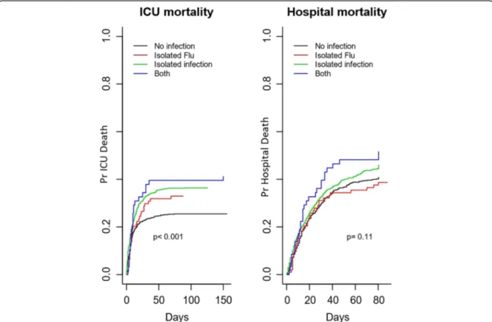
Outcomes in the different exposure groups in the crude analysis are summarized in Table 2. ICU mortality dif-fered between the four groups (P < 0.001), with the high-est mortality in patients with influenza plus co-infection (41%) and non-influenza infection (37%) and slightly lower mortality in influenza infection alone (33%) and in those without infection (26%). When the analyses were performed, after excluding those patients nega-tive for influenza but with no testing done, similar re-sults were found (Additional file 2: Table S2). Hospital and day 90 mortality showed a trend with the highest mortality in patients with influenza plus co-infection (52%) and non-influenza infection (46%) respectively (Table 2 and Additional file 2: Table S2). Survival curves for ICU and hospital stay for the four
Table 1 Influenza infection status and baseline characteristics at ICU admission
Baseline characteristics No infection (n
= 638)
Infection other than influenza (n = 820)
Influenza alone (n = 95)
Influenza co-infection
(n = 58) Pvalueb
Age (years), median [IQR] 63 [54–71] 63 [55–71] 65 [54–72] 64 [52–70] 0.80
Gender, male 351 (55) 512 (63) 59 (63) 32 (56) 0.04 Obesitya 108 (17) 151 (18) 21 (22) 12 (21) 0.21 Underlying disease Hematological disease 311 (49) 436 (53) 60 (63) 30 (51) 0.05 Solid tumor 252 (39) 285 (35) 18 (19) 12 (21) < 0.001
Solid organ transplantation 51 (9) 75 (10) 7 (8) 9 (16) 0.4
Systemic disease or other ID 102 (16) 133 (16) 25 (26) 18 (31) 0.002
Disease status at ICU admission
Newly diagnosed 161 (36) 154 (27) 12 (17) 7 (20) 0.0004
Remission 67 (15) 82 (14) 15 (21) 8 (23)
No remission 62 (14) 68 (12) 3 (4) 6 (17)
Allogeneic stem cell transplant 56 (9) 82 (10) 9 (9) 5 (9) 0.03
ECOGc
≥ 2 (severely disabled or bedridden) 210 (33) 299 (36) 36 (38) 23 (40) 0.15
Comorbidities
Cardiac 141 (24) 167 (22) 16 (18) 15 (27) 0.43
COPD 103 (17) 123 (15) 14 (15) 7 (12) 0.80
Kidney 88 (14) 117 (15) 16 (18) 10 (17) 0.75
Diabetes 108 (17) 161 (20) 21 (22) 14 (25) 0.30
Alcohol use disorder 63 (10) 76 (10) 5 (5) 4 (7) 0.48
Tobacco use 199 (33) 228 (29) 21 (23) 12 (21) 0.08
Duration of symptoms before ICU admission (days), median [IQR]
1 [0–4] 1 [0–3] 2 [1–7] 1.5 [1–4] <
0.001
Admission from emergency department 208 (33) 256 (32) 41 (43) 19 (33) 0.17
Neutropenia at admission 66 (11) 153 (20) 20 (21) 12 (21) <
0.001
Data are presented as median [IQR] orN (%)
a
Obesity grade I, II and extreme obesity
b
Chi-squared test of association with three degrees of freedom
c
groups are presented in Fig. 1. Hospital survival did not differ by group (P = 0.11).
For the propensity score-matched analysis, 152 pa-tients with influenza were matched. One patient with in-fluenza could not be matched and was excluded from the analysis. Imbalances in confounders were reduced after matching (Fig. 2). In the matched sample, there was no difference in hospital mortality attributable to in-fluenza infection (OR = 1.01, 95%CI 0.90–1.13, P = 0.85).
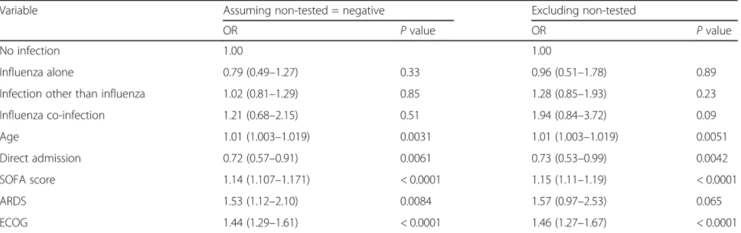
Table3 shows the results of multivariate analysis after multiple imputation by chained equations. The following factors were associated with hospital mortality: age, dir-ect ICU admission, severity manifested by SOFA score, diagnosis of ARDS, and performance status, with the lat-ter two demonstrating the strongest association with OR of hospital mortality of 1.53 and 1.44 respectively. None of the mechanism and/or type of immunosuppression was found as an independent risk factor for hospital
mortality. When the analysis was performed in patients negative for influenza and the test not done, similar re-sults were found (Table3).
Discussion
In summary, our multinational observational study ana-lyzed 1611 immunosuppressed patients from 68 centers and found that if a critically ill immunosuppressed pa-tient is infected with influenza, the outcome depends on the immunosuppression (independently of the mechan-ism and/or type of immunosuppression) rather than in-fluenza infection. We found that independent risk factors for hospital mortality were age, organ dysfunc-tion severity, direct admission to the ICU, and especially a diagnosis of ARDS and performance status. Influenza plus co-infection with bacterial or fungal pathogens was associated with the highest ICU mortality rate in our study. We did not observe a statistically significant
Table 2 Association between influenza infection status, clinical characteristics at day 1, and outcomes
Variables No infection (n =
638)
Infection other than influenza (n = 820)
Influenza alone (n = 95)
Influenza & co-infection (n
= 58) Pvaluea
At day 1
Maximum respiratory rate (breaths/min)
30 [24–36] 31 [25–37] 32 [28–36] 32 [26–38] 0.01
Liters/min O2 7 [3–15] 8 [5–15] 10 [4–15] 15 [2–15] 0.04
FiO2 50 [40–70] 50 [40–80] 50 [50–72] 59 [51–75] 0.03
PaO2/FiO2ratio 173 [115–215] 110 [79–173] 113 [110–204] 127 [87–170] <
0.001
ARDS at day 1 481 (75) 737 (90) 92 (97) 57 (98) <
0.001
SOFA at ICU admission 6 [4–9] 7 [4–11] 7 [4–10] 8 [6–10] <
0.001 Respiratory SOFA = 0 103 (17) 94 (12) 9 (10) 1 (2) < 0.001 Cardiovascular SOFA = 0 334 (53) 341 (42) 34 (37) 25 (43) < 0.001 Outcome
Intubation during the ICU stay 357 (56) 57 (60) 530 (65) 47 (81) <
0.001
Shock 171 (27) 429 (52) 47 (49) 32 (36) <
0.001
Renal replacement therapy 93 (15) 140 (17) 17 (17) 17 (29) 0.04
Steroidsb 187 (33) 272 (36) 27 (31) 27 (49) 0.09
ICU-acquired pneumonia 47 (7) 96 (12) 14 (15) 6 (10) 0.01
ICU length of stay (days) 5 [2–10] 7 [3–15] 8 [4–21] 10.5 [5–20] <
0.001
ICU mortality 165 (26) 302 (37) 31 (33) 24 (41) <
0.001
Hospital mortality 251 (41) 365 (46) 36 (38) 30 (52) 0.09
Day 90 mortality 291 (45) 410 (50) 38 (40) 32 (55) 0.06
Data are presented as median, IQR, orN (%)
a
Chi-squared test of association with three degrees of freedom
b
association between influenza infection status and hos-pital mortality, our primary outcome, in either crude or propensity score-matched analyses.
Influenza is a risk factor for acute respiratory failure in immunosuppressed patients. However, its role as an in-dependent risk factor for mortality in such a population has been questioned [9]. The number of immunosup-pressed patients hospitalized with influenza has in-creased in recent years, and we showed that while influenza alone may not increase mortality, influenza plus co-infection may be associated with higher ICU mortality. It might therefore be argued that empiric anti-biotic treatment for co-infection in such patients should be considered until the possibility of co-infection has been confidently ruled out. To facilitate earlier detection of co-infection, Rodriguez et al. [17] recently described that a low level of procalcitonin (PCT) has a high nega-tive predicnega-tive value (94%). However, clinicians may not be willing to tolerate even a low probability of untreated pulmonary co-infection in light of our observation that this category was associated with higher ICU mortality and length of stay.
In our cohort, two important factors stood out as in-dependent risk factors for death: the need for intubation during ICU stay and ARDS. While both features were associated with increased mortality in any immunosup-pressed patient, the mortality rate approached 100% when this occurred in patients with influenza and co-infection. Due to a known protective effect on mor-tality of direct admission from the emergency depart-ment to the ICU, it might be hypothesized that earlier assessment for severity and therefore earlier ICU admis-sion may improve the outcomes [18]. In a large popula-tion of patients with influenza, Álvarez-Lerma et al. [19] observed that ICU mortality was significantly higher among patients with late diagnosis as compared with early diagnosis (26.9% vs 17.1%, P < 0.001). Diagnostic delay was one independent risk factor for mortality (OR = 1.36, 95%CI 1.03–1.81, P < 0.001).
A common diagnostic challenge in immunosuppressed patients is the lack of clinical symptoms when develop-ing infections. In other words, an immunocompromised host is a patient who does not have the ability to re-spond normally to an infection due to an impaired or
Fig. 1 Hospital mortality and influenza infection status. Hospital mortality in the whole cohort according to influenza infection status categorized by four groups: (1) patients with influenza alone, (2) patients with influenza plus co-infections (clinically or microbiologically confirmed bacterial or fungal infection), (3) patients with infections other than influenza infection, and (4) patients without infection. Survival curves were compared using Cox regression
Fig. 2 Imbalances in confounders of mortality by influenza infection status before and after propensity score matching. Based on the matched sample, there was no evidence of any difference in hospital mortality across groups (OR = 1.01, 95%CI 0.90–1.13, p = 0.85). We developed a propensity score (PS) logistic model to have flu then matched the individuals on the basis of their PS using a 1:1 matching algorithm without replacement within a caliper of 0.15 standard deviation of the logit of the propensity score. To handle missing values in confounders, multiple imputation with chained equation was used for the PS model, where propensity score for each patient was averaged across 30 completed datasets while propensity score matching used these averaged scores to estimate the treatment effect. Only 1 patient with influenza could not be matched. Imbalances in confounders were reduced after matching
Table 3 Multivariate analysis of factors associated with hospital mortality after multiple imputations
Variable Assuming non-tested = negative Excluding non-tested
OR P value OR P value
No infection 1.00 1.00
Influenza alone 0.79 (0.49–1.27) 0.33 0.96 (0.51–1.78) 0.89
Infection other than influenza 1.02 (0.81–1.29) 0.85 1.28 (0.85–1.93) 0.23
Influenza co-infection 1.21 (0.68–2.15) 0.51 1.94 (0.84–3.72) 0.09 Age 1.01 (1.003–1.019) 0.0031 1.01 (1.003–1.019) 0.0051 Direct admission 0.72 (0.57–0.91) 0.0061 0.73 (0.53–0.99) 0.0042 SOFA score 1.14 (1.107–1.171) < 0.0001 1.15 (1.11–1.19) < 0.0001 ARDS 1.53 (1.12–2.10) 0.0084 1.57 (0.97–2.53) 0.065 ECOG 1.44 (1.29–1.61) < 0.0001 1.46 (1.27–1.67) < 0.0001
weakened immune system. This has been well reported in cases of bacterial or fungal infections, but little is known in the case of viral infections [20]. In our cohort, we found that patients with influenza were actually more likely to have a longer duration of symptoms prior to ICU admission.
Hypoxemia is a common clinical feature of patients with influenza, especially in immunosuppressed hosts. In a re-cent report from two cancer re-centers describing the out-comes in patients with hematological malignancies and influenza infection, severe hypoxemia was an independent risk factor (OR 5.87, 1.12–30.77) for 60-day mortality [21]. Similarly, hypoxemia was clearly a signal of illness severity in our study. In patients not intubated at admission to the ICU, oxygen requirements and ICU mortality rates were greatest in those with influenza plus co-infection.
Co-infection has previously been reported as an inde-pendent risk factor for poor outcome in patients with in-fluenza [9]. In our cohort, patients with co-infection were less likely to be cancer patients (have a hematological disease or solid tumor) but were more likely to have newly diagnosed immunosuppressive sys-temic disease or have poorer functional capacity. In this population, the criteria that suggest co-infection and therefore higher severity may be higher oxygen require-ments, greater tachypnea and work of breathing, and higher rates of mechanical ventilation.
Systemic immune mechanisms play a key role in the development of co-infection based on the complexity of the interaction of the host and the viral and bacterial pathogens. Several studies have been performed to de-termine the point prevalence of bacterial co-infection in influenza patients [9, 22–24]. In our cohort, almost half of the patients with co-infection received steroids. The use of steroids has been controversial and is currently not recommended in patients with influenza [25]. This is particularly relevant to our studied population because many patients were already receiving corticosteroid ther-apy for their primary disease. It appears plausible that steroids were given as a stress response treatment in pa-tients that were using longer-term steroids and not as a treatment for influenza per se. Importantly, we did not find steroids to be a risk factor for hospital mortality.
Some limitations should be mentioned. Vaccination status and information on antiviral regimen, dose, dur-ation, and delay in the start of therapy were not col-lected. Similar limitations apply to the determination of co-infection, which also could have led to misclassifica-tion error and bias. The sample contained primarily pa-tients with underlying hematological disease. Other subgroups of immunocompromised patients (particularly patients with lung transplant) may be underrepresented which may limit generalizability. Additionally, we did not completely account for the effect of the type of
immunosuppressive regimen in the adjusted analysis. The propensity score analysis aims at controlling for confounders, including those variables associated with the immunodeficiency that may affect the outcome. Nevertheless, one cannot assume that all confounders— possibly even not observed—have been taken into ac-count and that there may be residual confounders.
Conclusion
In summary age, severity score, ARDS, and performance status were all independent risk factors for ICU, hospital, and 90-day mortality in immunosuppressed patients ad-mitted to the ICU for acute hypoxemic respiratory failure. The main aim in this paper was to determine if influenza alone or co-infection played a role in the mortality in ICU patients. Category of infectious etiology of respiratory fail-ure (influenza, non-influenza, influenza plus co-infection, and non-infectious) was associated with ICU but not hos-pital mortality. In a propensity score-matched analysis, in-fluenza infection was not associated with the primary outcome of hospital mortality. Overall, influenza infection alone is not an independent risk factor for hospital mor-tality in immunosuppressed patients.
Additional files
Additional file 1: Table S1. Influenza infection status and baseline characteristics at ICU admission. Group no infection performed excluding 448 patients negative for influenza and testing not done. (DOCX 18 kb) Additional file 2: Table S2. Association between influenza infection status, clinical characteristics at day 1, and outcomes. (DOCX 36 kb)
Acknowledgements
The authors acknowledge the importance of the Nine-I (Caring for Critically Ill Immunocompromised Patients) group in executing this project, the GRRR-OH for funding the project, and all individuals involved in the data collection for the EFRAIM study.
Funding
The study was funded by the GRRR-OH (Groupe de Recherche en Réanima-tion Onco-Hématologique), an academic non-profit French organizaRéanima-tion. Availability of data and materials
Requests for data will be considered by the principal study investigators, based on the nature of the request and legal and ethical regulations. Authors’ contributions
IML, VL, EA, and PG were involved in the conception, design, and analysis phases and wrote the paper. MAM, PP, MS, AP, RBB, JR, PB, AL, FST, JS, PS, KR, NT, SM, MA, AK, PK, MV, PPL, FB, FP, VM, ASM, VS, GB, CG, UVAS, LM, FB, LBN, BG, and DM collected the data and provided significant scientific comments for the final manuscript. SC conducted the statistical analysis. EA and IML acted as guarantors of the project. All authors read and approved the final manuscript.
Ethics approval and consent to participate
Participating investigators obtained local institutional review board approval in accordance with local ethics regulations.
Consent for publication
Competing interests
The authors declare that they have no competing interest.
Publisher’s Note
Springer Nature remains neutral with regard to jurisdictional claims in published maps and institutional affiliations.
Author details
1Medical Intensive Care Unit, Hôpital Saint-Louis and Paris Diderot Sorbonne University, Paris, France.2Department of Intensive Care Medicine (710), Radboud University Medical Centre, Nijmegen, The Netherlands. 3
Department of Critical Care and Graduate Program in Translational Medicine, Programa de Pós-Graduação em Clínica Médica, D’Or Institute for Research and Education, Rio de Janeiro, Brazil.4Department of Intensive Care, Rigshospitalet, University of Copenhagen, Copenhagen, Denmark. 5
CIBERES, Universitat Autonòma de Barcelona, European Study Group of Infections in Critically Ill Patients (ESGCIP), Barcelona, Spain.6Pulmonary and Critical Care Medicine, Mayo Clinic, Rochester, MN, USA.7Division of Pulmonary and Critical Care, Penn State University College of Medicine, Hershey, PA, USA.8Department of Intensive Care, Hôpital Erasme, Université Libre de Bruxelles (ULB), Brussels, Belgium.9Department of Intensive Care Medicine, Multidisciplinary Intensive Care Research Organization (MICRO), St. James’s Hospital, Dublin, Ireland.10Department of Clinical Medicine, Wellcome Trust-HRB Clinical Research Facility, St. James Hospital, Trinity College, Dublin, Ireland.11Department of Medicine I, Medical University of Vienna, Vienna, Austria.12Department of Anesthesiology and Intensive Care Medicine and Institute for Medical Humanities, 1st Faculty of Medicine, Charles University in Prague and General University Hospital, Prague, Czech Republic.13Norwegian University of Science and Technology, Trondheim, Norway.14CHU Grenoble Alpes, Service de Réanimation Médicale, Faculté de Médecine de Grenoble, INSERM U1042, Université Grenoble-Alpes, Grenoble, France.15Department of Medicine and Interdepartmental Division of Critical Care Medicine, Sinai Health System, University of Toronto, Toronto, Ontario, Canada.16Agostino Gemelli University Hospital, Università Cattolica del Sacro Cuore, Rome, Italy.17Department of Medical Intensive Care Medicine, University Hospital of Angers, Angers, France.18Department of Immunology– Department of Emergencies and Critical Care, University of Oslo, Oslo, Norway.19Division of Intensive Care Medicine, Department of Anesthesiology, Intensive Care and Pain Medicine, Helsinki University Hospital, University of Helsinki, Helsinki, Finland.20Department of Critical Care, University Medical Center Groningen, Groningen, The Netherlands. 21Medical-Surgical Intensive Care Unit, Centre Hospitalier de Versailles, Le Chesnay, France.22Department of Anesthesiology I, Herlev University Hospital, Herlev, Denmark.23Medical ICU, Cochin Hospital, Assistance Publique-Hôpitaux de Paris and University Paris Descartes, Paris, France. 24Critical Care Department, King’s College Hospital NHS Foundation Trust, London SE5 9RS, UK.25Critical Care Center, CHU Lille, School of Medicine, University of Lille, Lille, France.26Terapia Intensiva, Hospital Maciel, Montevideo, Uruguay.27Department of Medical Intensive Care, Normandie Univ, UNIROUEN, EA-3830, Rouen University Hospital, F-76000 Rouen, France. 28ICU, Fundação Pio XII - Barretos Cancer Hospital, Barretos, Brazil.29Medical Intensive Care Unit, La Source Hospital - CHR Orléans, Orléans, France. 30Intensive Care Department, University of Southern Denmark, Sønderborg, Denmark.31Department of Anaesthesia and Intensive Care, Odense University Hospital, Odense, Denmark.32Medical Intensive Care Unit, Hôtel Dieu-HME-University Hospital of Nantes, Nantes, France.33Réanimation Polyvalente et Département d’Anesthésie et de Réanimation, Institut Paoli-Calmettes, Marseille, France.34ECSTRA Team, Biostatistics and Clinical Epidemiology, UMR 1153, INSERM, Paris Diderot Sorbonne University and Service de Biostatistique et Information Médicale AP-HP, Hôpital Saint-Louis, Saint-Louis, France.35Department of Intensive Care Medicine, St. James’s Hospital, St. James’s St, Dublin, Dublin 8, Ireland.
Received: 24 January 2019 Accepted: 9 April 2019
References
1. Azoulay E, Afessa B. The intensive care support of patients with malignancy: do everything that can be done. Intensive Care Med. 2006;32:3–5. 2. Nseir S, Di Pompeo C, Diarra M, Brisson H, Tissier S, Boulo M, Durocher A.
Relationship between immunosuppression and intensive care unit-acquired
multidrug-resistant bacteria: a case-control study. Crit Care Med. 2007;35: 1318–23.
3. Boomer JS, To K, Chang KC, Takasu O, Osborne DF, Walton AH, Bricker TL, Jarman SD, Kreisel D, Krupnick AS, Srivastava A, Swanson PE, Green JM, Hotchkiss RS. Immunosuppression in patients who die of sepsis and multiple organ failure. JAMA. 2011;306:2594–605.
4. Kash JC, Taubenberger JK. The role of viral, host, and secondary bacterial factors in influenza pathogenesis. Am J Pathol. 2015;185:1528–36. 5. Fowlkes A, Steffens A, Temte J, Di Lonardo S, McHugh L, Martin K, Rubino
H, Feist M, Davis C, Selzer C, Lojo J, Oni O, Kurkjian K, Thomas A, Boulton R, Bryan N, Lynfield R, Biggerstaff M, Finelli L, Influenza Incidence Surveillance Project Working Group. Incidence of medically attended influenza during pandemic and post-pandemic seasons through the influenza incidence surveillance project, 2009-13. Lancet Respir Med. 2015;3:709–18.
6. Díaz E, Rodríguez A, Martin-Loeches I, Lorente L, del Mar Martín M, Pozo JC, Montejo JC, Estella A, Arenzana A, Rello J. Impact of obesity in patients infected with 2009 influenza A(H1N1). Chest. 2011;139:382–6. 7. Napolitano LM, Angus DC, Uyeki TM. Critically ill patients with influenza
A(H1N1)pdm09 virus infection in 2014. JAMA. 2014;311(13):1289-90.https:// doi.org/10.1001/jama.2014.2116.
8. Martin-Loeches I, Díaz E, Vidaur L, Torres A, Laborda C, Granada R, Bonastre J, Martín M, Insausti J, Arenzana A, Guerrero JE, Navarrete I, Bermejo-Martin J, Suarez D, Rodriguez A. Pandemic and post-pandemic influenza A (H1N1) infection in critically ill patients. Crit Care. 2011;15:R286.
9. Martin-Loeches I, Schultz JM, Vincent J-L, Alvarez-Lerma F, Bos LD, Solé-Violán J, Torres A, Rodriguez A, Sole-Violan J, Torres A, Rodriguez A. Increased incidence of co-infection in critically ill patients with influenza. Intensive Care Med. 2017;43(1):48-58. https://doi.org/10.1007/s00134-016-4578-y. Epub 2016 Oct 5.
10. Cawcutt K, Kalil AC. Pneumonia with bacterial and viral coinfection. Curr Opin Crit Care. 2017;23:385–90.
11. White DB, Angus DC. Preparing for the sickest patients with 2009 influenza A(H1N1). JAMA. 2009;302:1905–6.
12. Visseaux B, Burdet C, Voiriot G, Lescure F-X, Chougar T, Brugière O, Crestani B, Casalino E, Charpentier C, Descamps D, Timsit J-F, Yazdanpanah Y, Houhou-Fidouh N. Prevalence of respiratory viruses among adults, by season, age, respiratory tract region and type of medical unit in Paris, France, from 2011 to 2016. PLoS One. 2017;12:e0180888.
13. Azoulay E, Pickkers P, Soares M, Perner A, Rello J, Bauer PR, van de Louw A, Hemelaar P, Lemiale V, Taccone FS, Martin Loeches I, Meyhoff TS, Salluh J, Schellongowski P, Rusinova K, Terzi N, Mehta S, Antonelli M, Kouatchet A, Barratt-Due A, Valkonen M, Landburg PP, Bruneel F, Bukan RB, Pène F, Metaxa V, Moreau AS, Souppart V, Burghi G, Girault C, et al. Acute hypoxemic respiratory failure in immunocompromised patients: the Efraim multinational prospective cohort study. Intensive Care Med. 2017;43(12): 1808-19.https://doi.org/10.1007/s00134-017-4947-1. Epub 2017 Sep 25. 14.
https://www.cdc.gov/flu/pdf/freeresources/healthcare/flu-specimen-collection-guide.pdfAcccesed 14 Mar 12, 2019.
15. Contejean A, Lemiale V, Resche-Rigon M, Mokart D, Pène F, Kouatchet A, Mayaux J, Vincent F, Nyunga M, Bruneel F, Rabbat A, Perez P, Meert A-P, Benoit D, Hamidfar R, Darmon M, Jourdain M, Renault A, Schlemmer B, Azoulay E. Increased mortality in hematological malignancy patients with acute respiratory failure from undetermined etiology: a Groupe de Recherche en Réanimation Respiratoire en Onco-Hématologie (GRRR-OH) study. Ann Intensive Care. 2016;6:102.
16. J T, Vincent JL, Moreno R, Takala J, Willatts S, De Mendonça A, Bruining H, Reinhart CK, Suter PM, Thijs LG. The SOFA (Sepsis-related Organ Failure Assessment) score to describe organ dysfunction/failure. On behalf of the working group on Sepsis-Related Problems of the European Society of Intensive Care Medicine. Intensive Care Med. 1996;22:707–10.
17. Rodríguez AH, Avilés-Jurado FX, Díaz E, Schuetz P, Trefler SI, Solé-Violán J, Cordero L, Vidaur L, Estella Á, Pozo Laderas JC, Socias L, Vergara JC, Zaragoza R, Bonastre J, Guerrero JE, Suberviola B, Cilloniz C, Restrepo MI, Martín-Loeches I, Cobo P, Martins J, Carbayo C, Robles-Musso E, Cárdenas A, Fierro J, Fernández DO, Sierra R, Huertos MJ, Carmona Pérez ML, Pozo Laderas JC, et al. Procalcitonin (PCT) levels for ruling-out bacterial coinfection in ICU patients with influenza: a CHAID decision-tree analysis. J Inf Secur. 2016;72:143–51.
18. Martin-Loeches I, Levy MMMM, Artigas A. Management of severe sepsis: advances, challenges, and current status. Drug Des Devel Ther. 2015;9: 2079–88.
19. Álvarez-Lerma F, Marín-Corral J, Vilà C, Masclans JR, Loeches IM, Barbadillo S, González de Molina FJ, Rodríguez A, H1N1 GETGAG/SEMICYUC Study Group. Characteristics of patients with hospital-acquired influenza A (H1N1)pdm09 virus admitted to the intensive care unit. J Hosp Infect. 2017; 95:200–6.
20. Zafrani L, Azoulay E. How to treat severe infections in critically ill neutropenic patients? BMC Infect Dis. 2014;14:512.
21. Vilar-Compte D, Shah DP, Vanichanan J, Cornejo-Juarez P, Garcia-Horton A, Volkow P, Chemaly RF. Influenza in patients with hematological
malignancies: experience at two comprehensive cancer centers. J Med Virol. 2018;90:50–60.
22. Martin-Loeches I, Rodriguez A, Sanchez-Corral A, Granada R, Zaragoza R, Albaya A, Cerda E, Catalan R, Luque P, Paredes A, et al.: Bacterial co-infection in critically ill patients infected with pandemic (H1N1) v influenza A infection. In Intensive Care Med. Volume 36; 2010:S369--S369. 23. Matos RG, Moreno RP, Diogo AC, Pereira JM, Martin-Loeches I, Cecconi M,
Lisboa T, Rhodes A, Rello J. Bacterial pneumonia complicating influenza A (H1N1) v viral pneumonia: results of the ESICM influenza A (H1N1) v registry. In: Intensive care med. Volume 36; 2010. p. S371.
24. Muscedere J, Ofner M, Kumar A, Long J, Lamontagne F, Cook D, McGeer A, Chant C, Marshall J, Jouvet P, Fowler R, ICU-FLU Group, Canadian Critical Care Trials Group. The occurrence and impact of bacterial organisms complicating critical care illness associated with 2009 influenza A(H1N1) infection. Chest. 2013;144:39–47.
25. Mc Mahon A, Martin-Loeches I. The pharmacological management of severe influenza infection - 'existing and emerging therapies'. Expert Rev Clin Pharmacol. 2017;10(1):81-95.https://doi.org/10.1080/17512433.2017. 1255550. Epub 2016 Nov 25.