1
Redifferentiation of iodine-refractory metastatic thyroid cancer: from bench to
bedside, through a network analysis
Dottorato di ricerca in Tecnologie Mediche Innovative in Medicina Clinica - XXXIII ciclo
Sapienza Università di Roma
Candidato: Dott.ssa Rosa Falcone
Tutor: Dott.ssa Antonella Verrienti
2 “Ogni visione è parziale. Non esiste un modo di vedere la realtà che non dipenda da una prospettiva. Non c’è punto di vista assoluto, universale.
I punti di vista tuttavia comunicano, i saperi sono in dialogo tra loro e con la realtà, nel dialogo si modificano, si arricchiscono, convergono, la nostra comprensione della realtà si approfondisce.” (Helgoland. C. Rovelli, 2020)
3 Outline ABSTRACT……….pag. 4 BACKGROUND……….………5 Preclinical data………....6 Clinical evidences ………....7 Bioinformatic analysis………..…8 AIMS………8
SECTION 1 – clinical study………...………..9
Materials and methods……….9
Results……….12
SECTION 2 – bioinformatic study……….17
Materials and methods………...17
Results……….19
SECTION 3 – in vitro study………...20
Materials and methods………...20
Results……….22
DISCUSSION……….27
4
ABSTRACT
BACKGROUND: Radioiodine-refractory (RAIR) differentiated thyroid cancers (DTC) lose the ability to efficiently concentrate iodide, rendering RAI (iodine-131; I-131) ineffective. Few studies have shown improved RAI uptake after MEK inhibitors (MEKi) and/or BRAF inhibitors (BRAFi). MicroRNA (miRNA) role in de- and re-differentiation process has been poorly investigated. METHODS: We retrospectively evaluated RAIR DTC patients treated with redifferentiation therapy using kinase inhibitor (KI) therapy with single-agent or combination MEKi and/or BRAFi. Our primary objectives were the percentage of patients with uptake on diagnostic (Dx) and post-treatment (post-Tx) whole body scan (WBS), and the radiologic response to I-131 achieved over that seen with KI. We exploited a bioinformatic approach (SWItchMiner, SWIM) to identify the key genes (switch genes) involved in BRAF-mediated carcinogenesis. MicroRNA Enrichment TURned NETwork (Mienturnet) was used to find the most important miRNAs able to regulate these switch genes. We validated our results by in vitro experiments.
RESULTS: A total of 41 patients with RAIR, advanced DTC (76% BRAF V600E mutant cancer) were included in the study. At post treatment scan, there was uptake, either optimal or suboptimal, in 95% of patients. After I-131, 73% of patients were deemed treatment successes, reaching stable or partial response, that lasted at least one year without treatment on systemic therapy. The median PFS was 29 months from the start of KI and the median TTP was 20 months from RAI administration. The investigation of mutational landscape of BRAF mutated papillaty thiroid cancer (PTC) by SWIM allowed to identify a subset of switch genes (227), 30% of which is regulated by hsa-miR-335-5p. In vitro experiments showed that miRNA-335-5p is lost in primary or continuous BRAF mutated cell lines. Its over-expression in thyroid tumor cell lines harboring BRAF mutation induces the activation of NIS protein and increases intra-cellular iodide uptake.
5 CONCLUSIONS: Redifferentiation therapy with MEKi and/or BRAFi resensitizes a subset of RAIR DTC patients to RAI and may result in prolonged stable disease off KI. Hsa-miR-335-5p may have a role in redifferentiation of RAIR BRAF-mutated papillary thyroid cancer. MiR-335-5p over-expression may contribute to restoring RAI responsiveness.
BACKGROUND
Radioiodine-refractory (RAIR) differentiated thyroid cancers (DTC) lose the ability to efficiently concentrate iodide, rendering RAI (iodine-131; I-131) ineffective 1. RAIR thyroid cancer has a dismal prognosis, with an estimated 10-year survival rate of 10% 2. Management options in this subset of patients include active surveillance, local treatments for metastatic sites, or kinase inhibitor (KI) therapy for rapidly progressing, symptomatic, or life-threatening disease.
A novel strategy, called “redifferentiation”, is currently being explored to restore the capacity of
RAIR tumors to trap and respond to a single administration of I-131, reducing or deferring the need of long-term KI therapies. Redifferentiation strategy consists of administration of KI (MEK and/or BRAF inhibitors) for a period of time to induce redifferentiation in RAIR DTC, thus re-sensitizes tumours to RAI, followed by therapeutic radioiodine. Patients continue KI until 2-3 days after RAI and then discontinue KI and start follow-up, off systemic treatment.
The first studies with the aim of redifferentiation, using retinoids 3, romidpesin 4, rosiglitazone 5 and sorafenib 6 have been demonstrated to have low clinical value. Grüning et al. did not observe any relevant improvement of RAI uptake in patients after isotretinoin treatment, and reported a disappointingly low response rate, in the order of 20% 3. No major responses were observed in the study with romidepsin and rosiglitazone 4, 5. An unexpected sudden death was reported during treatment with romidpesin, which led to the temporary suspension of the study. Authors did not recommend further investigation of study compounds as a single agent in RAI-refractory thyroid cancer.
6 A deeper understanding of the molecular processes who hide behind the lost of sensitivity to RAI (genetic and epigenetic alterations) has allowed to identify better therapeutic targets.
Preclinical data
Mutations in the mitogen-activated protein (MAP) kinase pathway, in particular BRAF V600E mutation, have been associated with reduced expression of key genes involved in iodine metabolism (iodide uptake, thyroid hormone synthesis and differentiation, i.e. PAX8, TTF-1, NIS, TPO, TG) 7-9.
Thyroid transcription factor-1 (TTF-1) and human paired box-8 (PAX-8) are involved in thyroid differentiation and control the expression of thyroglobulin (TG), thyroperoxidase (TPO), thyroid-stimulating hormone receptor (TSHr) and sodium/iodide symporter (NIS, also known as solute carrier family 5, member 5, SLC5A5) by binding to the promoters of these genes. PAX8 expression is frequently downregulated in thyroid carcinoma, and this decrease may correlate with the dedifferentiation of thyroid follicular cells, reflected by the loss or downregulated expression of genes involved in the cells’ ability to concentrate iodine, thereby contributing to tumor aggressiveness 10. NIS, located on the basolateral membrane of thyroid follicular cells, is the protein responsible of iodide uptake. Levels and activity of NIS on plasma membrane of thyroid cancer cells are highly variable. Down-regulated expression level of NIS has been observed in RAIR DTC 11. Different approaches have been used in the attempt to upregulate or re-activate NIS in RAIR tumours. Moreover, human thyroid cancers with BRAF mutations show greater reduction of NIS mRNA compared to tumors with other mutations or with no identifiable genetic changes 7. The restorability of iodide-metabolizing gene expression has been demonstrated using a MEK inhibitor (MEKi) or through the cessation of BRAF V600E expression in a thyroid cancer cell line 8. Chakravarty et al showed that in a thyroid cancer mouse model harbouring a BRAF V600E mutation, the pharmacological inhibition of either MEK or BRAF with selective inhibitors, is associated with restoration of gland architecture, tumor regression and recovery of RAI uptake 9.
7 BRAF is one of the best known driver genes, which encodes for a member of the RAF kinase family. It is a core component of the ERK signalling pathway which is involved in cell growth, differentiation, and survival. The most frequent mutation in the BRAF gene is the point mutation c.1799T>A, which is a substitution that results in an aminoacid change from valine (V) to glutamic acid (E) in the activation segment of the BRAF kinase (V600E), promoting its hyperactivation and resulting in constitutive activation of downstream ERK signalling. BRAF mutations are present in approximately 8% of human tumours. BRAF V600mutations occur in 37-50% of papillary thyroid cancers. BRAF V600mutations are associated with poor prognosis due to an aggressive disease phenotype, shortened overall survival, and minor response rates 12. Several BRAFi have been approved since 2011. The first one to be used was vemurafenib, approved first in patients with BRAF V600E mutant metastatic melanoma 13.
To date the molecular mechanism behind redifferentiation is not fully understood. MicroRNAs (miRNAs or miRs), small noncoding RNAs (∼22 nt) that regulate gene expression at mRNA
post-transcriptional level, are involved in physiological and pathological processes, including tumorigenesis, differentiation and progression 14. They predominantly bind to 3’UTR of target genes resulting in either degradation of target mRNA or inhibition of translation. Several studies reported a general downregulation of miRNAs in tumours, compared with normal tissues. Due to its repression effect, deregulation of specific miRNAs could lead to the repression of tumor suppressor genes and/or increase of oncogene expression. MiRNAs have been shown to be useful predictive biomarkers and targets of therapeutic options in thyroid cancers 15, 16. Few studies reported the association between RAI-uptake and miRNA expression profiles, and their association with response to RAI-treatment 17.
Clinical evidences
Previous preclinical observations provided the rationale for clinical trials with RAIR DTC patients treated with a MEK inhibitor and/or a BRAF inhibitor 18-21 to restore RAI uptake. These clinical trials
8 showed an improvement in RAI uptake after MEKi or/and BRAFi in RAS and BRAF mutant cancers, resulting in objective response to I-131 treatment. However, the time to progression and time off KIs is also of importance to physicians and patients. The ability to expose patients to KIs for a short period of time (instead of continuous treatment until it is no longer beneficial), exposes the patient to fewer potential adverse events. Furthermore, resistance to KIs after long-term exposure is inevitable, as demonstrated by the emergence, in thyroid cancer patients exposed to BRAF inhibitors, of mutations that confer resistance to treatment, leading to progression of disease 22.
Bioinformatic analysis
The analysis of the cancer molecular landscape, using bioinformatic tools, is emerging as a powerful approach to identify hidden, tissue-specific pathways 23, to predict effectiveness of existing therapeutic agents (repurposing) or to suggest new ones 24. Two tools, developed by physics and engineers at our Institution, were exploited for this study purpose: SWitchMiner (SWIM) and Mienturnet. SWIM is a software with a user-friendly graphical interface, developed in MATLAB. SWIM combines topological properties of co-expression networks with transcriptomic data and is able to identify a small pool of regulatory genes, called switch genes, which have been shown to be critically associated with drastic changes in cell phenotypes. This tool has been successfully applied in human cancers to identify genes that could be critically associated with the drastic changes in the physiological state of cells or tissues induced by cancer development, such as breast cancer and glioblastoma 25. Mienturnet is an interactive web tool for miRNA-target enrichment and network-based analysis 26. It fetches data of computationally predicted and experimentally validated miRNA-target interactions from two databases (TargetScan and miRTarBase).
AIMS
Here, we describe a population of patients with previously RAIR, advanced DTC, treated with either BRAFi and/or MEKi with the aim to restore RAI avidity (Section 1- clinical study). Thus, in this
9 study we sought to describe the long-term effects of redifferentiation therapy for those patients who were able to discontinue KI after receiving RAI. Assuming the pivotal role of BRAF mutation in oncogenesis, as well as in the de-differentiation process resulting in refractoriness to radioiodine therapy in thyroid cancer, we used a bioinformatic approach to identify the key genes involved in BRAF-mediated carcinogenesis. Then, we looked for the most important miRNAs able to regulate switch genes that might have a role in the redifferentiation process (Section 2-bioinformatic study). We validated our results by in vitro experiments (Section 3- in vitro studies).
SECTION 1 - clinical study
MATERIAL AND METHODS
Study design and participants
This was a single center retrospective analysis. Consecutive patients from October 2014 to April 2019 were considered for the study. Eligible patients were required to have RAI refractory DTC, histopathologically confirmed at MD Anderson Cancer Center. One of the following criteria for RAIR disease had to be met: 1) no RAI uptake at known sites of metastases, 2) progressive disease (PD) despite previous RAI treatment within 1 year of RAI therapy. Moreover, to be included in the study, patients needed to be treated with either single-agent or combination BRAFi and/or MEKi, and had to have a diagnostic (Dx) whole-body scan (WBS) while receiving this therapy. Patients who did not complete the redifferentiation strategy, i.e., patients who didn’t receive 131 or who received I-131 but continued the BRAFi and/or MEKi, were not included in the analysis. The study was approved by the institutional review board at The University of Texas MD Anderson Cancer Center, under a waiver of consent. Details about the demographics, tumor characteristics (i.e., histologic subtype and mutational status), volume and sites of disease, prior treatments (surgery, radiotherapy, RAI, kinase inhibitors) before redifferentiation therapy were collected from the charts of patients. Other information obtained from medical records were: name of BRAFi and/or MEKi used to induce
10 redifferentiation, duration of treatment, uptake on Dx and post treatment (post-Tx) WBS, activity of RAI administered, radiological response to BRAFi and/or MEKi and RAI, thyroglobulin (Tg) trend before, during and after redifferentiation strategy, and survival data.
Treatment schedule
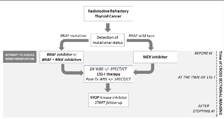
Patients with RAS (N-, H-, or K-) and no detectable driver mutations were treated with a MEKi. Patients with BRAF mutated DTC were treated with single agent BRAFi or the combination BRAFi + MEKi. Patients received I-131 after low iodine diet intake of 1 to 2 weeks and after rhTSH injections on two consecutive days. Dx and post-Tx WBS were performed for every patient and a SPECT/CT was available with Dx scan and/or with post-Tx scan (figure 1).
Figure 1. Treatment schedule. Patients with Radioiodine refractory thyroid cancer were
treated with MEK inhibitor (MEKi), BRAF inhibitor (BRAFi) or both, according to mutational status, in an attempt to induce redifferentiation (revert to RAI sensitive). Patients received I-131 after a median period of 3.9 months. Diagnostic (Dx) and post-treatment (post-Tx) whole body scan (WBS) were performed for every patient and a SPECT/CT was available with Dx scan and/or with post-Tx scan. The BRAFi and/or MEKi was discontinued 48-72 hours after I-131 administration. Cross-sectional imaging (CT, 18fdg PET/CT) was performed before starting kinase inhibitor (KI) and after I-131 administration. Target lesions were measured before KI, during KI (if available), at the time of RAI administration (using SPECT imaging or other cross-sectional imaging performed close to I-131) and after 131-I therapy (while off KI).
11 Preferably, post-Tx scans should be delayed for 5-14 days post-RAI, however, there was a lot of variability regarding the timing of the post-Tx scans. All scans were reviewed by the same expert nuclear medicine physician. Optimal uptake on WBS was defined as intensive accumulation of radioiodine in all tumor lesions. Suboptimal uptake was defined as either weak uptake or when some, but not all lesions, accumulated radioiodine. The BRAFi and/or MEKi was discontinued 48-72 hours after I-131 administration. Cross-sectional imaging (CT, 18fdg PET/CT) was performed before starting KI and after I-131 administration. For some patients (those who received KI for a longer period of time), cross-sectional imaging were available also during KI therapy. Radiologic response on cross-sectional imaging was measured using Response Evaluation Criteria in Solid Tumors, version 1.1. Target lesions were measured before KI, during KI (if available), at the moment of RAI administration (using SPECT imaging or other cross-sectional imaging performed close to I-131) and after RAI therapy while not receiving KI. Best response were recorded.
Levels of serum non-stimulated thyroglobulin (Tg) were collected before starting KI, before RAI administration, around 3, 6 and 12 months after administration of I-131, and at the moment of progression, when available. We excluded patients with positive anti Tg antibody (Ab) from the Tg analysis.
Data analysis
The primary endpoints of the study were the percentage of patients with uptake on Dx and post-Tx scan, and the best radiologic response to I-131 achieved over that seen with KI, confirmed at least after 6 months from radioiodine. The secondary endpoints of the study were progression free survival (PFS), time to progression (TTP), time on and off kinase inhibitors, percentage of treatment success, serum non-stimulated Tg level during redifferentiation strategy. An additional, exploratory end point was the assessment of differences in PFS and TTP between patients with BRAF mutation and patients without BRAF mutation (RAS-mutated or wild-type for a panel of 400 genes analyzed by Next
12 Generation Sequencing). PFS was defined as the time from the start of KI to progression according to RECIST 1.1 criteria. TTP is the time from RAI administration to progressive disease. Patients who did not progress, were censored at last follow-up visit. We defined treatment success as the absence of progression within one year from I administration and/or the discontinuation of KI after 131-I administration of at least 12 months. Survival times were calculated and compared using the Log-Rank test. A Wilcoxon signed-rank test for paired samples was used for the comparison of non-stimulated Tg levels at each follow-up assessment. Mann-Withney test was used to compare unpaired groups. P value < 0.05 was considered significant. The statistical analysis was performed using IBM SPSS Statistics 25.
Results
A total of 41 patients were considered for the analysis. Baseline characteristics of patients are listed in table 1a. Median age at diagnosis was 56 years. Twenty-four (59%) were women. Papillary thyroid cancer (PTC) was the prevalent histological subtype (80%), 5 (12%) patients had follicular thyroid cancer (FTC) and 3 (8%) patients had poorly differentiated cancer. Thirty-one patients (76%) had BRAF V600E mutant cancer, 9 patients had RAS mutations (2 KRAS, 5 NRAS, 2 HRAS), and one patient was negative for 400 tested genes, including BRAF and RAS. All but three patients had undergone at least one prior RAI therapy, with a median cumulative activity of 200 mCi. These three patients had no prior RAI treatments: one of them was deemed to be RAI refractory on the basis of a negative diagnostic scan at initial presentation to our institution, in the setting of biopsy-proven and 18FDG-PET avid distant metastases. In the other two cases, the patients had bulky disease and 18FDG-PET avid disease. The latter two patients were started on neoadjuvant systemic therapy prior to their initial thyroidectomy. Sixteen of 41 (39%) had bulky disease, defined as at least one lesion with a diameter ≥ 2 cm. Most of the patients (83%) had disease confined to lungs and nodes (neck
13 single agent BRAFi (56%) or a MEKi (24%), or the combination of BRAFi and MEKi (COMBO; 20%) (Table 1b).
Table 1. a) Study population characteristics (N=41). b) Kinase inhibitor (KI) treatment used for
redifferentiation therapy.
a)
BASELINE CHARACTERISTICS N°
Age at diagnosis in years, median (range) 56 (34–74) Sex (%) male Female 17 (41) 24 (59) Histology (%) PTC FTC PDTC 33 (80) 5 (12) 3 (8) Driver mutation (%) BRAF V600E
RAS (N-, H-, or K-) None detected
31 (76) 9 (22) 1 (2) Number of prior RAI treatments, median (range) 2 (0-3) Cumulative administered activity, median (range), mCi 200 (0-517) Prior treatment for thyroid cancer (%)
Reoperation Radiotherapy
Other systemic therapy
29 (71) 17 (41) 8 (20) Sites of disease (%)
Lungs, neck and/or mediastinum Other sites (liver, brain, pleura, bone)
34 (83) 7 (17)
Bulky disease* (%) 16 (39)
b)
Kinase Inhibitor N. of patients treated (%) Days of treatment, median (range)
BRAF inhibitor 23 (56) 147 (24-2344)
MEK inhibitor 10 (24) 58.5 (20-275)
COMBO** 8 (20) 221 (97-559)
Total 41 118 (20-2344)
PTC, papillary thyroid cancer; FTC, follicular thyroid cancer; PDTC, poorly differentiated thyroid cancer; RAI, radioactive iodine.
*At least one lesion with a diameter ≥ 2 cm **COMBO=BRAF inhibitor + MEK inhibitor
Of the 31 patients with BRAF V600E mutant DTC, 23 were treated with single agent dabrafenib (n=22) or vemurafenib (n=1), and 8 received the COMBO regimen (dabrafenib and trametinib). The other 10 patients (RAS mutant and the patient without a detectable mutation) were treated with single
14 agent MEKi (6 trametinib, 3 cobimetinib, 1 binimetinib). The median duration of KI therapy was 3.9 months (table 1b) with a shorter period of treatment for patients treated with MEKi compared to the other 2 groups (p=0.01). This difference can be partly explained by the fact that most patients who received MEKi started the therapy with the intent of redifferentiation.
Uptake on Diagnostic and Post-treatment WBS
Of 41 patients, after redifferentiation therapy, the Dx WBS was positive in 78% (32/41) of patients and it was considered negative in 22% (9/41) of patients. These nine patients with a negative Dx WBX were treated empirically with I-131. The median administered activity of I-131 was 157 mCi (100-255 mCi). The median time between I-131 treatment and post-Tx WBS was 5 days (range, 1-9). On post-Tx WBS, there was uptake in 39/41 (95%) of patients who received therapeutic doses of I-131 (17% of recovered uptake compared to Dx WBS). All RAS mutant thyroid cancer patients, who received therapeutic doses of I-131, had uptake, either optimal or suboptimal uptake on post-Tx scan. In BRAF mutant DTC, uptake on post-Tx WBS was seen in 29/31 (94%) of patients. In these two cases, the post-Tx WBS was performed within 48 hours from RAI therapy.
Radiological response
The median follow-up period was 13 (1-46) months. Out of 41 patients included in this study, 26 had a long enough follow-up to have at least one CT scan after RAI, thus were evaluable for response. Four out of the 26 patients (all with BRAF mutated cancers) had sub-centimeter lesions and were not evaluable with RECIST v1.1 criteria. This left 22 evaluable patients for response by RECIST v1.1 criteria.
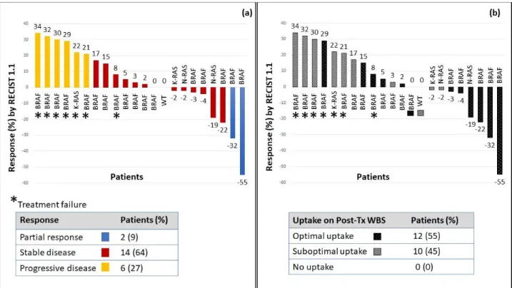
The best overall response comparing baseline imaging (prior to KI) to just prior to RAI therapy, was as follows: 32% (7/22) of patients had a partial response, 64% (14/22) had stable disease and 4% of patient (1/22) had progression. After I-131 administration, to evaluate the additional benefit from RAI over that seen on KI, we examined the response rate while off KI comparing the SPECT/CT imaging at the time of RAI administration to the imaging after stopping KI. Most of the study population
15 (64%; 14/22) had stable disease and 9% (2/22) of patients had a partial response (figure 2a), when compared to imaging taken at the time of RAI administration. Thirty-six percent (8/22) of patients had some degree of decrease in target lesions after RAI. The four patients not evaluable with RECIST criteria had stable disease during and after the redifferentiation strategy and were still off systemic therapy after one year from redifferentiation therapy. Nineteen of 26 patients (73%) were deemed treatment successes. The percentages of treatment successes among patients with optimal (11/13) and suboptimal (7/12) uptake on post-Tx WBS was 85% and 58%, respectively (figure 2b). The median (range) time off KI was 15 (2-39) months: 21 (12-39) months among treatment successes and 6 (3-21) months among treatments failures (p=0.02).
Response by tumor mutation
The single patient with no detectable mutations remained stable after RAI (and off KI) for 15 months. Among RAS mutant cancer, three patients (3/4, 75%) met criteria for treatment success, showing stable disease with some regression in target lesions. The other one (25%) failed, progressing after 6 months. In the study population of BRAF mutant RAIR DTC, 71% (15/21) were deemed treatment successes: most patients (83%, 10/12) who showed optimal uptake on post-Tx scan, had prolonged (more than one year) stable disease or partial response after RAI; among those patients with suboptimal uptake on post-Tx WBS, treatment success occurred in 50% (4/8).
16
Figure 2. a) Radiological response to I-131, off KI, according to RECIST 1.1 criteria.
Maximum percentage change in target lesion after RAI administration, using baseline cross sectional images or SPECT-CT at the time of RAI therapy. Blue bars represent patients who had partial response (PR) to RAI, red bars show stable disease (SD) and yellow bars show a response of progressive disease (PD). The figure shows response to RAI according to genotype. The asterisk marks patients who did not meet criteria for treatment success (the absence of progression within one year from 131-I administration and/or the discontinuation of KI after 131-I administration of at least 12 months), thus they are identified as treatment failures. b)
Combination of uptake on post-Treatment whole body scan (post-Tx WBS) and radiological response to 131-I. Uptake (none, suboptimal or optimal) on post-Tx scan for the cohort of patients
(n=22) evaluable for radiological response and according to cancer genotype. Optimal uptake on WBS was defined as intensive accumulation of radioiodine in all tumor lesions (striped bars). When not all lesions accumulated radioiodine or the signal was weak, it was considered as suboptimal uptake (black stippled bars).
Survival outcomes
The median PFS was 29 months (range: 15-43 months) from the start of KI and the median TTP was 20 months (range: 13-27 months) from RAI administration. There was no statistically significant difference in PFS (p=0.2) and TTP (p=0.5) between BRAFV600E and non-BRAFV600E mutant cancers. Sixty-three percent (26/41) did not restart KI at the time of last follow up visit and/or did not progress. The median PFS and median TTP in patients with optimal versus suboptimal uptake on post-Tx WBS was longer (36 months vs 17 months, 23 months vs 15 months, respectively), however, not statistically significant (p=0.12 and 0.3, respectively).
17 Biochemical response
A significant increase in non-stimulated Tg level was achieved after redifferentiation therapy, compared to the Tg before starting KI (p=0.01), without evidence of radiological progression. Moreover, a significant reduction in Tg was detected after 3 (p=0.001), 6 (p=0.007) and 12 months (p=0.02) after RAI administration. The Tg level increased again at progression.
SECTION 2 – Bioinformatic study
Materials and methods
To identify the switch genes putatively involved in BRAFV600E thyroid carcinogenesis, we analysed RNA-sequencing data, available on The Cancer Genome Atlas (TCGA; https://cancergenome.nih.gov/), of thyroid cancers harbouring the BRAF V600E mutation. We examined data obtained from 294 thyroid cancers (papillary thyroid cancers). Using SWItchMiner (SWIM), we compared data of BRAF V600E mutant thyroid cancers with their corresponding normal samples (59). SWIM software is available online and is downloadable from the supplementary material included in the reference 27. It works by extracting information contained in complex biological networks. Briefly, SWIM first computes the differentially expressed genes and builds the co-expression network by using the Pearson correlation coefficient between the expression profiles of any pair of genes. In this network, nodes are RNA transcripts and an edge occurs between two nodes if their expression profiles are highly correlated or anti-correlated (i.e. if the absolute value of the Pearson correlation coefficient between the expression profiles of two nodes exceeds a given cut-off). Details about how SWIM works are available online 27. The topological properties of the co-expression network were investigated by classifying each hub (i.e. nodes with degree at least equal to 5) on the basis of the average Pearson correlation coefficient (APCC) between its expression profile and that of its nearest neighbours. The extent to which hubs are co-expressed with their interaction partners leads to three classifications: date hubs (low positive APCC), party hubs (high positive
18 APCC), and fight-club hubs (negative APCC). In order to identify disease modules in the network, SWIM makes use of k-means clustering algorithm, employing SSE (sum of squared errors) values to determine the appropriate number of clusters. Then, to assign a role to each node in the network, the heat cartography map is created by evaluating two coordinates related to their intra- and inter-cluster interactions: the clusterphobic coefficient Kp (which measures the links of each node to nodes outside its own cluster) and the within-module degree zg (which measures how “well-connected” each node is within its own cluster). Nodes which have more external than internal links present high Kp values, whereas high zg values denote nodes that are hubs within their community (local hubs). The cartography contains seven regions corresponding to the seven different topological features of the network nodes. In the heat cartography map, each node (i.e. hubs and non-hubs) is coloured according to its APCC value. Switch genes are defined as the subset of fight-club nodes, which are coloured in light blue or blue, present in the R4 region of the map. In particular, switch genes are nodes which have the following features: 1) they mainly interact outside their own cluster (high values of Kp); 2) they are not local hubs (low values of zg), and 3) they are mainly anti-correlated with their interaction partners (negative APCC).
As second step, to identify a putative regulator of switch genes involved in BRAF carcinogenesis of thyroid cancers, we exploited MicroENA Enrichment TURned NETwork (Mienturnet), available at
http://userver.bio.uniroma1.it/apps/mienturnet/, to perform miRNA-target enrichment analysis of
switch genes identified in the comparison of thyroid cancers with their normal counterpart. This tool, receiving in input a list of genes, infers evidences, both computationally predicted and experimentally validated, of miRNA regulation 26, based on a statistical analysis for over-representation of miRNA-target interactions.
19 Results
SWIM identified 227 switch genes out of 2167 DEGs in the comparison between thyroid cancers versus normal thyroid tissue 23. Running SWIM on DEGs, we found that switch genes included protein-coding genes as well as long non-coding RNAs and pseudogenes.
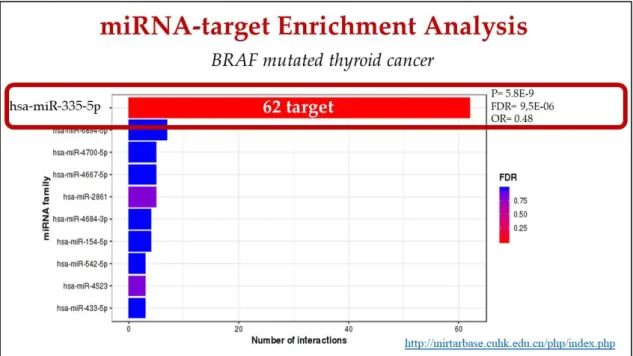
Exploiting Mienturnet to find a regulator of the 227 switch genes involved in BRAF carcinogenesis, miRNA 335-5p was identified as the one with the highest number of interactions. Indeed, about 30% (62 out of 227) switch genes were identified as regulated by miRNA 335-5p (figure 3, table 2).
Figure 3: miRNA-target enrichment analysis of switch genes involved in BRAF-mediated
thyroid cancer carcinogenesis, performed by Mienturnet.
SYT12 TNNI1 SERPIN SCN1B ABTB2 POU2F KRT19 SERINC DAPK2 GLS2 GRB7 MFGE8 TACSTD DHRS3 SLC34A PDE5A ACOT6 IYD ETV5 NR1D1 RNF157 CDH3 ABCC3 PRR15L PC ANO3 REN CELF4 SLCO4C CNFN TEKT4 S100A5 WDR17 HAPLN1 TMC6 PTPRE CATSPERB ST3GAL5 NUPR1 ASGR1 RDH5 VASN IL36RN PLEKHG4 SPTBN2 SYTL1 B3GNT3 NAB2 MXRA8 GDF15 PLCD3 PROS1 TPD52L1 DCSTAMP PDLIM4 B3GNT8 MYBPH IGF2BP2 UST SLC2A4 MYOT NPTXR
20
SECTION 3 – In vitro studies
Materials and methods
Expression levels of has-miRNA 335-5p were evaluated across several thyroid cell lines, both normal thyroid cell lines and thyroid disease cell lines (benign and tumoral disease).
Cell cultures: With regard to tumor cell lines, we made use of primary and immortalized thyroid cell lines. Primary tumour cell cultures were established, as previously described by our group 28, from the fresh-frozen specimens of patients with papillary thyroid cancer who underwent to surgical resection. Patients provided written informed consent, upon local ethics committee approval (Azienda Universitaria Policlinico Umberto I of Rome; project code Prot. 1184/17). Cell cultures were maintained in culture in specific condition 28-29.
Two commercial immortalized cells lines (8505c and K1; Gibco-BRL Division, Thermo Fisher Scientific, Waltham, Massachusetts, USA) were cultured in DMEM medium; the other ones (BCPAP and SW1736; Gibco-BRL Division, Thermo Fisher Scientific, Waltham, Massachusetts, USA) were cultured in RPMI media. Both of these media containe 10% FBS (Gibco-BRL Division, Thermo Fisher Scientific, Waltham, Massachusetts, USA) and Antibiotic–Antimycotic solution (Gibco-BRL Division, Thermo Fisher Scientific, Waltham, Massachusetts, USA). All cells were incubated at 37°C in an atmosphere of 5% CO2. The gene mutational profile of cell lines used in the present study was reported in Landa et al 30. All cancer thyroid cell lines harbor BRAF mutation.
Characterization of primary tumour cell lines: Patient-derived thyroid cancer cell lines were analysed for the presence of BRAF V600E point mutation by Sanger sequencing. DNA from cell lines was isolated using Nucleic Acid and Protein extraction (Macherey-Naghel) and Sanger sequencing analyses of DNA was performed as previously described31.
21 Nucleic acid isolation from tissues: DNA and total RNA (containing miRNAs) were simultaneously extracted from DTC tissues using the All Prep DNA/RNA Micro kit (Qiagen) and quantified with Qubit fluorescence-based assays for dsDNA and RNA (Qubit®,Thermo Fisher Scientific).
Cell treatments: Synthetic miR-335-5p (MISSION® microRNA Mimic hsa-miR-335-5p, Sigma Aldrich) or negative control (MISSION® miRNA, Negative Control 1, Sigma Aldrich) were transfected into primary and immortalized thyroid cancer cells lines, at final concentration of 30 nM using Lipofectamine RNAiMAX Transfection Reagent (Thermo Fisher Scientific) for 48h. The control of miR-335-5p overexpression was confirmed using the TaqMan™ MicroRNA Assay in qRT-PCR (Thermo Fisher Scientific).
RNA isolation and quantitative RT-PCR: RNA was isolated from cells using RNeasy Kit (Qiagen, Hilden, Germany) and quantified with Nanodrop 2000 (Thermo Fisher Scientific). The High Capacity cDNA reverse transcription kit (Applied Biosystems Life Technologies, TermoFisher) was used to synthesize cDNA from RNA according to manufacturer’s instructions (Applied Biosystems). mRNA
expression was analysed on cDNAs using the 7900 Real-Time PCR System, TaqMan Universal Master Mix, TaqMan gene expression Assay-on-Demand according to the manufacturer’s instructions (Applied Biosystems). Results were calculated using the 2−ΔΔCt method, normalized to corresponding control samples and expressed as mean±S.D. of three replicates. mRNA expression was evaluated through TaqMan® Gene Expression Assays, using the following assays:
SLC5A5 (Hs00166567_m1), TG (Hs00174974_m1), PAX8 (Hs00247586_m1), TPO (Hs00892519_m1), TSHR (Hs01053846_m1) and GAPDH (Hs02786624_g1) as endogenous control. MiRNA expression was assessed with TaqMan™ MicroRNA Assay for the miR-335-5p (Code: 00546) and U6 (Code: 001973) as endogenous control.
Immunofluorescence: Immunofluorescence experiments were performed using Labtek chamber slides as support. Cells were fixed with 4% paraformaldehyde for 20 min at room temperature,
22 permeabilized with Triton X-100 (0.1%) diluted in phosphate buffered saline (PBS) and incubated in blocking solution (BSA 3% in PBS). Cells were incubated overnight with the primary antibody diluited 1:50 in BSA 3% (rabbit anti-NIS, polyclonal, Genteck) and for 1h with secondary antibodies (dilution 1:200). Secondary antibodies (488-conjugated anti-mouse and anti-rabbit) were purchased from Thermo Fisher Scientific. Nuclei were Hoechst-counterstained. Images were acquired with a Leica microscope using the filter cube GFP ET.
Iodide uptake assay: Iodide uptake assay was performed as described by Walts et al. 32 Cells were seeded in 96-well microplates the day one; the day after, the cells were transfected with MISSION® miRNA and control, as describe above. After 48h from transfection, iodide uptake assay was performed. The culture medium was replaced twice by the uptake buffer (Hanks’ Balanced Salt solution, HBSS, and Hepes at concentration 10 mM). After washing cycle, 80 l of fresh uptake
buffer remained in each well of 96-well plate. Immediately 10 l of NaI solution (at the concentration of 100 µM) was added in each well. The assay plate was left to stand at 20°C for 60 minutes, in the dark. At the end of incubation, buffer was discarded, and cells were washed once with ice-cold HBSS (Thermo Fisher Scientific). Then, cells were lysed with 100 µl of 0,1 M NaOH solution. The whole lysate was used for “Nonradioactive Iodide Assay kit” according to manufacturer’s instructions
(Bertin pharma). The intracellular iodide concentrations of samples were determined using linear regression of the standard curve. Results were expressed as specific units of iodide accumulation relative to control.
Results
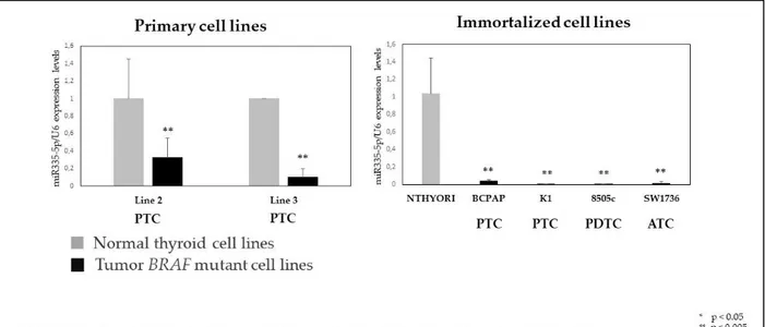
Hsa-miR-335-5p expression level in (both primary and immortalized) thyroid cancer cell lines, harboring BRAF V600E mutation, were significantly lower compared to their control (normal thyroid tissue) (figure 4).
23
Figure 4. Expression level of hsa-miR-335-5p in thyroid cell lines: normal versus tumor
cell lines harboring BRAF mutation.
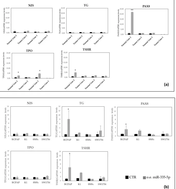
To verify if hsa-miR-335-5p acts on re-differentiation process of BRAF mutant thyroid cancer, modifying expression levels and/or function of thyroid specific genes (TG, TPO, NIS, PAX8, TSHR), we performed synthetic miR-335-5p re-expression through transfection.
After transient transfection with miR-335-5p in thyroid cell lines, its over-expression was controlled at transcriptional level in all selected cell lines. After 48 hours from transfection, the analysis of mRNA expression levels of thyroid-specific genes (NIS; thyroglobulin, TG; TSH receptor, TSH-R; TPO; PAX8) revealed that there was no difference in their expression in transfected cells versus controls. This result was similar in primary thyroid cell lines and in the immortalized ones (figure 5).
24
Figure 5: Expression levels of thyroid specific genes in primary thyroid cell lines (a) and
immortalized cell lines (b). CTR: control, o.e.:over-expression
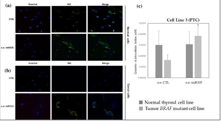
To further understand the role miR-335-5p on functional differentiation of thyroid cells, we performed immunofluorescence experiments and iodide uptake assay.
Immunofluorescence revealed anti-NIS staining in normal thyroid cell, as expected, and regardless of miR-335-5p overexpression (figure 6a). NIS appears at the plasma membrane level and in the
(a)
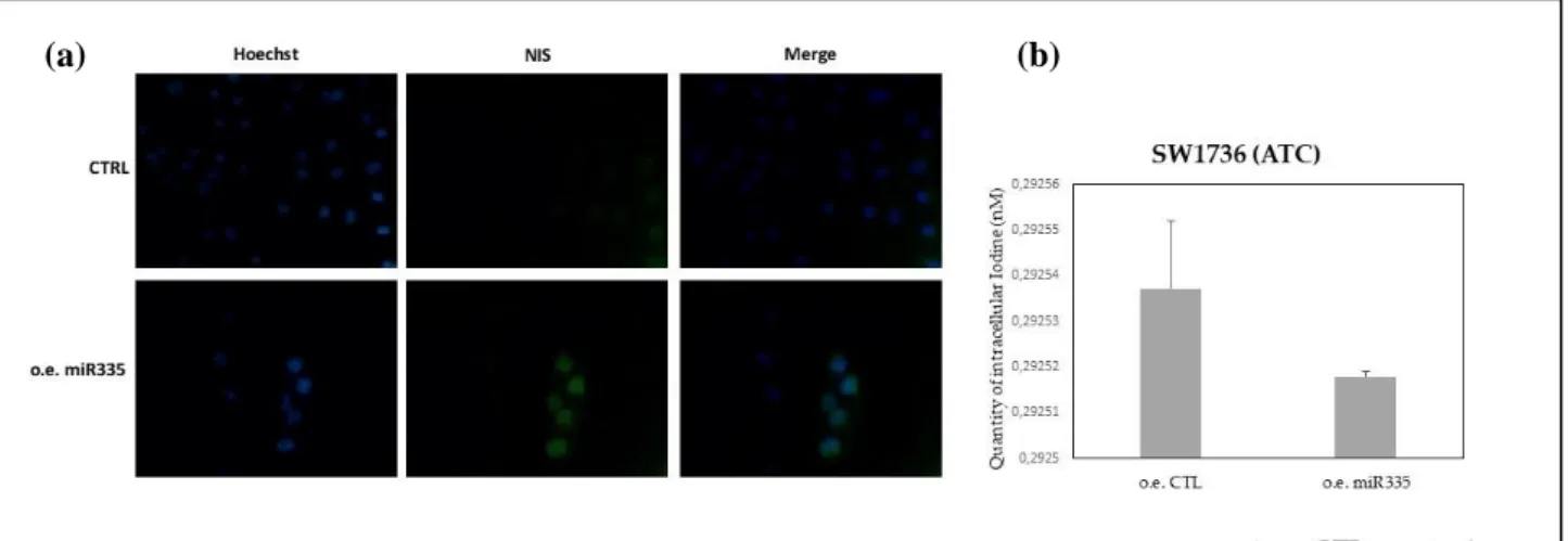
25 cytoplasm, that suggests the active and inactive form of NIS protein, respectively. NIS staining is absent in primary cell line 3 but it shows up after 48 hours from miR-335-5p transfection (figure 6b). A similar result was observed in K1 immortalized cell line (figure 7a). On the contrary, in less differentiated thyroid cell lines with a bulkier, thus more aggressive, mutational profile (8505c and SW1736), no substantial differences were observed between control cell lines and the ones over-expressing miR-335-5p (figures 7b, 8a).
Looking at quantity of intracellular iodide, evaluated by the uptake assay, in primary cell line 3 and K1, in line with the experiments of immunofluorescence, a statistically significant increase in intracellular iodide was observed after 48 hours from miR335-5p overexpression, compared to control (figures 6c, 7c). Iodide uptake was not significantly increased in poorly differentiated tumor lines (8505c and SW1736) (figures 7d, 8b).
Figure 6. (a) Immunofluorescence revealed anti-NIS staining in normal thyroid cell and regardless
of miR-335-5p overexpression; (b) NIS staining is absent in primary cell line 3 (ctrl: control) but it shows up after 48 hours from miR-335-5p transfection; (c) intracellular iodide, evaluated by the uptake assay, in primary tumor cell line 3 versus normal thyroid tissue, without (control) and after miR335-5p overexpression.
(a) (c)
26
Figure 7. (a-b) NIS staining by immunofluorescence in K1 (PTC: papillary thyroid cell) cell line and
8505c (PDTC: poorly differentiated thyroid cancer); (c-d) intracellular iodide, evaluated by the uptake assay, in these cell lines, after 48 hours from miR335-5p overexpression, compared to control.
Figure 8. (a) NIS staining by immunofluorescence in SW1736 cell line (ATC: anaplastic thyroid
cancer) in control ( CTRL) and after 48 hours from miR-335-5p transfection; (b) intracellular iodide, evaluated by the uptake assay, in SW1736 cell line without (control) and after miR335-5p overexpression. (a) (b) (c) (d) (a) (b)
27
DISCUSSION
Redifferentiation strategy with BRAFi and MEKi in metastatic RAIR DTC has been demonstrated to restore RAI uptake and result in objective response, both in pilot studies and retrospective experiences 18, 19, 21, 33. These studies showed an objective response to resensitization treatment (KI plus RAI) in a percentage ranging between 25 and 62% of patients who successfully completed the protocol schedule. These studies had small sample size and they did not report if the efficacy in redifferentiation therapy was associated with prolongation of TTP, PFS, time to start other KIs and overall survival. Our larger and retrospective experience with redifferentiation therapy followed by RAI administration confirms some previous finding and adds some data to the topic. We saw uptake, optimal or suboptimal, on post-Tx scan in 95% of patients included in the analysis. Ninety-four percent of BRAF mutated DTC patients had RAI uptake after redifferentiation therapy. For patients evaluable for radiological response after I-131, 73% of patients benefited from redifferentiation strategy, reaching stable or partial response for more than 12 months. TTP and PFS after redifferentiation therapy plus RAI were 20 and 29 months, respectively, with a median time off KI of 15 months. The results of this study should be interpreted with caution due to several limitations, including the retrospective nature of this study, the lack of control arm, the variability of the redifferentiation protocol, the different intent of starting KI therapy (not only for the purpose redifferentiation), and the timing of radiological evaluation. Development in the area of redifferentiation therapy is hampered by an absence of consensus regarding the definition of radioactive iodine-refractory differentiated thyroid cancer.
The investigation of mutational landscape of BRAF mutated DTC, allowed to identify a subset of switch genes, 30% of which is regulated by hsa-miR-335-5p.
A growing number of studies have been conducted to show that miR-335 is a significant cancer-associated miRNA. It has been demonstrated to be widely dysregulated in various human cancers
28
(breast cancer, hepatocellular cancer, colorectal cancer, meningioma and prostate cancer), playing
critical roles in tumorigenesis and cancer progression 34. The expression level of miR-335 is
upregulated or down-regulated depending on the type of cancer. MiR-335-5p role has been studied
in various biological process: tumorigenesis, proliferation, apoptosis, migration, metastasis, invasion34. It has also been studied as prognostic and diagnostic marker. Indeed, miR-335 expression levels have been correlated to clinical and pathological (TNM) stage, local recurrence, histologic grade, lymph node and distant metastases.
With regard to the issue of differentiation, studies on neuroblastoma showed that downregulation of miR-335 in non-neuronal cells (glial/smooth muscle precursor cells) modulates expression levels of HAND1 and JAG1, known modulators of neuronal differentiation35. Evidence suggests miR-335 maintains the non-neuronal features possibly by blocking neuronal differentiation. Other findings suggest that miR-335 is potently required for differentiation of malignant glioma cells induced by cAMP/PKA pathway activation, and it may act as an important fate determinant to control the
differentiation status of malignant gliomas36.
A few studies have explored miR-335-5p role in thyroid cancer. Previous experiences showed that the expression level of miR-335-5p in TPC-1 (human thyroid cancer cell line) was down-regulated, and miR-335-5p could inhibit the proliferation of thyroid cancer cells 37. MiR-335-5p has been demonstrated to be the most significant down-regulated miR comparing papillary thyroid cancer to adjacent non-tumor tissues38. A network-based integrative analysis of follicular thyroid carcinoma and benign follicular thyroid adenoma identified miR-335-5p as a molecular mechanism that differs between the two subtypes39.
So far, to the best of our knowledge, mir-335-5p role in dedifferentiation and redifferentiation process of thyroid cancer has not been investigated. We show for the first time that miRNA-335-5p is lost from normal thyroid tissue to primary or continuous BRAF mutated cell lines. Its over-expression in
29 thyroid tumor cell lines harboring BRAF mutation induces the activation of NIS protein and increases intra-cellular iodide uptake, supporting our hypothesis of miR-335 role in redifferentiation of BRAF mutated papillary thyroid cancer. This role seems to be more prominent in papillary thyroid cancer than in undifferentiated forms of thyroid cancer. The last result was predictable because the identification of miR-335-5p came from an analysis carried out in PTC from TCGA, where poorly differentiated and undifferentiated forms of cancer were not included. Further studies based on the evaluation of miR-335-5p levels in RAI-refractory thyroid cancer patients are needed to confirm that over-expression of this miRNA may have a role in restoring RAI responsiveness. At that point, a combinational approach, based on the inhibition of BRAF pathway and over-expression of miR-335-5p may be proposed to fight RAI refractoriness in thyroid cancer.
REFERENCES
1. Schlumberger M, Lacroix L, Russo D, Filetti S, Bidart JM. Defects in iodide metabolism in thyroid cancer and implications for the follow-up and treatment of patients. Nat Clin Pract Endocrinol Metab. Mar 2007;3(3):260-9. doi:10.1038/ncpendmet0449
2. Durante C, Haddy N, Baudin E, et al. Long-term outcome of 444 patients with distant metastases from papillary and follicular thyroid carcinoma: benefits and limits of radioiodine therapy. J Clin Endocrinol Metab. Aug 2006;91(8):2892-9. doi:10.1210/jc.2005-2838
3. Grüning T, Tiepolt C, Zöphel K, Bredow J, Kropp J, Franke WG. Retinoic acid for redifferentiation of thyroid cancer--does it hold its promise? Eur J Endocrinol. Apr 2003;148(4):395-402. doi:10.1530/eje.0.1480395
4. Sherman EJ, Su YB, Lyall A, et al. Evaluation of romidepsin for clinical activity and radioactive iodine reuptake in radioactive iodine-refractory thyroid carcinoma. Thyroid. May 2013;23(5):593-9. doi:10.1089/thy.2012.0393
5. Kebebew E, Lindsay S, Clark OH, Woeber KA, Hawkins R, Greenspan FS. Results of rosiglitazone therapy in patients with thyroglobulin-positive and radioiodine-negative advanced differentiated thyroid cancer. Thyroid. Sep 2009;19(9):953-6. doi:10.1089/thy.2008.0371
6. Hoftijzer H, Heemstra KA, Morreau H, et al. Beneficial effects of sorafenib on tumor progression, but not on radioiodine uptake, in patients with differentiated thyroid carcinoma. Eur J Endocrinol. Dec 2009;161(6):923-31. doi:10.1530/EJE-09-0702
7. Durante C, Puxeddu E, Ferretti E, et al. BRAF mutations in papillary thyroid carcinomas inhibit genes involved in iodine metabolism. J Clin Endocrinol Metab. Jul 2007;92(7):2840-3. doi:10.1210/jc.2006-2707 8. Liu D, Hu S, Hou P, Jiang D, Condouris S, Xing M. Suppression of BRAF/MEK/MAP kinase pathway restores expression of iodide-metabolizing genes in thyroid cells expressing the V600E BRAF mutant. Clin
Cancer Res. Feb 2007;13(4):1341-9. doi:10.1158/1078-0432.CCR-06-1753
9. Chakravarty D, Santos E, Ryder M, et al. Small-molecule MAPK inhibitors restore radioiodine incorporation in mouse thyroid cancers with conditional BRAF activation. J Clin Invest. Dec 2011;121(12):4700-11. doi:10.1172/JCI46382
10. Arturi F, Russo D, Bidart JM, Scarpelli D, Schlumberger M, Filetti S. Expression pattern of the pendrin and sodium/iodide symporter genes in human thyroid carcinoma cell lines and human thyroid tumors. Eur J
30 11. Trouttet-Masson S, Selmi-Ruby S, Bernier-Valentin F, et al. Evidence for transcriptional and posttranscriptional alterations of the sodium/iodide symporter expression in hypofunctioning benign and malignant thyroid tumors. Am J Pathol. Jul 2004;165(1):25-34. doi:10.1016/S0002-9440(10)63272-5
12. Tallini G, de Biase D, Durante C, et al. BRAF V600E and risk stratification of thyroid microcarcinoma: a multicenter pathological and clinical study. Mod Pathol. Oct 2015;28(10):1343-59. doi:10.1038/modpathol.2015.92
13. Chapman PB, Hauschild A, Robert C, et al. Improved survival with vemurafenib in melanoma with BRAF V600E mutation. N Engl J Med. Jun 2011;364(26):2507-16. doi:10.1056/NEJMoa1103782
14. Lu J, Getz G, Miska EA, et al. MicroRNA expression profiles classify human cancers. Nature. Jun 2005;435(7043):834-8. doi:10.1038/nature03702
15. Rosignolo F, Sponziello M, Giacomelli L, et al. Identification of Thyroid-Associated Serum microRNA Profiles and Their Potential Use in Thyroid Cancer Follow-Up. J Endocr Soc. Jan 2017;1(1):3-13. doi:10.1210/js.2016-1032
16. Haghpanah V, Fallah P, Tavakoli R, et al. Antisense-miR-21 enhances differentiation/apoptosis and reduces cancer stemness state on anaplastic thyroid cancer. Tumour Biol. Jan 2016;37(1):1299-308. doi:10.1007/s13277-015-3923-z
17. Li L, Lv B, Chen B, et al. Inhibition of miR-146b expression increases radioiodine-sensitivity in poorly differential thyroid carcinoma via positively regulating NIS expression. Biochem Biophys Res Commun. Jul 2015;462(4):314-21. doi:10.1016/j.bbrc.2015.04.134
18. Ho AL, Grewal RK, Leboeuf R, et al. Selumetinib-enhanced radioiodine uptake in advanced thyroid cancer. N Engl J Med. Feb 2013;368(7):623-32. doi:10.1056/NEJMoa1209288
19. Jaber T, Waguespack SG, Cabanillas ME, et al. Targeted Therapy in Advanced Thyroid Cancer to Resensitize Tumors to Radioactive Iodine. J Clin Endocrinol Metab. 10 2018;103(10):3698-3705. doi:10.1210/jc.2018-00612
20. Iravani A, Solomon B, Pattison DA, et al. Mitogen-activated protein kinase pathway inhibition for re-differentiation of radioiodine-refractory differentiated thyroid cancer: an evolving protocol. Thyroid. Oct 2019;doi:10.1089/thy.2019.0143
21. Dunn LA, Sherman EJ, Baxi SS, et al. Vemurafenib Redifferentiation of BRAF Mutant, RAI-Refractory Thyroid Cancers. J Clin Endocrinol Metab. May 2019;104(5):1417-1428. doi:10.1210/jc.2018-01478
22. Cabanillas ME, Dadu R, Iyer P, et al. Acquired Secondary RAS Mutation in BRAF. Thyroid. May 2020;doi:10.1089/thy.2019.0514
23. Falcone R, Conte F, Fiscon G, et al. BRAF. Endocrine. 05 2019;64(2):406-413. doi:10.1007/s12020-019-01890-4
24. Zhou Y, Hou Y, Shen J, Huang Y, Martin W, Cheng F. Network-based drug repurposing for novel coronavirus 2019-nCoV/SARS-CoV-2. Cell Discov. 2020;6:14. doi:10.1038/s41421-020-0153-3
25. Fiscon G, Conte F, Licursi V, Nasi S, Paci P. Computational identification of specific genes for glioblastoma stem-like cells identity. Sci Rep. 05 2018;8(1):7769. doi:10.1038/s41598-018-26081-5
26. Licursi V, Conte F, Fiscon G, Paci P. MIENTURNET: an interactive web tool for microRNA-target enrichment and network-based analysis. BMC Bioinformatics. Nov 2019;20(1):545. doi:10.1186/s12859-019-3105-x
27. Paci P, Colombo T, Fiscon G, Gurtner A, Pavesi G, Farina L. SWIM: a computational tool to unveiling crucial nodes in complex biological networks. Sci Rep. 03 2017;7:44797. doi:10.1038/srep44797
28. Pecce V, Sponziello M, Damante G, et al. A synonymous RET substitution enhances the oncogenic effect of an in-cis missense mutation by increasing constitutive splicing efficiency. PLoS Genet. 10 2018;14(10):e1007678. doi:10.1371/journal.pgen.1007678
29. Dima M, Pecce V, Biffoni M, et al. Molecular profiles of cancer stem-like cell populations in aggressive thyroid cancers. Endocrine. Jul 2016;53(1):145-56. doi:10.1007/s12020-015-0739-y
30. Schweppe RE, Klopper JP, Korch C, et al. Deoxyribonucleic acid profiling analysis of 40 human thyroid cancer cell lines reveals cross-contamination resulting in cell line redundancy and misidentification. J
Clin Endocrinol Metab. Nov 2008;93(11):4331-41. doi:10.1210/jc.2008-1102
31. D'Agostino M, Sponziello M, Puppin C, et al. Different expression of TSH receptor and NIS genes in thyroid cancer: role of epigenetics. J Mol Endocrinol. Apr 2014;52(2):121-31. doi:10.1530/JME-13-0160
31 32. Waltz F, Pillette L, Ambroise Y. A nonradioactive iodide uptake assay for sodium iodide symporter function. Anal Biochem. Jan 2010;396(1):91-5. doi:10.1016/j.ab.2009.08.038
33. Rothenberg SM, McFadden DG, Palmer EL, Daniels GH, Wirth LJ. Redifferentiation of iodine-refractory BRAF V600E-mutant metastatic papillary thyroid cancer with dabrafenib. Clin Cancer Res. Mar 2015;21(5):1028-35. doi:10.1158/1078-0432.CCR-14-2915
34. Luo LJ, Wang DD, Wang J, Yang F, Tang JH. Diverse roles of miR-335 in development and progression of cancers. Tumour Biol. Oct 2016;doi:10.1007/s13277-016-5385-3
35. Samaraweera L, Grandinetti KB, Huang R, Spengler BA, Ross RA. MicroRNAs define distinct human neuroblastoma cell phenotypes and regulate their differentiation and tumorigenicity. BMC Cancer. May 2014;14:309. doi:10.1186/1471-2407-14-309
36. Shu M, Zhou Y, Zhu W, et al. MicroRNA 335 is required for differentiation of malignant glioma cells induced by activation of cAMP/protein kinase A pathway. Mol Pharmacol. Mar 2012;81(3):292-8. doi:10.1124/mol.111.076166
37. Luo L, Xia L, Zha B, et al. miR-335-5p targeting ICAM-1 inhibits invasion and metastasis of thyroid cancer cells. Biomed Pharmacother. Oct 2018;106:983-990. doi:10.1016/j.biopha.2018.07.046
38. Zhang J, Liu Y, Liu Z, et al. Differential expression profiling and functional analysis of microRNAs through stage I-III papillary thyroid carcinoma. Int J Med Sci. 2013;10(5):585-92. doi:10.7150/ijms.5794 39. Hossain MA, Asa TA, Rahman MM, et al. Network-Based Genetic Profiling Reveals Cellular Pathway Differences Between Follicular Thyroid Carcinoma and Follicular Thyroid Adenoma. Int J Environ Res Public