Rhodanine derivatives as potent anti-HIV and
anti-HSV microbicides
Cristina Tintori1☯, Giulia Iovenitti1☯, Elisa Rita Ceresola2☯, Roberto Ferrarese2,
Claudio Zamperini1,3, Annalaura Brai1,3, Giulio Poli1, Elena Dreassi1, Valeria Cagno4,5, David Lembo4, Filippo Canducci2
*, Maurizio Botta1,3,6 *
1 Department of Biotechnology, Chemistry and Pharmacy, University of Siena, Siena, Italy, 2 Laboratory of Microbiology and Virology, Ospedale San Raffaele, Milan, Italy, 3 Lead Discovery Siena S.r.l., Castelnuovo Berardenga, Siena, Italy, 4 Laboratory of Molecular Virology and Antiviral Research. Department of Clinical and Biological Sciences, University of Torino, Orbassano, Torino, Italy, 5 Department of Molecular Microbiology, University of Geneva, Geneva, Switzerland, 6 Sbarro Institute for Cancer Research and Molecular Medicine, Center for Biotechnology, College of Science and Technology, Temple University, Philadelphia, PA, United States of America
☯These authors contributed equally to this work.
*[email protected](MB);[email protected](FC)
Abstract
Although highly active antiretroviral therapies (HAART) remarkably increased life expec-tancy of HIV positive people, the rate of novel HIV-1 infections worldwide still represent a major concern. In this context, pre-exposure prophylaxis (PrEP) approaches such as vagi-nal microbicide gels topically releasing antiretroviral drugs, showed to have a striking impact in limiting HIV-1 spread. Nevertheless, the co-presence of other genital infections, particu-larly those due to HSV-1 or 2, constitute a serious drawback that strongly limits the efficacy of PrEP approaches. For this reason, combinations of different compounds with mixed anti-viral and antiretroanti-viral activity are thoroughly investigated Here we report the synthesis and the biological evaluation of a novel series of rhodanine derivatives, which showed to inhibit both HIV-1 and HSV-1/2 replication at nanomolar concentration, and were found to be active also on acyclovir resistant HSV-2 strains. The compounds showed a considerable reduction of activity in presence of serum due to a high binding to serum albumin, as determined through in vitro ADME evaluations. However, the most promising compound of the series maintained a considerable activity in gel formulation, with an EC50comparable to that obtained for the reference drug tenofovir. Moreover, the series of compounds showed phar-macokinetic properties suitable for topical formulation, thus suggesting that the novel rhoda-nine derivatives could represent effective agents to be used as dual anti HIV/HSV
microbicides in PrEP approaches.
Introduction
Implementation of extensive patients’ treatment and prevention strategies in the global response against human immunodeficiency virus type 1 (HIV-1) epidemic has resulted in
a1111111111 a1111111111 a1111111111 a1111111111 a1111111111 OPEN ACCESS
Citation: Tintori C, Iovenitti G, Ceresola ER,
Ferrarese R, Zamperini C, Brai A, et al. (2018) Rhodanine derivatives as potent HIV and anti-HSV microbicides. PLoS ONE 13(6): e0198478.
https://doi.org/10.1371/journal.pone.0198478
Editor: Giovanni Maga, Istituto di Genetica
Molecolare, ITALY
Received: March 14, 2018 Accepted: May 18, 2018 Published: June 5, 2018
Copyright:© 2018 Tintori et al. This is an open access article distributed under the terms of the
Creative Commons Attribution License, which permits unrestricted use, distribution, and reproduction in any medium, provided the original author and source are credited.
Data Availability Statement: All relevant data are
within the paper and its Supporting Information file.
Funding: This work was supported by the
European Union collaborative project “CHAARM” (grant no. HEALTH-F3–2009–242135).https:// cordis.europa.eu/project/rcn/93644_en.html. Lead Discovery Siena srl provided support in the form of research materials but did not have any additional role in the study design, data collection and analysis, decision to publish, or preparation of the manuscript.
significant reduction of new infections worldwide. Nevertheless, sexually transmitted infec-tions (STIs), including HIV-1, represent a major global burden, particularly in resource lim-ited regions [1]. Moreover, since 2010, no significant reduction of novel HIV-1 infections was recoded in most countries, suggesting that current efforts, including the increase access to antiretroviral treatment are not sufficient to end the AIDS epidemic.
Microbicides delivering antiviral compounds in the vagina, in the rectum or in the oral cav-ity, were shown to reduce HIV-1 transmission and can thus be considered effective tools to implement the global response to HIV-1. In fact, the CAPRISA 004 trial showed that 1% teno-fovir (TFV) microbicide vaginal gel used as pre-exposure prophylaxis (PrEP) approach showed a 39% reduction of infection risk, demonstrating that topically used drugs can have a huge impact in limiting HIV-1 spread [2–7]. However, efficacy of currently planned PrEP approaches may be hampered by the presence of circulating strains already resistant to avail-able antiretroviral agents (present in the PrEP preparations) or the co-presence of other genital infections that increase patient’s susceptibility to HIV-1 infection [8–11]. This is the case of genital infections caused by herpes simplex virus type 2 (and type-1 in fraction of genital infec-tions). In some regions, especially in those with the highest prevalence of HIV-1 infection, such as in many African countries, up to 80% of young adults are serum-positive for genital herpes infection, with a significant fraction of herpes virus recurrences [12–15]. All currently available PrEP approaches, including vaginal gels containing antiretroviral or intravaginal rings with long-acting antiretroviral drugs or implantable and injectable formulations, selec-tively inhibit HIV-1 replication but have no or very limited activity against HSV-1 or 2 [16] thus their efficacy may be limited or not sufficient to prevent new infections especially in high prevalence settings for both viral infections. In our previous papers [17–19], we demonstrated that these molecules block very early step in HIV-1 replication, preventing viral entry into cells. This may be relevant to increase bioavailability of antiretroviral agents in topical prepara-tions. In fact, almost all currently available PrEP approaches target intracellular steps of virus replication, suggesting that available drugs will have to penetrate both the mucosal barrier and the target cell membranes in order to be effective when used in topical preparations. During the screening of ourin house library against a panel of sexually transmitted viruses, we
surpris-ingly found that compounds 1 and 2 (Fig 1) [17–19], potently inhibited the replication of both HSV-1 and HSV-2, without any antiviral activity on HPV-16, a not enveloped virus particu-larly relevant in sexually transmitted infections. As a continuation of our previous work, a novel series of rhodanine derivatives was synthesized. Biological evaluation showed that these compounds are able to inhibit HIV-1 and HSV-1 and 2 replication at nanomolar concentra-tion. We also characterized the binding affinity of these molecules to human albumin and their efficacy as antiviral agents in prototypical genital PrEP gel preparations. Furthermore, a preliminary ADME evaluation was performed by determining water solubility, passive mem-brane permeability and metabolic stability of selected derivatives.
Results and discussion
Chemistry
As shown inFig 2, the synthetic route to the final compounds 9a-f first entailed the prepara-tion of the target acid 6. Methyl 4-(5-formylfuran-2-yl)-2-hydroxybenzoate (5) was obtained through Suzuki reaction between commercially available 3 (methyl 4-iodosalicylate) and com-pound 4 (5-formyl-2-furanylboronic acid), subsequent basic hydrolysis furnished the acid ana-logue (6). Derivatives 9a-f, were synthesized using a recently published [18] multicomponent protocol that includes nucleophilic displacement between the opportune amine and
Competing interests: The commercial affiliation to
Lead Discovery Siena srl does not alter our adherence to PLOS ONE policies on sharing data and materials.
trithiocarbonate (7) to form the substituted rhodanine intermediates (8a-f), followed by Knoe-venagel condensation with aldehyde (6) catalysed by an excess of primary amine (Fig 2).
Antiviral activity and cytotoxicity on TZM-bl cell line infected with HIV-1
laboratory strains
The synthesized compounds (Fig 3) were testedin vitro, together with the parent compounds 1 and 2, to evaluate their ability to inhibit HIV replication on human TZM-bl cells infected
with HIV-1 NL4.3 (CXCR4-tropic strain) or AD8 (CCR5-tropic strain). All compounds showed antiviral activity at nanomolar concentrations with the parent compound 2 displaying the best inhibitory profile, having an EC50of 6.9 nM and 4 nM on AD8 and NL4.3 HIV strains,
respectively. Compound 9a showed a comparable inhibitory effect (7.5 nM and 5.4 nM on AD8 and NL4.3 HIV strains, respectively) but a worst safety profile with respect to the other compounds. Indeed, almost all compounds of the series showed low cytotoxicity in TZM-bl cell line, with CC50values higher than 20μM. Only for compound 9a and 9d lower CC50
val-ues were found (2.2μM and 12.3 μM, respectively). Interestingly, all compounds displayed a reduction of antiviral activity when pre-incubation with virus was performed in presence of complete medium containing fetal bovine serum (FBS). Particularly, the activity of compound
2 against AD8 strain decreased 90 times in presence of serum, shifting from 6.9 nM to 650
nM. Also compound 1 showed a high loss of activity against NL4.3 strain, with an EC50that
moved from around 50 nM to 3μM, corresponding to a 64-fold activity reduction.
Fig 1. 2D structures of HIV-1 inhibitors previously published.
https://doi.org/10.1371/journal.pone.0198478.g001
Fig 2. Synthesis of aldehyde 6 and derivatives 9a-f. Synthesis scheme of aldehyde 6:i) Pd(PPh3)2Cl2, Na2CO3, DMF/EtOH, RT, 1h; ii) 1N NaOH
(aq), MeOH/THF, reflux 2h. Synthesis scheme of derivatives 9a-f:iii) DME, Et3N, MW (300 W), 90˚C, 10 min.iv) aldehyde 6, MW (300 W), 110˚C,
5 min.
Compounds 9b-f showed moderate to low loss of activity, in the range of 5 to 38 times. Com-mercially available drugs such as maraviroc [20] and raltegravir [21] did not show any alter-ation of antiviral activity when pre-incubated with serum.
Antiviral activity and cytotoxicity on human CD4
+lymphocytes
The antiviral activity and cytotoxicity of the most promising compound (2) was evaluated also on freshly purified human CD4+cells. CD4+T lymphocytes were infected with the two refer-ence laboratory wild type strains NL4.3 and AD8, as well as with two recombinant viruses bearing mutations of specific integrase residues (Q148H, G140S and Y143R) that confer resis-tance to raltegravir (RAL). As reported inTable 1, compound 2 was found to be active at low nanomolar concentrations (11–13 nM) on both NL4.3 and AD8 HIV strains also in CD4+ human lymphocytes, while showing a CC50of 810 nM and thus a selectivity index of 62.
Inter-estingly, in contrast to raltegravir, compound 2 maintained similar EC50values also on
inte-grase mutating recombinant viruses, thus suggesting that the strong antiviral activity of the series of rhodanine derivatives could not be ascribed to integrase inhibition [18].
Antiviral activity and cytotoxicity on Vero cell line infected with HSV-2
laboratory strain and HeLa infected with HPV-16 PsVs
The rhodanine derivatives were also testedin vitro to evaluate their antiviral activity toward
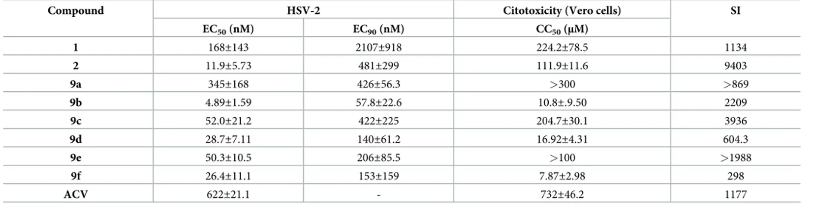
HSV-2 on Vero cells and HPV-16 in HeLa cells. No antiviral activity was shown for all the compounds up to 50μM for HPV-16. Instead, the compounds were subjected to a plaque reduction assay against HSV-2, in which they were pre-incubated with the virus for 1h at 37˚C; then the mixtures were added on cells. As shown inTable 2, all the compounds were found to be active at nanomolar concentration and showed EC50values lower than that
obtained for acyclovir, which was used as reference compound. Particularly, the tested rhoda-nines proved to be up to 130-fold more active than acyclovir (compound 9b) while presenting a very good safety profile, with selectivity indexes ranging from 600 (9d) to 9400 (2). Com-pound 2 showed the best HSV-2 antiviral profile, with an EC50in the low nanomolar range
(11.9 nM) and the highest selectivity index. Therefore, we further investigated the antiviral activity of this compound on HSV-1 and an acyclovir resistant strain of HSV-2.
Compound 2 was found to be about 10 times less active on HSV-1 with respect to HSV-2 (Table 3). Anyway, this rhodanine derivative showed an EC50(132 nM) slightly lower than
that obtained for acyclovir (168 nM) and maintained a good safety profile (SI > 752.7). More-over, compound 2 completely retained its antiviral activity toward the acyclovir resistant strain
Fig 3. Antiviral activity of the novel series of rhodanine derivatives on TZM-bl cell line infected with two laboratory strains (NL4.3 and AD8).
Maraviroc (MAR) and raltegavir (RAL) were used as reference compounds. Values represent mean±S.D of three independent experiments. Differences between pre-incubation in complete medium containing FBS are shown, together with the corresponding fold change (ratio). EC50= Half maximal effective
concentration. CC50= Half maximal cytotoxic concentration.apre-incubation in complete medium containing fetal bovine serum (FBS).
https://doi.org/10.1371/journal.pone.0198478.g003
Table 1. CC50and EC50values of the reference compound 2 and the control drug raltegravir (RAL) on human CD4+T lymphocytes.
Compound NL 4.3 WT EC50(nM) AD8 WT EC50(nM) Q148H, G140S EC50(nM) Y143R EC50(nM) CC50(nM) SI
2 13±3 11±1 9±2 8±3 810±20 62
RAL 3±2 2±3 800±12 460±10 n.a n.a
SI = Selectivity Index. EC50= Half maximal effective concentration. CC50= Half maximal cytotoxic concentration. Values represent mean±S.D of three independent
experiments.
of HSV-2, suggesting a different mechanism of action and highlighting the potential of this series of rhodanines for the treatment of HSV-2 infections resistant to acyclovir. However, as expected, compound 2 showed a remarkable reduction of HSV-2 inhibitory activity when pre-incubated with the virus in presence of serum.
Binding to bovine serum albumin in a cellular model
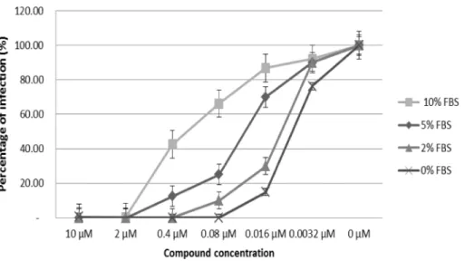
To better elucidate the mechanism underlying the loss of activity observed for the rhodanine compounds during incubation with complete medium, the selected compound 2 and the con-trol drug maraviroc were pre-incubated with the AD8 strain of HIV-1 in presence of concen-trations of FBS ranging from 0% to 10%. As shown inFig 4, the increase of FBS concentration from 2% to 10% corresponded to a progressive loss of activity, corroborating the inhibitory effect of serum on compound 2. Such alteration was not seen on maraviroc, whose antiviral efficacy did not change with the modification of FBS concentration (data not shown).
To evaluate if the inhibitory effect of serum on compound 2 was due to the binding to serum albumin, compound 2 and the reference drug maraviroc were pre-incubated with the virus and increasing concentrations of purified bovine serum albumin (BSA), starting from the physiological concentration present in the bovine serum up to ten-fold values. A physio-logical concentration of albumin altered only modestly the antiviral activity of compound 2 (20% loss), when tested at a concentration of 2μM (Fig 5). However, at a concentration of 400 nM the reduction of compound’s activity was found to be stronger (about 50%) and increasing
Table 2. Antiviral activity of the rhodanine derivatives on Vero cell line infected with HSV-2.
Compound HSV-2 Citotoxicity (Vero cells) SI
EC50(nM) EC90(nM) CC50(μM) 1 168±143 2107±918 224.2±78.5 1134 2 11.9±5.73 481±299 111.9±11.6 9403 9a 345±168 426±56.3 >300 >869 9b 4.89±1.59 57.8±22.6 10.8±.9.50 2209 9c 52.0±21.2 422±225 204.7±30.1 3936 9d 28.7±7.11 140±61.2 16.92±4.31 604.3 9e 50.3±10.5 206±85.5 >100 >1988 9f 26.4±11.1 153±159 7.87±2.98 298 ACV 622±21.1 - 732±46.2 1177
Acyclovir (ACV) was used as reference compound. Values represent mean±S.D of two independent experiments. EC50= Half maximal effective concentration. EC90=
90% maximal effective concentration. CC50= Half maximal cytotoxic concentration. The selectivity index (SI) was calculated by dividing the CC50by the EC50value.
https://doi.org/10.1371/journal.pone.0198478.t002
Table 3. Antiviral activity of compound 2 on Vero cell line infected with HSV-1, HSV-2 with serum and HSV-2 acyclovir resistant strains.
Compound Virus EC50(nM) Vero cell CC50(μM) SI
2 HSV-1 131±59.9 98±23.1 >752.7
ACV HSV-1 168±41.3 698±72.5 4155
2 HSV-2 with serum 719±157.7 n.d. n.d.
2 HSV-2 acyclovir resistant 10.7±6.34 111.9±11.6 >10,400
ACV HSV-2 acyclovir resistant 31915±1159 732±46.2 22.93
Acyclovir (ACV) was used as reference compound. Values represent mean±S.D of two independent experiments. EC50= Half maximal effective concentration. CC50=
Half maximal cytotoxic concentration. The selectivity index (SI) was calculated by dividing the CC50by the EC50value.
the BSA concentration of 5 to 10 folds led to a complete loss of activity. As expected, the antivi-ral activity of the control drug maraviroc was not influenced by any concentration of BSA.
Antiviral activity of compound 2 in a microbicide gel formulation
Considered the promising antiviral activity of compound 2 on both HIV-1 and HSV-1/2, as well as the negative effect of serum on its efficacy, we envisioned that a topical administration of the compound would have overcome the problem of albumin binding and represented a suitable strategy for a pre-exposure prophylaxis (PrEP) approach for the treatment of HIV infection. For this reason we evaluated the activity of compound 2 and the reference com-pound tenofovir in a microbicide gel formulation using a transwell experiment. The usage of
Fig 4. Effect of different concentrations of fetal bovine serum (0%, 2%, 5% and 10%) on the antiviral activity of compound 2. Compound 2 was tested in TZM-bl cells infected with AD8 HIV-1 laboratory strain.
https://doi.org/10.1371/journal.pone.0198478.g004
Fig 5. Effect of different concentrations of purified bovine serum albumin on the antiviral activity of compound 2 and maraviroc. Compounds were tested on TZM-bl cells infected with AD8 HIV-1 strain. No BSA = absence of BSA;
1X = 35 mg/mL BSA; 5X = 175 mg/mL BSA; 10X = 350 mg/mL BSA.
such experiment allowed to test the compounds in a gel formulation at a high concentration (50%) for all drug dilutions without affecting cell viability and antiretroviral activity evaluation. As shown inFig 6, compound 2 maintained its antiviral activity in the gel formulation, despite the increase in EC50observed in the transwell experiment (EC50of 13μM), which was however
less than 3-fold higher than that observed for tenofovir (EC50of 4.5μM).
In vitro ADME studies
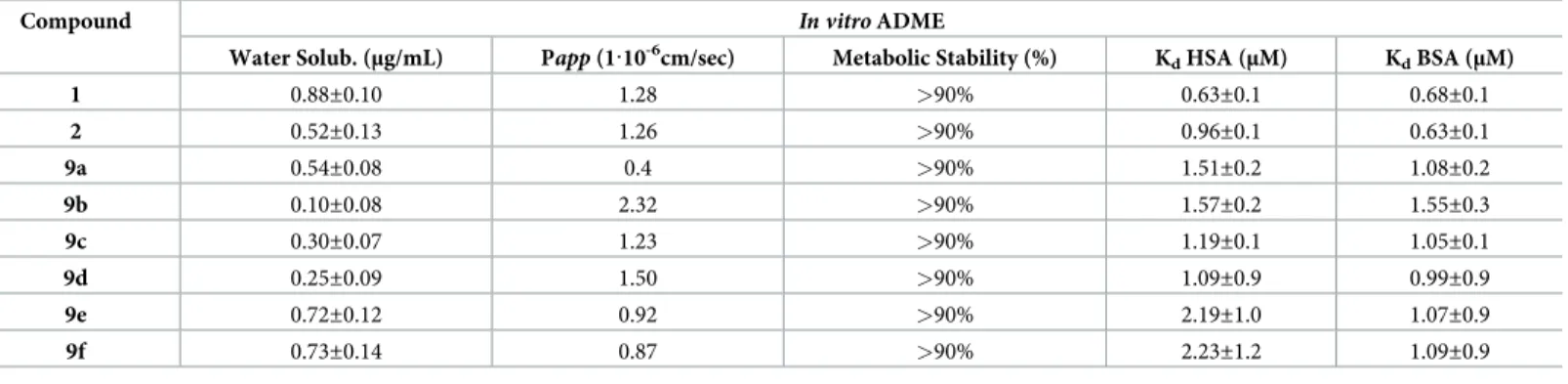
Solubility, permeability and metabolic stability. Selected compounds from the
rhoda-nine series were profiledin vitro for aqueous solubility (thermodynamics solubility), liver
microsomal stability and membrane permeability (Table 4). Although the aqueous solubility of the compounds (ranging from 0.1 to 0.88μg/mL) was found to be rather low, this aspect did not prevent the efficacy of the gel formulation according to the results obtain from solubility assessment (data not shown). In fact, the concentrations of the compounds in the pre-gel tions obtained using our experimental conditions were identical to those of the control solu-tion. In the same way, passive membrane permeability in a PAMPA assay indicated a low
Fig 6. Anti-HIV-1 activity of compound 2 (black line) and tenofovir (T, grey line) in gel, formulation, in human TZM-bl cell line. Each concentration of both compounds was evaluated in triplicate.
https://doi.org/10.1371/journal.pone.0198478.g006
Table 4. Results ofin vitro ADME analysis for selected rhodanine derivatives.
Compound In vitro ADME
Water Solub. (μg/mL) Papp (1·10-6cm/sec) Metabolic Stability (%) KdHSA (μM) KdBSA (μM)
1 0.88±0.10 1.28 >90% 0.63±0.1 0.68±0.1 2 0.52±0.13 1.26 >90% 0.96±0.1 0.63±0.1 9a 0.54±0.08 0.4 >90% 1.51±0.2 1.08±0.2 9b 0.10±0.08 2.32 >90% 1.57±0.2 1.55±0.3 9c 0.30±0.07 1.23 >90% 1.19±0.1 1.05±0.1 9d 0.25±0.09 1.50 >90% 1.09±0.9 0.99±0.9 9e 0.72±0.12 0.92 >90% 2.19±1.0 1.07±0.9 9f 0.73±0.14 0.87 >90% 2.23±1.2 1.09±0.9
Values represent mean±S.D of three independent experiments
membrane permeability value for all compounds (ranging from 0.4 to 2.32· 10−6cm/s). How-ever, this feature is considered to be drug-like, since it increases the time of exposure of the microbicide to the site of application, thus increasing its local activity. Moreover, stability tests disclosed that all compounds showed a good metabolic stability in human liver microsomes (>90%).
Albumin binding fluorimetric assay. The selected method was first validated with drugs
(paracetamol, diazepam and warfarin) having known Kdfor human serum albumin (HSA)
and experimental results were in good agreement with literature data [22] (S1 Table). Each compound analyzed by fluorimetric titration showed decreased intrinsic fluorescence of Tryp-tophan and the percentage of bound albumin (HAS and BSA) at different concentrations was calculated. The percentages obtained were plotted against the concentrations used and the obtained Kdvalues are reported inTable 4. Overall, all the compounds showed low Kdvalues,
demonstrating a high binding affinity to serum albumin. In fact, it is well known that com-pounds showing Kdvalues lower than 10μM are characterized by binding affinity to plasmatic
proteins greater than 90% [22].
Storage Stability of compound 2 Gel Formulation. The sample, at final concentration of
50μM, was found to be stable at 25˚C stored in the dark. Compound 2 recovery after 1 month of storage amounted to about 100%. In addition, the apparent viscosity and pH of this gel remained stable during storage (283.31± 8.21 and 7.41 ± 0.08 respectively).
Conclusions
Several studies confirmed the efficacy of topical PrEP approaches to limit the burden of novel HIV-1 infections and to enhance global efforts to eradicate this virus. However, several co-fac-tors may limit the efficacy of PrEP approaches. Patients’ compliance due to the need of fre-quent or fastidious topical applications was initially shown as a critical cause of protection failure [9,10]. Therefore, the usage of long-acting formulations or slow releasing devices signif-icantly ameliorated patients’ compliance and PrEP efficacy. Unfortunately, independently from the type of vehicle and release kinetic of current PrEP formulations, the presence of cir-culating strains already resistant to available antiretroviral agents (present in the PrEP prepara-tions) or the co-presence of other genital infections such as those due to HSV-1 or 2, which increase patient’s susceptibility to HIV-1 infection, remain unsolved problems [11]. During HSV-1 or 2 infections, the mucosal barrier damage and the increased number of inflammatory cells susceptible to HIV-1 infection facilitate HIV-1 transmission. Interestingly, despite the very limited anti HSV-2 activity of tenofovir [16], in the CAPRISA 004 study, TFV PrEP reduced occurrence of HSV-2 infection by 50%, possibly enhancing the overall observed effi-cacy against HIV-1. Thus, multipurpose combinations of different compounds with antiviral and antiretroviral activity are intensively investigated, sometimes also associated with unin-tended pregnancy prevention compounds [23–28]. Compounds acting on both viruses such as the rhodanine derivatives described in this paper, represent effective agents to be used in can-didate PrEP approaches without the need of dual-agents preparations. We demonstrated in our previous paper that these molecules block very early step in virus replication, preventing viral entry into cells. The inactivity against HPV-16, a not enveloped virus implicated in sexu-ally transmitted infections suggests that the viral envelope could be implicated in the mode of action of our compounds. Preliminary data in our possess corroborate our hypothesis demon-strating that our compounds act on specific components of the viral envelope. These results will be reported in a subsequent more specific manuscript. Compounds targeting efficiently the entry of both HIV-1 or HSV-1 and 2 may represent a very attractive strategy to prevent tar-get cell infection, with limited cell toxicity and reduced need of tissue penetration.
Moreover, PrEP devices undergoing clinical trials are based on already available antiretrovi-ral molecules or their derivatives, thus running the risk of being ineffective to prevent trans-mission of previously selected drug-resistant strains from the donor patients. In fact, as expected, mutations which confer resistance to TFV have been reported in oral TFV PrEP reg-imens [11]. Similar observations may arise in the future in the case of other reverse transcrip-tase or integrase inhibitors PrEP-based devices, in parallel with the increased usage of molecules within these drug classes (with cross-resistance profiles) in currently used highly active antiretroviral therapies (HAART) worldwide, including developing countries. Thus, molecules with novel mechanisms of action and targeting very early steps of virus life cycle may represent potent anti-HIV-1 agents able to block the formation of a viral reservoir of chronically infected cells.
The activity of such inhibitors decreased of several times (from 20 to 100) once they were pre-incubated with serum before thein vitro assay. To better elucidate this behavior, the
bind-ing affinities of the new derivatives to the Human and Bovine Serum Albumin (HSA/BSA) were determined by a fluorescence-spectroscopy measurement and the Kdvalues were
esti-mated. As a result, high affinity binding of the studied molecules to albumin was found, which could explain the loss of activity observedin vitro in the case of serum or purified recombinant
albumin pre-incubation. Remarkably, the salicylic acid group resulted to be crucial for both antiviral activity and albumin binding [18]. These pharmacokinetic characteristics, which will be better explored in the future to modulate a long-acting release or activity of this compound even for systemic antiretroviral treatments, could be extremely useful to reduce toxicity of these novel compounds when used in topical preparations. Indeed, high locally active drug concentrations may be reached (favored also by the drug-like features observed in the ADME experiments) at the mucosal interface, with a significantly reduced risk of systemic toxicity due to the rapid albumin binding and inactivation of the compounds.
Materials and methods
Chemistry
General information. All commercially available chemicals were used as purchased.
Anhydrous reactions were run under a positive pressure of dry N2. Thin-layer
chromatogra-phy (TLC) was carried out using Merck TLC plates: silica gel 60 F254. Chromatographic puri-fications were performed on columns packed with Merck 60 silica gel, 23–400 mesh, for the flash technique.1H and13C NMR spectra were recorded at 400 MHz on a Bruker Avance DPX400 spectrometer. Melting points were measured using a Gallenkamp melting point appa-ratus and are uncorrected. Microwave irradiation experiments were conducted using a CEM Discover Synthesis Unit (CEM Corp., Matthews, NC, USA). The instrument consists of a con-tinuous focused microwave power delivery system with operator-selectable power output from 0 to 300 W. The temperature of the contents of the vessel was monitored with a calibrated IR temperature control mounted under the reaction vessel. All experiments were performed using a stirring option, whereby the contents of the vessel are stirred by a rotating magnetic plate located below the floor of the microwave cavity and a teflon-coated magnetic stir bar in the vessel.
Methyl 4-(5-formylfuran-2-yl)-2-hydroxybenzoate (5). Methyl-4-iodosalycilate 3 (1.00
mmol) and 5-formyl-2- furan boronic acid 4 were dissolved in 10 mL of DMF and 15 mL of EtOH. The reaction mixture was stirred for 10 minutes under N2, then Pd(PPh3)2 Cl2(0.10
mmol) was added and finally Na2CO32M (6.00 mmol). The reaction mixture (light-orange)
was stirred under N2at room temperature. After 1h the reaction went to completion
mixture was stirred until the two layers became clear. The aqueous layer was extracted three times with EtOAc, then the organic phase were washed several times with H2O and brine,
dried over Na2SO4, filtered and evaporated under reduced pressure. The crude product was
purified by flash chromatography using PE/EtOAc = 4:1 as eluent to yield the wishes product
5 as a light orange solid (yield: 96%); mp = 150˚C (decomposition);1H NMR (CDCl3, 400 MHz):ð = 10.84 (s, 1H), 9.69 (s, 1H), 7.91–7.88 (d, 1H, J = 12 Hz), 7.40–7.39 (d, 1H, J = 4 Hz), 7.35–7.32 (m, 3H), 6.94–6.93 (d, 1H,J = 4Hz), 3.97 (s, 3H);13C (CDCl3, 100 MHz):ð = 177.47,
161.69, 157.37, 152.53, 135.17, 130.58,122.56, 115.74, 113.73, 112.82, 109.84, 52.40, 29.59; MS (ES): m/z 245.0 [M-H]-; Anal. (C13H10O5) C, H, N.
4-(5-Formylfuran-2-yl)-2-hydroxybenzoic acid (6). Compound 5 was dissolved in 25
mL of CH3OH, then a solution of NaOH 1M (5.00 mmol) was added dropwise, after the
reac-tion mixture was heated at reflux. The reacreac-tion mixture was stirred overnight until complereac-tion (TLC). Organic solvent was removed under reduced pressure, then some water was added, and the aqueous layer was extracted three times with Et2O; the aqueous layer was then
acidi-fied to pH 1 with HCl 6N and a precipitated appeared. (6) was obtained as a brown-red solid (yield: 95%); mp = 230˚C (decomposition);1H NMR (DMSO, 400 MHz):ð = 9.63 (s, 1H), 7.88–7.86 (d, 1H,J = 8 Hz), 7.66–7.65 (d, 1H, J = 4 Hz), 7.45–7.39 (m, 3H);13C NMR (DMSO, 100 MHz):ð = 178.72, 171.68, 161.69, 156.82, 152.63, 135.11, 131.72, 125.14, 116.02, 114.06, 113.29, 111.53; MS (ES): m/z 231.0 [M-H].
General Procedure for the synthesis of final compounds 9a-f. To a solution of
bis(car-boxymethyl)trithiocarbonate (0.22 mmol) in DME (1.0 mL) were added TEA (0.22 mmol) and the opportune amine (0.22 mmol). The reaction mixture was heated at 90˚C for 10 min under microwave irradiation. After this time, the aldehyde 6 (0.22 mmol) was added, and the mixture was heated at 110˚C for 5 min under microwave irradiation. The reaction mixture was evapo-rated to dryness and the residue was additioned with MeOH and a drop of HCl 2N; the final rhodanine derivatives were obtained as a pure precipitate, isolated by filtration, washed with water and hexane, and finally dried under high vacuum.
(Z)-4-(5-((3-(4-fluorophenethyl)-4-oxo-2-thioxothiazolidin-5-ylidene)methyl)furan-2-yl)-2-hydroxybenzoic acid (9a). (yield: 30%); Yellow solid. Mp = 292˚C (decomposition),
1 H NMR: (400 MHz, DMSO-d6) d = 7.92–7.90 (d, 1H,J = 8.0 Hz), 7.61 (s, 1H), 7.42–7.41 (d, 1H,J = 3.2 Hz), 7.38–7.33 (m, 3H),7.26–7.22 (m, 2H), 7.11–7.06 (m, 2H), 4.26.4.22 (m, 2H), 2.99–2.95 (m, 2H) ppm.13C NMR (100 MHz, DMSO-d6): d = 194.20, 171.72,166.65, 161.84, 156.71, 154.49, 150.43, 134.83, 134.20, 131.78, 131.00,130.91, 123.19, 119.91, 118.46, 116.16, 115.72, 115.51, 113.76, 113.04,112.41, 45.63, 31.68 ppm. MS (ES): m/z 468.0 [M-H]-. HPLC: tr = 4.58 min; conditions: temp = 25˚C, mobile phase composed of (A)70% acetonitrile and (B) 30% water with 0.5% formic acid at a flowrate of 1.0 mL/min; purity: 96.5%.
(Z)-2-hydroxy-4-(5-((3-(4-methylphenethyl)-4-oxo-2-thioxothiazolidin-5-ylidene) methyl)furan-2-yl)benzoic acid (9b). (yield: 79%); Orange solid. Mp = 258˚C
(decomposi-tion);1H NMR: (400 MHz DMSO-d6) d = 7.91–7.89 (d, 1H,J = 8.0 Hz), 7.60 (s, 1H), 7.42–7.33
(m, 4H), 7.10–7.08 (m, 4H), 4.21–4.18 (m, 2H), 2.92–2.88 (m, 2H), 2.25 (s, 3H) ppm. 13C NMR (100 MHz, DMSO-d6): d = 194.03, 178.56, 171.56, 166.68, 161.84, 156.79, 150.52, 135.99,
134.93, 131.77, 129.44, 128.88, 122.95, 120.15, 118.36, 115.56, 114.37, 113.86, 112.91, 112.52, 11.32, 45.70, 32.16, 20.96 ppm. MS (ES): m/z 464.0 [M-H]-. HPLC: tr = 4.65 min; conditions: temp = 25˚C, mobile phase composed of (A) 70% acetonitrile and (B) 30% water with 0.5% formic acid at a flow rate of 1.0 mL/min; purity:95.9%.
(Z)-2-hydroxy-4-(5-((4-oxo-3-(3-phenylpropyl)-2-thioxothiazolidin-5-ylidene)methyl) furan-2-yl)benzoic acid (9c). (yield: 91%); Orange solid. Mp = 248˚C (decomposition);
1HNMR: (400 MHz, DMSO-d6) d = 7.90–7.88 (d, 1H,J = 8.4 Hz), 7.61 (s,1H), 7.44–7.43 (d,
1.98–1.92 (m, 2H) ppm. 13C NMR (100 MHz, DMSO-d6): d = 194.23, 171.74, 166.91, 161.79,
156.60, 150.53, 141.20, 134.94, 131.77, 128.68, 128.55, 126.30, 122.99, 120.28, 118.28, 115.53, 113.49, 113.06, 112.39, 44.39, 32.72, 28.16 ppm. MS (ES): m/z 464.0 [M-1]-. HPLC: tr = 4.36 min; conditions: temp = 25˚C, mobile phase composed of (A) 70% acetonitrile and (B) 30% water with 0,5% formic acid at a flow rate of 1.0 mL/min; purity: 96.8%.
(Z)-4-(5-((3-(4-ethylphenethyl)-4-oxo-2-thioxothiazolidin-5-ylidene)methyl)furan-2-yl)-2-hydroxybenzoic acid (9d). (yield: 80%); Brown solid. Mp = 273˚C
(decomposi-tion);1H NMR: (400 MHz, DMSO-d6) d = 7.89–7.87 (d, 1H,J = 8.0 Hz), 7.58 (s, 1H), 7.42–7.41 (d, 1H,J = 3.6 Hz), 7.34–7.30 (m, 2H), 7.12–7.11 (m, 5H), 4.18–4.14 (m, 2H), 2.90–2.86 (m, 2H), 2.57–2.52 (q, 2H,J = 7.6 Hz), 1.15–1.11 (t, 3H, J = 7.6 Hz) ppm.13 C NMR (DMSO-d6100 MHz): d = 193.44, 171.74, 166.58, 161.80, 156.66, 150.42, 149.51, 142.40, 135.13, 134.85, 131.71, 128.92, 128.28, 123.10, 119.97, 118.35,115.49, 113.54, 113.01, 112.39, 45.69, 32.16, 28.16, 15.98 ppm. MS (ES): m/z 478.0 [M-H]-. HPLC: tr = 4.23 min; conditions: temp = 25˚C, mobile phase composed of (A) 70% acetonitrile and (B) 30% water with 0.5% formic acid at a flow rate of 1.0 mL/min; purity:96.5%.
(Z)-4-(5-((3-(3-chloro-4-fluorophenyl)-4-oxo-2-thioxothiazolidin-5-ylidene)methyl) furan-2-yl)-2-hydroxybenzoic acid (9e). (yield 30%) red solid,1H NMR (400 MHz,
DMSO-d6)δ11.32 (s, 1H), 7.89 (d, J = 8.1 Hz, 1H), 7.77 (d, J = 6.4 Hz, 1H), 7.68 (s, 1H), 7.62 (t, J = 8.9
Hz, 1H), 7.54–7.47 (m, 1H), 7.43 (d,J = 3.3 Hz, 1H), 7.36–7.32 (m,3H)ppm;13C NMR (DMSO-d6100 MHz):δ 198.0, 171.60, 167.62, 156.59, 155.80, 152.63, 152.07, 135.06, 134.53,
129.32, 126.76, 125.48, 124.06, 122.43, 120.60, 117.57, 115.92, 115.62, 111.62, 111.29, 103.95 ppm MS (ES): m/z 474.0 [M-H]-. HPLC: tr = 4.61 min; conditions: temp = 25˚C, mobile phase composed of (A) 70% acetonitrile and (B) 30% water with 0,5% formic acid at a flow rate of 1.0 mL/min; purity:97.1%.
(Z)-4-(5-((3-(4-chloro-3-fluorobenzyl)-4-oxo-2-thioxothiazolidin-5-ylidene)methyl) furan-2-yl)-2-hydroxybenzoic acid (9f). (yield 57%) brown solid,1H NMR (400 MHz, DMSO-d6)δ 11.28 (s, 1H), 7.85 (d, J = 8.0 Hz, 1H), 7.62 (s, 1H), 7.52 (t, J = 7.8 Hz, 1H), 7.39 (s,
1H), 7.36–7.24 (m, 3H), 7.15 (d,J = 8.0 Hz, 1H), 5.18 (s, 2H)ppm,13
C NMR (101 MHz, DMSO-d6)δ 204.26, 181.73, 176.87, 171.78, 168.65, 166.82, 165.98, 160.40, 146.86, 144.67,
141.68, 140.82, 135.19, 133.36, 129.92, 129.20, 128.86, 126.69, 126.19, 125.22, 123.56, 123.03, 122.07, 56.51ppm. MS (ES): m/z 488.0 [M-H]-. HPLC: tr = 4.28 min; conditions: temp = 25˚C, mobile phase composed of (A) 70% acetonitrile and (B) 30% water with 0.5% formic acid at a flow rate of 1.0 mL/min; purity:96.6%.
Antiviral activity and cell toxicity
Phenotypic analyses with fully replicating recombinant HIV-1 strains and clonal viral variants selected in patients failing integrase inhibitors on TZM-bl cells and human CD4+ lymphocyte. The human TZM-bl indicator cell line (obtained from the NIH AIDS reagents
program, cat. Nr 8129 (www.aidsreagent.org) and maintained at 37˚C and 5% CO2in
Dulbec-co’s modified Eagle’s medium (DMEM) medium containing 10% fetal bovine serum, 50μg/ mL penicillin, and 50μg/mL streptomycin. The HIV-1 laboratory strains NL(AD8) (NIH AIDS reagents program, cat.nr 11346), NL4.3 (NIH AIDS reagents program, cat. Nr. 114) were titrated as previously reported. Briefly, serial 5-fold dilutions of each virus were made in quadruplicate wells in 96-well culture plates, in a total volume of 100μL of growth medium (DMEM), for a total of 8 dilution steps. Freshly trypsinized cells (20,000 cells in 100μL of growth medium containing 75μg/mL DEAE-dextran) were added to each well, and the plates were incubated at 37˚C in a humidified 5% CO2-95% air environment. After 48 h of
(CPRG) assay (Roche). Twenty thousand TZM-bl cells/well were seeded in 96-well plates in complete DMEM supplemented with 30μg/mL DEAE-dextran (Sigma-Aldrich). Three hun-dred times the 50% tissue culture infective dose (TCID50)/mL of each strain was pretreated for 1 h at 37˚C with six serial dilutions (20000 nM to 3.2 nM) of each compound and then added to the cells, as previously described [29–32]. Vehicle (0.1% dimethyl sulfoxide [DMSO])-treated cells served as a negative control. A CCR5 inhibitor (maraviroc) and an integrase inhibitor (ral-tegravir) were used as positive-control drugs. After 2 days, viral infection was quantified using a CPRG assay (Roche). The inhibitory curves were fitted by nonlinear regression, allowing for the calculation of the 50% inhibitory concentration (IC50) using the Prism software. To evaluate the
cell toxicity of the compounds, the metabolic XTT [2,3-bis-(2-methoxy-4-nitro-5-sulfophenyl)-2H-tetrazolium-5-carboxanilide] test (Sigma-Aldrich) was performed according to the
manu-facturer’s instructions.
The antiviral activity and cytotoxicity of compounds 2, 9e and 9f was tested also on freshly purified human CD4+T lymphocytes obtained from healthy blood donors (informed consent available) as described previously on HIV laboratory strain NL4.3 and AD8 [33,34].
HSV-1 and HSV-2 antiviral activity in vero cells. African green monkey fibroblastoid
kidney cells (Vero, ATCC CCL-81) were grown as monolayers in Eagle’s minimal essential medium (MEM) (Gibco/BRL, Gaithersburg, MD) supplemented with 10% heat inactivated fetal calf serum (FCS) and 1% antibiotic-antimycotic solution (Zell Shield, Minerva Biolabs GmbH, Berlin, Germany).
Clinical isolates of HSV-1 and HSV-2 were kindly provided by Prof. M. Pistello, University of Pisa, Italy. HSV-1 and HSV-2 strains were propagated and titrated by plaque assay on Vero cells. A HSV-2 strain with phenotypic resistance to acyclovir was generated by serial passage in the presence of increasing concentrations of acyclovir, as previously described [35].
Acyclovir was purchased from Sigma Aldrich (Milan, Italy).
The effect of the rhodanine derivatives on HSV infection was evaluated by plaque reduction assay. Vero cells were pre-plated 24 h in advance in 24-well plates at a density of 105 cells. Increasing concentrations of compounds were mixed with HSV-2 (MOI 0.001 pfu/cell) or HSV-2 acyclovir resistant (MOI 0.001) or HSV-1 (MOI 0.0005) or and incubated for 1 hour at 37˚C. The mixtures were subsequently added to the cells, which were then incubated at 37˚C for 2 h. The virus inoculum was then removed and the cells washed and overlaid with a medium containing 1.2% methylcellulose (Sigma). After further incubation at 37˚C for 24 h (HSV-2 and HSV-2 R Acy) or 48 h (HSV-1), cells were fixed and stained with 0.1% crystal vio-let in 20% ethanol and viral plaques counted. The effective concentration producing 50% reduction (EC50) and 90% reduction (EC90) in plaque formation was determined using Prism
software by comparing drug-treated with wells treated with medium and solvent. The selectiv-ity index (SI) was calculated by dividing the CC50by the EC50value.
All results are presented as the mean values from three independent experiments. The EC50
values for inhibition curves were calculated by regression analysis using the software Graph-Pad Prism (GraphGraph-Pad Software, San Diego, California, U.S.A.) by fitting a variable slope-sig-moidal dose–response curve. Acyclovir was purchased from Sigma Aldrich (Milan, Italy).
Vero cell viability. Cell viability was measured using the MTS
[3-(4,5-dimethylthiazol-2-yl)-5-(3-carboxymethoxyphenyl)-2-(4-sulfophenyl)-2H-tetrazolium] assay. Cell cultures were seeded in 96-well plates were incubated with different concentrations of compounds in triplicate under the same experimental conditions described for the antiviral assays. Cell viabil-ity was determined using the CellTiter 96 Proliferation Assay Kit (Promega, Madison, WI, USA) according to the manufacturer’s instructions. Absorbances were measured using a Microplate Reader (Model 680, BIORAD) at 490 nm. The effect on cell viability at different concentrations of the compound was expressed as a percentage, by comparing absorbances of
treated cells with those of cells incubated with culture medium and equal volumes of vehicle. The 50% cytotoxic concentrations (CC50) and 95% confidence intervals (CIs) were determined
using Prism software (Graph-Pad Software, San Diego, CA).
Human papilloma pseudovirus (HPV16 PsV) production
Plasmids and 293TT cells used for pseudovirus (PsV) production were kindly provided by John Schiller (National Cancer Institute, Bethesda, MD). 293TT cell line, derived from human embryonic kidney cells transformed with the simian virus 40 (SV40) large T antigen, was cul-tured in DMEM (Gibco-BRL, Gaithersburg, MD) supplemented with heat inactivated 10% FCS (Gibco- BRL), Glutamax-I 1% (Invitrogen, Carlsbad, CA) and nonessential amino acids 1% (Sigma Aldrich, Steinheim, Germany). HPV-16 PsVs were produced as described in Cagno et al 2015. 293TT cells were transfected with plasmid expressing the papillomavirus major and minor capsid proteins (L1 and L2, respectively), together with a reporter plasmid expressing GFP. Capsids were allowed to mature overnight in cell lysate; the clarified superna-tant was then loaded on top of a density gradient of 27 to 33 to 39% Optiprep at room tempera-ture for 3 h. The material was centrifuged at 28000 rpm for 16 h at 4˚C in an SW41.1 rotor (Beckman Coulter, Inc., Fullerton, CA) and then collected by bottom puncture of the tubes. Fractions were inspected for purity in 10% sodium dodecyl sulfate (SDS)–Tris–glycine gels, titrated on 293TT cells to test for infectivity by GFP detection, and then pooled and frozen at -80˚C until needed. The L1 protein content of PsV stocks was determined by comparison with bovine serum albumin standards in Coomassie-stained SDS-polyacrylamide gels.
HPV-16 inhibition assay in HeLa cells
HeLa, Human Adenocarcinoma cells purchased from ATCC CCL-2, were cultured with DMEM (Gibco-BRL, Gaithersburg, MD) supplemented with heat inactivated 10% FCS (Gibco- BRL), Glutamax-I 1% (Invitrogen, Carlsbad, CA). Compounds were serially diluted and incubated with HPV-16 (MOI 0.1) for 1 h at 37˚C. Then the mixture was added to 8E4 HeLa cells grown as monolayers in a 96-well plate. Three days post-infection, cells were observed with a confocal microscope Zeiss LSM 510 and positive cells were counted.
Inhibition of antiviral activity by fetal bovine serum. To evaluate the binding of
com-pounds to plasma proteins also in a cell based assay, the infection with the HIV laboratory strain AD8 was performed in absence or in presence of different concentrations of Fetal Bovine Serum (FBS): 2%, 5% and 10%. The representative compound 2 was pre-incubated at different concentrations ranging from 10μM to 3.2 nM with 300 TCID50 /mL of AD8 in DMEM supplemented with 50μg/mL penicillin, 50 μg/mL streptomycin, 30 μg/mL DEAE-dextran and FBS at the concentration described above. Maraviroc was used as control. After an hour the mixture was added to 40.000 TZM-bl cells/well plated in a 96-well plate and after two days the infection was quantified using a CPRG assay (Roche) as previously described.
Inhibition of antiviral activity by purified bovine serum albumin. To confirm the
bind-ing of compound 2 to serum albumin, infection was performed in presence of different con-centrations of purified bovine serum albumin (BSA, Sigma Aldrich) starting from the concentration that is usually present in FBS serum (35 mg/mL, 1X) and increasing to a five-fold concentration (175 mg/mL) up to a ten-five-fold concentration (350 mg/mL). In this experi-ment the pre-incubation between HIV and two selected concentrations of compound 2 and maraviroc (2000 nM and 400 nM) was performed in DMEM without FBS and supplemented with BSA at the concentrations described above. After one hour, the mixture was added to 40.000 TZM-bl cells/well plated in DMEM supplemented with 50μg/mL penicillin, 50 μg/mL
streptomycin, 30μg/mL DEAE-dextran and 10% FBS in a 96-well plate. After two days the infection was evaluated through a CPRG assay.
ADME assay
Chemicals and excipients. All solvents, L-α-phosphatidylcholine, hydroxyethylcellulose (HEC), and propionic acid were from reagents, were from Sigma-Aldrich Srl (Milan,Italy). Dodecane was purchased from Fluka (Milan, Italy). Pooled Male Donors 20 mg/mL HLM were from BD Gentest-Biosciences (San Jose, California). Milli-Q quality water (Millipore, Milford, MA, USA) was used. Hydrophobic filter plates (MultiScreen-IP, Clear Plates, 0.45μm diameter pore size), 96-well microplates, and 96-well UV-transparent microplates were obtained from Millipore (Bedford, MA, USA).
UV/HPLC-MS method. LC analyses were performed by Agilent 1100 LC/MSD VL
sys-tem (G1946C) (Agilent Technologies, Palo Alto, CA) constituted by a vacuum solvent degas-sing unit, a binary high-pressure gradient pump, an 1100 series UV detector and a 1100 MSD model VL benchtop mass spectrometer was used. The Agilent 1100 series mass spectra detec-tion (MSD) single-quadrupole instrument was equipped with the orthogonal spray API-ES (Agilent Technologies, Palo Alto, CA). Nitrogen was used as nebulizing and drying gas. The pressure of the nebulizing gas, the flow of the drying gas, the capillary voltage, the fragmentor voltage and the vaporization temperature were set at 40 psi, 9 L/min, 3000 V, 70 V and 350˚C, respectively. UV detection was monitored at 254 nm. The LC-ESI-MS determination was per-formed by operating the MSD in the negative ion mode. Spectra were acquired over the scan range m/z 50–1500 using a step size of 0.1 u. Chromatographic analysis was performed using a Kinetex EVO C18 100A column (150 x 4.6 mm, 5μm particle size) at room temperature. Anal-ysis was carried out using a gradient elution of acetonitrile (ACN) and an aqueous solution (HCOOH 0.1%v/v): t = 0min ACN 5%, t = 3min ACN 5%, t = 12min ACN 95%, t = 25 min ACN 95%. The analysis was performed at flow rate of 0.6 mL/min and injection volume was 20μL.
Aqueous solubility. Each solid compound (1 mg) was added to 1 mL of water. Each
sam-ple was mixed at 20˚C, in a shaker water bath for 24 h. The resulting suspension was filtered through a 0.45μm nylon filter (Acrodisc). The concentration of solubilized compounds weres determined by UV/LC-MS (performed in triplicate) by comparison with the appropriate cali-bration curve that was obtained from samples of the compound dissolved in methanol at dif-ferent concentrations.
Parallel Artificial Membrane Permeability Assay (PAMPA). Donor solution (0.5 mM)
was prepared by diluting 1 mM dimethylsulfoxide (DMSO) compound stock solution using phosphate buffer (pH 7.4, 0.025 M). Filters were coated with 5μL of a 1% (w/v) dodecane solu-tion of L-α-phosphatidylcholine. Donor solusolu-tion (150 μL) was added to each well of the filter plate. To each well of the acceptor plate were added 300μL of solution (50% DMSO in phos-phate buffer). All compounds were tested in three different plates on different days. The sand-wich was incubated for 5 h at room temperature under gentle shaking. After the incubation time, the plates were separated, and samples were taken from both receiver and donor sides and analyzed using UV/HPLC-MS gradient method above reported. Permeability (Papp) for PAMPA, were calculated according to the following equation, obtained from Wohnsland and Faller [36] and Sugano [37] equation with some modification in order to obtain permeability values in cm s-1,
Papp¼
VDVA
ðVDþVAÞAt
where VAis the volume in the acceptor well, VDis the volume in the donor well (cm3), A is the
“effective area” of the membrane (cm2), t is the incubation time (s) and r the ratio between drug concentration in the acceptor and equilibrium concentration of the drug in the total vol-ume (VD+VA). Drug concentration is estimated by using the peak area integration.
Metabolic stability in HLM (Human Liver Microsomes). The incubation mixture (total
volume of 500μL) was constituted by the following components: HLM (0.2 mg/mL), a NADPH regenerating system (NADPH 0.2 mM, NADPH+1 mM, D-glucose-6-phosphate 4 mM, 4 unit/mL glucose-6-phosphate dehydrogenase and MgCl248 mM), 50μM of each
com-pound in DMSO and phosphate buffer (pH 7.4, 25 mM, up to a final volume of 500μL). The mixture was incubated at 37˚C for 1 h. The reaction was cooled down and quenched with ace-tonitrile (1.0 mL). After centrifugation (4000 rpm, 10 min), the supernatant was taken, dried under nitrogen flow, suspended in 100μL of methanol and analyzed by UV/LC-MS to deter-mine the percentage of compound that was not metabolized.
Solubility assessment. The solubilizing capacity of formulation media was measured in
the presence of 2% v/v of DMSO. Stock solution of compound 2 and tenofovir, were added to obtain a final concentration of 50, 25 and 10μM in the final volume of 1 mL of buffer solution (pH 7.4 25 mM). After sonication, the samples were shacked in a shaker bath at room tempera-ture for 24 h to reach equilibrium conditions. Each pre-gel solution was analyzed before and after filtration by a 0.45-μm nylon filter (Acrodisc). The solubilized compound was deter-mined using LC-UV-MS method above reported.
Gel formulation. All the semi-solid formulations were prepared adding at the pre-gel
solution reported above hydroxyethyl cellulose (HEC) (1.8% w/v). In all samples, 0.2% v/v pro-pionic acid was added to aqueous solution in order to preserve form bacteria and mold con-tamination. HEC was dispersed in the pre-gel solution at 25˚C under magnetic stirring, overnight.
Storage stability and rheological characterization. Gel containing compound 2 was
stored at 25˚C in the dark. At predetermined data points, the formulations were visually inspected to evidence aggregate formations and the pH was measured. Rheological evaluations were performed using a rotational viscometer (Bohlin Model Visco 88, Bohlin Instruments, UK). Appropriate measuring spindles (C14 DIN) were used during viscosity measurements. Samples were loaded into the cup and allowed to equilibrate for 10 min at desired temperature (25± 0.5˚C) and apparent viscosity (mPa s) was determined using a shear rate of 552 s-1over a 1 month period.
Binding fluorimetric assay. The binding of each compounds to HSA (human serum
albu-min) and BSA (bovine serum albualbu-min) were monitored by fluorescence spectroscopy in order to determine the dissociation constant (Kd). A quantitative analysis of the potential interaction was performed by fluorimetric analysis using 96 multiwell plates: in each well a fixed concentra-tion of HSA or BSA (10μM in phosphate buffer 1 mM), was added with different amounts of tested compound (0.1μM to 500μM by stock solutions in DMSO). Plates were gently shacked and after allowing 30 minutes at room temperature for equilibration, after excitation at 295nm, spectra were recorded from 300 to 400 nm with a Perkin Elmer EnVision Multilabel Reader 2014 spectrofluorimeter, spectra were acquired with EnVision Manager ver.1.13 software. The obtained fluorescence quenching percentages were plotted against drug concentrations and the relative Kdvalues were obtained using GraphPad software (version 6.0).
Antiviral activity of compound 2 in a gel formulation
Antiviral activity of compound 2 on NL4.3 HIV strain was evaluated also in a gel formulation. To reproduce more physiological conditions, TZM-bl cells were seeded into transwells and the
gel was applied onto the cell monolayer at 50% fixed concentration with serial drug dilutions as previously described. Briefly, 4x10⁴TZM-bl cells were plated into each transwell apical chamber of a 24-well plate (pore size 3μm and diameter 6,5 mm, Corning) containing medium in the bottom plate and cultured overnight. For efficacy testing, 60 TCID50 of virus was added to each apical well in the presence of 100μL of compound 2 gel, tenofovir gel or blank gel at the final drug concentrations of 25μM, 10 μM and 5 μM. Inhibition of infection was deter-mined as previously based on deviations from blank gel.
Supporting information
S1 Table. Binding studies realized to validate the used fluorimetric assay.
(PDF)
Acknowledgments
Tenofovir was obtained through the NIH AIDS Reagent Program, Division of AIDS, NIAID, NIH. We thank Marika Tiberi for excellent technical assistance.
Author Contributions
Conceptualization: Cristina Tintori, Filippo Canducci, Maurizio Botta. Data curation: Elisa Rita Ceresola, Filippo Canducci.
Formal analysis: Roberto Ferrarese, Elena Dreassi.
Investigation: Giulia Iovenitti, Elisa Rita Ceresola, Roberto Ferrarese, Claudio Zamperini,
Annalaura Brai, Valeria Cagno, David Lembo.
Methodology: Giulia Iovenitti, Claudio Zamperini, Annalaura Brai, Elena Dreassi, Valeria
Cagno, David Lembo.
Project administration: Cristina Tintori, Maurizio Botta. Supervision: Maurizio Botta.
Visualization: Giulio Poli.
Writing – original draft: Giulio Poli, Filippo Canducci.
References
1. World Health Organization. Global health sector strategy on sexually transmitted infections 2016–2021. 2016. Available from:http://apps.who.int/iris/bitstream/10665/246296/1/WHO-RHR-16.09-eng.pdf
2. Cates W Jr. After CAPRISA 004: time to re-evaluate the HIV lexicon. Lancet. 2010; 376: 495–496.
https://doi.org/10.1016/S0140-6736(10)61200-7PMID:20709216
3. Cates W Jr, Kashuba AD. The North Carolina contribution to the CAPRISA 004 study: the Global Health Initiative in action. N C Med J. 2010; 71: 490–491. PMID:21473558
4. Karim QA, Kharsany AB, Frohlich JA, Baxter C, Yende N, Mansoor LE et al. Recruitment of high risk women for HIV prevention trials: baseline HIV prevalence and sexual behavior in the CAPRISA 004 tenofovir gel trial. Trials. 2011; 12: 67.https://doi.org/10.1186/1745-6215-12-67PMID:21385354
5. Valley-Omar Z, Sibeko S, Anderson J, Goodier S, Werner L, Arnev L, et al. CAPRISA 004 tenofovir microbicide trial: no impact of tenofovir gel on the HIV transmission bottleneck. J Infect Dis. 2012; 206: 35–40.https://doi.org/10.1093/infdis/jis305PMID:22551813
6. Redd AD, Mullis CE, Wendel SK, Sheward D, Martens C, Bruno D, et al. Limited HIV-1 superinfection in seroconverters from the CAPRISA 004 Microbicide Trial. J Clin Microbiol. 2014; 52: 844–848.https:// doi.org/10.1128/JCM.03143-13PMID:24371237
7. Kashuba AD, Gengiah TN, Werner L, Yang KH, White NR, Karim QA, et al. Genital Tenofovir Concen-trations Correlate With Protection Against HIV Infection in the CAPRISA 004 Trial: Importance of Adher-ence for Microbicide Effectiveness. J Acquir Immune Defic Syndr. 2015; 69: 264–269.https://doi.org/ 10.1097/QAI.0000000000000607PMID:26181703
8. Grobler A, Abdool Karim S. Declining adherence is a more likely explanation than frailty of the apparent decline in efficacy in the CAPRISA 004 trial. AIDS. 2012; 26: 2261; author reply 2262–2263.https://doi. org/10.1097/QAD.0b013e328355ce08PMID:23123519
9. Gengiah TN, Mansoor LE, Upfold M, Naidoo A, Yende-Zuma N, Kashuba AD, et al. Measuring adher-ence by visual inspection of returned empty gel applicators in the CAPRISA 004 microbicide trial. AIDS Behav. 2014; 18: 820–825.https://doi.org/10.1007/s10461-014-0749-4PMID:24623069
10. Mansoor LE, Abdool Karim Q, Yende-Zuma N, MacQueen KM, Baxter C, Madlala BT et al. Adherence in the CAPRISA 004 tenofovir gel microbicide trial. AIDS Behav. 2014; 18: 811–819.https://doi.org/10. 1007/s10461-014-0751-xPMID:24643315
11. Lehman DA, Baeten JM, McCoy CO, Weis JF, Peterson D, Mbara G, et al. Risk of drug resistance among persons acquiring HIV within a randomized clinical trial of single- or dual-agent preexposure pro-phylaxis. J Infect Dis. 2015; 211: 1211–1218.https://doi.org/10.1093/infdis/jiu677PMID:25587020
12. Wald A, Link K. Risk of human immunodeficiency virus infection in herpes simplex virus type 2-seropos-itive persons: a meta-analysis. J Infect Dis. 2002; 185: 45–52.https://doi.org/10.1086/338231PMID:
11756980
13. Renzi C, Douglas JM Jr, Foster M, Critchlow CW, Ashley-Morrow R, Buchbinder SP, et al. Herpes sim-plex virus type 2 infection as a risk factor for human immunodeficiency virus acquisition in men who have sex with men. J Infect Dis. 2003; 187: 19–25.https://doi.org/10.1086/345867PMID:12508142
14. Reynolds SJ, Risbud AR, Shepherd ME, Zenilman JM, Brookmeyer RS, Paranjape RS, et al. Recent herpes simplex virus type 2 infection and the risk of human immunodeficiency virus type 1 acquisition in India. J Infect Dis. 2003; 187: 1513–1521.https://doi.org/10.1086/368357PMID:12721931
15. Mugo N, Dadabhai SS, Bunnell R, Williamson J, Bennett E, Baya I, et al. Prevalence of herpes simplex virus type 2 infection, human immunodeficiency virus/herpes simplex virus type 2 coinfection, and asso-ciated risk factors in a national, population-based survey in Kenya. Sex Transm Dis. 2011; 38: 1059– 1066.https://doi.org/10.1097/OLQ.0b013e31822e60b6PMID:21992985
16. Tan D. Potential role of tenofovir vaginal gel for reduction of risk of herpes simplex virus in females. Int J Womens Health. 2012; 4: 341–350.https://doi.org/10.2147/IJWH.S27601PMID:22927765
17. Tintori C, Corradi V, Magnani M, Manetti F, Botta M. Targets looking for drugs: a multistep computa-tional protocol for the development of structure-based pharmacophores and their applications for hit dis-covery. J Chem Inf Model. 2008; 48: 2166–2179.https://doi.org/10.1021/ci800105pPMID:18942779
18. Rinaldi M, Tintori C, Franchi L, Vignaroli G, Innitzer A, Massa S, et al. A versatile and practical synthesis toward the development of novel HIV-1 integrase inhibitors. ChemMedChem. 2011; 6: 343–352.https:// doi.org/10.1002/cmdc.201000510PMID:21246739
19. Botta L, Maccari G, Calandro P, Tiberi M, Brai A, Zamperini C, et al. One drug for two targets: Biological evaluation of antiretroviral agents endowed with antiproliferative activity. Bioorg Med Chem Lett. 2017; 27: 2502–2505.https://doi.org/10.1016/j.bmcl.2017.03.097PMID:28408224
20. Woollard SM, Kanmogne GD. Maraviroc: a review of its use in HIV infection and beyond. Drug Des Devel Ther. 2015; 9: 5447–5468.https://doi.org/10.2147/DDDT.S90580PMID:26491256
21. Temesgen Z, Siraj DS. Raltegravir: first in class HIV integrase inhibitor. Ther Clin Risk Manag. 2008; 4: 493–500. PMID:18728839
22. Parikh HH, McElwain K, Balasubramanian V, Leung W, Wong D, Morris ME, et al. A rapid spectrofluori-metric technique for determining drug-serum protein binding suitable for high-throughput screening. Pharm Res. 2000; 17: 632–637. PMID:10888318
23. Friend DR. Drug delivery in multiple indication (multipurpose) prevention technologies: systems to pre-vent HIV-1 transmission and unintended pregnancies or HSV-2 transmission. Expert Opin Drug Deliv. 2012; 9: 417–427.https://doi.org/10.1517/17425247.2012.668183PMID:22385316
24. Fernandez-Romero JA, Deal C, Herold BC, Schiller J, Patton D, Zydowsky T, et al. Multipurpose pre-vention technologies: the future of HIV and STI protection. Trends Microbiol. 2015; 23: 429–436.https:// doi.org/10.1016/j.tim.2015.02.006PMID:25759332
25. Friend DR. An update on multipurpose prevention technologies for the prevention of HIV transmission and pregnancy. Expert Opin Drug Deliv. 2016; 13: 533–545.https://doi.org/10.1517/17425247.2016. 1134485PMID:26742698
26. Hynes JS, Sales JM, Sheth AN, Lathrop E, Haddad LB. Interest in multipurpose prevention technolo-gies to prevent HIV/STIs and unintended pregnancy among young women in the United States. Contra-ception. 2018; 97: 277–278.https://doi.org/10.1016/j.contraception.2017.10.006PMID:29055782
27. Quaife M, Terris-Prestholt F, Vickerman P. The promise of multipurpose pregnancy, STI, and HIV pre-vention. Lancet Infect Dis. 2017; 17: 21–22.
28. Smith JM, Moss JA, Srinivasan P, Butkyavichene I, Gunawardana M, Fanter R, et al. Novel multipur-pose pod-intravaginal ring for the prevention of HIV, HSV, and unintended pregnancy: Pharmacokinetic evaluation in a macaque model. PLoS One. 2017; 12: e0185946.https://doi.org/10.1371/journal.pone. 0185946PMID:28982161
29. Tiberi M, Tintori C, Ceresola ER, Fazi R, Zamperini C, Calandro P, et al. 2-Aminothiazolones as anti-HIV agents that act as gp120-CD4 inhibitors. Antimicrob Agents Chemother. 2014; 58: 3043–3052.
https://doi.org/10.1128/AAC.02739-13PMID:24614386
30. Esposito F, Tintori C, Martini R, Christ F, Debyser Z, Ferrarese R, et al. Kuwanon-L as a New Allosteric HIV-1 Integrase Inhibitor: Molecular Modeling and Biological Evaluation. Chembiochem. 2015; 16: 2507–2512.https://doi.org/10.1002/cbic.201500385PMID:26360521
31. Tintori C, Corona A, Esposito F, Brai A, Grandi N, Ceresola ER, et al. Inhibition of HIV-1 Reverse Tran-scriptase Dimerization by Small Molecules. Chembiochem. 2016; 17: 683–688.https://doi.org/10.1002/ cbic.201500668PMID:26946324
32. Martini R, Esposito F, Corona A, Ferrarese R, Ceresola ER, Visconti L, et al. Natural Product Kuwanon-L Inhibits HIV-1 Replication through Multiple Target Binding. Chembiochem. 2017; 18: 374–377.https:// doi.org/10.1002/cbic.201600592PMID:27992102
33. Canducci F, Ceresola ER, Boeri E, Spagnuolo V, Cossarini F, Castagna A, et al. Cross-resistance pro-file of the novel integrase inhibitor Dolutegravir (S/GSK1349572) using clonal viral variants selected in patients failing raltegravir. J Infect Dis. 2011; 204: 1811–1815.https://doi.org/10.1093/infdis/jir636
PMID:21984737
34. Canducci F, Ceresola ER, Saita D, Castagna A, Gianotti N, Underwood M, et al. In vitro phenotypes to elvitegravir and dolutegravir in primary macrophages and lymphocytes of clonal recombinant viral vari-ants selected in patients failing raltegravir. J Antimicrob Chemother. 2013; 68: 2525–2532.https://doi. org/10.1093/jac/dkt220PMID:23798668
35. Donalisio M, Nana HM, Ngane RA, Gatsing D, Tchinda AT, Rovito R, et al. In vitro anti-Herpes simplex virus activity of crude extract of the roots of Nauclea latifolia Smith (Rubiaceae). BMC Complement Altern Med. 2013; 13:266.https://doi.org/10.1186/1472-6882-13-266PMID:24131916
36. Wohnsland F, Faller B. High-throughput permeability pH profile and high-throughput alkane/water log P with artificial membranes. J Med Chem. 2001; 44: 923–930. PMID:11300874
37. Sugano K, Hamada H, Machida M, Ushio H. High throughput prediction of oral absorption: improvement of the composition of the lipid solution used in parallel artificial membrane permeation assay. J Biomol Screen. 2001; 6: 189–196.https://doi.org/10.1177/108705710100600309PMID:11689115