1 UNIVERSITÀ DEGLI STUDI DELLA TUSCIA DI VITERBO
Dipartimento di Scienze Ecologiche e Biologiche
Corso di dottorato di ricerca in Evoluzione Biologica e Biochimica XXIV Ciclo
Resistance and survival of endolithic microorganisms in outer
space and Mars conditions simulated in space
Coordinatore: Professoressa Laura Zucconi Tutor: Professor Silvano Onofri
2 Indice
Prefazione 4
1. Introduction 5
1.1. What is astrobiology? 5
1.2. Life on Earth and beyond: there’s a limit of Biosphere? 6 1.2.1.Boundaries of Biosphere 7 1.3. The dawn of the livings: Astrobiological Exposure Facilities. 8 1.3.1. EXPOSE-E facility 10 1.4. Model Organisms selected for EXPOSE-E LIFE experiments 12 1.5. The aim: Planetary Protection issues 12
2. Survival of rock-colonizing organisms after 1.5 year in outer space. EXPOSE-E special issue, Astrobiology (in press) 14
2.1. Introduction 15
2.2. Material and Methods 17
2.3. Results 22
2.4. Discussion 29
3. LIFE Experiment: Isolation of cryptoendolithic organisms from Antarctic colonized sandstone exposed to space and simulated Mars conditions on the International Space Station. (2012) Origins of Life and Evolution of Biospheres
(accepted) 31
3.1. Introduction 32
3.2. Material and Methods 34
3.3. Results 39
3
4. Evaluation of Procedures for the Collection, Processing, and Analysis of Biomolecules from Low-Biomass Surfaces 2011 Applied and Environmental Microbiology
77(9):2943-53. 45
4.1. Introduction 46
4.2. Material and Methods 49
4.3. Results 58
4.4. Discussion 64
5. Discussion 68
4 Prefazione
Questa tesi si basa sull’analisi della sopravvivenza, e dell’eventuale danno subito, di due specie di funghi neri meristematici e di comunità criptoendolitiche antartici successivamente all’ esposizione per un anno e mezzo a condizioni spaziali reali e marziane simulate all’esterno della Stazione Spaziale Internazionale (ISS).
A seguito dell’esperimento, l’interesse del candidato si è esteso a quei microrganismi che, essendo presenti nelle “Spacecraft Assembly Facilities” (SAF), potrebbero potenzialmente contaminare sistemi extraterrestri considerati “incontaminati” dall’intervento antropico. Per ognuna delle tematiche affrontate, i risultati elaborati e discussi sono stati inviati a delle riviste peer-reviewed con IF per la valutazione e la pubblicazione. Ciascuno dei capitoli da 2 a 4 è in ogni sua parte conforme al manoscritto inviato ad una rivista (capitolo 2 in press, capitolo 3 accepted e capitolo 4 published), ad eccezione della bibliografia, la quale rappresenta il capitolo finale della tesi. Infine, nel capitolo 5 è presentata una discussione generale delle tematiche affrontate e dei risultati ottenuti e la loro rilevanza ai fini degli obiettivi dello studio.
5
1. Introduction
1.1 What is Astrobiology?
With the development of space technology, astrobiology has been established as a scientific discipline. Classical biological research has concentrated on the only example of “life” so far known, i.e. life on Earth. In contrast, astrobiology extends the boundaries of biological investigations beyond the Earth, to other planets, comets, meteorites and space at large. To date, the overriding objective of “exobiological” research is to attain a better understanding of the principles leading to the emergence of life from inanimate matter, its evolution, and its distribution on Earth and throughout the Universe (Klein 1986). To reach this goal, astrobiological research has focused on the different steps of the evolutionary pathway through cosmic history that maybe related to the origin, evolution and distribution of life. Since it involves many disciplines, in this study emphasis will be laid to the results obtained through a biological approach.
The present thesis would in one hand discuss the bewildering resistance of microorganisms exposed to real space and Mars simulated conditions, and on the other hand clarify with a methodological approach, the new procedures to minimize the biological cross-contamination resulting from the exploration of the solar system.
6 1.2 Life on Earth and Beyond. There’s a limit of Biosphere?
Despite an ever-expanding understanding of the limits of life in Earth’s most extreme environments, little remains known about life’s resolve when removed from Earth and the all-encompassing biosphere that cradles it. For example, can life resist to a transfer between planets?
Based on the wide distribution of cosmic dust, the theory of Panspermia (Arrhenius, 1903, 1908) postulates that microscopic forms of life, e.g. spores, can be propagated in space driven by radiation pressure from the sun (Robertson. 1937), thereby seeding life from one planet to another. Since its formulation, panspermia has been subjected to several criticisms with arguments such as (1) it cannot be experimentally tested (Lederberg, 1960), (2) it shunts aside the question of the origin of life (Dose, 1986; Allamandola et al., 1989; Haynes, 1990), and (3) spores will not survive long-time exposure to the hostile environment of space, especially vacuum and radiation.
Richter (1865) and Arrhenius (1903)first proposed the panspermia theory, which speculates about the transfer of life between planets. Although panspermia still remains little more than an idea and there is no evidence that it has occurred, the various steps required for the transfer of organisms from one planet to another have been the focus of experimental testing (Cockell, 2008). A variety of recent discoveries have shed new light on the likelihood of viable transfer in space such as (i) the detection of meteorites, some of lunar and some of Martian origin; (ii) the detection of organics and the still highly debated supposition of microbial fossils in one of the Martian meteorite; (iii) the probability of small particles of diameters between 0.5 μm and 1 cm boulder-sized rocks reaching escape velocities by the impact of large comets or asteroids on a planet, e.g., on Earth (Melosh 1988) or Mars (Vickery and Meloish 1996); (iv) the ability of bacterial spores to survive to a certain extent the shock waves of such a simulated impact (Horneck et al., 2001); (v) the high UV resistance of microorganisms at the low temperatures of deep space, tested at temperatures down to 10 K; (vi) the reported survival of bacterial spores over millions of years, if enclosed in amber or salt stocks, or in space over periods extending up to 6 years (Horneck et al., 1994); (vii) the paleogeochemical evidence of a very early appearance of life on Earth in the form of metabolically advanced microbial prokaryotic ecosystems leaving not more than approximately 0.4 Ga for the evolution of life from the simple precursor molecules to the level of a prokaryotic, photoautotrophic cell; (viii) the biochemical evidence of a common ancestor for all life forms on Earth; (ix) the likelihood of artificial or directed transport by probes sent to other planets (Crick and Orgel, 1973). Viable
7
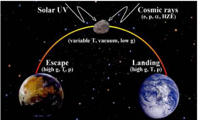
transfer from one planet to another requires that life, probably of microbial nature, survives the following three steps: (i) the escape process, i.e. ejection into space, e.g., caused by a large impact on the parent planet; (ii) the journey through space, i.e. time scales in space comparable with those experienced by the Martian meteorites (approximately 1-15 Ma); and (iii) the landing process, i.e. non-destructive deposition of the biological material on another planet (Fig. 1).
Fig. 1 Scenario of an interplanetary transfer of life in the solar system
Although it will be difficult to prove that life could be transported through our solar system, estimates of the chances for the different steps of the process to occur can be obtained from measurements, calculations, and experiments.
1.2.1 Boundaries of Biosphere
Distribution of air spora including their identity, behavior, movements and survival in the troposphere, as well as their impact on public health and agriculture are well understood (Horneck and Brack, 1992). They comprises viruses, bacteria, algae, microfungi, fungal spores of moss and fern, pollen, minute seeds, and protozoan cystis, up to the concentration of 100s-1000s per m3. It has been shown that spores as well as pigmented species are especially adapted to survive in inhospitable environment, characterized by low temperature, drought,
8
low pressure and solar radiation (Mancinelli and Shulls, 1978). With increasing height, the concentration of air spora rapidly falls off. In a few early experiments, using balloons or meteorological rockets with especially designed analysers, viable microorganisms were collected up to 77 km (Rogers and Meier, 1938; Imshenetsky et al., 1976, 1978, 1979). They
collected predominantly black conidia and spores of fungi. It is assumed that pigmentation
offers a selective advantage for the spores, because it protects them against the intense solar UV radiation. No viable microorganisms were collected from Earth orbit by use of collectors on Gemini 9A and 12. A pivotal role for the understanding of limits of life has been played by the progress of space technology.
1.3 The dawn of the livings: Astrobiological Exposure Facilities.
Space technology has provided opportunities to expose terrestrial specimens, such as prebiotic compounds, organic molecules and organisms, to this unique environment of space or to selected space conditions (Horneck and Brack 1992). Questions to be tackled include:
the chemistry of precursors of life in space, e.g. in the interstellar medium or in comets;
the role of the extraterrestrial short wavelength UV radiation, at present absorbed by the Earth’s ozone layer, on prebiotic and biological evolution;
the survival of microorganisms, when travelling in space, e.g. as blind passengers inside of meteorites (lithopanspermia hypothesis);
the survival of microbial contaminants on space craft on outbound missions to other planets (planetary protection requirements);
the limits of life.
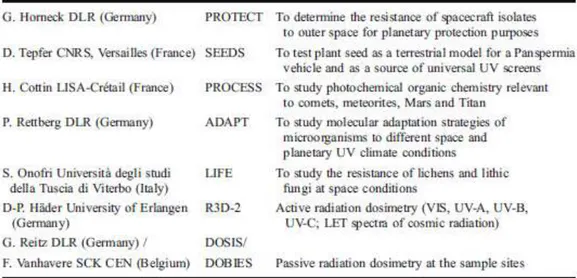
During previous space missions, such as Gemini, Apollo 16 (Taylor et al. 1974), Spacelab 1 (Horneck et al. 1984a and b), Spacelab D2 (Horneck et al. 1994a; 1996), LDEF (Long Duration Exposure Facility) (Horneck et al. 1994b), MIR (Rettberg et al. 2002), EURECA (EUropean REtrievable CArrier) (Horneck et al. 1995) and several Foton missions (Horneck et al. 2001, Rettberg et al. 2004, Sancho et al. 2007) (Table 1), exposure of various microorganisms to selected or combined space conditions demonstrated the lethal effects of extraterrestrial short wavelength solar UV radiation, but also the enormous resistance of
9
selected species against LEO vacuum when UV shielded (Horneck 1998). Recent experiments on board of the BIOPAN facility extended the list of organisms surviving exposure to space vacuum in LEO with lichens (Sancho et al. 2007) and tardigrades (Jönsson et al. 2008). Both are eukaryotic and multicellular. The macroscopic lichens even survived UV-C exposure during their two-week flight in LEO.
Except for the two free flying satellite missions LDEF and EURECA that were planned to stay in orbit for approximately 1 year and with LDEF accidentally remaining in space for nearly 6 years, exposure of most exobiological experiments in space did not last longer than approximately 2 weeks (Foton, Spacelab, Gemini). With the availability of the International Space Station (ISS), more extended exposure experiments have become possible. The European Space Agency has provided the exposure facility EXPOSE for astrobiology experiments in Low Earth Orbit (LEO).
This technical availability has led to several studies, under real space conditions (Buecker and Horneck, 1970; Mancinelli and Klovstad, 2000; De la Torre et al., 2003) in spaceflight experiments (Horneck, 1993; Fajardo-Cavalzos et al., 2005; Sancho et al., 2007; De los Rìos et al., 2010; Horneck et al., 2010). The one of the main objectives of these astrobiological experiments has been to test whether different kinds of organisms can survive in the extremely hostile conditions of interplanetary space with particular attention to the space vacuum that causes dehydration of the samples and the high intensity of cosmic rays and solar extraterrestrial UV radiation, the latter being especially harmful to DNA. The survival capacity of exposed organisms is an interesting feature that can indirectly support or deny the old panspermia theory and recent revisions (Friedmann et al., 2001; Fajardo-Cavalzos et al., 2005).
10 Fig. 2 International Space Station
1.3.1EXPOSE Facility
Following an European Space Agency announcement of opportunity in 1996 for ”Externally mounted payloads for 1st utilization phase” on the International Space Station (ISS), scientists working in the fields of astrobiology proposed experiments aiming at long-term exposure of a variety of chemical compounds and extremely resistant microorganisms to the hostile space environment. The ESA exposure facility EXPOSE was built and an operations´ concept was prepared. The EXPOSE experiments were developed through an intensive pre-flight experiment verification test program. 12 years later, two sets of astrobiological experiments in two EXPOSE facilities have been successfully launched to the ISS for external exposure for 1.5 years. The present study will focus on EXPOSE-E Facility LIFE experiment, that will be discribed in depth in the next chapters.
11 Table 1. Experiments selected and accommodated in EXPOSE-E
12 1.4 Model Organisms selected for EXPOSE-E LIFE experiments
In view of the harsh conditions of space, the choice of suitable test organisms is of great importance. Some authors, such as Horneck (1993), Horneck et al. (1994, 2001), and Mancinelli et al. (1998), have worked with bacterial endospores and halophiles, respectively, because of their known exceptionally high resistance to harsh terrestrial climatic conditions, while, in recent years, lichens have also been tested (Sancho et al., 2007, 2008; De la Torre et al., 2010). During the Foton M2 and Foton M3 missions, Rhizocarpon geographicum, as well
as Xanthoria elegans, survived for 10 days in the LICHENS experiment, while Aspicilia fruticulosa survived for a 16 days journey in Low Earth Orbit (LEO) space, demonstrating
resistance in a short term space experiment.
All the while, the lack of data in response to long term exposure in outer space remains a task of great interest.
In addition to the previous test systems mentioned, other model organisms have been elected as test samples to assess the possible survival in LEO space.
Since some of the most resilient life forms ever encountered are cryptoendolithic fungi and epilithic lichens originally discovered in the extremely cold, hyper-arid dry valleys of Antarctica (Friedman et al., 1982), and after positive results during Experiment Verification Tests (EVTs) (Onofri et al. 2008), Antarctic black meristematic fungi Cryomyces antarcticus (CCFEE 515), Cryomyces minteri (CCFEE 5187), as well as whole Antarctic criptoendolithic communities have been exposed for 1.5 years in real space and Mars simulated conditions in the ESA EXPOSE-E facility.
The first two chapters of the present thesis will focus on the after-retrieval analyses of the LIFE test systems. In particular, it has been investigated the survival of the test systems as well the survivals of those organisms embedded in their natural substratum.
1.5 The aim: Planetary Protection issues
LIFE experiment as well as EXPOSE-type facilities have different implications in the future life detection missions, besides the understanding of the likelihood of lithopanspermia. Since the last 50 years, human activities in space exploration have become another potential source of spreading microorganisms between planets. Nearly 40 robotic missions have been launched with Mars as their destination (Horneck et al. 2007). In order to prevent the introduction of microbes from the Earth to another celestial body or vice versa, a concept of contamination
13
control has been elaborated by the Committee on Space Research (COSPAR) under consideration of specific classes of mission/target combinations, which have been recommended to be followed by each space-faring organization (COSPAR 2011). Lander missions to Mars require especially strict measures of cleanliness and partial sterility of the spacecraft (COSPAR 2011). The test systems that have been used in the LIFE experiment, though unlikely to be found on spacecraft, can be considered as models for demonstrating how life, if accidentally transferred from Earth to outer space, may resist and contaminate other celestial bodies and planets (i.e. Mars), thereby interfering with future life detection missions. Moreover EXPOSE itself it has been used as “testbed” for experiments in support of upcoming Planetary Protection mission.
To control the bioload of Spacecrafts and their related environments (Spacecraft Assembly Facilities), the fields of applied microbiology and molecular biology have made enormous technological advancements over the past two decades. The development of DNA-based methodologies has resulted in applications ranging from quantitative polymerase chain reaction (PCR) assays that are sensitive down to a single cell, to high-throughput methods that can simultaneously identify the widest possible range of biosignatures in a sample (Brodie et al. 2006; Sogin et al. 2006). Despite advances in the specificity and sensitivity of molecular biological technologies, however, the ability to efficiently collect (Bruckner and Venkateswaran 2007) and purify nucleic acids (La Duc et al. 2007b; Moissl et al. 2007b) from low-biomass environments remains a significant challenge for accurately describing the true microbial diversity of these samples. To this end, the third chapter of the present study will focus on suitable protocols and procedures involved with the collection, processing, and molecular analysis of contaminant biomolecules from clean-room surfaces.
14
2. Survival of rock-colonizing organisms after 1.5 year in outer
space*
Abstract
Cryptoendolithic microbial communities and epilithic lichens have been considered as appropriate candidates for the scenario of Lithopanspermia, which proposes a natural interplanetary exchange of organisms by means of rocks that have been impact ejected from their planet of origin. So far, the hardiness of these terrestrial organisms in the severe and hostile conditions of space has not been tested over extended periods of time. A first long-term (1.5 years) exposure experiment in space was performed with a variety of rock-colonizing eukaryotic organism at the International Space Station on board of the European EXPOSE-E facility. Organisms were selected that were especially adapted to cope with the environmental extremes of their natural habitat. It was found that some – but not all - of those most robust microbial communities from extremely hostile regions on Earth are also partially resistant against the even more hostile environment of outer space, including high vacuum, temperature fluctuation, the full spectrum of extraterrestrial solar electromagnetic radiation and cosmic ionizing radiation. Although the reported experimental period of 1.5 years in space is not comparable with the time spans of thousands or millions of years believed to be required for Lithopanspermia, our data provide first evidence of the differential hardiness of cryptoendolythic communities in space.
Keywords: Astrobiology, Lithopanspermia, Radiation Resistance, Survival, Vacuum
*Silvano Onofri, Rosa de la Torre, Jean-Pierre de Vera, Sieglinde Ott, Laura Zucconi, Laura Selbmann, Giuliano Scalzi, Kasthuri J. Venkateswaran, Elke Rabbow, Francisco J. Sanchez Iñigo, and Gerda Horneck. EXPOSE-E special Issue, Astrobiology 2012 (in press)
15
2.1 Introduction
The Lithopanspermia hypothesis suggests that impact-ejected rocks could transfer living organisms through space from one planet to another. This scenario implies that rock-embedded organisms need to survive the following three phases: Firstly (phase-I), the ejection into space inside of rock fragments, caused by an impact of a cosmic projectile on one planet (Melosh, 1984); secondly (phase-II), the journey through space for a long time (hundreds, even thousands or millions, of years) (Gladman et al., 1996); and lastly (phase-III), the capture by and landing on another planet (Mileikowsky et al., 2000; Horneck et al., 2008; Nicholson, 2009). This hypothesis dates back to Lord Kelvin’s Presidential address to the British Association in 1871 (Thomson, 1871). However, it was dismissed by most contemporary scientists because it was the general opinion that outer space would kill any living being exposed to it; and there was no way at that time to test it experimentally. Another severe criticism was that it anyhow just shifts the problem of the origin of life to another planet. Only in the last decade, after the detection of several meteorites that originated from Mars (Nyquist et al., 2001; Fritz et al., 2005; Shuster & Weiss, 2005; The Meteoritical Society, 2011), Lithopanspermia was again seriously considered(Sancho et al., 2007; Stöffler et al, 2007; Horneck et al 2008; 2010; Nicholson, 2009; de la Torre et al., 2010).
Shock recovery experiments performed to test phase-I of Lithopanspermia showed that spores of Bacillus subtilis and the lichen Xanthoria elegans could survive pressures up to 40 GPa, which are comparable to those experienced by the Martian meteorites (Stöffler et al, 2007; Horneck et al., 2008). Space technology provided the opportunity to study a variety of biological specimens after exposure to space. Among the systems tested, bacterial spores (B.
subtilis) and the lichens Rhizocarpon geographicum and X. elegans stood out due to their high
resistance to the hostile space environment (Sancho et al., 2007; Horneck et al., 2010; de la Torre et al., 2010). However eukaryotes have never been studied after long-term exposures to space conditions.
To investigate the fate of lithic organisms and communities during long-term travel in space, we used ESA’s EXPOSE-E facility (Rabbow et al., 2009; Rabbow et al., this issue). This facility was attached to the balcony of the Columbus module of the International Space Station (Fig. 1A). EXPOSE-E was designed to expose a variety of biological systems to selected parameters of space over time-spans of one year and more. All biological test
16
systems of the LIFE (Lichens and Fungi Experiment) experiment were rock-dwelling organisms from hostile regions: Antarctic cryptoendolithic (dwelling inside rocks) communities in their natural sandstone, microcolonial black cryptoendolithic fungi (Cryomyces antarcticus and Cryomyces minteri) isolated from Antarctic sandstone, and high mountain epilithic lichens (R. geographicum and X. elegans) (de Vera et al., 2003; 2008; Selbmann et al., 2005; Sancho et al., 2007; de la Torre et al., 2010). These lichens were selected as test systems due to their high resistance to space conditions demonstrated during short-term (10-16 days) exposures (LICHENS Experiment on ESA’s FOTON M2 Mission 2005 and LITHOPANSPERMIA Experiment on ESA’s FOTON M3 Mission 2007) (Sancho et al., 2007; de la Torre et al., 2010). The biological samples were accommodated in small chambers (1.4 cm in diameter) of the EXPOSE-E facility (Fig. 1B). During the space mission they were exposed either to the full space environment (vacuum from 10-7 to 10-4 Pa, fluctuations of temperature between -21.5 and +59.6 C°, cosmic ionizing radiation up to 190 mGy, and solar extraterrestrial electromagnetic radiation up to 6.34 × 10⁸ Jm-2) or they were shielded from insolation. After 1.5 years in space, the samples were retrieved and their viability was investigated. During the mission the sun-exposed LIFE samples had been exposed to 1,879 eSCh (estimated Solar Constant hours) (Rabbow et al. 2012).
17
2.2 Material and Methods
Experiment hardware and biological samples of the LIFE experiment on board of the International Space Station.
The EXPOSE-E facility is part of the European Technology Exposure Facility (EuTEF) (Fig. 1A), which was designed for testing different materials under selected parameters of space. EuTEF with EXPOSE-E accommodating the biological samples of the LIFE experiment (Fig. 1B), was launched on February 7, 2008 with Space Shuttle STS-122 to the International Space Station (ISS). On February 15, 2008 it was mounted onto the outside balcony of the Columbus module by Extravehicular Activity (EVA). EXPOSE-E was decommissioned on September 1, 2009, retrieved by EVA on September 2, 2009 and returned to Earth on September 12, 2009 with STS-128. During the 1.5 year mission, the samples were exposed to space vacuum (10-7 to 10-4 Pa) (Horneck et al., 2010), galactic cosmic radiation (≤190 mGy) (Berger et al., this issue) and to the full spectrum of solar extraterrestrial electromagnetic radiation (λ>110 nm) with fluences of 9.19 ×10⁵
Jm-² (below a 0.1% transmission neutral density filter) and 6.34 ×10⁸ Jm-² (100% transmission insolated samples). All fluences were calculated for the biologically active UV range of 200 nm < > 400 nm, and depending on the orientation of the ISS to the Sun. Temperature varied between -21.5 °C and +59.6 °C (Rabbow et al., 2012).
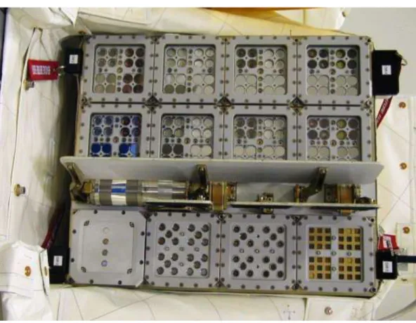
18 Fig. 1. Experiment hardware and biological samples of the LIFE experiment. (A) EXPOSE-E
facility (arrow) attached to the Columbus module of the International Space Station (ISS) during orbital flight, accommodating the LIFE samples. (B) Samples of LIFE experiment accommodated in one of the compartments of EXPOSE-E facility (wells diameter 1.4 cm). Vertical rows show: X. elegans (b1), and R. geographicum (b2) on their natural rock habitat; dried cultures of the lichen fungus (mycobiont) of X. elegans (1st and 3rd sample from the top in b3); sandstone fragments colonized by a stratified cryptoendolithic microbial community (2nd and 4th samples from the top in b4), and the fungi C. antarcticus and C. minteri (from the top 2nd and last sample in b3, upper and 3rd sample in b4).
19
Test systems of the LIFE experiment
The lichen Xanthoria elegans was collected from alpine habitats between 2000 and 3000 m altitudes at Zermatt, Switzerland (45°59’N, 7°48’E) (de Vera et al., 2003; 2008); Rhizocarpon
geographicum was collected from the Plataforma de Gredos, Spain (40º17’N, 5º14’W) at an
altitude of 2020 m (de la Torre et al., 2010); sandstone fragments colonized by a stratified cryptoendolithic microbial community were collected by L. Zucconi at Battleship Promontory (76°54’37.6”S 160°55’27.5”E), Southern Victoria Land, Antarctica in January 2004, and the microcolonial black yeast-like fungi Cryomyces antarcticus CCFEE 515 and Cryomyces
minteri CCFEE 5187, both dwelling cryptoendolithically, were isolated from sandstone
collected in McMurdo Antarctic Dry Valleys (Selbmann et al., 2005). The survival of those cryptoendolithic organisms is of special interest in term of Lithopanspermia, because rocks may supply an additional external protection to face the impact-driven ejection into space (Horneck et al., 2008; Meyer et al., 2011) and transfer from one planet to another.
Viability assays of the LIFE test systems
The photosynthetic activity of the lichens X. elegans and R. geographicum was measured after reactivating the samples in a climatic chamber under the following controlled conditions: constant temperature of 10°C, 12 h light and 12 h dark cycles for 96 h (X. elegans) or 72 h (R.
geographicum). Irradiation with photosynthetic active light was performed using a mercury
lamp with a 100 μmol m
-² s-1 photosynthetic photon flux density. For rehydration samples were sprayed twice a day with deionized water. After reactivation, the activity of the PhotoSystem II (PSII) of the photobiont was measured by use of a Mini-PAM fluorometer (Heinz Walz GmbH), as described previously (Sancho et al., 2007; de Vera et al., 2008). Lichens were rewetted immediately before each measurement. The optimum quantum yield of chlorophyll a was determined by fluorescence measurements after 20 minutes of dark adaptation according to the equation:
Fv/Fm = (Fm - Fo)/ Fm
with Fv = variable fluorescence yield, Fm = maximal fluorescence yield, and Fo = minimal fluorescence yield. This optimum quantum yield of PSII was taken as an indication of the PSII activity of the photobiont of the lichen system after the exposure to space parameters. The percentage (n=2 for space 100% insolated, and for space 0.1% insolated, and n=4 space dark samples) of PSII activity was determined from the ratio of the Fv/Fm of the same flight
20
sample before and after flight. Control data are given for the same sample measured before space flight (pre-flight) (Fig. 2A and B).
The survival of C. antarcticus and C. minteri (Fig. 3A and B) was determined from their colony forming abitiliy as percentages of CFU (Colony Forming Units). Growth tests were performed by suspending fungal cells from rehydrated colonies in a 0.9% NaCl solution, inoculating them on Malt Agar (MA) Petri dishes (5 replicates), and incubating them at 15°C for 30 days. Control data were obtained from fresh colonies. Statistical analyses were performed by one-way analysis of variance (Anova) and pairwise multiple comparison procedure (Tukey test), carried out using the statistical software SigmaStat 2.0 (Jandel, USA). The means (n=5) ± s.d. are plotted. *, P=0.001; Power of performed test with α=0.050: 1.000 (Fig. 3A and B).
The Propidium Monoazide (PMA) assay was used to check the integrity of the cell membranes after spaceflight. Fractions of DNA extracted from intact cells of cryptoendolythic fungi C. antarcticus and C. minteri colonies were compared with the fraction of DNA extracted from cells isolated from the space exposed colonized sandstone fragments. It was performed by adding PMA (Biotium, Hayward, CA) at a final concentration of 200 μM to the re-hydrated fungal colonies or to powdered rock suspensions in PBS (Phosphate Buffer Saline) solution. PMA penetrates only damaged cell membranes, crosslinks then to DNA after light exposure and thereby prevents Polymerase Chain Reaction (PCR). Following DNA extraction and purification (Maxwell 16 automatised DNA extraction instrument, Promega, Madison, WI), quantitative PCR (Biorad CFX96 real time PCR detection system) was used to quantify the number of fungal Internal Transcribed Spacer (ITS) ribosomal DNA fragments present in both PMA treated and non-treated samples. For all reactions, 1 μl of purified genomic DNA was added to 23 μl of PCR cocktail containing 1X Power Sybr-Green PCR Master Mix (Applied Bios, Foster City, CA), as well as NS91 forward (5‘-gtc cct gcc ctt tgt aca ca3‘) and ITS51 reverse (5‘-acc ttg tta cga ctt tta ctt cct c-3‘) primers, each at 0.02 M final concentration. These primers amplify a 203 bp product spanning the 18S/ITS1 region of rRNA encoding genes.
A standard Q-PCR cycling protocol consisting of a hold at 95°C for 10 min, followed by 40 cycles of denaturing at 95°C for 15 s, annealing at 58°C for 20 s, and elongation at 72°C for 15 s, was performed. Fluorescence measurements were recorded at the end of each annealing step. At the conclusion of the 40th cycle, a melt curve analysis was performed by recording changes in fluorescence as a function of raising the temperature from 60-95°C in 0.5°C per 5
21
s increments. These protocols were applied to the processing of both fungal colonies, and cryptoendolithic sandstone samples.
Control data have been obtained for an identical sample stored on ground in the laboratory during the mission (Control). Because cryptoendolithic communities are not uniformly distributed within the rocks, different quantities of total fungal DNA were obtained from different rock samples (Fig. 3C, D and E).
Space data are given for samples shielded from extraterrestrial UV radiation (Space Dark), exposed to (>110 nm) at a fluence of 9.19 105 Jm-² beneath a 0.1 % transmission MgF2
filter (Space 0.1% insolated), or at a higher fluence of 6.34 108 Jm-² (Space 100% insolated). Statistical analyses were performed by one-way analysis of variance (Anova) and pairwise multiple comparison procedure (Tukey test), carried out using the statistical software SigmaStat 2.0 (Jandel, USA)*, P=0.001. **, P>0.05. Power of performed test with α=0.050: 1.000. The means (n=3) and ± s.d. are plotted (Fig. 3C, D and E).
Confocal Laser Scanning Microscopy (CLSM) imaging: Viability of X. elegans analysed by LIVE/DEAD staining kit FUN 1
Adult lichen thallus, young thallus and the isolated mycobiont of X. elegans were stained by FUN 1 to determine their viability. A green and yellow color of the cells indicates that they still maintain vitality. A change from green to yellow color in the cytoplasm and from green to red color in the vacuoles is an indication of physiological activity expressed by accumulation of the dye in the vacuoles. Dead cells cannot be stained and therefore red crystals are not formed in the vacuoles.
Instruments used for imaging are: LSM 510 META of Carl Zeiss Mikroskopsysteme Jena GmbH with objective lenses 10 x and 40 x/oil emersion, scanning resolution 1024 x 1024 Pixel and time 64μs to 1,76μs, scan zoom 0.7x – 2.8, image bit depth 12 bit, experimental temperature condition 22°C, UV-Laser with emission wavelength 488/561/633nm and VIS-Laser HeNe with 633nm excitation and 5mW power, Filters with ChS1 679-754; Ch2 BP 505-550; Ch3 BP 575-615. For operation of the instrument, the Carls Zeiss Jena GmbH software system at the Institute of Genetics of the Heinrich-Heine-University Düsseldorf was used.
22
2.3 Results
First visual inspections assured that the “space samples” had not changed in shape and color compared to their pre-flight appearances. Tailored to each test system, different viability assays were applied: (i) photosynthetic activity of the lichenized alga (photobiont) of X.
elegans and R. geographicum (Fig. 2A and B), (ii) colony forming ability of C. antarcticus
and C. minteri (Fig. 3A and B), (iii) fraction of DNA amplified from intact cells of C.
antarcticus and C. minteri and of cryptoendolithic communities inside sandstone fragments
(Fig. 3C, D and E), and (iv) viability of X. elegans and the fungus of the lichen (mycobiont, cultured without the algal symbiont and dried) by means of vital staining (Fig. 4).
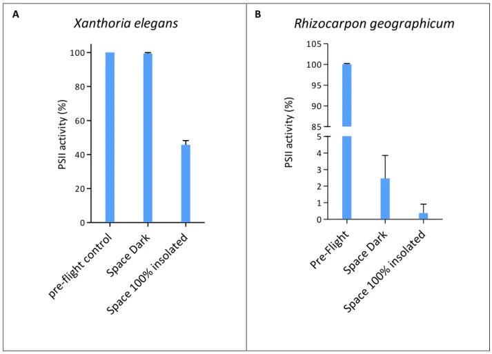
Among those space samples that were shielded from extraterrestrial insolation, but were exposed to space vacuum, cosmic radiation and temperature fluctuations (Space Dark in Figs. 2 and 3), the lichen X. elegans excelled by a PhotoSystem II (PSII) activity of (99.35 ± 0.59%) compared to the pre-flight data of the same samples (Fig. 2A). This high viability was not reached by the other space test systems kept in the dark during the mission: (2.46 ± 1.39%) for the PSII activity of the lichen R. geographicum (Fig. 2B), and (8.04 ± 3.05%) and (0.13 ± 0.07%) surviving cells for C. antarcticus and C. minteri respectively (Fig. 3A, and B). PMA assay showed (98 ± 4.67%) and (8.14 ± 0.35%) of DNA amplified from intact cells from colonies of C. antarcticus and C. minteri respectively (Fig. 3C, and D), in comparison to total extracted DNA; the percentage of DNA amplified from intact fungal cells in sandstone fragments was (18 ± 0.18%) (Fig. 3E).
23 Figure 2
Xanthoria elegans
pr e-fligh t con trol Spac e Da rk Spac e 10 0% in sola ted 0 20 40 60 80 100 P SI I a ct iv it y (% )Rhizocarpon geographicum
Pre-Fligh t Spac e Da rk Spac e 10 0% in sola ted 0 1 2 3 4 5 85 90 95 100 105 P SI I a ct iv it y (% )Fig. 2. Viability of the lichens (A) X. elegans and (B) R. geographicum, determined by
fluorescence measurements of the photosynthetic activity of the photobiont. Control data are given for the same sample measured before space flight (pre-flight), as 100% viability. Space data are given for samples shielded from extraterrestrial UV-radiation (space dark), and insolated (>110 nm) at a fluence of 6.34 108 Jm-² (space 100% insolated).
24 Figure 3
C. antarcticus
Fresh Cells Spac e Da rk Spac e 0.1% insola ted 0 20 40 60 80 100 Su rv iv al ( % )*
*
C. minteri
Fresh Cells Spac e Da rk Spac e 0.1% insola ted 0.0 0.5 1.0 1.5 2.010 20 30 40 Su rv iv al ( % )*
*
A B25 C
26 Fig. 3. Viability of the fungi (A), C. antarcticus and (B), C. minteri, as percentage of survival,
determined as CFU (Colony Forming Units). Control data were obtained from fresh colonies. No CFU were obtained from the samples “Space 100 % insolated”. The means ± s.d. are plotted. *, P=0.001; Power of performed test with α=0.050: 1.000.
Fraction of DNA amplified from intact cells of C. antarcticus and C. minteri (C and D) colonies, and from sandstone fragments (E) after different space conditions, compared to total extracted DNA. PMA penetrates only damaged cells’ membranes, crosslinking to DNA after light exposure, and thereby preventing PCR amplification. Following DNA extraction and purification, quantitative PCR was used to quantify the number of fungal ITS rDNA genes (203 bp) amplifiable in samples treated and non-treated with PMA. The means (n=3) and ± s.d. are plotted. Statistical significance was calculated using the Tukey test. *, P=0.001. **, P>0.05. Power of performed test with α=0.050: 1.000.
E
* * * *
27
The viability of culturable cells was not further decreased in the black fungi [(12.5 ± 4.11%) for C. antarcticus and (0.46 ± 0.24%) for C. minteri] that were insolated with the full extraterrestrial spectrum (>110 nm) of the Sun, having received an UV-irradiation of 9.19 105 Jm-² (Space 0.1% insolated, Fig. 3A, and B). The highest fraction of intact fungal cells (35 ± 0.15%) was accomplished by a sandstone sample that had received the full influx of solar electromagnetic radiation of 6.34 108 Jm-² (Space 100% insolated, Fig. 3E). When comparing all test systems exposed to outer space, including the high influx of full solar extraterrestrial radiation (6.34 108 Jm-²), again the lichen X. elegans, with a PSII activity of (45 ± 2.50%) (Fig. 2A) and C. antarcticus with (80 ± 0.82%) of DNA from intact cells (Fig. 3C), were the most resistant test systems of LIFE. However, black Antarctic cryptoendolithic fungi lost colony-forming ability after exposure to full insolation: no survivors were detected in the space 100 % insolated samples. Resistance of X. elegans and its mycobiont after exposure to the combined action of all space parameters tested – including full insolation - was confirmed by vital staining and Confocal Laser Scanning Microscopy. Fig. 4 shows the high capacity of the lichenized fungus (part of the symbiotic lichen association) in a young thallus (B), and of the lichen fungus of X. elegans in pure culture (C), to resist space exposure. The fungal cells in the cortex (Cx) of the young thallus were still vital, although a protecting mucilage layer had not been formed and the Parietin layer appeared to be very thin. The same features appeared to (C), the fungal cells of the mycelium.
28 Fig. 4. Confocal Laser Scanning Microscopy (CLSM) imaging: Viability of X. elegans
analyzed by LIVE/DEAD staining kit FUN I. (A), adult lichen thallus; (B), young thallus and (C), lichen fungus isolated in pure culture (mycobiont) of X. elegans, all dried and exposed to 100 % insolation and space vacuum. Green to yellow coloured cells are stained by FUNI, indicating vital cells. Turning from green to yellow and finally to red, indicates physiological activity expressed by the accumulation of the dye in the vacuoles. The high degree of maintained viability is due to a cortex Cx, a mucilage layer Mc and crystal deposits of Parietin P on the surface of the lichen. These layers are able to protect interior cells of the alga layer A and the medulla M.
29
2.4 Discussion
The LIFE experiment has provided for the first time data on the viability of rock-dwelling organisms and microbial communities after a long-term exposure to space parameters. These conditions cannot easily be simulated in the laboratory, if at all. The test systems, collected from hostile conditions, such as Antarctica and high mountain regions, are adapted to cope with high radiation intensities, arid phases, and extreme temperature fluctuations, somehow similar to those experienced in space. In C. antarcticus, for example, globular cells are enveloped in a thick melanised cell wall, protecting them from radiation and desiccation. Their meristematic way of producing colonies (i.e. dividing in all directions) further supports their resistance (Selbmann et al., 2005; Sterflinger, 2005; Onofri et al., 2008; 2009). Special protection against environmental extremes is also granted for the photobiont, the green alga
Trebouxia sp. of the lichen X. elegans. In this symbiotic organization, the fungus forms a
cortex with an upper layer incrusted with parietin and a mucilage layer enveloping the alga cells in a medulla matrix (Fig. 4A and B) (de Vera et al., 2003; 2008).
All organisms selected for the LIFE experiment are poikilohydric, i.e. they are able to dehydrate till most biochemical activities stop. In this state they are highly tolerant to stresses and may resume their metabolism once water becomes available again. Specifically, exposure to vacuum should inhibit any oxidative process related to their metabolism; this particular effect could protect against other damaging effects induced, e.g., by solar UV radiation, cosmic ray ions and temperature extremes.
The LIFE experiment has demonstrated that some – but not all - of those most robust microbial communities from extremely hostile regions on Earth are also partially resistant against the even more hostile environment of outer space. In this experiment the following species stood out as the most persistent survivors after 1.5 years in outer space: the black fungus C. antarcticus – as determined from PMA assay - and the symbiotic X. elegans - as determined from PSII activity- and its mycobiont - as determined by LIFE-DEAD staining. However, the CFU test did not yield any survivors of C. antarcticus flight samples that were exposed to the un-attenuated solar extraterrestrial spectrum (space 100 % insolated) and less than 10% survivors for the space dark samples. This means, even if the cell membrane seemed to be intact – as indicated by the PMA test -, the cells had lost their ability to grow and divide.
30
Earlier studies showed that the circumpolar and alpine red lichen X. elegans was able to retain its photosynthetic activity almost completely after 14 days in space (Sancho et al., 2007; de la Torre et al., 2010). This observation has been confirmed in this study for a much longer exposure time of 565 days in space, although this high viability was only observed, if the lichen had been shielded from solar electromagnetic irradiation, maintaining 45% PSII activity after 100% insolation exposure. Particularly, the mycobiont seems to play a fundamental role in maintaining the viability of the entire lichen system, because 565 days in space appeared not to have any effect on its physiological activity, even after 100% insolation (Fig. 4C). Interestingly, it has been earlier shown that X. elegans resisted also shock pressures, comparable to those experienced by the Martian meteorites during impact ejection (Horneck et al., 2008; Meyer et al., 2011), as required for phase-I of Lithopanspermia.
Although we have demonstrated that some rock-dwelling species are capable of partially withstanding the harsh environment of outer space – or certain parameters of it - for at least 1.5 years, the data are insufficient for drawing any consequences for the likelihood of Lithopanspermia The possibility of surviving a much longer journey in space, as would be required for a natural travel from Mars to Earth or vice-versa, still remains an open question. So far, our studies have provided a first long-term experimental test on the survival of cryptoendolithic microbial communities in space, over a time period as long as currently available by space technology. These experiments demonstrate that outer space can act as a selection pressure on the composition of microbial communities (Cockell et al. 2011).
31
3. LIFE Experiment: Isolation of cryptoendolithic organisms from
Antarctic colonized sandstone exposed to space and simulated
Mars conditions on the International Space Station*
Abstract
Desiccated Antarctic rocks colonized by cryptoendolithic communities were exposed on the International Space Station (ISS) to space and simulated Mars conditions (LiFE - Lichens and Fungi Experiment). After 1.5 years in space samples were retrieved, rehydrated and spread on different culture media. Colonies of a green alga and a pink-coloured fungus developed on Malt-Agar medium; they were isolated from a sample exposed to simulated Mars conditions beneath a 0.1% T Suprasil neutral density filter or from a sample exposed to space vacuum without solar radiation exposure, respectively. None of the other flight samples showed any growth after incubation. The two organisms able to grow were identified at genus level by Small SubUnit (SSU) and Internal Transcribed Spacer (ITS) rDNA sequencing as
Stichococcus sp. (green alga) and Acarospora sp. (lichenized fungal genus) respectively. The
data in the present study support the possibility of eukaryotic life transfer from one planet to another by means of rocks and of survival in Mars environment.
Key words: Antarctic colonized rocks, EXPOSE-E, International Space Station,
Lithopanspermia, Lichens and Fungi Experiment.
*Giuliano Scalzi, Laura Selbmann, Laura Zucconi, Elke Rabbow, Gerda Horneck, Patrizia Albertano and Silvano Onofri. Origins of Life and Evolution of Biospheres (Accepted for publication on December 21st 2011)
32
3.1 Introduction
The Lithopanspermia hypothesis suggests that impact-ejected rocks could transfer living organisms through space from one planet to another (Thomson 1871). This scenario implies that rock-embedded organisms need to survive (i) the ejection into space within rock fragments, (ii) the journey through space for a long time (hundreds up to thousands or millions of years) and (iii) the landing on another planet (Gladman et al. 1996; Mileikowsky et al. 2000; Horneck et al. 2008; Nicholson 2009).
Despite an ever-expanding understanding of the limits of life in the Earth’s most extreme environments, little remains known about life’s potential to survive when removed from Earth and the all-encompassing biosphere that harbors it. Scientific literature has numerous reports on microbial survival and proliferation in the most inhospitable environments that our planet has to offer (Rothschild and Mancinelli 2001; Venkateswaran et al. 2001; Canganella and Wiegel 2011). All the while, reports addressing the uppermost limits of microbial survival in near-Earth orbit (Horneck et al. 2010), in the interplanetary (Mileikowsky et al. 2000) or interstellar space are scarce (Valtonen et al. 2009). Most early studies of long-term survival in outer space have dealt with prokaryotes, mostly bacterial endospores (e.g., Horneck et al. 1994). Later on, studies were extended to eukaryotes exposed to either simulated space conditions or space during short-term flights, such as the experiments in the framework of ESA’s Biopan missions (Sancho et al. 2007; de la Torre et al. 2010; Raggio et al. 2011). The lichens Rhizocarpon geographicum, Xanthoria elegans and Aspicilia fruticulosa were exposed to space environment for about 10 days; long term experiments in space on eukaryotic test organisms have been done for the first time within this LIFE experiment. To ascertain the uppermost extreme limits of life, it is important to start examining a wider spectrum of microbiota, including those rock-embedded communities from extreme environments such as Antarctic deserts, and expose them to space in their integrity, in their natural substrate.
Cryptoendolithic communities are among the most resistant life forms ever encountered, originally discovered in the extremely cold, hyper-arid Dry Valleys of Antarctica (Friedmann 1982). In structural cavities of Antarctic sandstones lichenized and non-lichenized fungi and algae (cryptoendoliths), bacteria and cyanobacteria, form the lichen-dominated cryptoendolithic community (Friedmann 1982). Their extreme environment is considered the
33
most similar terrestrial environment to Mars surface (Wynn-Williams and Edwards 2000; Onofri et al. 2004); it is characterized by very low temperatures, ranging from -20 to -50°C in winter, with annual average temperature below 0°C, wide thermal fluctuations, frequent freeze/thaw cycles, dryness due to the lack of snow or ice cover and rare precipitations (<100 mm Water Equivalent per year), high salt concentrations, low nutrient availability and high radiation including UV (Onofri et al. 2007). Molecular studies of environmental DNA extracts from these communities suggested the identity of an endolithic fungal phylotype with adjacent epilithically growing Buellia spp. (>97% similarity of SSU rDNA sequences; de la Torre et al. 2003). The same authors highlighted the microbial biodiversity in these communities comprising lichen mycobionts, free-living fungi, algae, as well as
Actinobacteria, Alphaproteobacteria, Gammaproteobacteria and some other unidentified
bacterial phylotypes. Some new genera and species of black meristematic fungi have also been described, but most taxa have not yet formally described (Selbmann et al. 2005; Selbmann et al. 2008). The increasing knowledge about diverse eukaryotes thriving in such harsh conditions suggested us to test in a space flight experiment the survival of a whole rock community under space and simulated Martian conditions.
In this LIFE experiment, fragments of sandstone colonized by cryptoendolithic communities from the McMurdo Dry Valleys (Antarctica) were flown in the European EXPOSE-E facility attached to the exterior of the ISS. Their suitability was tested before flight in ground-based Experiment Verification Tests (Onofri et al. 2009). The samples were exposed for 18 months to the environment of space in Low Earth Orbit (LEO) and to simulated Mars conditions. Once returned to Earth they were subjected to both, molecular and cultivation-based assays to estimate survival and viability (Onofri et al. 2012) including growth ability.
34
3.2 Material and Methods
Colonized Sandstone
A sample of sandstone was collected under sterile conditions by L. Zucconi at Battleship Promontory (76°54’37.6”S 160°55’27.5”E), Southern Victoria Land, Antarctica in January 2004; it showed under a stereomicroscopic inspection a well developed and stratified cryptoendolithic community with typical color bands, including the green one, due to living organisms. They were stored under sterile conditions at -20°C. Well colonised sandstone fragments (11 mm wide, maximum 6 mm thick, meanly 437 mg) including significant portions of microbial biomass were removed under sterile conditions by striking the sandstone sample lengthwise with a rock hammer. Excised fragments were dehydrated at room temperature and glued, using Wacker silicone glue RTV-S 69, on sterile teflon disks (12 mm diameter). These fragments were successively accommodated in different trays of the EXPOSE-E facility.
EXPOSE-E facility
The European Space Agency (ESA) has recently developed the multi-user research facility EXPOSE-E as an exterior portion of the ISS (Rabbow et al. 2009) (Fig. 1). This facility is designed to foster and promote astrobiology research including studies on the survival of, and genotypic and phenotypic changes in, model organisms (plant seeds, bacterial spores, black meristematic fungi, lichens, and cryptoendolithic communities) when exposed to the outer space environment.
The EXPOSE-E facility is a multi-user facility having a box-shaped structure, accommodating samples in three different and separate compartments, called trays (Figure 1). Each tray is subdivided into four sample carriers composed of 16 wells disposed on two levels, to face either outward towards space and radiations or kept at the same conditions but in the darkness. The LIFE samples were exposed either in Tray 1 that was vented with access to space vacuum or in Tray 2 that was sealed and filled with a simulated Mars gas mixture. The carriers were covered by an optical filter system to control intensity and spectral range of solar UV irradiation. The conditions to which the Antarctic sandstone fragments were exposed are listed in Table 1. During the 1.5 year lasting mission the rock samples experienced space vacuum (10-7to 10-4 Pa) (Horneck et al. 2010), cosmic radiation (≤190
35
mGy) (Berger et al. 2012) and the full spectrum of solar extraterrestrial electromagnetic radiation (for space exposed samples with a long pass cut-off at 110 nm, and for simulated Mars condition samples with a long pass cut off at 200 nm). The UV (200-400 nm) fluences reached 9.19 × 10⁵ Jm-2 (below a 0.1% transmission neutral density filter) and 6.34 × 10⁸ Jm-2 (100% transmission insolated samples). All fluences were calculated for the biologically active UV range of 200 nm < λ > 400 nm for the whole mission and depended on the orientation of the ISS to the sun. Temperature varied between -21.5 °C and +59.6 °C (Rabbow et al. 2012; Onofri et al. 2012).
36
Cultivability tests
Treated samples (only one third of the whole sample analyzed, meanly 140 mg), ground controls (mean weight 91,2 mg) and fresh colonized rocks (weight about 500 mg) were gently homogenized in sterile physiological solution (0.9% NaCl w/v) and plated on MA (Malt Agar and incubated at 15°C for 3 months. The MA medium was selected because it gave best growth for colonies from fresh colonized rock samples. Each test was performed in triplicate.
The organisms that were able to grow where isolated and stored in pure culture at 10°C.
Table 1 Exposure conditions during the 1.5 year lasting LIFE experiment.
Sample specification Parameter
Space Dark Space vacuum (10-7 to 10-4 Pa) Space 0.1% insolated
Space vacuum and solar UV (>110 nm) screened with a MgF2 neutral density filter of 0.1%
transmission Space 100% insolated
Space vacuum and solar UV (>110 nm) Mars Dark
1000 Pa CO2 gas mixture
Mars 0.1% insolated 1000 Pa CO2 and solar UV (>200 nm) screened
with a Suprasil neutral density filter of 0.1% transmission
Mars 100% insolated
1000 Pa CO2 gas mixture and solar UV (>200 nm)
Ground Control Dark, room temperature, 1 atm
DNA extraction and sequencing
DNA was extracted from colonies grown for 6 months at 10°C, using Nucleospin Plant Kit (Macherey nagel, Duren, Germany) according to the user’s manual. Polymerase Chain
37
Reactions (PCR) were prepared in 25 µl tubes adding BioMix (BioLine GmbH, Luckenwalde, Germany), 5 µM of each primer and 40 ng of template. Internal Transcribed Spacers (ITS) were amplified using ITS1 and ITS4 primers (Table 2) for fungi with the following PCR protocol: an initial denaturation step at 95°C for 2 min, followed by 35 cycles of denaturation at 95°C for 30s, annealing at 55°C for 30s, and an extension at 72°C for 30s. At the end of the last cycle, an additional extension at 72°C for 5 min was performed. Algal Internal Transcribed Spacer was amplified using ITS1T and ITS4T primers (Table 2) with the following protocol: initial denaturation at 94°C for 2 min, 30 cycles of denaturation at 94°C for 1 min, annealing at 50°C for 1 min, and an extension at 72°C for 2 min; an additional extension at 72°C for 7 min was performed. Algal SSU was amplified with a PCR spanning the whole rRNA Small Subunit region using primers Euk A and Euk B (Table 2) with the following protocol: an initial denaturation at 94°C for 3 min, 30 cycles of denaturation at 94°C for 45 s, annealing at 55°C for 1 min, and extension at 72°C for 3 min, and a final extension at 72°C for 5 min. For SSU sequencing primers in Table 2 have been used. The DNA sequences were assessed by direct sequencing of the PCR product (Macrogen, South Korea).
Sequence assembling was performed by using the software ChromasPro v1.41 (2003-2007 Conor McCarthy School of Health Science, Griffith University, Southport, Queensland, Australia). Sequences were compared in the public domain (NCBI) using BLASTn algorithm.
38 Table 2 Nuclear SSU and ITS primers used for algal and fungal DNA amplification and
sequencing (Katana et al. 2001; Moro et al. 2009)
Name Sequence 5’-3’ Organism
EukA AACCTGGTTGATCCTGCCAGT Algae 898-919R TAAATCCAAGAATTTCACCTCT Algae 1936-57R GGTAGGAGCGACGGGCGGTGTG Algae CHLORO F TGGCCTATCTTGTTGGTCTGT Algae EukB TGATCCTTCTGCAGGTTCACCTAC Algae ITS1T GGAAGGATCATTGAATCTATCGT Algae ITS4T GGTTCGCTCGCCGCTACTA Algae ITS1 TCCGTAGGTGAACCTGCGG Fungi ITS4 TCCTCCGCTTATTGATATGC Fungi
39
3.3 Results
The first test in the LIFE experiment was to observe the effects of dehydration. The growth ability of the flight samples, i.e. Antarctic sandstones hosting cryptoendolithic communities that were exposed to different conditions in space was compared to that of the dehydrated Ground Control and a fresh sample of sandstone stored at -20°C. Table 3 shows the colony formers after cultivation on MA. The highest colony numbers were obtained from the fresh samples: 52 ± 10.5 algae, 1 ± 0 black fungi, 90 ± 27 pink fungi and 12.6 ± 2.0 yeasts. The GC sample, which was stored for 1.5 years in the laboratory, showed only 20.56 ± 3.5 colonies of pink fungi.
A pink fungal colony with a pale pink mycelium (Fig. 2) composed of appressed hyphae of 4 µm in width was obtained from a flight sample that was exposed to space vacuum, but kept in the dark (Table 3: Space Dark).
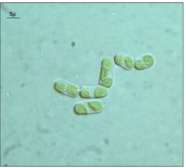
A colony of a green unicellular alga (Fig. 3) grew from a flight sample that was exposed to simulated Mars atmosphere and the Martian spectrum of solar radiation > 200 nm beneath a suprasil neutral density filter (Table 3: Mars 0.1% insolated). All other flight samples did not result in any colonies after incubation (Table 3).
Macroscopic observations and microscopic morphological analyses showed that both, the pink fungus from the Space Dark sample as well as the green unicellular alga from the Mars 0.1% insolated sample seemed to be identical with those isolated from the fresh and the GC samples.
40 Table 3 Growth studies of the LIFE experiment after the 1.5 years lasting space mission:
Colonies formed after cultivation on MA (Flight samples and controls)
Exposure conditions
Colony formation Total
colonies/m g Algae Black colonies Filamentous pink fungi Yeasts Space Dark 0 0 3* 0 0.021 Space 0.1% insolated 0 0 0 0 0 Space 100% insolated 0 0 0 0 0 Mars Dark 0 0 0 0 0 Mars 0.1% insolated 1* 0 0 0 0.007 Mars 100% insolaed 0 0 0 0 0 Ground Control 0 0 20.5± 3.5 0 0.18 Fresh sample (dark,-20°C, 1 atm) 52 ± 10.5 1 ± 0 90 ± 27.0 12.66 ± 2.0 0.29
*Total CFU obtained from 3 replicates
To identify the fungal isolate, PCR amplification of ITS rDNA portions and sequencing were performed. The sequences obtained gave low identity (94%), with E-value = 0, in NCBI GenBank with the lichenized genus Acarospora A. Massal. The closest related sequence was
41 Fig. 2 Hyphal network of the mycelium isolated from a rock exposed for 1.5 years to space
vacuum (10-7 to 10-4 Pa) in the dark (Space Dark sample), seen at the light microscope. Usually lichenized fungi, when separated from their photobionts, lose all their morphological peculiarities and grow as common mycelia.
that of Acarospora rosulata (accession number GU184116.1). Based on this data the fungus was identified at the genus level only.
To further identify the green alga, the same analyses (PCR amplification of ITS rDNA portions and sequencing) showed that the ITS sequences matched with different algal genera with similarities ranging from 95% to 97%, E-value = 0. They belonged to the Class
Trebouxiophyceae, 5 algae showed similarities to the Order Chlorellales, and 1 to the Order Prasiolales, genus Stichococcus (97% identity). SSU sequencing showed a similarity ranging
from 94% to 96%, E-value = 0, with different algal species in the genus Stichococcus, the closest related sequence (96%) was S. deasonii UTEX 1706 (accession number DQ275460).
42
This strain grew mostly as single cells or four-celled short uniseriate and unbranched, straight or sigmoid, filaments. However, the morphology of the cylindrical cells, 5 m wide and 10
m long on average, with broadly rounded poles, does not completely match the morphological description of the genus Stichococcus Nägeli (1849), in which only one unlobed parietal chloroplast is usually present, while our strain was almost always characterized by two chloroplasts.
Fig. 3 Green alga isolated from colonized sandstone exposed to simulated Mars conditions in
space beneath a Suprasil neutral density filter with a cut off at 200 nm and 0.1 % transmission (Space 0.1% insolated). Phylogenetic analyses indicate that the alga belongs to the genus
43
3.4 Discussion
LIFE is the first experiment where cryptoendolithic microorganisms within their natural substratum have been exposed for long time to space and simulated Martian conditions in Low Earth Orbit. After exposure to the conditions applied during the LIFE experiment, a few cryptoendolithic organisms grew after recovery, namely in one Space Dark sample (3 pink fungi) and in one Mars 0.1% insolated sample (1 green alga). The pink lichenized fungus seemed to belong to the genus Acarospora and the green alga seemed to belong to the genus
Stichococcus. Due to the scarcity of algal ITS and SSU sequences in nucleotide databases, it
was only possible to address the alga to that genus. Considering the generic level of the data obtained, an extended study based on morphology and multi-gene molecular phylogeny is in progress.
All other flight samples did not result in any growth under the conditions tested. It should be noted that the exposed samples were very small and the rock fragments used for growth tests exiguous. Due to the not uniform distribution of the organisms composing the community within the Antarctic sandstone fragments, it was not possible to know before flight which organisms were present in each subsample. This may explain the differences recorded in cultural tests between the Ground Control, where only pink fungi were isolated, and fresh samples, from which a variegated number of fungal and algal colonies (Table 3) were obtained. It is interesting to note that although no algal growth was observed in the ground control samples, one algal colony was isolated from one Mars 0.1% insolated sample. It should again be stressed that due to the small size of the analyzed samples, it was not guaranteed that all members of the rock community were represented. We have shown that at least two microorganisms, a fungus and an alga, of a rock colonizing community survived after 1.5 years in space.
Antarctic cryptoendolithic organisms have been suggested as good study model for survival in space because of their ability to cope with the complex interplay of extremely low temperatures, thermal fluctuations, arid phases and high radiation intensities (Selbmann et al. 2005; Ruisi et al. 2007; Selbmann et al., 2011). Moreover, it has been assumed that organisms living within rocks could enter a cryptobiotic state after being ejected into space (Onofri et al., 2012).
44
This feature is possibly aided by the presence of abundant extracellular polymeric substances (EPS) in many endolithic species. EPS production allows the community to survive long dry periods, it may be abundant in fungi (Selbmann et al. 2005), in lichen thalli (de Vera et al. 2004, de los Ríos et al. 2005), and in the whole cryptoendolithic community (de los Ríos et al. 2004). Previous studies ascertained that the presence of sugars such as glucose or buffer salts on the surface of Bacillus subtilis spores act as chemical protectants, highly increasing their survival in space (Horneck et al. 1994). Furthermore Mancinelli and Klovstad (2000) showed that under 1mm of Martian analog soil a B. subtilis monolayer did not exhibit loss of viability after exposure to 12.3 kJm-2 of UV radiation from deuterium source, and some protection was even afforded by a layer 12 µm thick. The few rock-colonizing organisms that were isolated after retrieval of EXPOSE-E could have been survived because of: (i) the ability of cryptoendolithic organisms to tolerate desiccation and radiation and (ii) the shielding of even a small layer of rock material thereby preventing loss of viability after simulated Martian UV irradiation. However, the study showed also that most of the rock colonizing species did not survive after 1.5 years in space or in a simulated Martian climate. Nevertheless extremophilic endolithic Eukarya composing this Antarctic community may be suitable model organisms to investigate the resistance of life in the framework of Lithopanspermia. After astrobiology research has centered - at least in the past decades - almost exclusively on prokaryotic microorganisms, these recent findings of the LIFE experiment allow us to select models from a wider spectrum of living organisms.