European Commission E.C - Structural Funds
Ministero dell’Università e della Ricerca Scientifica e tecnologica
Università degli Studi di Catania
UNIVERSITÁ DEGLI STUDI DI CATANIA
DOTTORATO DI RICERCA INTERNAZIONALE
IN NEUROBIOLOGIA Ciclo XXVIII
Sedi consorziate: Università di Catania, Roma e Pavia
Sede amministrativa: Università di Catania
Dott.ssa Floriana D’Angeli
Biomolecular effects and bioclinical applications of
PARPs inhibitors
TESI DI DOTTORATO
COORDINATORE: Chiar.mo Prof. Roberto Avola
TUTOR: Chiar.ma Prof.ssa V. Spina-Purrello
Index
Index
Section I
Background………...pag.1 1. PARPs Family……….……….…………..pag.1 2. MAR- or PAR-generating PARPs ………...……….…………..pag.3 3. PARPs functions...………...pag.5 4. PARPs localization……….pag.6 5. PARP-1...pag.8 6. PARP-1 and DNA repair……….pag.9 7. DNA-independent PARP-1 activation………..pag.11 8. PARP and Angiogenesis………...pag.13 9. PARPs Inhibitors………..pag.15 Aim of investigation………pag.17 1. Introduction………...pag.19 2. Materials and Methods………...pag.21 2.1 Chemicals and antibodies………...pag.21 2.2 Cell cultures ………...pag.21 2.3 Preparation of C6 glioma CMs………pag.22 2.4 MTT assay………..…...………..pag.22 2.5 Cell migration.………..………..pag.22
Index
2.6 Western analysis ………...……….pag.23 2.7 RT-PCR ……….pag.24 2.8 Immunofluorescence analysis.………pag.24 2.9 Cell imaging by confocal microscopy ………pag.25 2.10 Statistic Analysis ………..pag.26 3. Results………..pag.27 3.1 Viability of GP8.3 cells in presence of PJ-34 and of CM alone or with PJ-34. pag.27 3.2 Effects of PARP-1 Inhibitor PJ-34 on stimulated GP8.3 cell migration……..pag.28 3.3 PARP-1 expression and PJ-34 effect ………..pag.30 3.4 Modulation of phospho-ERK levels by PARP-1 inhibitor PJ-34………pag.31 3.5 Modulation of phospho-Elk-1 levels by PARP-1 inhibitor PJ-34...…………pag.32 3.6 Effects of MEK inhibitor PD98059 on PARP-1 mRNA levels and on PARP-1 protein expression……….pag.33 3.7 Laser scanning microscopy.………pag.35 3.8 PARP-1 and phospho-ERK interaction.………...pag.38 4. Discussion.………...pag.39 5. Conclusion.………..pag.43 References………...pag.44Index
Section II
Background……….pag.52 1. Macro PARPs……….……….………….pag.52 2. Macro PARP: PARP-14………...……….………pag.53 3. PARP-14 acts as a pro-survival signal in multiple myeloma...………...pag.54 4. The 2nd generation PARP inhibitors: PJ-34 acts as pan PARP inhibitor……...pag.55 5. Diabetes...pag.57 5.1 The pancreatic Islets………...pag.57 5.2 Diabetes Mellitus: definition and description……….pag.58 5.3 Immune-mediated diabetes ………pag.58 5.4 Molecule effectors in immune-mediated diabetes ………..pag.59 5.5 Induction of Insulitis………...pag.59 5.6 ER stress in β cells and antigen presentation………...pag.62 5.7 Role of PARP in Type 1 Diabetes: The Okamoto model……….pag.64 5.8 Involvement of pancreatic α-cell in Diabetes………..pag.65 5.9 Glucagon secretion in mouse α-cells………...pag.66 5.10 The role of α-cell in immune-mediated diabetes………...pag.68 Aim of investigation………..…...pag.70 1. Introduction.………..……….pag.72
Index
2. Materials and Methods………...………pag.74 2.1 Cell cultures and treatment with cytokines ……….pag.74 2.2 MTT assay.……….pag.74 2.3 Apoptosis assay………..pag.75 2.4 Imaging Flow Cytometer analysis………..pag.75 2.5 RT-PCR ……….pag.77 2.6 Immunofluorescence………..pag.77 2.7 Confocal microscopy imaging………pag.78 2.8 Statistical Analysis ……….pag.79 3. Results………..pag.80 3.1 RT-PCR...………...pag.80 3.1.1 PARP expression in β-TC1 cells treated with inflammatory cytokines……pag.80 3.1.2 PARP expression in α-TC1.6 cells treated with inflammatory cytokines….pag.82 3.1.3 mRNA expression of PARP-14 on pancreatic β-TC1 and α-TC1.6 cells….pag.83 3.2 Confocal microscopy analysis.………...pag.84 3.3 Cell Viability.……….pag.86 3.3.1 Effect of PJ-34 on α-TC1.6 cells viability………pag.86 3.3.2 Effect of PJ-34 on β-TC1.6 cells viability………pag.88 3.4 Apoptosis assay………...pag.89Index
3.4.1 Caspase-3 activity on α-TC1.6 cells treated with cytokines, in presence or absence of PJ-34………...pag.89 3.4.2 Caspase-3 activity on β-TC1 cells treated with cytokines, in presence or absence of PJ-34………...pag.91 3.5 Flow Cytometry………..pag.93 3.5.1 Effect of PJ-34 on apoptotic α-TC1.6 cells death: flow cytometry analysis.pag.93 3.5.2 Effect of PJ-34 on apoptotic β-TC1 cells death: flow cytometry analysis…pag.98 3.6 Graphical Abstract………pag.103 4. Discussion………..pag.104 5. Conclusion……….pag.110 References……….pag.111Section I
Section I
Biomolecular effects and bioclinical
applications of PARPs inhibitors:
“PJ-34 inhibits PARP-1 expression and ERK
phosphorylation in glioma-conditioned brain
Section I: Background
Background
1. PARPs Family
ADP-ribosyltransferases (ARTs) comprise a family of structurally conserved enzymes that catalytically cleave NAD+ and transfer the ADP-ribose moiety to acceptor residues of target proteins (Steffen J.D. et al., 2013). Originally PARP-1 was the only known enzyme with poly(ADP-ribosylation) activity, however, studies over the past decade have identified a family of as many as 17 proteins that share homology to the catalytic domain of PARP-1 (Ame, J.C. et al., 2004) (Figure 1).
Figure 1. Schematic domain structures of human PARP proteins. Numbers to the bottom right of
the protein schematic indicate the total length, in amino acids, of each protein. BRCT: BRCA1 C terminus domain; VIT: vault inter-trypsin domain; vWA: von Willebrandfactor type A; MVP-BD: major vault protein binding domain. PARPs are categorized as either poly-ADP-ribosyltransferases (Poly-), mono-ADP-ribosyltransferases (Mono-) or inactive based on presence of conserved motifs and, when available, data using enzymatic assays. PARP-9 and PARP-13 lack one or more catalytic residues conserved in all other PARPs and are therefore predicted to lack catalytic activity, although it is unknown whether they still bind ADP-ribose or ADP-ribosylated proteins. (Daugherty M.D. et al., PLOS Genetics 2014).
Section I: Background
Currently, only the first six members of this family (ARTs 1–6) are regarded as having poly(ADP-ribosyl)ation activity: PARP-1, PARP-2, PARP- 3, PARP-4 (vPARP), PARP-5a (TNKS1), and PARP-5b (TNKS2) (Figure 2). The remaining ARTs 7–17, although originally considered PARPs (PARPs 6–16) (Steffen J.D. et al., 2013), are only capable of producing mono-ADP-ribose modifications and are referred to as mono- ARTs (MARTs). ARTs 9 (PARP-9; BAL-1) and 13 (PARP-13) have yet to confirm any sort of catalytic activity like PARPs or MARTs.Figure 2. Domains of human PARPs. Sequence and structural representation of six PARPs. Each
PARP has a catalytic domain, containing an ADP-ribosyl transferase domain (ART) and conserved catalytic glutamic acid residue. In addition, PARPs 1–4 contain a helical domain (HD) that serves in allosteric regulation. PARPs 1–3 contain a WGR domain, which is important in DNA-dependent catalytic activation. The BRCT domain (Breast Cancer Susceptibility Protein-1) C-terminus is commonly found in DNA-repair and check-point proteins: it resides in the automodification domain of PARP-1 and is also present in PARP-4. Zinc-fingers Zn1 and Zn2 of PARP-1 are important in binding DNA, while the third zinc-finger (Zn3) is important in DNA-dependent catalytic activation. Other domains and sequences represented include: centriole-localization signal (CLS), vault protein inter-alpha-trypsin (VIT), vonWillebrand type A (vWA), major vault particle interaction domain (MVP-ID), His-Pro-Ser region (HPS), ankyrin repeat clusters (ARCs), sterile alpha motif (SAM), and nuclear localization signal (NLS). (Steffen J.D. et al., Frontiers in Oncology 2013).
Section I: Background
2. MAR- or PAR-generating PARPs
Multiple characteristics of the PARP catalytic domain are important in determining whether a PARP generates PAR or MAR modifications. These characteristics include the specific amino acid residues that bind to NAD+ and catalyse the transfer reaction, as well as structural elements that define the substrate and acceptor binding pockets (Figure 3; Table 1).
Table 1. ARTD, ADP-ribosyltransferaseARTD, ADP-ribosyltransferase diphtheria-toxin-like; BAL, B
aggressive lymphoma; MAR, mono(ADP-ribose); miRNA, microRNA; ND, not determined; NF-κB, nuclear factor‑κB; PAR, poly(ADP-ribose); PARP, PAR polymerase; RISC, RNA-induced silencing complex; TNKS, tankyrase; UPR, unfolded protein response; WWE, Trp-Trp-Glu; ZAP, zinc finger antiviral protein; ZC3HAV1, zinc finger CCCH-type antiviral protein 1. *Catalytic activity is based on the ability of PARPs to automodify when incubated with NAD+. (Vyas S. and Chang P.,
Section I: Background
PAR-generating PARPs contain a His-Tyr-Glu (HYE) motif in which histidine and tyrosine residues are involved in NAD+ binding and coordination, whereas glutamate is required for PAR transfer and elongation activity (Marsischky G. T. et al., 1995). Most PARP family members lack this glutamate and instead contain leucine, isoleucine or valine and are predicted (and in some cases have been shown) to generate in vitro, MAR, using auto-modification reactions containing purified PARP and labelled NAD+ (Table 1). In addition, PARP-9 (also known as B aggressive lymphoma 1 (BAL1) and PARP-13 (also known as ZC3HAV1) lack the histidine residues and are predicted to be enzymatically inactive; they do not show auto-modification activity (Kleine H. et al., 2008; Otto H. et al., 2005) (Table 1).Structural characteristics of the substrate and acceptor binding pockets, which affect enzymatic activity, include the donor loop (D-loop) that interacts with the substrate NAD+ and is thought to function as a ‘lid’ to hold NAD+ within the catalytic pocket (Wahlberg E. et al., 2012) (Figure 3). Additionally, the acceptor pocket is partly lined by the loop between β-sheets 4 and 5, which is referred to as the acceptor loop (Figure 3).
Figure 3. Sequence and structural elements of the poly(ADP-ribose) polymerase (PARP) catalytic domain. The ribbon structure shows the donor (yellow) and acceptor (orange) loops of PARP1 (protein
data bank ID: 3L3M117), which shape the substrate and acceptor binding pockets, respectively. The His Tyr Glu (HYE) motif is shown in magenta. A cocrystallized NAD+ analogue inhibitor (A927929)
Section I: Background
This loop is implicated in the binding of protein substrate for MAR- and PAR-generating PARPs, or incoming ADP-ribose units for PAR-PAR-generating PARPs (Han S. and Tainer, J. A., 2002; Ruf A. et al., 1998). Both PAR and MAR function as traditional post-translational modifications, which can alter the functions of target proteins.3. PARPs functions
PARP-1, the founding member of the PARP family, is a molecular sensor of DNA breaks, playing a key role in the spatial and temporal organization of break repair through the local synthesis of poly(ADP-ribose) (PAR) at damaged sites. Indeed, PARP-1 is activated by single- and double-strand breaks, being one enzyme critical in the base excision repair (BER) pathway (Dantzer, F. et al., 1999). In addition to its critical involvement in cellular response to DNA damage, poly(ADP-ribosyl)ation has been ascribed to regulate various biological processes such as transcription, mitotic segregation, chromatin modification, telomere homeostasis, cell proliferation, transformation, and cell death (Figure 4) (Schreiber et al., 2006). In particular, PARP-1 and, to a lesser extent, PARP-2 are important in maintaining telomere length and chromosomal stability. PARP-1 also forms part of the Groucho/TLE1 co-repressor complex, and has been implicated as a transcriptional regulator of androgen receptor expression. Other PARPs function in the repair of DSBs and in progression of mitosis (PARP-3), and some have potential roles in Wnt signalling and telomere maintenance (PARP-5 and PARP-6). PARP-1 is also a regulator of NHEJ, a mechanism of DSB repair. PARP-1 acts on mitochondria and, depending on the extent of oxidative stress, DNA damage and PARP-1 activation, different cell death pathways may be triggered.
Section I: Background
Figure 4. In addition to the classic activity of PARP in BER, the PARP family members have diverse
functions in otherbiological processes, including transcriptional regulation, chromatin modification, mitosis (mitotic-spindle formation), and apoptosis, as well as intracellular trafficking, and energy metabolism (not shown). Abbreviations: APC, adenomatous polyposis coli protein; BER, base-excision repair; DSB, double-strand break; DVL, dishevelled homologue; GSK-3β, glycogen synthase kinase-3β; NHEJ, non homologous end joining; PARP, poly(ADP-ribose) polymerase; TLE1, transducin-like enhancer protein 1 (Groucho homologue). (Sonnenblick A. et al., Nat Rev Clin Oncol. 2015).
4. PARPs localization
PARP family members are localized in various cellular compartments, including nucleus, cytoplasm, mitochondria, Golgi Apparatus, endoplasmic reticulum and stress granules, although the function of many of the PARPs are unknown (Figure 5) (Krishnakumar R. and Kraus W. L., 2010). The primary nuclear PARPs are PARP-1, PARP-2 (the closest paralog to PARP-1), PARP-3, and tankyrases 1 and 2 (PARP-5a and -5b). Others PARPs have been found in the cytoplasm, although not exclusively, are vPARP (PARP4), PARP6, PARP9, the Bal proteins Bal 13 (PARP13, 14, -15), and PARP-10. In addition, PARP-9 and PARP-14 exhibited enriched localization at the cell periphery. Both proteins were later confirmed to co-localize with actin filaments, motile elements of the actin cytoskeleton that are enriched at the cell
Section I: Background
periphery. PARP-12 localizes to the Golgi apparatus, while PARP-13 is assembled in to stress granules in the cytoplasm. Finally, PARP16 exhibits reticular membrane localization, identifying it as an endoplasmic reticulum protein (Vyas S. et al., 2013).Figure 5. PARPs localization. Human PARPs have various cellular localizations. Some of them are
cytoplasmic as 4, 6, 10 and 15; others are localized in the Nucleus as PARP-1, PARP-7, PARP-8 (nuclear envelope) and PARP-11. PARP-2, PARP-3 and TNK-1 and TNK-2 are localized in both cytoplasm and nucleus. PARP-9 and PARP-14 co-localize with actin filaments, while PARP-12 is localized to the Golgi. PARP-13 is assembled in to stress granules in the cytoplasm and PARP-16 exhibited reticular membrane localization.
The best-studied PARP is the founding member PARP-1 that catalyze the formation of long, branched chains of ADP-ribose known as poly-ADP-ribose (PAR) (Daugherty M.D. et al., 2014; Gibson B.A. and Kraus W.L., 2012; Hassa P.O. and Hottiger M.O., 2008; Hottiger M.O. et al., 2010; Schreiber V. et al., 2006). PARP-1 and other poly(ADP-ribosyl) transferases are localized not only in the nucleus but also in the cytoplasm (Motta et al., 2015) and in the mitochondria.
Section I: Background
In fact, it has been recently reported that intramitochondrial poly-(ADP-ribosylation)contributes to NAD+ depletion and cell death induced by oxidative stress in neurons
(Nguewa P.A. et al., 2003).
5. PARP-1
Poly(ADP-ribose) polymerases (PARPs) are defined as cell signaling enzymes that catalyze the transfer of ADP-ribose units from NAD+ to a number of acceptor proteins. Poly(ADP-ribose) polymerase-1 (PARP-1), also known as poly(ADP-ribose) synthetase and poly(ADP-ribose) transferase, is the main member of the PARP enzyme family (Nguewa P.A. et al., 2003). PARP-1 is a highly conserved protein of ~116 kDal (D’Amours D. et al., 1999). Like many other chromatin- and transcription related proteins, it has a modular structure comprising multiple independently folded domains. The major functional units of PARP-1 are an amino-terminal DNA-binding domain (DBD), a central automodification domain (AMD), and a carboxyterminal catalytic domain (CD) (Hakme A. et al., 2008; Schreiber V. et al., 2006) (Figure 6). The DBD contains two Cys-Cys-His-Cys zinc fingers (FI/Zn1 and FII/Zn2) that mediate binding to DNA, a newly discovered third zinc binding domain (FIII/Zn3) that mediates interdomain contacts important for DNA-dependent enzyme activation (Langelier M.F. et al., 2008; Langelier M.F. et al., 2010), a nuclear localization signal (NLS), and a caspase-3 cleavage site (Hakme A. et al., 2008; Schreiber V. et al., 2006). The AMD contains a BRCT (BRCA1 C terminus) fold, which mediates protein-protein interactions (e.g., with DNA repair enzymes). The CD, which is the most conserved domain across the PARP family, contains a PARP signature motif, which binds
Section I: Background
NAD+, as well as a ‘‘WGR’’ motif, which is named after the most conserved amino acid sequence in the motif (Trp, Gly, Arg) and has an unknown function.Together, the structural and functional domains of PARP-1 confer the activities required for the broad range of functions of PARP-1 in the nucleus.
Figure 6. Structure of poly(ADP-ribose) polymerase-1 (PARP-1). A, schematic representation of the
modular organization of human PARP-1 (hPARP-1). B, ribbon representation of chicken's PARP-1 catalytic fragment (C-terminal end, amino acids 662 to 1014), which was cocrystallized with the NAD analog carba-NAD. The ribbon diagram shows the interaction of carba-NAD (inhibitor substrate analog) with the NAD+-binding site of PARP-CF. The observed bound ADP moiety of carba-NAD is shown; it marks the acceptor site. (Adapted from Ruf A, Rolli V, de Murcia G, and Schulz GE (1998) The mechanism of the elongation and branching reaction of poly(ADP-ribose) polymerase as derived from crystal structures and mutagenesis. J Mol Biol278:57-65. Copyright © 1998 Academic Press. Used with permission.) C, structure of carba-NAD: the ring oxygen of the nicotinamide ribose is replaced by a methylene group, which prevents ADP-ribosyl transfer and hydrolysis of the nicotinamide moiety by cleavage of the β-glycosidic bond. (Nguewa P.A. et al., Mol Pharmacol. 2003).
6. PARP-1 and DNA repair
PARP-1 is activated by single- and double-strand breaks, being one enzyme critical in
the base excision repair (BER) pathway (Dantzer F. et al., 1999). After the induction
of certain types of DNA damage, PARP-1 is rapidly recruited to the altered DNA and its catalytic activity increases 10- to 500-fold, resulting in the synthesis of
protein-Section I: Background
conjugated long branched pADPr chains (Dantzer F. et al., 1999; Haince J.F. et al., 2008; Hassa P.O. and Hottiger M.O., 2008).The addition of pADPr interferes with the functions of modified proteins, such as histones, topoisomerase I and DNA protein kinase (DNA-PK). Notably, however, the bulk of pADPr is attached to PARP1. Once formed, this polymer could recruit
hundreds of other proteins (Gagné J.P. et al., 2008; Gottschalk A.J. et al., 2009). Some
of these recruited proteins, typified by XRCC1, the scaffolding protein that assembles
and activates the DNA base excision repair (BER) machinery(El-Khamisy S.F. et al.,
2003; Masson M. et al., 1998), bind directly to pADPr, whereas others are indirectly recruited because they interact with pADPr-binding proteins (figure 7).
Figure 7. The biochemical pathway of poly(ADP-ribosyl)ation. PARP detects and rapidly binds to
DNA strand breaks and catalyzes poly(ADP-ribosyl)ation mainly of itself using NAD+ as substrate. Upon the formation of long, branched polymers, PARP is released from DNA. Upon the formation of long, branched polymers, PARP is released from DNA and the polymers are degraded by the PARG enzyme, permitting access of the DNA repair machinery to the lesion and its repair.
Section I: Background
At the same time, formation of pADPr diminishes the affinity of PARP1 and histones for DNA, providing a mechanism for removing PARP1 from damaged DNA and forthe local modulation of chromatin compaction (Timinszky G. et al., 2009;Tulin A.
and Spradling A., 2003). In vitro studies suggest that removal of PARP-1 provides access for repair proteins and suppresses further pADPr synthesis (Satoh M.S. and Lindahl T., 1992). Further polymer growth is also antagonized by two enzymes that hydrolyse pADPr, poly(ribose) glycohydrolase (PARG) and, possibly, the
ADP-ribose hydrolase ARH3 (Meyer-Ficca M.L. et al., 2004; Oka S. et al., 2006). The
concerted action of these enzymes removes pADPr from PARP1, restoring its ability to recognize DNA strand breaks and initiate a new round of damage signalling.
7. DNA-independent PARP-1 activation
Recent findings point to the involvement of PARP-1 activation in processes that are not necessarily related to DNA repair (Cohen-Armon M. et al., 2007). These findings showed that the C-terminal of PARP-1 containing the conserved catalytic domain of PARP enzymes (Amè J.C. et al., 2004) is involved in the interaction of PARP-1 with phosphorylated ERK-2. In cell-free systems, recombinant human PARP-1 was activated and highly auto-polyADP-ribosylated by a direct interaction with phosphorylated ERK2 constructs in the absence of DNA and ATP (one molecule of PARP-1 per two molecules of phosphorylated ERK2) (Cohen-Armon M. et al., 2007). PARP-1 activated by phosphorylated ERK2 has a higher affinity for its substrate,
NAD+, than the affinity of PARP-1 activated by nicked DNA (Cohen-Armon M. et al.,
2007; Mendoza-Alvarez H. and Alvarez-Gonzalez R., 1993).
Thus, PARP-1 activated by phosphorylated ERK2 was highly
Section I: Background
PolyADP-ribosylated PARP-1 bound to phosphorylated ERK2 acts as a scaffold protein, dramatically enhancing ERK2-catalyzed phosphorylation of the transcription factor Elk1(Cohen-Armon M. et al., 2007).Elk1, one of the ternary complex transcription factors, forms a ternary complex with the serum response factor (SRF) and the serum response element (SRE) in the promoter of its target genes (Buchwalter G. et al., 2004; Herdegen T. and Leah J.D. 1998). Transcription factor Elk1 is a prominent substrate of phosphorylated
mitogen-activated protein kinases (MAPKs) (Buchwalter G. et al., 2004). Phosphorylation of
transcription factor Elk1 activates the histone acetyl transferase (HAT) activity of CBP/p300 (Buchwalter G. et al., 2004; Li Q.J. et al., 2003). This results in core histone acetylation, and transcription of the Elk1 target gene, c-fos (Buchwalter G. et al., 2004; Li Q.J. et al., 2003) (Figure 8). ERK induced acetylation of core histones and the expression of immediate early gene c-fos were both suppressed after treatment with either PARP inhibitors or PARP-1- targeted siRNA (Cohen-Armon M. et al., 2007), indicating that PARP-1 activation mediates or significantly enhances transcription induced by ERK phosphorylate. Thus, in the absence of DNA damage, PARP-1 activation by phosphorylated ERK2 might actually mediate proliferation and differentiation regulated by the ERK phosphorylation cascade. In fact, transcription factor c-Fos protein has been implicated in cell proliferation, both in normal and in
transformed cells.In view of the stimulatory effect of ERK-induced PARP-1 activation
on c-fos expression (Cohen-Armon M. et al., 2007), PARP-1 activation in the ERK signalling pathway is a promising target for anti-proliferation drugs in malignancies caused by enhanced and uncontrolled activation of the ERK phosphorylation cascade.
Section I: Background
Figure 8. PARP-1 meet phosphorylated ERK2 in the nucleus. ERK is phosphorylated via diverse
signal transduction mechanisms initiated by phosphorylation of receptor tyrosin kinases (Trk) or stimulation of G-protein-coupled receptors (GPCR), resulting in PLC activation, Ras activation, phosphorylation of Ca+2 dependent kinases, PKC and CAMK, and activation of the Raf1-MEK-ERK phosphorylation cascade. Phosphorylated ERK shuttles between the cytoplasm and nucleus. The interaction of phosphorylated ERK2 with PARP-1 in the nucleus enhances PARP-1 activation and auto polyADP-ribosylation. PolyADP-ribosylated PARP-1 acts as an anchoring protein for phosphorylated ERK2 in the nucleus. It also acts as a scaffold protein, enhancing ERK-catalyzed phosphorylation of transcription factor Elk1. This results in an enhanced HAT activity CBP, promoting histone acetylation and expression of Elk1 target gene, c-fos.(Cohen-Armon M. et al., Mol. Cell. 2007).
8. PARP and Angiogenesis
Angiogenesis, the process of new blood vessel formation, is crucial for the development and progression of pathophysiological changes associated with a variety of disorders, including various cancers, tumor metastases, and retinopathies.
Section I: Background
Recently, a number of reports from various laboratories have led to a novel and unexpected effect of PARP inhibitors, showing a relationship between PARP and angiogenesis, and to the proposition of PARP inhibitors as antiangiogenic agents. Sofar at least five PARP inhibitors have been efficiently used in vitro (Pyriochou A. et
al., 2008; Rajesh, M. et al., 2006a; Rajesh M. et al., 2006b; Tentori L. et al., 2007) to inhibit vascular endothelial growth factor (VEGF)-induced proliferation, migration, and tube formation in human umbilical vein endothelial cells (HUVECs) and in tumor
models (Martin-Oliva D. et al., 2006). The PARP inhibitors 3-AB and PJ-34 have been
shown to do this in HUVECs in a dose-dependent manner (Rajesh, M. et al., 2006a). Moreover, PARP inhibitors prevented the sprouting of rat aortic ring explants in an ex
vivo assay of angiogenesis (Rajesh M. et al., 2006b). The PARP inhibitor PJ-34 was
also shown to efficiently inhibit the chicken chorioallantoic membrane model of angiogenesis when used at low concentrations (Pyriochou A. et al., 2008).
Further, PARP activity has the ability to modulate the expression of genes involved in angiogenesis, particularly the hypoxia inducible factor (HIF), whose activity is impaired when tumors are induced either in presence of PARP inhibitor DPQ or in parp-1-knockout mice. HIF-α has been largely involved in tumor progression by promoting a global response to hypoxia, including new vessel formation. There are results suggesting that the absence of PARP-1 modulates HIF-α accumulation by reducing both NO and oxidative stress (Martinez-Romero R. et al., 2008) (Figure 9); however, the ultimate molecular link between HIF-α and PARP-1 has to date not been established clearly and further work will be necessary to unravel this mechanism.
Section I: Background
Figure 9 (A) Tumor progression. Hypoxia stimulates the expansion and remodeling of the existing
vasculature to enhance blood flow to oxygen-deprived tissues. This process is accomplished primarily through the activation of HIF target genes involved in various steps of angiogenesis, such as vascular endothelial growth factor (VEGF) and other growth factors. (B) PARP inhibitors promote a delay in
tumor formation and a dramatic reduction in tumor size. PARP inhibitors have an antiangiogenic
effect, and they might be an interesting target for the treatment of cancer. (Peralta-Leal A. et al., Free Radic Biol Med. 2009).
9. PARPs Inhibitors
Most of the PARP inhibitors in development mimic the nicotinamide moiety of NAD+.
PARP catalyzes the cleavage of NAD+ into ADP and ADP-ribose and attaches several
molecules of the latter to the target protein in a process called poly(ADP-ribosyl)ation.
Therefore, molecules that mimic NAD+ block the binding of the NAD+ to the enzyme,
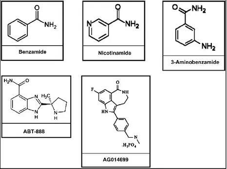
inhibiting PARP activity. First-generation inhibitors were developed 30 years ago: nicotinamide, benzamide, and substituted benzamide, in particular 3-aminobenzamide (3-AB), were shown to be competitive inhibitors of PARP (Peralta-Leal A. et al., 2009) (Figure 10). Initial research demonstrated that all the benzamides are more potent
Section I: Background
However, these classical inhibitors lacked specificity and potency.Figure 10. Inhibitors of PARP. The classical inhibitors are nicotinamide, benzamide, and substituted
benzamide, in particular 3-aminobenzamide. Important inhibitors in clinical trials include ABT-888 and AG014699. (Peralta-Leal A. et al., Free Radic Biol Med. 2009).
They affect cell viability, glucose metabolism, and DNA synthesis, and in the case of 3-AB used in combination with chemotherapy or radiotherapy, millimolar
concentrations are needed, which have a toxic effect (Milam K.M. and Cleaver J. E.
1984). A second generation of very potent PARP inhibitors was developed in the 1990s, producing 170 specific inhibitors. All these inhibitors may be classified as analogues of benzamide that act in the reaction between PARP and NAD+ and they are used in the micromolar range (Banasik M. et al., 1992).
A third generation of inhibitors, benzamidazoles, not only had potency but also allowed the elucidation of the PARP inhibitor structure–activity relationship. These new agents exhibit increased potency and specificity relative to earlier inhibitors (Zaremba T. and Curtin N.J., 2007). The development of specific, potent, effective, and safe PARP inhibitors has become an area of active research and much recent excitement in the PARP field.
Section I: Aim of investigation
Aim of investigation
Inhibitors of PARP-1(Poly(ADP-ribose) polymerase-1) act by competing with NAD+,
the enzyme physiological substrate, which play a protective role in many pathological conditions characterized by PARP-1 overactivation. It has been shown that PARP-1 also promotes tumor growth and progression through its DNA repair activity. Since angiogenesis is an essential requirement for these activities, we sought to determine whether PARP inhibition might affect rat brain microvascular endothelial cells (GP8.3) migration, stimulated by C6-glioma conditioned medium (CM). Through wound-healing experiments and MTT analysis, we demonstrated that PARP-1 inhibitor PJ-34 [N-(6-Oxo-5,6-dihydrophenanthridin-2-yl)-N,N-dimethylacetamide] abolishes the migratory response of GP8.3 cells and reduces their viability. PARP-1 also acts in a DNA independent way within the Extracellular-Regulated-Kinase (ERK) signaling cascade, which regulates cell proliferation and differentiation. By western analysis and confocal laser scanning microscopy (LSM), we analysed the effects of PJ-34 on PARP-1 expression, phospho-ERK and phospho-Elk-1 activation. The effect of MEK (mitogen-activated-protein-kinase-kinase) inhibitor PD98059 (2-(2-Amino-3-methoxyphenyl)-4H-1-benzopyran-4-one) on PARP-1 expression in unstimulated and in CM-stimulated GP8.3 cells was analyzed by RT-PCR. PARP-1 expression and phospho-ERK activation were significantly reduced by treatment of GP8.3 cells with PJ-34 or PD98059. By LSM, we further demonstrated that PARP-1 and phospho-ERK are coexpressed and share the same subcellular localization in GP8.3 cells, in the cytoplasm as well as in nucleoplasm. Based on these data, we propose that PARP-1 and phospho-ERK interact in the cytosol and then translocate to the nucleus, where they trigger a proliferative response.
Section I: Aim of investigation
We also propose that PARP-1 inhibition blocks CM-induced endothelial migration by interfering with ERK signal-transduction pathway.Section I: Introduction
1. Introduction
PARP-1 (E.C. 2.4.2.30) is the most thoroughly studied protein within the eukaryotic PARP family, which in mammals is comprised of eighteen members identified to date (Amè et al., 2004; Citarelli et al., 2010; D’Amours et al., 1999; Hassa et al., 2008; Hottiger et al., 2010). It catalyzes the attachment of ADP-ribose moieties from its substrate NAD+ to target proteins, modulating their molecular structure and biochemical activity. The enzyme alters chromatin structure, making damaged sites more accessible to members of the DNA Repair Apparatus: these are recruited at lesion sites and undergo ADP-ribosylation (Hassa et al., 2006; Nguewa et al., 2005). Accordingly, PARP-1 plays an important role in genome stability and expression, cell cycle regulation, cell metabolism (Lange et al., 2010). PARP-1 overactivation, with
consequent NAD+ depletion, has been related to inflammation and cell death (Gobell
S. Et al., 2001; Spina-Purrello et al., 2008). In fact, its inhibition reduces severity of asthma, colitis, diabetes mellitus, experimental autoimmune encephalomyelitis, Parkinson’s disease (Boulares et al., 2003; Burkart et al., 1999; Chiarugi, 2002; Eliasson et al., 1997; Iwashita et al., 2004; Jijon et al., 2000). Furthermore, PARP-1 inhibition causes a decrease in the activity of proangiogenic factors, as vascular endothelial growth factor (VEGF), transmembrane signaling protein syndecan-4 (SDC-4), platelet/endothelial cell adhesion molecule (PECAM1/CD31), and hypoxia inducible factor (HIF): this is due to a block of ERK2 target gene stimulation and ensuing reduction of angiogenesis and inflammation (Lacal et al., 2009; Martin-Oliva et al., 2006; Pyriochou et al., 2008; Tentori et al., 2008). These data suggest the implication of PARP-1 in ERK signaling in addition to its known involvement in DNA repair.
Section I: Introduction
Interestingly, the activity of ERK/MEK inhibitors in blocking the ERK signaling network may be increased by PARP inhibitors (Kerr et al., 2003; Morris et al., 2013; Tai et al., 2007; Yeh et al., 2007). It is well known that glioma is characterized by an active production of proangiogenic factors (Giurdanella et al., 2011). In the present study, we performed cell culture experiments in which GP8.3 cells were incubated with CM in order to study the antiangiogenic effects of PJ-34. In fact, we are convinced that understanding endothelial cell metabolism and the signaling mechanisms that underlie angiogenesis is important, as it provides potential therapeutic targets to inhibit or enhance angiogenesis. We demonstrate here that PJ-34 significantly reduces migration and cell viability of CM-stimulated endothelial cells. To verify the involvement of PARP-1 in ERK signaling pathway, we evaluated PARP-1 expression by RT-PCR, ERK and Elk-1 phosphorylation, by western blotting analysis. In addition by LSM we demonstrated that PARP-1 and phospho-ERK are coexpressed and share the same subcellular localization in GP8.3 cells, both in the cytoplasm as in nucleoplasm. The data obtained demonstrates that PJ-34 (a classical pharmacological PARP-1 inhibitor) lowers ERK and Elk-1 phosphorylation levels, while PD98059, (a well known MEK inhibitor) downregulates PARP-1 expression, confirming an intriguing regulatory loop between PARP-1 and phospho-ERK, which mediates endothelial cell growth and migration.Section I: Materials and Methods
2. Materials and Methods
2.1 Chemicals and antibodies
Reagent grade chemicals were purchased from Sigma Chemicals Co. (St. Louis, MO) or E. Merck (Darmstadt, Germany). MEK inhibitor PD98059 [2-(2-Amino-3-methoxyphenyl)-4H-1-benzopyran-4-one] and PARP-1 inhibitor PJ-34
[N-(6-Oxo-5,6-dihydrophenanthridin-2-yl)-N,N-dimethylacetamide.HCl] were from
Calbiochem. (La Jolla, CA). Primary antibodies against PARP-1 (rabbit polyclonal antibody); rabbit polyclonal antibody against ERK1 or ERK2; mouse monoclonal antibody to phospho-ERK1/2; mouse monoclonal antibody against phospho-Elk-1; rabbit polyclonal antibody against Elk-1; mouse monoclonal antibody against actin; were purchased from Santa Cruz Biotechnology, Inc. (CA). Reagents for RT-PCR: Trizol, deoxyribonuclease 1 (DNase I Amplification Grade), High Capacity RNA-to-cDNA Kit, Power SYBR® Green PCR Master Mix, were from Lifetechnologies™, Foster-City, CA, USA).
2.2 Cell cultures
Immortalized rat brain microvascular endothelial cells GP8.3 were fed with Ham’s F-10 medium supplemented with F-10% fetal calf serum (FCS), 80 µg/ml heparin, 2 mM glutamine, 100U/ml penicillin, and 100 µg/ml streptomycin. The cell line was already characterized, and our cell cultures were prepared and characterized following previously described procedures (Anfuso et al., 2007). Primary microvascular endothelial cells from bovine brain (BBEC) were purchased from European Collection of Cell Cultures (ECACC).
Section I: Materials and Methods
C6 glioma (rat brain astroglioma) cells were grown in F-12 medium containing 2mM glutamine, antibiotics and 10% FCS (Anfuso et al., 2007; Giurdanella et al., 2011). C6 glioma cells were purchased from European Collection of Cell Cultures (ECACC).2.3 Preparation of C6 glioma CMs
C6 glioma (rat brain astroglioma) cells (1x10⁶) were seeded in a 100-mm dish with
F-12 medium supplemented with heat inactivated 10% fetal calf serum (FCS) overnight. In cells cultured to sub-confluence, the medium was replaced with 1% serum F12-F10 HAM’s (1:1) plus glutamine and antibiotics medium, and the tumor cells were incubated for 48 h. The culture supernatant (CM) was collected, centrifuged at 500xg for 10 min and filtered with 0.2 µm filter. The aliquots were stored at -80°C until use. In all experiments, CM was used without any dilution (Giurdanella et al., 2011).
2.4 MTT assay
To quantify cell viability, the 3-[4,5-dimethylthiazol-2-yl]-2,5-diphenyl tetrasodium bromide (MTT) assay was used (Chemicon, Temecula, CA). Controls (cells grown with 1% FCS, with or without 10µM PJ-34 for 24h) and treated cells (cells grown with CM, with or without 10µM PJ-34 added simultaneously for 24h), were seeded in 96-well plates at 3000 cells/96-well, to obtain optimal cell density throughout the experiment. In all assays, cells were first incubated at 37°C with MTT for 4h; then, 100μl dimethyl sulfoxide was added and absorbance was measured. The absorbance was read in a plate reader (Synergy 2-bioTek) with a test wavelength of 570 nm.
2.5 Cell migration
GP8.3 cells migration was measured using a standard wound healing assay, essentially performed as previously reported (Giurdanella et al., 2011).
Section I: Materials and Methods
To better evaluate GP8.3 cells migration in our experimental conditions, GP8.3 cells were incubated with or without (data not shown) 10 µg/ml of mitomycin C for 2 h (37°C)and then washed twice with PBS, to render them incapable of cell division (Anfuso et al., 2014).Migration was followed by an inverted Leica DM IRB microscope equipped with CCD camera. Time zero represents the time after the scratch for all conditions: controls (cells grown with 1% FCS, with or without 10µM PJ-34 for 24h) and treated cells grown with CM, with or without 10µM PJ-34 added simultaneously.
2.6 Western analysis
PARP-1, expression, phospho-ERK and phospho-Elk1 levels were evaluated by western blot analysis. Cells were grown for 24h with 1% FCS: controls; CM, either in presence or absence of inhibitors (10μM PJ-34 or 25μM PD98059 added simultaneously), GP8.3 cells were lysed as previously described (Anfuso et al., 2007; Lupo et al., 2005). Cell lysate proteins were quantified with a bicinchoninic acid (BCA) protein assay kit (Pierce, Thermo Scientific). Immunoblots (30μg nuclear proteins and cell lysate proteins) were performed as described elsewhere (Anfuso et al., 2007). Membranes were incubated with primary antibodies against total ERK1/2 (rabbit polyclonal, 1:500 dilution), phospho-ERK (mouse monoclonal, 1:500 dilution) and PARP-1 (rabbit polyclonal, 1:500), total Elk-1 rabbit polyclonal antibody (1:100 dilution), phospho Elk-1(mouse monoclonal antibody 1:200 dilution). The membranes were then incubated with secondary antibodies for 1h at 20°C, and the immunocomplexes were detected by enhanced chemiluminescence reagent (ECL, Amersham). All blots were controlled for equal loading by actin mouse monoclonal antibody (1:500 dilution).
Section I: Materials and Methods
2.7 RT-PCR
Total RNA was extracted, quantified, DNase-treated, reverse-transcribed and amplified through real-time PCR as previously described (Barbagallo et al., 2014). PPIA was used as reference gene to normalize PCR data. Primer sequences are reported in table 1:
Table 1
Primers used for amplification of PARP1 and PPIA
2.8 Immunofluorescence analysis
To detect the expression and localization of PARP-1 and phospho-ERK, by confocal microscopy, GP8.3 cells fed with 1% FCS for 24h as follows: control; +10µM PJ-34; CM; CM+10µM PJ-34 (added simultaneously), were grown on a sterile circular microcover glass (12 mm diameter, from Electron Microscopy Sciences), and inserted on a 24-well plate. After 24h of incubation with or without CM, either in presence or absence of 10µM PJ-34, the cells were processed as previously reported (Scalia et al., 2013). GP8.3 cells were fixed with 3% paraformaldehyde in PBS (phosphate buffered saline), and permeabilized in 0,2% Triton (100-X concentration) for 10 min. The non specific-sites were blocked by incubation in 5% BSA (bovine serum albumin) and subsequently the GP8.3 cells were incubated overnight at 4°C , with the first primary rabbit polyclonal antibody against PARP1 (diluted 1:100 in PBS containing 1% BSA) in a moist chamber.
Gene
Name RefSeq ID Forward Reverse
PARP1 NM_007415.2 CTCTCCAATCGCTTCTACAC GTTGTCTAGCATCTCCACCT
Section I: Materials and Methods
Following three washing steps with PBS, antirabbit TRITC-conjugated secondary antibody (Santa Cruz) diluted 1:100 in PBS was added for 1h at room temperature in a dark chamber.After washing in PBS, the cells were incubated overnight at 4°C with the second primary antibody mouse monoclonal phospho-ERK (1:100 dilution in PBS containing 1% BSA). The slides were then washed with PBS and incubated for 1h at room temperature in a dark chamber with the second anti-mouse FITC-conjugated secondary antibody(Santa Cruz) diluted 1:50 in PBS containing 1% BSA. After the fluorescent labeling procedures, the slides were washed three times (5 min each) with PBS, dried on air and finally mounted up-side down on glass slides and covered with a drop of DAPI solution (Electron Microscopy Sciences) to counterstain the nucleus. Negative controls included the omission of both primary antibodies.
2.9 Cell imaging by confocal microscopy
GP8.3 cells fed with 1% FCS for 24h as follows: control;+10µM PJ-34; CM; CM+10µM PJ-34 (added simultaneously), were treated with the immunofluorescent antibodies as described above and then analyzed by confocal microscopy. Imaging was obtained using an Olympus FV1000 confocal laser scanning microscope (LSM), equipped with UV/visible lasers: 405 nm diode, multiline Argon laser (458/488/515 nm), HeNe(G/R) lasers (543/633 nm); oil immersion objective (60xO PLAPO) and spectral filtering system. Acquisition parameters were: 405 nm excitation at 32% laser power, emission filter SDM490 (band pass) 425–475 nm, PMT voltage at 390 V (channel 1, blue); 488 nm excitation at 19% laser power, emission filter SDM560, 500–540 nm, PMT voltage at 775 V (channel 2, green), and 543 nm excitation line at 21% laser power, emission filter BA560IF, PMT 730V.
Section I: Materials and Methods
Image analysis was carried out using the public domain, Java-based image processing program ImageJ (version 1.46e). Statistical analysis was performed with a one-way Anova test by using Microcal Origin (version 8.6).2.10 Statistic Analysis
Data are expressed as mean ± standard error of the means (S.E.M.). We evaluated the statistical significance of our data by applying the one way ANOVA analysis, to assess significance of at least three sample groups from three different experiments (i.e., biological and technical triplicates). Intensity data (LSM) were analysed by one-way Anova test by using Microcal Origin (version 8.6).
Section I: Results
3. Results
To verify the involvement of PARP-1 inhibitor PJ-34 in viability and migration of GP8.3 cells, both in basal conditions and after treatment with CM, we performed MTT analysis and wound-healing experiments. PARP-1 expression, phospho-ERK and phospho-Elk-1 levels, following inhibition by 10µM PJ-34 or 25 µM PD98059 in both conditions, were analyzed by RT-PCR or western analysis. Subcellular localization of PARP-1 and phospho-ERK was identified by electron confocal microscopy.
3.1 Viability of GP8.3 cells in presence of PJ-34 and of CM alone or
with PJ-34
Viability of GP8.3 cells in our experimental conditions was assessed through MTT analysis (Fig. 1A).
Addition of 10µM PJ-34 for 24h to culture medium containing 1% FCS, did not cause any significant effect in cell viability compared to control.
Fig. 1 Panel A. MTT assay on GP8.3 microvascular rat brain endothelial cells. GP8.3 were cultured
with 1% FCS medium for 24h: control; 10μM PJ-34; CM; CM+10µM PJ-34 (added simultaneously). *P<0.001 CM vs control; **P<0.001 CM+10μM PJ-34 vs CM. Analysis
Section I: Results
Moreover, in these experimental conditions, without addition of CM, no activation of PARP-1 was observed. Addition of CM to GP8.3 cells induced a statistically significant increase in cell viability. Interestingly, the addition of PARP-1 inhibitor PJ-34 simultaneously to CM-treated GP8.3 cells for 24h significantly reduced cell viability (Fig. 1A): this clearly confirms that the PARP-1 inhibitor is able to counteract the increase in cell viability induced by CM.3.2 Effects of PARP-1 Inhibitor PJ-34 on stimulated GP8.3 cell
migration
Fig. 1B shows representative micrographs of GP8.3 cells 24h after monolayer wounding in cells incubated with or without (data not shown) 10 µg/ml of mitomycin C for 2h (37°C) to render them incapable of cell division.
Fig. 1 Panel B. Effect of CM and PJ-34 on GP8.3 cells wound healing in vitro. To better evaluate
GP8.3 migration in our experimental conditions, GP8.3 cells were cultured with 1% FCS medium for 24h and incubated with or without (data not shown) 10µg/ml of mitomycin C for 2h (37°C), to render them incapable of cell division. The small box inside panel (a) shows time 0 after scratch. GP8.3 cells were wounded as described in Materials and Methods in the following conditions: control (a); 10μM PJ-34 (b); CM (c); CM+10µM PJ-34 (d) added simultaneously.
Section I: Results
When cell monolayer was wounded and incubated in medium containing 1% FCS, alone or in the presence of 10μM PJ-34, the migration of unstimulated GP8.3 cells partially reduced the wound size (Fig. 1B, panels a and b). The image in the small upper box shows the culture at time 0 after scratching. The wound heals in a stereotyped fashion: cells polarize toward the wound, initiate protrusion, migrate, and close the wound during a 36-48h time period. The addition of CM to cultures induced a significant enhancement of crossing cells and a faster migration of GP8.3 cells into the denuded area (Fig. 1B, panel c). GP8.3 cells treated with CM fully traversed the wound in 24h. The addition for 24h of CM+10μM PJ-34 added simultaneously, significantly arrested wound edge advancement, reproducing the same situation present at time 0 (Fig. 1B, panel d).Section I: Results
3.3 PARP-1 expression and PJ-34 effect
To evaluate the effect of PARP-1 inhibition, 10μM PJ-34 was added to GP8.3 cells, grown in culture medium with 1% FCS for 24h: (control+/-PJ-34). This addition caused a minor reduction of PARP-1 expression (Fig. 2A).
When GP8.3 cells were fed for 24h with CM, a remarkable increase of PARP-1 expression compared to controls was observed, demonstrating the effect of growth factors present in conditioned medium (Fig. 2A). Once again, the addition of PARP-1 inhibitor to cells, added simultaneously with CM, downregulates PARP-1 level.
Fig. 2 Effect of PARP-1 inhibitor PJ-34 on PARP-1 expression (Panel A). GP8.3 cells were grown
in culture medium containing 1% FCS for 24h: control; 10μM PJ-34; CM; CM+10µM PJ-34 (added simultaneously). Total cell lysates were blotted as reported in Materials a and Methods section. *P<0.001 CM vs control; **P<0.001 CM+10μM PJ-34 vs CM. Analysis was determined with one way ANOVA test (**P<0.001). The error bars indicate ±S.E.M. (S.E.M.=standard error of measurement).
Section I: Results
3.4 Modulation of phospho-ERK levels by PARP-1 inhibitor PJ-34
To verify PARP-1 activation within ERK signalling pathway, we evaluated phospho-ERK expression in endothelial cells by western analysis. Cells were grown in the presence of 1% FCS for 24h (Controls) or to CM for the same time. In both conditions, GP8.3 cells were co-treated with 10µM PJ-34 (Fig. 2 B).GP8.3 cells, stimulated for 24h with CM, expressed phospho-ERK at statistically significant higher levels than unstimulated GP8.3 cells (Fig. 2B).
Fig. 2 Effect of PARP-1 inhibitor PJ-34 on phospho-ERK levels (Panel B). GP8.3 cells were grown
in culture medium containing 1% FCS for 24h: control; 10μM PJ-34; CM; CM+10µM PJ-34 (added simultaneously). Differently from phospho-ERK, CM and PARP-1 inhibitor did not affect ERK1/2 total protein expressions. Total cell lysates were blotted as reported in Materials and Methods section. *P<0.001 CM vs control; **P<0.001 CM+10μM PJ-34 vs CM. Analysis was determined with one way ANOVA test (**P<0.001). The error bars indicate ±S.E.M. (S.E.M.=standard error of measurement).
Section I: Results
When GP8.3 cells were treated with 10µM PJ-34, we detected reduced phospho-ERK levels in both unstimulated and CM-treated cells (Fig. 2B); this reduction was highly remarkable in GP8.3 cells maintained for 24h in presence of CM+10µM PJ-34 (Fig. 2B). CM did not induce any change in endothelial ERK1/2 total protein expression (Fig. 2B).3.5 Modulation of phospho-Elk-1 levels by PARP-1 inhibitor PJ-34
Through western blot analysis, we evaluate the effect of PJ-34 on phospho-Elk-1 levels (Fig. 2C).Fig. 2 Effect of PARP-1 inhibitor PJ-34 on phospho-Elk-1 levels (Panel C). GP8.3 cells were grown
in culture medium containing 1% FCS for 24h: control; 10μM PJ-34; CM; CM+10µM PJ-34 (added simultaneously). Differently from phospho-Elk-1, CM and PARP-1 inhibitor did not affect Elk-1 total protein expressions. Total cell lysates were blotted as reported in Materials and Methods section. *P<0.001 CM vs control; **P<0.001 CM+10μM PJ-34 vs CM. Analysis was determined with one way ANOVA test (**P<0.001). The error bars indicate ±S.E.M. (S.E.M.=standard error of measurement).
Section I: Results
A double fold increase, compared to control was obtained, when CM was added to GP8.3 cells for 24h. On the other hand, a significative reduction of phospho-Elk-1 level was observed in presence of 10µM PJ-34, added simultaneously with CM for 24h. It is clear that, PARP-1 inhibitor is able to modulate phopho-Elk-1 activation, suggesting an interesting involvement of PARP-1 in Elk-1 phosphorylation.3.6 Effects of MEK inhibitor PD98059 on PARP-1 mRNA levels and
on PARP-1 protein expression
To confirm the possible interaction between PARP-1 and phospho-ERK, we tested in cells grown in the same experimental conditions, PD98059 (25µM) on PARP-1 expression using RT-PCR and western analysis approach (Fig. 3A and 3B respectively).
Fig. 3 Effect of MEK inhibitor PD98059 on PARP-1 mRNA level (Panel A). RT-PCR was
performed as described in Materials and Methods section. GP8.3 cells were grown in culture medium containing 1% FCS for 24h: control; 25µM PD98059; CM; CM+25µM PD98059, added simultaneously. Panel A: Box plots with whiskers from minimum to maximum represent -1*(ΔCt) values. ▪P<0.05 CM vs control; **P<0.001 CM+25µM PD98059 vs CM.
Section I: Results
PARP-1 mRNA resulted significantly differentially expressed (DE) in GP8.3 cells exposed for 24h to CM, compared to the controls. PARP-1 mRNA resulted even more significantly DE in cells exposed to CM+PD98059 compared to CM (Fig. 3A). A remarkable increase of PARP-1 expression was achieved when GP8.3 cells were cultured for 24h with CM (Fig. 3B). This increase was reversed in presence of 25µM PD98059 (added simultaneously with CM). This downregulation by PD98059 on PARP-1 expression strongly suggests that the two proteins PARP-1 and phospho-ERK may interact. On the other hand, a slight decrease compared to control was obtained when PD98059 was added to the GP8.3 cells for 24h (Fig. 3B).
Fig. 4 Effect of MEK inhibitor PD98059 on PARP-1 protein expression (Panel B). Total cell lysate
immunoblottings were performed as described in Materials and Methods section. GP8.3 cells were grown in culture medium containing 1% FCS for 24h: control; 25µM PD98059; CM; CM+25µM PD98059, added simultaneously.
Panel B: *P<0.001 CM vs control; **P<0.001 CM+25µM PD98059 vs CM. Analysis was determined with one way ANOVA test. The error bars indicate ±S.E.M. (S.E.M.=standard error of measurement).
Section I: Results
3.7 Laser scanning microscopy
We investigated the interaction between PARP-1 and phospho-ERK by a laser scanning microscopy using double fluorescence antibodies (respectively, green emission for phospho-ERK and red for PARP-1).
Fig. 5. Confocal LSM of phospho-ERK and PARP-1 expression and localization in GP8.3 cells.
GP8.3 cells were cultured in medium containing 1% FCS for 24h: control (a); 10 M PJ-34 (b); CM (c); CM+10 M PJ-34 (d), (added simultaneously), as described in Materials and Methods. Cell monolayers were washed, fixed, permeabilized and stained with a rabbit polyclonal PARP-1 antibody (coupled to a red fluorescent-labeled secondary antibody), and a mouse monoclonal phospho-ERK antibody (coupled to a green fluorescent-labeled secondary antibody). The blue fluorescence is due to the labeling with DAPI to counterstain the nucleus. The merge shows the co-localization of the three dyes. The images were recorded at the following conditions of excitation/emission wavelengths: 405/425-475 nm (blue); 488/500-540 nm (green); 543/560-700 nm (red). Scale bar = 20µm.
Section I: Results
Fig. 4 displays representative fluorescence images of the cells dye-labeled in the nucleus and in cytoplasm: in control (Fig. 4 panel a) GP8.3 immunofluorescence signal for PARP1(red) or phospho-ERK (green) are almost detectable in both the cytoplasm and in the nucleus. No significant variation of fluorescence was detected in control+PJ-34 (Fig. 4 panel b).The CM-culture condition very efficiently enhances the fluorescence intensity red (TRITC secondary antibody) for PARP-1 and green (FITC secondary antibody) for phospho-ERK. The treatment with CM+PJ-34 significantly decreases fluorescence (Fig. 4 panel d). Such effect is more evident for phospho-ERK than PARP-1 labeling (see merged fluorescence images).
Quantitative analysis of confocal micrographs was carried out to analyze the fluorescence differences recorded for the two secondary antibodies FITC and TRITC, with subcellular resolution at the level of intra and extra-nuclear regions (Fig. 5a and 5b). The mean values of fluorescence measured for phospho-ERK indicate a significant increase of intensities by CM as well as a significant decrease of fluorescence upon the simultaneous addition of CM and PJ-34, both inside and outside the nuclei (Fig. 5a). A similar trend was found for the PARP-1 labeled-samples (Fig. 5b). What it is relevant to note is the comparable PARP-1 expression inside and outside the nucleus in presence of CM. PARP-1 expression is drastically reduced below control values in CM+PJ-34 (both added simultaneously), mainly inside the nucleus. Furthermore, phospho-ERK levels were much higher outside the nucleus in GP8.3 cells grown with CM; the addition of PJ-34 simultaneously with CM to the cells for 24h, caused a significant reduction of phospho-ERK levels, demonstrating once again the effect of PJ-34 also on ERK phosphorylation (Fig. 5a).
Section I: Results
Fig. 5. Quantitative analysis of Confocal LSM data. The graph shows mean intensity values (a.u.)
of phospho-ERK (a) and PARP-1 (b) fluorescence inside (in) and outside (out) the nuclear areas, as measured on the confocal LSM . GP8.3 cells were grown in culture medium containing 1% FCS for 24h: control; 10μM PJ-34; CM; CM+10µM PJ-34 (added simultaneously), respectively (a) for phospho-ERK, (b) for PARP-1. One-way analysis of variance (ANOVA; ▪P<0.05, **P<0.001) was performed by using the data from 4–6 randomly chosen fields and a minimum of 10 cells in each field. The error bars indicate ± S.E.M. (S.E.M.=standard error of measurement).
Section I: Results
3.8 PARP-1 and phospho-ERK interaction
In Fig. 6 we propose a model based on our data . PARP-1 activation by phosphorylated ERK could contribute to the mechanism of proliferation and migration induced by the MAPK cascade of ERK, and Elk-1 (Fig. 6). Activated PARP-1 increases ERK-catalyzed Elk1 phosphorylation. This leads to an increases of migration, proliferation and vascular permeability. Based on our LMS analysis (Fig. 4 and Fig. 5), we propose that PARP-1 and phospho-ERK interact in the cytosol before their migration into the nucleus (Fig. 6).
Fig. 6 PARP-1 interacts
with phosphorylated ERK in the cytosol and then the complex migrates in the nucleoplasm (a schematic model). Binding of a growth factor to tyrosine kinase receptors leads to their phosphorylation and ensuing activation of ERK transduction pathway. In our model, we propose that PARP-1 and phosphorylated ERK (phospho-ERK) interact in the cytosol before their migration to the nucleus. Here, PARP-1 bound to phospho-ERK is activated by auto-polyADP-ribosylation and behaves as a scaffold protein, promoting ERK-catalyzed phosphorylation of Elk1. This signaling cascade induces endothelial
proliferation, migration and vascular
Section I: Discussion
4. Discussion
PARP-1 is the most extensively investigated among eighteen PARP family members; it is activated by single and double-strand DNA breaks. Its activity is critical within the base excision repair (BER) pathway (D’Amours et al., 1999; Dantzer et al., 1999; Peralta-Leal et al., 2009). Recently, PARP-1 activation in processes unrelated to DNA repair has attracted much interest. It has been demonstrated that phosphorylated ERK2 interacts with a conserved catalytic domain of PARP-1 C-terminal domain (Choen-Armon et al., 2007). In cell-free systems devoid of DNA and ATP, recombinant human PARP-1 was demonstrated to be activated and highly auto-polyADP-ribosylated by direct interaction with phosphorylated ERK2 constructs, even at low nanomolar NAD+ concentrations (Choen-Armon et al., 2007; Mendoza-Alvarez and Alvarez-Gonzalez, 1993). Binding of polyADP-ribosylated PARP-1 to phosphorylated ERK2 markedly enhances phosphorylation of the transcription factor Elk1 by ERK2 (Weaver and Yang, 2013). It is well established that the activation of CBP/p300 is a consequence of Elk1 phosphorylation. One of the Elk1 target genes is c-fos, whose transcription results from core histone acetylation (Buchwalter et al., 2004; Herdegen and Leah. 1998; Li et al., 2003). After treatment with either PARP-1 inhibitors or PARP-1 targeted siRNAs, the expression of ERK and c-fos were both repressed: this demonstrates that PARP-1 activation is involved in ERK transcription (Choen-Armon 2007; Choen-Armon et al., 2007). Even in the absence of DNA damage, PARP-1 activation by phosphorylated ERK2 could contribute to the mechanism of proliferation and migration induced by the MAPK (mitogen-activated protein kinase) cascade at ERK level (Weaver and Yang, 2013).
Section I: Discussion
PARP-1 inhibitors could find a potential therapeutic application as anti-proliferative agents within the signal transduction pathway, which leads to PARP-1 activation by ERK and modulates c-fos expression (Choen-Armon et al., 2007). The pathway described above involves PARP-1 in a wide range of signaling transduction networks, including angiogenesis. In human umbilical vein endothelial cells (HUVECs), PARP-1 inhibitors were able to counteract in a dose-dependent manner migration, proliferation, and tube formation induced by VEGF (Pyriochou et al, 2008; Rajesh et al., 2006a; Rajesh et al., 2006b; Rodriguez et al., 2013;Tentori et al., 2007). Furthermore, the expression of genes involved in angiogenesis, as the hypoxia inducible factor (HIF), can be modulated by PARP-1 activity. HIF is a transcription factor expressed in tumors following oxygen deprivation: this allows new blood vessels formation (Peralta-Leal, A.et al., 2009). It has been reported that in presence of PARP-1 inhibitor DPQ [3,4-Dihydro-5-[4-(1-piperidinyl) butoxy]-1(2H)-isoquinoline] or in PARP-1 knockout mice, the hypoxia inducible factor (HIF) activity is reduced as well as the size of the tumor (Martin-Oliva et al., 2006; Peralta-Leal, A.et al., 2009). In our previous studies, we demonstrated the effects of DPQ and PJ-34 on human glioblastoma (GBM) cells in a proinflammatory state induced by Lipopolysaccharide (LPS) and Interferon-γ (INF-γ). In that case, we demonstrated that DPQ and PJ-34 reduced cell inflammation and damage following PARP-1 overexpression, while they increased cell survival, similar to other well characterized drugs (Scalia et al., 2013). In addition, the use of PARP inhibitors in multidrug regimens also prevents inflammation associated to diverse side effects of traditional chemotherapeutics (Korkmaz et al., 2008; Vyas and Chang, 2014).Section I: Discussion
Moreover, two related features of cancer such as proliferative signaling and metastasis can be inhibited when PARP-1 activity is blocked (Huang et al., 2001; Ohanna et al., 2011; Simbulan-Rosenthal et al., 1998). Our data demonstrates the interaction between PARP-1 and phospho-ERK, supporting what is actually known in literature (Inbar-Rozensal et al., 2009). On the basis of our confocal results, quantitative fluorescence analysis by LSM demonstrates that phospho-ERK increases in presence of CM, whereas it decreases upon the addition of PJ-34, both inside and outside the nuclei (Fig. 5a). A similar trend was also detected for PARP-1 (Fig. 5b). What is relevant to note is the comparable PARP-1 expression inside and outsite the nucleus in presence of CM. PARP-1 expression is drastically reduced in CM+PJ-34, mainly inside the nucleus. We speculate that PARP-1 and phospho-ERK interact in the cytosol, where they are colocalized, and then they migrate into the nucleus as a complex, where they trigger a cellular response. Furthermore, we report here evidence that PARP-1 inhibitor PJ-34 acts as an antiangiogenic agent: through a wounding technique and cell viability assay on GP8.3 cells, we clearly showed the effects of PARP-1 inhibition on cell migration and viability in a glioma tumor environment. It is well known that an effective system of microcirculation is needed for the growth of tumors. This process is due to a production of an extensive range of angiogenic molecules, such as growth factors, pro-inflammatory factors, angiogenic enzymes, cytokines, chemokines and endothelial receptors (Giurdanella et al., 2011; Tentori et al. 2007; Tentori et al. 2014). The most aggressive form of malignant astrocytoma is glioblastoma, characterized by a highly abnormal vasculature. Analysis carried out on supernatants from highly-invasive glioma cells showed the presence of several proangiogenic factors, whose activity may be effectively counteracted by antiangiogenic molecules as PJ-34 and PD98059.Section I: Discussion
It is well emphasized that modulating the accessibility to DNA through the modification of chromatin structure, PARP-1 can alter gene expression (Ba and Garg, 2011). In addition it has been reported that the activity of the DNA methyltransferase 1 Dnmt1 is regulated by PARP-1 expression (Caiafa et al., 2009; Caiafa and Zlatanova, 2009). Concomitant binding of PARP-1 to multiple nucleosomes allowed the formation of a supranucleosomal structure and caused chromatin packing and transcription repression (Kim et al., 2004). Instead, PARylation of core histones implies charge repulsion, which is responsible for chromatin relaxation allowing the transcription machinery to access DNA (Kraus and Lis, 2003; Krishnakumar and Kraus, 2010). PARP-1 possesses two different activities, protein binding and enzymatic functions: pharmacological inhibition of PARP-1 influences both (Ba and Garg, 2011; Weaver and Yang, 2013). This bring us to the question of how PARP inhibitors can modify the activity and expression of many proteins, and how in our experimental model they can negatively modulate PARP-1 expression, phospho-ERK, and phospho-Elk-1 levels. It is noteworthy that our RT-PCR experiments demonstrated that PD 98059 (MEK inhibitor) downmodulates PARP-1 mRNA expression in the presence of CM (Fig. 3A). In any case, more studies are needed to better understand the molecular mechanism underlying the action of PARP-1 and MEK inhibitors.Section I: Conclusion
5. Conclusion
Our results demonstrate the interaction between PARP-1 and phospho-ERK, supporting the hypothesis that this pathway could be implicated in tumor-induced endothelial cells migration and proliferation. By demonstrating here that PARP-1 inhibitors efficiently reduce phospho-ERK levels and MEK inhibitors show similar effects on PARP-1, our data could represent an interesting contribution to this field. Accordingly, it may be proposed that also MEK inhibitors deserve to be appropriately tested in clinical trials.