Review Article
From the Neurology Service, Hospital Clinic and Neuroimmunology Program, August Pi i Sunyer Biomedical Research Institute, University of Barcelona (J.D., F.G.), and the Catalan Institution for Re-search and Advanced Studies (J.D.) — both in Barcelona; and the Department of Neurology, University of Pennsylvania, Philadelphia (J.D.). Address reprint re-quests to Dr. Dalmau at the Hospital Clinic, University of Barcelona, Casanova 143, Fl. 3a, Barcelona 08036, Spain, or at jdalmau@ clinic . cat.
N Engl J Med 2018;378:840-51. DOI: 10.1056/NEJMra1708712 Copyright © 2018 Massachusetts Medical Society.
A
ntibody-mediated encephalitides constitute a group ofin-flammatory brain diseases that are characterized by prominent neuropsy-chiatric symptoms and are associated with antibodies against neuronal cell-surface proteins, ion channels, or receptors (Table 1).1 Common clinical
fea-tures include a change in behavior, psychosis, seizures, memory and cognitive deficits, abnormal movements, dysautonomia, and a decreased level of conscious-ness. There are, however, no systemic manifestations other than autonomic dys-function, and this group of diseases is separable from traditional autoimmune disorders such as systemic lupus erythematosus, which may affect the nervous system. Also separate from this group of antibody-mediated encephalitides are several disorders, some of which are paraneoplastic, such as cerebellar degenera-tion,2 neuromyelitis optica,3 and stiff-person spectrum diseases,4 that are
associ-ated with antibodies against neuronal or glial cell-surface antigens but that are rarely associated with the aforementioned symptoms.
The antibody-mediated encephalitides occur in persons of all ages, with some types affecting predominantly children and young adults. Certain syndromes are recognizable on clinical grounds, and their autoimmune cause can be established with laboratory tests. Despite the severity of symptoms, prompt diagnosis and treatment lead to improvement or full recovery in most cases. This review focuses on the encephalitides associated with autoantibodies against neuronal cell-surface antigens, for which there is compelling evidence that the antibodies have direct pathogenic effects.
Fr equency, Immunologic Fe atur es, and Associated Disor der s
The estimated annual incidence of all types of encephalitis is approximately 5 to 8 cases per 100,000 persons, and in 40 to 50% of the cases, the cause cannot be established.5 A prospective, multicenter, population-based study suggests that
auto-immune disorders are the third most common cause of encephalitis, after infec-tions, usually viral, and acute disseminated encephalomyelitis, which is typically a postinfectious disorder.5 A study from a center that is specifically concerned with
the epidemiology of encephalitis showed that the frequency of the most common form of autoimmune encephalitis, the type with antibodies against the N-methyl-d-aspartate receptor (NMDAR), surpassed the frequency of any individual viral cause
of encephalitis in young persons,6 and in one retrospective study, anti-NMDAR
encephalitis accounted for 1% of all admissions of young adults to an intensive
care unit.7 A retrospective Dutch study showed that encephalitis characterized by
antibodies against leucine-rich, glioma-inactivated 1 (LGI1) was the second most frequent autoimmune encephalitis, with an incidence of 0.83 cases per 1 million persons.8
Allan H. Ropper, M.D., Editor
Antibody-Mediated Encephalitis
Josep Dalmau, M.D., Ph.D., and Francesc Graus, M.D., Ph.D.Table 1. Clinical and Immunologic Features and Antibody Effects of Antibody-Mediated Encephalitis.* Antibody (No. of Patients)† Median Age (Range); Male:Female Ratio Main Clinical Features on Presentation Main Syndrome Findings on MRI (% of Patients)‡ Frequency of Cancer (% of Patients) Predominant IgG Class In Vitro Antibody Effects NMDAR (>1500) 21 yr (2 mo–85 yr ); 1:4 Children: seizures, dyskine -sias; adults: behavioral
changes, psychiatric symptoms
NMDAR encephalitis Normal findings (70) or nonspecific changes Varies with age and sex; ovarian terato -ma in women 18–45 yr old (58)§ IgG1 Internalization of NMDAR, disruption of NMDAR interaction with ephrin-B2 receptor AMPAR (80) 56 yr (23–81); 1:2.3 Confusion, memory loss; in rare cases, psychiatric symptoms Limbic encephalitis Increased signal in medial temporal lobes (67) SCLC, thymoma, or breast cancer (56) IgG1 Internalization of AMPARs GABA B R (80) 61 yr (16–77); 1.5:1 Seizures, memory loss, confusion Limbic encephalitis, prominent seizures Increased signal in medial temporal lobes (45) SCLC (50) IgG1 Blocking of agonist effect of baclofen on GABA B R LGI1 (400) 64 yr (31–84); 2:1 Memory loss, faciobrachial dystonic seizures, hypo -natremia Limbic encephalitis Increased signal in medial temporal lobes (83) Thymoma (<5) IgG4 Inhibition of LGI1 interaction with ADAM22 and ADAM23; decrease in postsynaptic AMPAR CASPR2 (120) 66 yr (25–77); 9:1 Memory loss, insomnia, dys -autonomia, ataxia, pe -ripheral-nerve hyperexcit -ability, neuropathic pain Limbic encephalitis¶‖ Increased signal in medial temporal lobes (67) Varies with the syn -drome (<5 overall)** IgG4 Alteration of gephyrin clusters in inhibitory synapses mGluR5 (11) 29 yr (6–75); 1.5:1 Confusion, psychiatric symptoms Encephalitis Normal findings in 5 of 11 patients Hodgkin’s lympho -ma in 6 of 11 pa -tients IgG1 Decrease in density of surface mGluR5 D2R (25) 6 yr (2–15); 1:1 Parkinsonism, dystonia, psychiatric symptoms Basal ganglia encephalitis Increased signal in basal ganglia (50) No associated cancer Unknown Receptor internalization and decrease in D2R surface density DPPX (45) 52 yr (13–76); 2.3:1 Confusion, diarrhea, weight loss Encephalitis, myoclonus, trem -ors, hyperekplexia¶ Normal findings or non -specific changes (100) B-cell neoplasms (<10) IgG4 Decrease in density of surface DPPX and Kv4.2 GABA A R (70) 40 yr (2 mo– 88 yr); 1:1 Seizures, confusion, behav -ioral changes Encephalitis, frequent status epilepticus Cortical and subcortical FLAIR signal abnormal -ities involving two or more brain regions (77) Thymoma (27) IgG1 Selective reduction of GABA A R at synapses Neurexin-3α (6) 44 yr (23–57); 2:4 Confusion, seizures Encephalitis Normal findings in 4 of 6 patients No associated cancer Unknown Decrease in density of surface neurexin-3α and total num -ber of synapses in neurons undergoing development *
Data are from a review of studies.
1 ADAM denotes a disintegrin and metalloproteinase; AMPAR α-amino-3-hydroxy-5-methyl-4-isoxazolepropionic acid receptor; CASPR2
contactin-associated protein–like 2; D2R dopamine 2 receptor; DPPX dipeptidyl-peptidase–like protein 6; GABA γ-aminobutyric acid; GABA
A
R GABA type A receptor; GABA
B
R GABA type B
receptor; LGI1 leucine-rich, glioma-inactivated 1; mGluR5 metabotropic glutamate receptor 5; NMDAR
N
-methyl-d
-aspartate receptor; and SCLC small-cell lung cancer.
†
The number of patients is the approximate number reported.
‡
Data on brain abnormalities are based on T
2
-weighted MRI of the head with fluid-attenuated inversion recovery (FLAIR). Unless otherwise indicated, MRI showed normal featu
res or
nonspecific changes.
§
The association with teratoma is sex- and age-dependent. Young women frequently have an ovarian teratoma, but the presence of a
tumor is uncommon in children and young men.
¶
Most patients have progressive symptoms over a period of more than 3 months.
‖
CASPR2 antibodies are frequently associated with Morvan’s syndrome, a chronic disorder characterized by neuromyotonia, cognitiv
e deterioration, sleep dysfunction (agrypnia excitata),
and autonomic features.
**
The frequency of an underlying tumor in patients with CASPR2 antibodies varies according to the syndrome; although patients wit
h limbic encephalitis rarely have an underlying tum
or
(but if they do, the type of tumor may vary from patient to patient), 40% of patients with Morvan’s syndrome have an underlying
Beginning in the 1980s, studies of paraneo-plastic neurologic syndromes associated with antibodies against intracellular neuronal anti-gens informed subsequent clinical and labora-tory research on the autoimmune
encephaliti-des.9 The distinction between these two groups
of disorders is important because some of the triggers and syndromes are similar but their pathogenic mechanisms and outcomes are dif-ferent. A comparison of the antibodies associated with these two categories is shown in Figure 1A through 1F. In the autoimmune encephalitides, the antibodies bind to extracellular epitopes of cell-surface proteins and cause reversible neuro-nal dysfunction.1 These features may explain the
better outcomes for patients with autoimmune encephalitides, as compared with the outcomes for patients with neurologic syndromes relat-ed to antibodies against intracellular proteins, in which neuronal loss is frequent and
cyto-toxic T-cell mechanisms predominate10 (Fig. 1G
through 1J).
Most autoimmune encephalitides occur in patients with no apparent immunologic triggers, leading some investigators to postulate a genetic predisposition to these disorders. Two studies showed an association of anti-LGI1 encephalitis with HLA class II genes, including HLA-DRB1*07
(DR7) and HLA-DRB4 in a Dutch population11
and DRB1*07:01–DQB1*02:02 in a Korean
popu-lation.12 In the same two studies, no specific
HLA association was found with anti-NMDAR
encephalitis,12 but another study suggested a
genetic predisposition in Maori and Pacific Island populations.13
Two potential triggers of autoimmune encepha-litides are tumors (Table 1) and viral encephali-tis. Some of the implicated tumors contain nerve tissue or the tumor cells express the neuronal
proteins targeted by the autoantibodies,14
sug-gesting that the ectopic expression of these proteins may play a role in initiating the autoim-mune response. Herpes simplex encephalitis, and possibly other viral encephalitides, can trig-ger antibodies against the NMDAR and other neuronal cell-surface proteins; such antibodies might explain relapsing neurologic symptoms that arise weeks after the onset of herpes sim-plex encephalitis.15,16 This delayed complication
affects approximately 20% of patients with her-pes simplex encephalitis and is manifested
pre-dominantly as choreoathetosis in children and as psychiatric and behavioral alterations in adults.15,17
Immunotherapy with glucocorticoids, plasma ex-change, intravenous immune globulin, or ritux-imab is partially effective during relapse and does not appear to confer a predisposition to reactivation of the herpes simplex virus.18
Clinical Syndromes
In most cases of autoimmune encephalitis, the clinical presentation and findings on magnetic
Figure 1 (facing page). Antibody Reactivity and Pathological Features of Encephalitis Associated with Antibodies against Neuronal Cell-Surface Antigens as Compared with Encephalitis Associated with Antibodies against Intracellular Antigens.
In encephalitis associated with antibodies against cell-surface antigens, the antibodies have access to the epi-topes and can potentially alter the structure and function of the cognate antigen (Panel A), whereas in encephalitis associated with antibodies against intracellular antigens, the antibodies cannot reach the intracellular epitopes, and cytotoxic T-cell mechanisms are predominantly involved (Panel B). N-methyl-d-aspartate receptor
(NMDAR) antibodies (Panels C and E) are examples of the group of antibodies against cell-surface antigens, and Hu antibodies (Panels D and F) are examples of the group of antibodies against intracellular antigens. In immunofluorescence studies of rodent brain with tis-sue permeabilized to allow entry of antibodies, NMDAR antibodies are characterized by a pattern of neuropil-like immunolabeling (Panel C, green staining), whereas Hu antibodies have a discrete pattern of cellular immuno-labeling (Panel D, green staining). In contrast, with live cultured neurons, NMDAR antibodies have access to the target antigen (Panel E, intensive immunolabeling), whereas Hu antibodies cannot reach the intracellular antigen (Panel F, no immunolabeling). Autopsy studies have shown that patients with anti-NMDAR encephali-tis have moderate brain inflammatory infiltrates along with plasma cells (Panel G, cells stained brown with a CD138 antibody), deposits of IgG (Panel H, diffuse brown staining with an antihuman IgG antibody), and microglial proliferation (Panel H inset, microglial cells stained red with a CD68 antibody), without evidence of T-cell–mediated neuronal loss (not shown). In contrast, patients with anti-Hu paraneoplastic encephalitis have extensive neuronal loss and inflammatory infiltrates (not shown); the T cells are in direct contact with neu-rons (Panel I, arrows; hematoxylin and eosin), probably contributing to neuronal degeneration through perforin and granzyme mechanisms (Panel J, arrow; granzyme B staining). All human tissue sections (Panels G through J) were obtained from the hippocampus.
resonance imaging (MRI) of the head and cere-brospinal fluid (CSF) assessment resemble those in cases due to viral infection.19 Symptoms
prog-ress over a period of days or weeks, with the ex-ception of some patients who have autoimmune encephalitis with antibodies against
contactin-associated protein–like 2 (CASPR2),8
dipeptidyl-peptidase–like protein 6 (DPPX),20 or LGI1,21
which may have a more indolent course. Ap-proximately 60% of patients with autoimmune encephalitis have prodromal low-grade fever, malaise, or headache. Some prodromal
symp-Cytotoxic T cell Cell-surface antigen Antibody Intracellular antigen Cytokine release Synapse
Encephalitis Associated with Cell-Surface Antigens Encephalitis Associated with Intracellular Antigens
N E U R O N N E U R O N B R A I N B R A I N 5 µm 5 µm 20 µm 20 µm 20 µm 10 µm 10 µm 1 mm 1 mm N E U R O N 10 µmµ 20 µmµ 10 µmµ 20 µmµ Cell-surface Cell-surface Intracellular Intracellular Antibody Antibody A C E G H I J D F B Synapse Synapse Antibody Antibody
toms are characteristic of particular types of autoimmune encephalitides — for example, fa-ciobrachial dystonic seizures and paroxysmal dizzy spells occur with anti-LGI1 encephali-tis,22,23 and severe diarrhea and weight loss occur
in the prodromal phase of anti-DPPX encephalitis24
(Table 1).
The disorders most frequently recognized on clinical grounds are anti-NMDAR encephalitis and limbic encephalitis. Anti-NMDAR encepha-litis affects predominantly children and young adults (median age, 21 years), with a predomi-nance of cases in females (4:1) that becomes less evident after the age of 45 years.25 Up to 58% of
affected young female patients have an ovarian teratoma (extragonadal teratomas are a rare cause); in men and children, the association
with tumors is less frequent.25 Young children
typically present with insomnia, seizures, abnor-mal movements, or a change in behavior such as irritability, temper tantrums, agitation, and re-duction of verbal output. Teenagers and adults more often present with psychiatric symptoms, including agitation, hallucinations, delusions, and catatonia, which may lead to hospital ad-mission for psychosis. The disease progresses in a period of days or weeks to include reduction of speech, memory deficit, orofacial and limb dys-kinesias, seizures, decreased level of conscious-ness, and autonomic instability manifested as excess salivation, hyperthermia, fluctuations of blood pressure, tachycardia, or central
hypoven-tilation.26 Bradycardia and cardiac pauses are
infrequent but require a temporary pacemaker in some patients. One month after disease onset, regardless of the symptoms at presentation, most children and adults have a syndrome that combines several of the above-mentioned symp-toms; in approximately 5% of patients, the dis-ease may remain monosymptomatic (e.g., psychi-atric symptoms).25
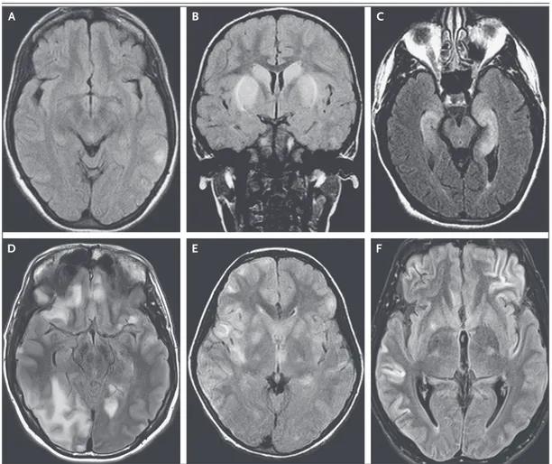
MRI of the head is abnormal in 30% of af-fected patients, mainly showing increased fluid-attenuated inversion recovery (FLAIR) signal involving the cortical, subcortical, or cerebellar regions (Fig. 2A).25 The diagnosis of anti-NMDAR
encephalitis is confirmed by the detection of CSF antibodies against the GluN1 subunit of the NMDAR; serum testing is less reliable, with false negative results in up to 14% of cases.27 In
children who have symptoms suggestive of
anti-NMDAR encephalitis but with discordant MRI changes involving the basal ganglia and brain stem, the possibility of encephalitis due to anti-bodies against the dopamine 2 receptor should be considered (Fig. 2B).28
In contrast to anti-NMDAR encephalitis, lim-bic encephalitis can result from immune re-sponses against several different neuronal cell-surface proteins (Table 1).19 Patients with limbic
encephalitis are usually older than 45 years, with a sex predominance that varies with the type of antibody (Table 1). Symptoms include confusion, behavioral changes, seizures, and inability to form new memories, with relative preservation of the old ones. The MRI scan shows increased FLAIR signal in the medial aspect of the tempo-ral lobes, which in rare cases is enhanced with gadolinium infusion. In some cases, the MRI scan is normal or shows unilateral changes (Fig. 2C). If only one temporal lobe is involved, the differential diagnosis includes cortical edema from ongoing seizures, glioma, and herpes sim-plex encephalitis.
The likelihood and type of underlying tumor and the response to treatment differ according to the type of limbic encephalitis. LGI1 antibod-ies account for the majority of cases of limbic encephalitis, and hyponatremia is a feature of 65% of these cases; an underlying tumor is rare.21 Limbic encephalitis associated with
anti-bodies against γ-aminobutyric acid (GABA)
type B receptor (GABABR) and that associated
with antibodies against α-amino-3-hydroxy- 5-methyl-4-isoxazolepropionic acid receptor (AMPAR) are the next most frequent types of limbic encephalitis; 50 to 60% of patients with limbic encephalitis due to one of these antibod-ies have cancer (Table 1).29,30 Limbic
encephali-tis can also be a manifestation of the aforemen-tioned conventional paraneoplastic syndromes with antibodies against intracellular antigens
(e.g., Hu and Ma2)9 or the 65-kD isoform of
glutamic acid decarboxylase (GAD65). These syndromes usually respond less well to immu-notherapy than do the autoimmune encepha-litides.19
Other autoimmune encephalitides (Table 1) have less distinctive symptoms and MRI find-ings. Certain clinical features nevertheless sug-gest a specific type of autoimmune encepha-litis, such as refractory status epilepticus with
GABA type A receptor (GABAAR) antibodies; encephalopathy, insomnia, dysautonomia, ataxia, peripheral-nerve hyperexcitability, and
neuropathic pain with CASPR2 antibodies23,31;
and myoclonus, tremors, and exaggerated
star-tle responses (hyperekplexia) with DPPX anti-bodies.20,24
In most autoimmune encephalitides, the MRI is normal or shows nonspecific inflammatory changes; two exceptions are limbic encephalitis
Figure 2. MRI Findings in Antibody-Mediated Encephalitis.
Shown are representative MRI scans from patients with several types of autoimmune encephalitides. Anti-NMDAR encephalitis often is present despite normal MRI findings or mild signal abnormalities on fluid-attenuated inversion recovery (FLAIR) images (Panel A). Basal ganglia encephalitis associated with dopamine 2 receptor antibodies typi-cally affects the striatum (Panel B). Limbic encephalitis may result from several different immune responses and is typically indicated by FLAIR signal increases in the medial temporal lobes (Panel C). In contrast, encephalitis with antibodies against γ-aminobutyric acid (GABA) type A receptor (GABAAR ) is usually associated with multiple
corti-cal and subcorticorti-cal FLAIR signal changes (Panel D). Patients with various acute inflammatory demyelinating diseases may have clinical and MRI findings that are indistinguishable from the findings in patients with autoimmune encepha-litis. For example, an MRI scan showing extensive, bilateral FLAIR signal abnormalities was obtained from a patient in whom sudden-onset confusion and encephalopathy developed that were caused by acute disseminated encepha-lomyelitis associated with antibodies against myelin oligodendrocyte glycoprotein (Panel E). The clinical and radio-logic features of autoimmune encephalitides can occasionally be misleading. For example, a young man was admit-ted for severe encephalitis and refractory seizures that required pharmacologically induced coma. Studies showed a large thymoma, α-amino-3-hydroxy-5-methyl-4-isoxazolepropionic acid receptor (AMPAR) antibodies, and MRI find-ings (Panel F) that suggested widespread cortical damage and a poor prognosis. However, removal of the tumor and immunotherapy resulted in complete clinical recovery.
A B C
E F
and anti-GABAAR encephalitis. In GABAAR en-cephalitis, which occurs predominantly in chil-dren and young adults, FLAIR images show multifocal cortical and subcortical signal abnor-malities, mainly in the frontal and temporal lobes and less frequently in the cerebellum and basal ganglia.32 These lesions do not show
diffu-sion restriction or contrast enhancement and resemble the lesions in acute disseminated en-cephalomyelitis (Fig. 2D).
Most patients with autoimmune encephalitis have moderate CSF lymphocytic pleocytosis (<100 cells per cubic millimeter), but the absence of pleocytosis does not rule out the diagnosis. The encephalitides with LGI1 or DPPX antibodies are the ones that most frequently occur with normal MRI findings and normal results of standard CSF studies (cell count and protein and glucose
levels).20,21 In most patients with autoimmune
encephalitis, even those with normal findings on standard CSF studies, neuronal autoantibodies
are detected in CSF.19 However, some patients
with anti-LGI1 encephalitis have antibodies that are detectable only in serum23 or only in CSF.21
The differential diagnosis of autoimmune encephalitis, which is extensive,19 includes acute
disseminated encephalomyelitis, characterized by MRI abnormalities throughout white and gray matter and frequent detection of autoantibodies against myelin oligodendrocyte glycoprotein (Fig. 2E), and neuromyelitis optica spectrum dis-orders, in which the MRI abnormalities are often adjacent to periventricular and ependymal regions and most patients have antibodies against the water channel aquaporin-4, which is expressed in the endfeet of astrocytes. For unexplained rea-sons, these demyelinating syndromes can de-velop concurrently with anti-NMDAR encephali-tis, resulting in overlapping clinical syndromes.33
The clinical and MRI features of autoimmune encephalitides can occasionally suggest a neuro-degenerative process21 or irreversible brain
dam-age reflected by restricted diffusion in regions of the cerebral cortex (Fig. 2F). Clinical recognition of these atypical presentations of autoimmune encephalitides is important because they are potentially treatable with immunotherapy.
Mechanisms of Dise ase The target antigens in autoimmune encephaliti-des are cell-surface proteins involved in neuronal
signaling and synaptic plasticity.1 The associated
syndromes show substantial resemblance to the syndromes observed when the function of the same proteins is altered by genetic modification or pharmacologic antagonists. For example, many clinical features of anti-NMDAR encepha-litis resemble those observed with the
adminis-Figure 3 (facing page). Proposed Mechanisms of Disease and Functional Interactions of Autoantibodies with Neuronal Surface Proteins.
A multistep process results in antibody-mediated neu-ronal cell dysfunction; some of the steps have been shown in reported studies, whereas others are based on proposed hypotheses. Two well-known triggers of autoimmune encephalitides are represented: herpes simplex virus (Panel A) and systemic tumors (Panel B). It is postulated that antigens released by virus-induced neuronal cell destruction or apoptotic tumor cells are loaded into antigen-presenting cells (dendritic cells) and transported to regional lymph nodes.9 In the lymph nodes, naive B cells exposed to the processed antigens, in conjunction with CD4+ T cells, become antigen- conditioned and differentiate into antibody-producing plasma cells. After entering the brain, memory B cells undergo restimulation, antigen-driven affinity matura-tion, clonal expansion, and differentiation into antibody-producing plasma cells (Panel C).35 The contribution of systemically produced antibodies to the pool of anti-bodies present in the brain is unclear and may depend on systemic antibody titers and the integrity of the blood–brain barrier. On the basis of experimental models with cultured neurons, the presence of anti-bodies in the brain may lead to neuronal dysfunction through various mechanisms, including functional blocking of the target antigen (GABA type B receptor [GABABR] antibodies, Panel D), receptor cross-linking
and internalization (NMDAR antibodies, Panel E), and disruption of protein–protein interactions (leucine-rich, glioma-inactivated 1 [LGI1]), potentially affecting the function of the voltage-gated potassium channels and leading to a decrease in the levels of AMPAR (Panel F).1 These mechanisms are influenced by the type of anti-bodies; for example, IgG1 antibodies frequently cross-link and internalize the target antigen, but IgG4 anti-bodies are less effective in cross-linking the target and more often alter protein–protein interactions. A graph based on an in vivo model (Panel G) shows how anti-bodies from patients with anti-NMDAR encephalitis cause symptoms. In this mouse model, passive cere-broventricular infusion of antibodies during 14 days was associated with a progressive increase in brain-bound human antibodies, which was maximal on day 18. The antibodies caused a progressive decrease in syn-aptic NMDAR and loss of memory. All findings were reversed a few days after cessation of the antibody in-fusion, including a gradual decrease in levels of anti-bodies in the mouse hippocampus and restoration of the density of synaptic NMDAR and memory function.36
100 75 50
Percentage Relative to Day 0
Days Ventricular infusion of NMDAR antibodies Intensity of human IgG immunostaining Key
Density of synaptic NMDAR
(no. of synaptic clusters/µm3
of dendrite) Memory (novel-object recognition index) 25 0 0 18 36
A Herpes Simplex Virus
D GABABR Antibodies
G
C
E NMDAR Antibodies F LGI1 Antibodies
Damaged neuron B Systemic Tumor Apoptotic tumor cell Transport to regional lymph nodes Dendritic cell Dendritic cell Antigen uptake Antigen Antigen Virus CD4+ T cell Naive B cell Memory B cell Plasma cell Plasma cell Systemic antibodies Antibodies Differentiation Antigen exposure Differentiation Transport to the brain Maturation L Y M P H N O D E B R A I N P O S T S Y N A P T I C B U L B P R E S Y N A P T I C B U L B P O S T S Y N A P T I C B U L B P O S T S Y N A P T I CB U L B B R A I N Functional blocking of target antigen GABA GABABR G proteins Effector proteins Effector proteins Blocking of receptor signaling Disruption of protein–protein interactions Decrease in AMPAR AMPAR LGI1 ADAM23 Voltage-gated K+ channel ADAM22 Reduced NMDAR density NMDAR Glycine Glutamate Ca2+ and Na+ Cross-linking and internalization α β γ Inactive G proteins α β γ Neuronal Dysfunction Transport to regional lymph nodes nodes Transport to regional lymph regional lymph regional lymph nodes nodes Differentiation Differentiation Differentiation Differentiation Differentiation Differentiation Maturation Maturation Maturation Maturation Memory B cell B cell antibodies antibodies Memory Memory B cell B cell B cell B cell Transport Transport to the brain to the brain Antigen Antigen Antigen Antigen Antigen Antigen Antigen Antigen Antigen Antigen Antigen Antigen Antigen Antigen Antigen Antigen Antigen Antigen Antigen Antigen Antigen Antigen Antigen Antigen Antigen Antigen Antigen Decrease
tration of noncompetitive NMDAR antagonists
(ketamine or phencyclidine).34 The ways in
which the immune response is initiated and the antibodies reach or are produced in the brain are starting to be elucidated. It has been postulated that the autoimmune response is initiated by antigens released by the viral de-struction of neurons (e.g., in herpes simplex encephalitis) (Fig. 3A), by tumors (Fig. 3B), or by unknown mechanisms. In the case of anti-NMDAR encephalitis, there is preliminary evi-dence that memory B cells reach the brain, where they undergo restimulation, antigen-driven affinity maturation, clonal expansion, and differentiation into antibody-producing plasma cells (Fig. 3C). This is supported by brain biopsy and autopsy studies showing plas-ma cells (Fig. 1G), deposits of IgG (Fig. 1H), and
reduced levels of NMDAR37,38 and by CSF
stud-ies showing an ongoing, antigen-driven, intra-thecal immune response characterized by clon-ally expanded plasma cells producing antibodies
against NMDAR.35 Similar mechanisms may
ap-ply to those autoimmune encephalitides that are also characterized by intrathecal synthesis of antibodies, little clinical evidence of blood– brain barrier disruption, and low or undetect-able serum antibody levels in patients with se-vere deficits.
For all autoimmune encephalitides, patho-genic effects of the antibodies have been shown in primary cultures of neurons. These effects include blocking of receptor function (e.g., in the case of GABABR), cross-linking and internal-ization of receptors (NMDAR),38,39 and
interfer-ence with protein–protein interactions (LGI1)40
(Table 1 and Fig. 3D, 3E, and 3F). Even though some antibodies are of subclass IgG1 or IgG3, there is limited evidence that complement fixa-tion plays a major role in autoimmune encepha-litides.10,37 In a mouse model involving passive
cerebroventricular transfer of antibodies from
the CSF of affected patients36,41 or of a human
recombinant antibody derived from CSF plasma cells,35 the antibodies disrupted the interaction
between NMDAR and the ephrin-B2 receptor, leading to receptor internalization, impairment of long-term synaptic plasticity, memory defi-cits, anhedonia, and depressive behaviors. These alterations gradually resolved after the antibody
infusion was stopped (Fig. 3G).36 The
pathoge-nicity of NMDAR antibodies from affected pa-tients has been suggested in other experimental
models.42,43 No animal models are available for
other autoimmune encephalitides.
Tr e atments and Outcome Treatment recommendations are based largely on retrospective series and expert opinion, since few clinical trials have been conducted. The cur-rent approach includes immunotherapy and re-moval of the immunologic trigger, such as tera-toma or another tumor, when applicable. Early tumor treatment is particularly important in
achieving a good outcome.25,29 In most
autoim-mune encephalitides, antibody production and inflammatory changes occur behind the blood– brain barrier, which probably explains the lim-ited effectiveness of plasma exchange and of intravenous immune globulin, in contrast to the beneficial effects of these interventions in sys-temic antibody-mediated diseases such as my-asthenia gravis. Nevertheless, in practice, most patients are treated with glucocorticoids, intra-venous immune globulin, or plasma exchange, and if there is no clinical response, rituximab
and cyclophosphamide are used.25 Rituximab is
usually effective in refractory cases, and it ap-pears to reduce the risk of a clinical relapse,25
which accounts for its increasing use as an
initial treatment.44 Although hyperthermia,
muscle rigidity, mutism, and coma may develop in patients with anti-NMDAR encephalitis in-dependent of the use of neuroleptic agents, studies suggest an increased susceptibility to the adverse effects of these drugs (e.g., the neuroleptic malignant syndrome); the mecha-nisms underlying this complication are
un-known.45
The speed of recovery, degree of residual deficit, and frequency of relapse vary according to the type of autoimmune encephalitis. In a series of 577 patients with anti-NMDAR en-cephalitis, 53% had clinical improvement within 4 weeks, and 81% had substantial recovery (i.e.,
mild or no residual symptoms) at 24 months.25
Another study showed that patients with anti-LGI1 encephalitis had a more rapid response but that only 70% had substantial recovery at 24
months.21 For autoimmune encephalitides that
are frequently associated with cancer, such as
anti-AMPAR and anti-GABABR encephalitides,
the rate of response to immunotherapy is lower, particularly when additional paraneoplastic mechanisms such as antibodies and cytotoxic T-cell responses against intracellular antigens are identified.29
For all types of autoimmune encephalitides, prompt immunotherapy has been associated with a favorable outcome; spontaneous clinical
improvement is infrequent.25 The frequency of
clinical relapse in the encephalitides associated with antibodies against NMDAR, AMPAR, LGI1, CASPR2, or DPPX ranges from 12 to 35%.8,21,24,25,29
Relapses often occur when immunotherapy is
reduced or discontinued.23 There is anecdotal
evidence that cases of anti-LGI1 or anti-NMDAR encephalitis can relapse many years after the first episode. Relapses may herald recurrence of the associated tumor or a tumor that was missed
in the initial episode.25 Immunotherapy and
treatment of the tumor, if it was missed initially, usually result in improvement.
Futur e S tudies
The discovery of the category of autoimmune encephalitides has changed the diagnostic and treatment approach to many neurologic or psy-chiatric syndromes that were previously consid-ered to be idiopathic. The rapid increase in the number of syndromes and autoantibodies iden-tified over the past 10 years suggests that other autoimmune encephalitides have yet to be
dis-covered.1 Antibody titers correlate imperfectly
with the course of the disease and may remain detectable (albeit at a low titer) after clinical
re-covery,27 indicating the need to identify
biomark-ers for prognosis and treatment decisions. The usefulness of neuropsychological testing,
elec-troencephalography,46 advanced neuroimaging,47
and 18F-fluorodeoxyglucose–positron-emission
tomography48 in the diagnosis of autoimmune
encephalitides, assessment of treatment effi-cacy, and prognosis requires investigation. Pre-liminary data suggest that the protracted clini-cal course of anti-NMDAR encephalitis is due to antibody production by long-lived plasma cells in the brain,35 along with the antibody
ef-fects on brain circuitry.41 Further studies are
needed to confirm these hypotheses and deter-mine whether they apply to other autoimmune encephalitides. Studies of how autoantibodies alter the structure and function of synaptic proteins and cause symptoms are critical for an understanding of the underlying pathogenic mechanisms, which in turn could lead to the development of new treatment strategies. For example, the observation that NMDAR anti-bodies alter the interaction between NMDAR
and the ephrin-B2 receptor49 and that a soluble
agonist of the ephrin-B2 receptor antagonizes the antibody effects suggests a potential
treat-ment strategy.41 Finally, knowing how
antibod-ies cause symptoms, such as the psychosis caused by anti-NMDAR antibodies, may help to understand psychiatric diseases in which the same receptors may be altered by other mecha-nisms.50
Disclosure forms provided by the authors are available with the full text of this article at NEJM.org.
We thank Drs. Russell Dale and Shekeeb Mohammad (Chil-dren’s Hospital at Westmead, Australia) and Drs. Leslie Benson and Mark Gorman (Boston Children’s Hospital) for providing MRI scans and Dr. Myrna R. Rosenfeld for reviewing an earlier version of the manuscript.
References
1. Dalmau J, Geis C, Graus F. Autoanti-bodies to synaptic receptors and neuronal cell surface proteins in autoimmune dis-eases of the central nervous system. Physiol Rev 2017; 97: 839-87.
2. de Graaff E, Maat P, Hulsenboom E, et al. Identification of delta/notch-like epidermal growth factor-related receptor as the Tr antigen in paraneoplastic cere-bellar degeneration. Ann Neurol 2012; 71: 815-24.
3. Hinson SR, Romero MF, Popescu BF,
et al. Molecular outcomes of neuromyelitis optica (NMO)-IgG binding to aquaporin-4 in astrocytes. Proc Natl Acad Sci U S A 2012; 109: 1245-50.
4. Carvajal-González A, Leite MI, Waters P, et al. Glycine receptor antibodies in PERM and related syndromes: character-istics, clinical features and outcomes. Brain 2014; 137: 2178-92.
5. Granerod J, Ambrose HE, Davies NW, et al. Causes of encephalitis and differ-ences in their clinical presentations in
England: a multicentre, population-based prospective study. Lancet Infect Dis 2010; 10: 835-44.
6. Gable MS, Sheriff H, Dalmau J, Tilley DH, Glaser CA. The frequency of auto-immune N-methyl-D-aspartate receptor encephalitis surpasses that of individual viral etiologies in young individuals enrolled in the California Encephali-tis Project. Clin Infect Dis 2012; 54: 899-904.
Retrospective analysis of NMDA receptor antibodies in encephalitis of unknown origin. Neurology 2010; 75: 1735-9. 8. van Sonderen A, Petit-Pedrol M, Dal-mau J, Titulaer MJ. The value of LGI1, Caspr2 and voltage-gated potassium channel antibodies in encephalitis. Nat Rev Neurol 2017; 13: 290-301.
9. Darnell RB, Posner JB. Paraneoplastic syndromes involving the nervous system. N Engl J Med 2003; 349: 1543-54. 10. Bien CG, Vincent A, Barnett MH, et al. Immunopathology of autoantibody-asso-ciated encephalitides: clues for patho-genesis. Brain 2012; 135: 1622-38. 11. van Sonderen A, Roelen DL, Stoop JA, et al. Anti-LGI1 encephalitis is strongly associated with HLA-DR7 and HLA-DRB4. Ann Neurol 2017; 81: 193-8.
12. Kim TJ, Lee ST, Moon J, et al. Anti-LGI1 encephalitis is associated with unique HLA subtypes. Ann Neurol 2017; 81: 183-92.
13. Jones HF, Mohammad SS, Reed PW, et al. Anti-N-methyl-D-aspartate receptor encephalitis in Māori and Pacific Island children in New Zealand. Dev Med Child Neurol 2017; 59: 719-24.
14. Lancaster E, Lai M, Peng X, et al. Anti-bodies to the GABA(B) receptor in limbic encephalitis with seizures: case series and characterisation of the antigen. Lan-cet Neurol 2010; 9: 67-76.
15. Armangue T, Moris G, Cantarín- Extremera V, et al. Autoimmune post-herpes simplex encephalitis of adults and teenagers. Neurology 2015; 85: 1736-43.
16. Linnoila JJ, Binnicker MJ, Majed M, Klein CJ, McKeon A. CSF herpes virus and autoantibody profiles in the evaluation of encephalitis. Neurol Neuroimmunol Neuroinflamm 2016; 3(4): e245.
17. Hacohen Y, Deiva K, Pettingill P, et al. N-methyl-D-aspartate receptor antibodies in post-herpes simplex virus encephalitis neurological relapse. Mov Disord 2014; 29: 90-6.
18. Nosadini M, Mohammad SS, Corazza F, et al. Herpes simplex virus-induced anti-N-methyl-D-aspartate receptor en-cephalitis: a systematic literature review with analysis of 43 cases. Dev Med Child Neurol 2017; 59: 796-805.
19. Graus F, Titulaer MJ, Balu R, et al. A clinical approach to diagnosis of auto-immune encephalitis. Lancet Neurol 2016; 15: 391-404.
20. Tobin WO, Lennon VA, Komorowski L, et al. DPPX potassium channel antibody: frequency, clinical accompaniments, and outcomes in 20 patients. Neurology 2014; 83: 1797-803.
21. Ariño H, Armangué T, Petit-Pedrol
M, et al. Anti-LGI1-associated cognitive impairment: presentation and long-term outcome. Neurology 2016; 87: 759-65. 22. Irani SR, Stagg CJ, Schott JM, et al. Faciobrachial dystonic seizures: the influ-ence of immunotherapy on seizure con-trol and prevention of cognitive impair-ment in a broadening phenotype. Brain 2013; 136: 3151-62.
23. Gadoth A, Pittock SJ, Dubey D, et al. Expanded phenotypes and outcomes among 256 LGI1/CASPR2-IgG-positive patients. Ann Neurol 2017; 82: 79-92. 24. Hara M, Ariño H, Petit-Pedrol M, et al. DPPX antibody-associated encephalitis: main syndrome and antibody effects. Neurology 2017; 88: 1340-8.
25. Titulaer MJ, McCracken L, Gabilondo I, et al. Treatment and prognostic factors for long-term outcome in patients with anti-NMDA receptor encephalitis: an ob-servational cohort study. Lancet Neurol 2013; 12: 157-65.
26. de Montmollin E, Demeret S, Brulé N, et al. Anti-N-methyl-D-aspartate receptor encephalitis in adult patients requiring intensive care. Am J Respir Crit Care Med 2017; 195: 491-9.
27. Gresa-Arribas N, Titulaer MJ, Tor-rents A, et al. Antibody titres at diagnosis and during follow-up of anti-NMDA re-ceptor encephalitis: a retrospective study. Lancet Neurol 2014; 13: 167-77.
28. Dale RC, Merheb V, Pillai S, et al. Anti-bodies to surface dopamine-2 receptor in autoimmune movement and psychiatric disorders. Brain 2012; 135: 3453-68. 29. Höftberger R, van Sonderen A, Ley-poldt F, et al. Encephalitis and AMPA receptor antibodies: novel findings in a case series of 22 patients. Neurology 2015; 84: 2403-12.
30. Jeffery OJ, Lennon VA, Pittock SJ, Gregory JK, Britton JW, McKeon A. GABAB receptor autoantibody frequency in ser-vice serologic evaluation. Neurology 2013; 81: 882-7.
31. Joubert B, Saint-Martin M, Noraz N, et al. Characterization of a subtype of autoimmune encephalitis with anti-con-tactin-associated protein-like 2 antibod-ies in the cerebrospinal fluid, prominent limbic symptoms, and seizures. JAMA Neurol 2016; 73: 1115-24.
32. Spatola M, Petit-Pedrol M, Simabukuro MM, et al. Investigations in GABAA re-ceptor antibody-associated encephalitis. Neurology 2017; 88: 1012-20.
33. Titulaer MJ, Höftberger R, Iizuka T, et al. Overlapping demyelinating syn-dromes and anti–N-methyl-D-aspartate receptor encephalitis. Ann Neurol 2014; 75: 411-28.
34. Krystal JH, Karper LP, Seibyl JP, et al.
Subanesthetic effects of the noncompeti-tive NMDA antagonist, ketamine, in hu-mans: psychotomimetic, perceptual, cog-nitive, and neuroendocrine responses. Arch Gen Psychiatry 1994; 51: 199-214. 35. Malviya M, Barman S, Golombeck KS, et al. NMDAR encephalitis: passive trans-fer from man to mouse by a recombinant antibody. Ann Clin Transl Neurol 2017; 4: 768-83.
36. Planagumà J, Leypoldt F, Mannara F, et al. Human N-methyl D-aspartate recep-tor antibodies alter memory and behav-iour in mice. Brain 2015; 138: 94-109. 37. Martinez-Hernandez E, Horvath J, Shiloh-Malawsky Y, Sangha N, Martinez-Lage M, Dalmau J. Analysis of comple-ment and plasma cells in the brain of patients with anti-NMDAR encephalitis. Neurology 2011; 77: 589-93.
38. Hughes EG, Peng X, Gleichman AJ, et al. Cellular and synaptic mechanisms of anti-NMDA receptor encephalitis. J Neurosci 2010; 30: 5866-75.
39. Kreye J, Wenke NK, Chayka M, et al. Human cerebrospinal fluid monoclonal N-methyl-D-aspartate receptor autoanti-bodies are sufficient for encephalitis pathogenesis. Brain 2016; 139: 2641-52. 40. Ohkawa T, Fukata Y, Yamasaki M, et al. Autoantibodies to epilepsy-related LGI1 in limbic encephalitis neutralize LGI1-ADAM22 interaction and reduce synaptic AMPA receptors. J Neurosci 2013; 33: 18161-74.
41. Planagumà J, Haselmann H, Mannara F, et al. Ephrin-B2 prevents N-methyl-D-aspartate receptor antibody effects on memory and neuroplasticity. Ann Neurol 2016; 80: 388-400.
42. Li Y, Tanaka K, Wang L, Ishigaki Y, Kato N. Induction of memory deficit in mice with chronic exposure to cerebro-spinal fluid from patients with anti-N-methyl-D-aspartate receptor encephalitis. Tohoku J Exp Med 2015; 237: 329-38. 43. Wright S, Hashemi K, Stasiak L, et al. Epileptogenic effects of NMDAR antibod-ies in a passive transfer mouse model. Brain 2015; 138: 3159-67.
44. Nosadini M, Mohammad SS, Ram-anathan S, Brilot F, Dale RC. Immune therapy in autoimmune encephalitis: a systematic review. Expert Rev Neurother 2015; 15: 1391-419.
45. Lejuste F, Thomas L, Picard G, et al. Neuroleptic intolerance in patients with anti-NMDAR encephalitis. Neurol Neuro-immunol Neuroinflamm 2016; 3(5): e280. 46. Mohammad SS, Soe SM, Pillai SC, et al. Etiological associations and out-come predictors of acute electroencepha-lography in childhood encephalitis. Clin Neurophysiol 2016; 127: 3217-24.
47. Heine J, Prüss H, Bartsch T, Ploner CJ, Paul F, Finke C. Imaging of autoimmune encephalitis — relevance for clinical practice and hippocampal function. Neu-roscience 2015; 309: 68-83.
48.Probasco JC, Solnes L, Nalluri A, et al. Abnormal brain metabolism on
FDG-PET/CT is a common early finding in autoimmune encephalitis. Neurol Neu-roimmunol Neuroinflamm 2017; 4(4): e352.
49.Mikasova L, De Rossi P, Bouchet D, et al. Disrupted surface cross-talk between NMDA and Ephrin-B2 receptors in
anti-NMDA encephalitis. Brain 2012; 135: 1606-21.
50.Weickert CS, Fung SJ, Catts VS, et al. Molecular evidence of N-methyl-D-aspar-tate receptor hypofunction in schizophre-nia. Mol Psychiatry 2013; 18: 1185-92. Copyright © 2018 Massachusetts Medical Society.