Conservative Treatment of
Spondylodiscitis: Possible
Therapeutic Solution in Case of
Failure of Standard Therapy
Enrico Maria Bonura1, David Joaquin Ortolà Morales1, Domenico Fenga1, Giuseppe Rollo2, Luigi Meccariello2, Danilo Leonetti1, Francesco Traina1, Francesco Centofanti3, Michele Attilio Rosa1
ABSTRACT
Introduction: Spondylodiscitis (SD) is an uncommon disease but not rare, because it
rep-resents around 3–5% of all cases of osteomyelitis. Late diagnosis and/or inadequate treat-ment often cause irreversible damage to cause neurological deficit. Most require only conser-vative treatment, sometimes a surgical approach is required. Aim: The purpose of this study is to propose a conservative protocol to treat spondylodiscitis when the standard conservative treatment has failed. This alternative treatment has been for a long time at the Codivilla-Putti Institute. Methods: We performed a prospective cohort study of 192 consecutive patients who underwent paravertebral intramuscular injections of antibiotic associated with standard treatment at our Center from January 2010 to December 2015 with SD. Of this 192 patients we selected 98 who had already undergone standard antibiotic therapy at another hospital without resolution of the disease. All patients have performed our protocol that provides a total of 3 cycles, each of 3 weeks, repeated at approximately 5 weeks apart. For each patient we evaluated Erythrocyte Sedimentation Rate (ESR), C-Reactive Protein (CRP), White Blood Cells (WBC) indexes, SF36 and VAS Score at the beginning and at the end of the treatment.
Results: At a mean follow up of 22 months (range 60-12), clinical healing was achieved in
87 patients (88,9%) of cases with significant reduction in back pain and functional limita-tion. The VAS Score and the SF36 were better at the end of treatment compared to previous “GOLD STANDARD” treatments in the previous hospitalization in another hospital. In most cases there were slightly reduced in inflammatory indexes. Conclusion: There are no studies in the literature demonstrating the effective efficacy of local infiltrative treatment with anti-biotics, associated with standard treatment protocol. We believe that our protocol in treating SD, favors an early functional recovery, and be able to offer more chance of success than the standard treatment.
Keywords : Spondylodiscitis; Pyogenic spondylodiscitis; Spinal infection; Postoperative spondylodiscitis; Vertebral osteomyelitis; Antibiotic therapy; Conservative treatment.
1. INTRODUCTION
The Spondylodiscitis (SD), also known as vertebral osteomyelitis, represent an infrequent but not rare disease, constitute about 3-5 % of all osteomyelitis with a bimodal distri-bution with peak incidence below 10 years and between 50-70 years of age, and mainly affect males to wom-en (1-3). The incidwom-ence of this disease is constantly increasing, especially in patients with comorbidities such as diabetes mellitus, immunodeficien-cy, tumors, rheumatoid arthritis, alcoholism, hemodialysis, bacterial endocarditis, treatment with immu-nosuppressants and impaired nutri-tional status (4-5).
Staphylococcus aureus is the most frequently isolated etiologic agent in pyogenic vertebral osteomyelitis (PVO), present in 50% of patients (6), followed by gram- bacteria (E. Coli, P. aerouginosa, P. Mirablis, Enteroc-cocchi) (7-9). The treatment of this pathology is not currently standard-ized and is in continuous evolution (10-13).
The gold standard treatment is based, today, on conservative and/ or surgical procedures (5), in cases where there is an important struc-tural deformity (kyphosis above 15°, destruction of the vertebral body), presence of epidural abscess, neuro-logical deterioration, chronic rachis pain, spondylodiscitis secondary to
ORIGINAL PAPER
doi: 10.5455/medarh.2019.73.39-43 MED ARCH. 2019 FEB; 73(1): 39-43
RECEIVED: DEC 23, 2018 | ACCEPTED: JAN 28, 2019
1Department of Biomedical, Dental and
Morphological and Functional Images, Section of Orthopedics and Traumatology, University of Messina, Messina, Italy
2Department of Orthopedics and Traumatology,
Vito Fazzi Hospital, Lecce, Italy
3Center for Osteo-Articular Infections, Codivilla
Putti Istitute, Cortina d’Ampezzo, Italy Corresponding author: Luigi Meccariello, MD. Department of Orthopedics and Traumatology, Vito Fazzi Hospital, Piazzetta Filippo Muratore, Block: A- Floor:V, Lecce, Italy. E-mail: [email protected]. ORCID: https://orcid. org/0000-0002-3669-189X.
© 2019 Enrico Maria Bonura, David Joaquin Ortolà Morales, Domenico Fenga, Giuseppe Rollo, Luigi Meccariello, Danilo Leonetti, Francesco Traina, Francesco Centofanti, Michele Attilio Rosa
This is an Open Access article distributed under the terms of the Creative Commons Attribution Non-Commercial License (http://creativecommons.org/ licenses/by-nc/4.0/) which permits unrestricted non-commercial use, distribution, and reproduction in any medium, provided the original work is properly cited.
surgical treatment or failure of conservative treatment ( 9, 14-15).
The cornerstone of conservative treatment consists of targeted antibiotic therapy lasting between 4 and 12 weeks associated with bed rest and the use of orthopedic busts (9, 14-15). Proper diagnosis and individual therapy can improve clinical outcomes and decrease the likeli-hood of failure and promote the healing. Still, the per-centage of failure reported in the literature goes from 6.2 to 32% (9, 16-22) and the complete recovery from both the neurological and the analgesia point of view is very variable from 50 to 91% of cases (9, 21-25).
2. AIM
The aim of our work is to provide an alternative when the standard conservative treatment has failed, propos-ing a new procedure able to increase the percentage of healing and favor a return to daily life, reducing the pos-sible complications due to the disease itself.
3. METHODS
We performed a prospective cohort study of 192 con-secutive patients who underwent paravertebral intra-muscular injections of antibiotic associated with oral treatment at our Center from January 2010 to December 2015 with PVO. Of this 192 patients we selected 98 ac-cording to our criteria as follows:
Inclusion criteria:
• SD pyogenic haematogenous or post-surgical (not indicated in the exclusion criteria) not responsive to standard treatment;
• Germ isolation causes infection;
• Clinic with rachialgia and functional limitation at the time of admission
• Informed consent to patient treatment and inclu-sion in our study;
Exclusion criteria:
• SD tubercular;
• SD post-surgical with the help of synthetic means and/or grafts;
• Structural deformity or vertebral collapse; • Patients who have not completed the
therapeu-tic protocol, without a minimum follow up of 24 months;
• Serious neurological deficits and/or paravertebral abscesses;
• Allergic to antibiotics;
• Concomitant infections, hepatic and/or renal fail-ure and malignant tumors;
• Use of psychotropic drugs.
We evaluated the Indices of inflammation, white blood cells, VAS Score and SF36 at the beginning and at the end of the treatment. The study was approved by the Eth-ics committee of our institution and informed consent was required.
Therapeutic Protocol
It consists of daily injections of targeted antibiotic, administered by intramuscular paravertebral in corre-spondence of the affected vertebral segment, these are performed in synergy with the standard conservative
treatment. The protocol provides a total of 3 cycles, each of 3 weeks, repeated at approximately 5 weeks apart in which the antibiotic is taken orally.
Antibiotic and Infiltration Preparation Method
A specific antibiotic is diluted with a 1 cc of mepiv-acaine hydrochloride at 2% and then introduced into a syringe of 5 cc. The preparation procedure is performed under conditions of complete sterility, the sterile fenes-trated drape is placed, the skin is disinfected in the af-fected area with iodopovidone (iodine at 10%).
Position of the Patient
The patient is placed in prone decubitus, the syringe containing the diluted antibiotic is placed in the paraver-tebral region of the affected verparaver-tebral tract with needle inclined at an angle of 45° with respect to the horizontal plane.
Choice of the Antibiotic to be Injected by Muscle
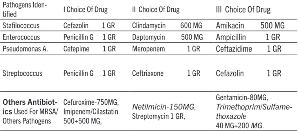
The choice of the antibiotic is based on the interpreta-tion of the antibiogram of the isolated germ, in collabo-ration with the department of infectious diseases, using the breakpoints of the EUCAST guidelines (The Europe-an Committee on Antimicrobial Susceptibility Testing), adapting the values to the sensitivity and resistance of the germs present in our Region. Taking into consider-ation the possibility that the patient may be allergic and/ or is affected by resistant bacteria to a specific drug of first choice, we have been able to implement the protocol equally, administering drugs of second or third choice (Table 1). We have created this scheme based on the breakpoints present in the EUCAST guidelines.
Anesthetic
The use of the antibiotic intramuscularly associated with an anesthetic makes it less painful to inject some antibiotics. In our study we used Mepivacaine hydro-chloride which has an average duration of action of 1,5-2 h to 5 hours, and a Emivita of 2 hours, has a molecular form very similar to that of lidocaine, compared to this lastly, it does not need to be associated with the adren-aline which has an important vasoconstrictive action at the injection site.
Statistical Analysis
Statistical analysis was performed using SPSS software (version 24.0, SPSS, Chicago, USA). Normal distribution was determined by the Kolmogorov-Smirnov test. Cat-egorical variables were compared using the Chi-square test or the non-parametric Mantel-Haenzel test, and continuous variables with Student’s t-test for paired samples.
Data were presented as odds ratio (OR) with 95% con-fidence interval (CI). A value of P <0.05 was considered statistically significant for all tests.
4. RESULTS
The 98 patients are so divided (51 males and 47 fe-males) of average age 57 years (range 27-82 years), the most affected vertebral district was the lumbar side with 80 cases, followed by the dorsal and the cervical respec-tively with 16 and 2 cases. In 29 patients (29,59%) the cause of PVO was secondary to surgery (hemilamino-artrectomy, microdiscectomy, decompression for
verte-bral stenosis). At a mean follow up of 22 months (range 60-12), clinical healing was achieved in 87 patients (88,9%) of cases with a signifi cant reduction in back pain and functional limitation. In the literature, it is shown that in the course of vertebral infection, the values of Erythro-cyte Sedimentation Rate (ESR), C-Reactive Protein (CRP) and White Blood Cells (WBC) are high (26, 27).
In our study, the assessed data were shown to be uniform to those present in the literature
with ESR values at admission which were 40,05 ±(33.72), while at the end of our treatment were ESR 14.14 ± (11.61) and were results statistically signifi cant with a P value (P) less than 0.00 with IC to 95% (25.01 - 38.82); Th e CRP values at admission were 8.09 ± (33.72), while at the end of our treatment were PCR 2.75 ± (2.34) and were results statistically signifi cant with (P) less than 0.003 with a IC to 95% (1.90 - 8.69); Th e WBC values at admission were WBC 6.59 ± (2.08), while at the end of our treatment were WBC 5.34 ± (1.77) and were results statistically signifi cant with (P) less than 0.00 with a IC to 95% (1.72 – 5.23).
Th e microbiology results were based on blood culture, CT guided biopsy or open biopsy. Th e most common off ending organism was methicillin sensitive Staphylo-coccus aureus (MSSA) (n=29), followed by Streptococ-cus (n=16), Methicillin resistant StaphylococStreptococ-cus aureus (MRSA) (n=11), and Escherichia coli (n=7), MSSA was the most common organism found in the treatment group (Table 1, Figure 2).
For each patient were evaluated SF36, VAS Score at the entrance to the hospital, at the end of the third cycle and during follow-up. Th e VAS Score and the SF36 were better at the end of treatment, compared to the previous “GOLD STANDARD” treatment carried out elsewhere. Th e patients in our study are those patients who have not had symptomatic resolution. Th ese had previously been treated at other centers with standard protocol without success.
We found that of these 98 selected patients, treated with our therapeutic protocol, the 88.9% (87 patients out of 98) has healed and is well. We had a 11.1% failure rate (11 patients). Over 80% of patients before starting treat-ment had continuous pain even in the obligatory supine
position. During the fi rst 5 days of treatment, 78% of these patients showed a signifi cant reduction in the VAS Score and managed to assume the position sitting in bed, always with the aid of the spinal brace.
5. DISCUSSION
After careful bibliographic research, we can state that there are no studies to prove the eff ective effi cacy of treatment with paravertebral muscle injection with an-tibiotic, associated with the standard therapeutic proto-col. Th e standard conservative treatment, as understood in the literature, consists in the administration of oral and parenteral antibiotics for a duration between 6 and 12 weeks associated to the immobilization in discharge (8, 9).
In the literature is still debated the optimal duration of conservative treatment with antibiotics by endovenous (EV) and oral administrations (OS) (9, 28-36). In 2015, Rutges indicated that specifi c antibiotic therapy based on the antibiogram by OS and EV it must be adminis-tered for at least 6 weeks (2 weeks EV + 4 weeks OS ), obtaining similar results compared to longer treatments (12 weeks) (9, 20, 34-41).
When possible, the antibiotic therapy should be tar-geted on a well- identifi ed microorganism to treat the PVO. Considered the multiplicity of microorganisms, it is essential to identify the specifi c agent causing the disease to obtain successful therapy. If the patient is neu-rologically intact and has structurally stable lesions, the antibiotic therapy should be postponed until the micro-organism has been identifi ed (2, 9). Once the etiological diagnosis has been performed, the targeted antibiotic therapy requires an assessment of an infectious special-ist. Th e medical treatment fails when the symptoms, es-pecially pain and functional impotence persist or wors-en, the levels of ESR, CRP and WBC may be normal or remain elevated, depending on the state of the infection, whether in the planktonic or quiescent phase of the mi-croorganism, instrumental examinations can help us to demonstrate an evolution of bone damage after specif-ic antibiotspecif-ic therapy during pyogenspecif-ic infections (9, 22, 35). For nonspecifi c SD the duration of treatment varies according to the germ that causes it and the precocious-ness of the diagnosis.
Pathogens
Iden-tifi ed I Choice Of Drug II Choice Of Drug III Choice Of Drug Stafi lococcus Cefazolin 1 GR Clindamycin 600 MG Amikacin 500 MG
Enterococcus Penicillin G 1 GR Daptomycin 500 MG Ampicillin 1 GR
Pseudomonas A. Cefepime 1 GR Meropenem 1 GR Ceftazidime 1 GR
Streptococcus Penicillin G 1 GR Ceftriaxone 1 GR Cefazolin 1 GR
Others Antibiot-ics Used For MRSA/ Others Pathogens Cefuroxime-750MG, Imipenem/Cilastatin 500+500 MG, Netilmicin-150MG, Streptomycin 1 GR, Gentamicin-80MG, Trimethoprim/Sulfame-thoxazole 40 MG+200 MG.
Table 1. Scheme of antibiotic choice, based on the interpretation of the antibiogram, using the breakpoints of the EUCAST guidelines.
Graphic 1.Pathogens isolated in percentage.
Graphic 2. Comparison graphic of the VAS score (left side) and SF 36 (right side) at the beginning and at the end of the treatment
S.Aure us S. Epider midis Strepto
coccusStafilococcus MRSA Enterococcus Pseudo monas A. Escheri chia Coli Anaero bic Bacteri a Polymi crobica l Non Identif. % Pathogens isolated 30% 7% 16% 6% 11% 5% 3% 7% 6% 7% 0,00% 0% 5% 10% 15% 20% 25% 30% 35% 9 10 7 8 3 2 3 1 0 2 4 6 8 10
Beginning of the treatment End of the Treatment
0 20 40 60 80 100 SF 36 Begin SF 36 End Physical health 40 90 Mental health 25 85
The favorable evolution of the treated cases leads to the disappearance of the pains, to the improvement of the general physical state, to the normalization of the inflammation indexes, to the thickening of the bone le-sions, and to the more or less complete ankylosis of the involved vertebrae. From the literature, it is clear that non-tbc SD “usually” have a favorable outcome, benefit-ing only from traditional conservative treatment (8, 14).
In our study, all patients had undergone, previously, a standard targeted antibiotic therapy (parenteral and oral) without resolution of the disease, in 100% of cas-es the germ was isolated by histological examination on percutaneous tc-guided biopsy, test of culture on the sample biopsy, on intraoperative swab or hemoculture. Intramuscular injections are a convenient alternative when medications can not be taken by mouth (some pa-thologies alter their absorption) or there are problems with swallowing. In addition, the intramuscular pathway is a good way to inject drugs that are inactivated by the gastric juices or hepatic level. This way guarantees a bio-availability of the drug almost comparable to the intra-venous one (37). Once the injection at the level of the paravertebral muscles of the affected vertebral tract has been carried out, the blood vessels supplying that par-ticular district, distribute the drug, through the venous vertebral plexuses, to the systemic cardiovascular circu-lation (38); We must however remember the study of Wi-ley and Trueta in which it is noted that the metaphyses and the cartilaginous end plates of the vertebrae repre-sent the most frequent site for the establishment of he-matogenous infection (39); this area consists of multiple slow-flowing anastomoses which, due to their predis-position, allow the drug, via the intramuscular pathway, to remain in the site of infection at the maximum of its concentration and at the same time for a relatively longer time compared to other anatomical districts (38-40).
The efficacy of the treatment was assessed by monitor-ing the indices of blood inflammation and by means of serial radiological controls (X-Ray, CT and MRI) (8, 41, 42). The laboratory findings in association with radiolog-ical images constitute the pivotal points for the diagnosis of vertebral column infections. The speed of erythro-cyte sedimentation (ESR) and levels of C reactive pro-tein (CRP) are fundamental for initial assessment and to follow patient response during and after therapy (2, 26, 27). All patients completed the 3 treatment cycles with duration of each cycle of 3 week, repeated after about 5 weeks, in which the antibiotic was taken exclusively orally.
However, it is documented in the literature that the standard therapeutic protocol has a non-negligible per-centage of failure ranging from 6.2 to 32% (9, 16-22), characterized by persistence of rachialgia and functional limitation; The patients in our study are those patients already treated with the standard therapeutic protocol, who did not get the cure from the disease; we found that in these 98 selected patients, treated with our therapeu-tic protocol, 88,9% (87 patients out of 98) recovered and were well.
Furthermore, by analyzing our statistical data, evaluat-ed through the SPSS program, we found that, the values of inflammation (ESR, CRP and white blood cells), the Vas Score and the SF36, achieved statistically significant results to consider our therapeutic proposal as a further possibility of treatment, effective in treating the non re-sponsive PVO to the standard conservative protocol.
6. CONCLUSION
We believe our protocol in the care of the PVO: • It represents a valid alternative to be considered
in cases where the targeted standard therapy has failed;
• Promote a functional recovery and return to nor-malization of inflammation indices;
• Reduce the pain symptomatology and functional impotence;
• Can offer a chance of success when standard treat-ment has failed.
• Further controlled randomized studies are need-ed in double blind with larger case to demonstrate their actual efficacy and then propose it as a possi-ble standard treatment in the therapy of pyogenic spondylodiscitis.
• Author’s contribution: E.M.B., D.J.O., D.F., G.R., L.M., D.L., F.T., F.C. and M.A.R. gave substantial contribution to the conception or design of the work and in the acquisition, analysis and interpretation of data for the work, E.M.B. G.R. and L.M. had role in drafting the work and revising it critically for important intellectual content. Each author gave final approval of the version to be published and they are agree to be accountable for all aspects of the work in ensuring that questions related to the accuracy or integrity of any part of the work are appropriately investigated and resolved.
• Declaration of patient consent: The authors certify that they have obtained all appropriate patient consent forms.
• Conflicts of interest: None declared. • Financial support and sponsorship: None.
REFERENCES
1. Zarghooni K, Röllinghoff M, Sobottke R, et al. Treatment of spon-dylodiscitis. Int Orthop. 2012; 36: 405-411.
2. Alfonso M. Patologia de la columna vertebral, infecciones de la columna vertebral. 2016; (37): 525-535.
3. Sur A, Tsang K, Brown M, et al. Management of adult sponta-neous spondylodiscitis and its rising incidence. Ann R Coll Surg Engl. 2015 Sep; 97(6): 451-455.
4. Carragee EJ. Pyogenic vertebral osteomyelitis. J Bone Joint Surg Am. 1997; 79: 874-880.
5. Dimar JR, Carreon LY, Glassman SD. et al. Treatment of pyogenic vertebral osteomyelitis with anterior debridement and fusion fol-lowed by delayed posterior spinal fusion. Spine. 2004; 29: 326-332. 6. Torda AJ, Gottlieb T, Bradbury R. Pyogenic vertebral osteomy-elitis: analysis of 20 cases and review. Clin Infect Dis. 1995; 20: 320-328.
7. L. Homagk, N. Homagk, J. R. Klauss, et al. Marmelstein. Spon-dylodiscitis severity code: scoring system for the classification and treatment of non-specific spondylodiscitisEur Spine J. 2016 Apr;25(4):1012-20.
Spon-dylitis beim Erwachsenen. Radiologe. 1996;36:795–804. 9. Rutges JP et al. Outcome of conservative and surgical treatment
of pyogenic spondylodiscitis: a systematic literature review.Eur Spine J. 2016 Apr;25(4):983-99.
10. Stengel D, Bauwens K, Sehouli J, et al. Systematic review and me-ta-analysis of antibiotic therapy for bone and joint in-fections. Lancet Infect Dis 2001;1:175-88.
11. Gasbarrini A, Boriani L, Nanni C, et al. Spinal infection mul-tidisciplinary management project (SIMP): from diagnosis to treatment guideline. Int J Immuno- pathol Pharmacol. 2011;24(1 Suppl 2):95–100.
12. Societe de Pathologie Infectieuse de Langue Francaise (SPLIF) Primary infectious spondylitis, and following intradiscal pro-cedure, without prothesis. Med Mal Infect. 2007; 37:554–572. 13. Sobottke R, Seifert H, Fatkenheuer G, et al. Current
diagno-sis and treatment of spondy- lodiscitis. Dtsch Arztebl Int. 2008;105(10):181–187.
14. D’Agostino C, Scorzolini L, Massetti AP, et al. A seven-year pro-spective study on spondylodiscitis: epidemiological and micro-biological features. Infection 2010;38:102-7.
15. Di Martino A, Papapietro N, Lanotte A, et al. Spondylodiscitis: standards of current treatment. Curr Med Res Opin 2012;28:689-99.
16. Mylona E, Samarkos M, Kakalou E, et al. Pyogenic vertebral os-teomyelitis: a systematic review of clinical characteristics. Semin Arthritis Rheum 2009;39:10-17.
17. McHenry MC, Easley KA, Locker GA. Vertebral osteomyelitis: long-term out- come for 253 patients from 7 Cleveland-area hos-pitals. Clin Infect Dis 2002;34:1342-50.
18. Priest DH, Peacock Jr JE. Hematogenous vertebral osteomyelitis due to Staphylococcus aureus in the adult: clinical features and therapeutic out-comes. South Med J 2005;98:854-62.
19. Kim J, Kim YS, Peck KR et al. Outcome of culture-negative pyo-genic vertebral osteomyelitis: comparison with microbiologi-cally confirmed pyogenic vertebral osteomyelitis. Semin Arthri-tis Rheum. 2014 Oct;44(2):246-52.
20. Roblot F, Besnier JM, Juhel L, et al. Optimal duration of antibi-otic therapy in vertebral osteomyelitis. Semin Arthritis Rheum. 2007; 36(5):269–277.
21. Mavrogenis AF, Megaloikonomos PD, Igoumenou VG, et al. Spon-dylodiscitis revisited. EFORT Open Reviews. 2017;2(11):447-461. 22. Gupta A, Kowalski TJ, Osmon DR, et al. Long-term out-come of pyogenic vertebral osteomyelitis: a cohort study of 260 patients.Open Forum Infect Dis. 2014 Dec 5;1(3):ofu107. 23. Gouliouris T, Aliyu Sh, Brown Nm. Spondylodiscitis:
up-date on diagnosis and management. J Antimicrob Chemother 2010;65:iii11-iii24.
24. Kwon J-W, Hyun S-J, Han S-H, et al. Pyogenic Vertebral Osteo-myelitis: Clinical Features, Diagnosis, and Treatment. Korean Journal of Spine. 2017;14(2):27-34.
25. Khan IA, Vaccaro AR, Zlotolow DA: Management of vertebral diskitis and osteomyelitis. Orthopedics 22:758-765, 1999 26. Meyer B, Schaller K, Rohde V, et al. The C-Reactive Protein For
Detection Of Early Infections After Lumbar Microdiscectomy. Acta Neurochir (Wien) 1995;136:145-50.
27. Mok Jm, Pekmezci M, Piper Sl, et al. Use Of C-Reactive Pro-Tein
After Spinal Surgery: Comparison With Erythrocyte Sedi-Men-tation Rate As Predictor Of Early Postoperative Infectious Com-plications. Spine (Phila Pa 1976) 2008;33:415-21.
28. Grados F, Lescure FX, Senneville E, et al . Suggestion for man-aging pyogenic (non- tuberculous) discitis in adults. Joint Bone Spine. 2007; 74:133–139.
29. Euba g, narváez Ja, nolla JM, et al. Long-term clinical and radio-logical magnetic resonance imaging outcome of abscess-associ-ated spontaneous pyogenic vertebral osteomyelitis under con-servative management. Semin Arthritis Rheum 2008;38:28-40. 30. Rossbach BP, Niethammer TR, Paulus AC, et al. Treatment of
pa-tients with spondylodiscitis and neurological deficits caused by spinal epidural abscess (SEA) is a predictor of clinical outcome. J Spinal Disord Tech. 2014;27(7):395–400.
31. Sobottke R, Zarghooni K, Krengel M, et al. Treatment of spondy-lodiscitis in human immunodeficiency virus-infected patients: a comparison of conservative and operative therapy. Spine. 2009; 34(13):E452–E458.
32. Valancius K, Hansen ES, Hoy K, et al. Failure modes in conser-vative and surgical management of infectious spondylodiscitis. Eur Spine J. 2013; 22(8):1837–1844.
33. Legrand E, Flipo RM, Guggenbuhl P, et al. Management of non-tubercu- lous infectious discitis. Treatments used in 110 patients admitted to 12 teaching hospitals in France. Joint Bone Spine. 2001;68(6):504–509.
34. Mulleman D, Philippe P, Senneville E, et al. Streptococcal and en-terococcal spondylodiscitis (vertebral osteomyelitis). High inci-dence of infective endocarditis in 50 cases. J Rheumatol. 2006; 33(1):91–97.
35. Aagaard T, Roed C, Dragsted C, et al. Microbio- logical and ther-apeutic challenges in infectious spondylodiscitis: a cohort study of 100 cases, 2006-2011. Scand J Infect Dis. 2013; 45(6):417–424. 36. Bernard L, Dinh A, Ghout I, et al. Antibiotic treatment for 6 weeks versus 12 weeks in patients with pyogenic vertebral osteomyeli-tis: an open-label, non-inferiority, randomised, controlled trial. Lancet. (2014)
37. Goonetilleke AK1, Dev D, Aziz I, Hughes C, et al. A comparative analysis of pharmacokinetics of ceftriaxone in serum and pleu-ral fluid in humans: a study of once daily administration by in-tramuscular and intravenous routes. J Antimicrob Chemother. 1996 Dec;38(6):969-7.
38. X.Demondion, G.Lefebvre, O.Fisch, et al. Radiographic anat-omy of the intervertebral cervical and lumbar foramina (ves-sels and variants). Diagnostic and Interventional Imaging.2012 Sep;93(9):690-697.
39. Wiley AM, Trueta J. The vascular anatomy of the spine and its re-lationship to pyogenic vertebral osteomyelitis. J Bone Joint Surg Br 1959; 41-B: 796–809.
40. Mahesh V. Jayaraman. Preoperative and Therapeutic En-dovascular Approaches for Spinal Tumors: Tumors of the Spine.2008 May:(14).
41. An Hs, Seldomridge Ja. Spinal Infections: Diagnostic Tests And Imaging Studies. Clin Orthop Relat Res 2006;444:27-33. 42. Modic Mt, Feiglin Dh, Piraino Dw, Et Al. Vertebral