UNIVERSITÀ DEGLI STUDI DI CATANIA
TESI DI DOTTORATO IN FARMACOLOGIA PRECLINICA E CLINICA CICLOXXIV
__________________________________________________ ______________________________________
DOTT.SSA LAURA SCUDERI
PEGYLATED-INTERFERON AND RIBAVIRIN THERAPY OF HCV-RELATED TYPE II MIXED CRYOGLOBULINEMIA
—————
TESI SPERIMENTALEDI DOTTORATO
——
Coordinatore:
Chiar.mo Prof. Renato Bernardini
Tutor:
Chiar.ma Prof.ssa Leonarda Mariani
INTRODUCTION………...p. 3 METHODS Study design………...…….p. 20 Selection of patients………p. 20 Statistical analysis……….…..p. 31 RESULTS
Characteristics of the HCV-related
type II MC patients……….p. 32 Treatment-related data and outcomes……….…p. 38 Virological response……….…..p. 38 Clinical response……….……p. 40 Immunological response……….………p. 45 Tolerance of therapy………...p. 49 MLR analyses……….p. 50 DISCUSSION……….p. 51 CONCLUSIONS………...p. 57 REFERENCES………..……..p. 59
INTRODUCTION
Hepatitis C virus (HCV) is an enveloped RNA virus
belonging to the Flaviviridae family [1]. It has been classified into six major genotypes and more than 50 subtypes [2]. It causes chronic infection in 200 million people worldwide [3]. As a hepatotropic virus, almost 80-90% of the infected patients develop Chronic Hepatitis (CH), followed by Hepatic Cirrhosis (HC) in 10–20% and Hepatocellular Carcinoma (HCC) in 1–5% [4-5]. In some cases, HCV infection cannot be associated with hepatic disease. HCV+ patients without hepatic disease have not clear clinical, biochemical and histological signs of liver involvement. Resolution of HCV infection is possible in 10% of cases, while chronic HCV infection defined by persistent HCV
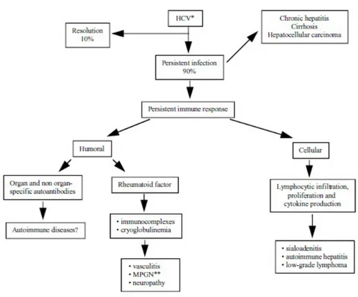
RNA in the serum is observed in 90% of cases and it is responsible of a persistent humoral and cellular immune response (Table 1) [6].
Table 1. Spectrum of immunologic perturbations and extrahepatic diseases after persistent hepatitis C virus infection
∗∗Membranoproliferative glomerulonephitis.
Humoral immune response: antibody response to HCV may
lead to formation of immune complexes. They comprise monoclonal immunoglobulin M (IgM) with rheumatoid factor (RF) activity, polyclonal immunoglobulin G (IgG) with anti-HCV activity, HCV RNA and fractions of complement. Deposition of immune complexes in organs and tissues is considered responsible of the multiorgan disease [7]. Moreover, a variety of autoimmune disorders have been described in patients with HCV infection, including autoimmune hepatitis [8-11], Sjogren syndrome [12], lichen planus [13], thyroiditis [14-15], glomerulonephritis [16] and polyarteritis nodosa [17]. The prevalence of autoantibodies and immunologic abnormalities is significantly higher in patients with HCV-related CH (CCH) than
in patients with liver diseases of different etiology, suggesting that HCV could play a role in their pathogenesis [18].
Cellular immune response: HCV binds to B lymphocytes via CD81 facilitating the production of autoantibodies [19]. The role of T lymphocytes in HCV infection has not been well elucidated [20]. Focal lymphocytic aggregates, composed by a core (B lymphocytes mixed with CD4+ T helper lymphocytes) surrounded by T suppressor/cytotoxic lymphocytes, occur in portal tracts in 59% to 85% of liver biopsy specimens of patients with chronic hepatitis C; the high prevalence of lymphoid follicles in liver is considered a specific histologic feature of the disease [21-22], suggesting that persistent viral infection leads to a lymphocytic proliferation followed by infiltration of organs and tissues and production of cytokines [21;23]. The role of cytokines is to inhibit viral replication and eradicate infection [24].
However, this mechanism is not sufficient to achieve a complete viral clearance.
Extrahepatic diseases: occur in approximately 40% of HCV
infected patients (Table 2) [6;25].
CLOSELY ASSOCIATED TO HCV INFECTION PROVISIONALLY ASSOCIATED TO HCV INFECTION Glomerulonephritis (membranous membranoproliferative) Cutaneous vasculitis (leukocytoclastic) Low grade non-Hodgkin lymphoma Neuropathy Mixed cryoglobulinemia Polyarteritis nodosa Sjogren syndrome Polymyositis (dermatomyositis) Arthritis
Idiopathic pulmonary fibrosis Pulmonary and intestinal vasculitis Ocular (Mooren’s corneal ulcers) Hematologic (aplastic anemia) Thyroiditis
Lichen planus
Table 2. Extrahepatic diseases of chronic HCV infection
The most frequent extrahepatic disease in HCV+ patients is
Mixed Cryoglobulinemia (MC), a potentially life-threatening,
systemic vasculitis affecting small and, less frequently, medium caliber arteries and veins, characterized by deposition of immune complexes on endothelial surface, eliciting vascular inflammation through poorly understood mechanisms [26-27].
Cryoglobulinemia is defined as the presence in the serum of one (monoclonal cryoglobulinemia) or more immunoglobulins (mixed cryoglobulinemia), called cryoglobulins, which reversibly precipitate at temperatures below 37 °C and redissolve on re-warming [28-31]. According to immunoglobulin composition, cryoglobulinemia is traditionally classified into three subgroups [31-32]:
Type I cryoglobulinemia composed by a single monoclonal
immunoglobulin, usually IgM or, less frequently, IgG. It accounts for 10% to 15% of people with cryoglobulinemia. It is mainly found in patients with lymphoproliferative disorders (immunocytoma/Waldenström macroglobulinemia, multiple myeloma) [28-31;33].
Type II and Type III mixed cryoglobulinemia [31;34-35]
are composed by polyclonal IgGs and mono- or polyclonal IgMs, respectively. They occur in almost 80-85% of patients with cryoglobulinemia: type II in 50-60% and type III in 20-30%. The IgMs typically has rheumatoid factor (RF) activity. MC can be associated with different infectious, immunological and neoplastic diseases [28-31;33].
“Essential” MC refers to patients for whom no cause of MC
has been identified. Since the identification of HCV in 1989 (HCV is the most cause of MC), less of 5% of cases are considered “essential” [33;36-37].
The prevalence of HCV infection in MC patients varies geographically, with high values (over 90%) in the Mediterranean area [7;33;36]. MC is found in 10% to 70% of patients with chronic HCV infection, with only 5% to 10% of them developing clinical symptoms of MC.
Several epidemiological and clinico-pathological observations suggest that MC is the result of a multifactorial and multistep pathogenetic process [27;36;38]. While the immune-complex-mediated vasculitis is the final step of this complex process, B-lymphocyte expansion [29] may represent the remote
disorder responsible for production of pathologic quantities of immunoglobulins with rheumatoid factor activity that form cryoglobulins. The deposition of cryoglobulins on endothelial surface of vessels elicits a vascular inflammation inducing vascular damage. The mechanisms responsible for the B-lymphocyte expansion remain still unknown [27;36;38-40]. A direct role of HCV in the B-cell expansion has been hypothesized on the basis of the high frequency of HCV-RNA positive lymphocytes in cryoglobulinemic patients demonstrated by RT-PCR (reverse transcription–polymerase chain reaction), in situ hybridization and immunohistochemical techniques [27;36;38-40]. Since HCV is a RNA virus without reverse trascriptase activity, viral genome cannot integrate in the host genome and thus cannot regulate the cell replication and/or survival. Probably, HCV may exert its oncogenic potential, indirectly, through viral
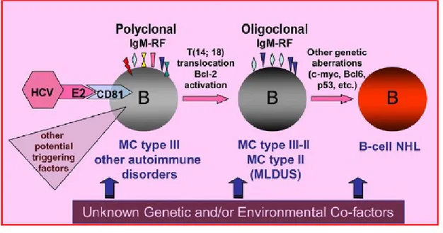
epitopes, autoantigen production, and/or molecular mimicry mechanism [7;27;36;38-49]. Several pathogenetic hypothesis were suggested but yet the exact role of HCV in the lymphoproliferation is not completely clarified [27]. Studies in vitro showed an interaction between the E2 protein of the viral envelop and the CD81 receptor of infected B-cells; this interaction might increase genetic rearrangements [27], as showed by the high frequency of t(14;18) translocation or Bcl-2 rearrangement in infected B-cells [50]. The subsequent activation of the protooncogene Bcl2, with antiapoptotic activity, could be responsible for the prolonged survival of lymphocytes. Similarly, the prolonged B-cell survival might represent a predisposing condition for further genetic aberrations, which might lead to frank B-cell malignancy [51].
Fig. 1. B-cell proliferation represents the biological substrate of mixed cryoglobulinemia (MC). The evolution from a simple serological alteration to clinical immunological manifestations and ultimately to frank B-cell non-Hodgkin's lymphoma (B-NHL) is a multistep and multifactorial process. HCV is the main triggering agent; it represents a chronic stimulus through the interaction of HCV-E2 antigen (and/or other viral antigens) with CD81 on B-cell surface. But other infectious, environmental, and genetic factors are probably involved. RF: rheumatoid factor; MLDUS: monotypic lymphoproliferative disorder of undetermined significance [27].
In 1966, Meltzer described MC as the clinical triad of palpable purpura, arthralgia, and asthenia accompanied by multiple organ involvement and elevated serum RF [30]. It is now
known that this triad is rare. In more recent studies, other clinical features were added, including an evolution to low-grade non-Hodgkin lymphoma, observed in few patients (Table 3) [6;52].
Fatigue 100%
Monoclonal rheumatoid factor 99%
Reduced C4 level 95% Cutaneous vasculitis 95% Palpable purpura 90% Hepatitis 85% Arthralgias 75% Glomerulonephritis 55% Peripheral neuropathy 50% Splenomegaly 40% Raynaud phenomenon 25% Sjogren syndrome 25% Neurocognitive impairment 25% Lymphoadenomegaly 15% Skin ulcers 15% Thyroiditis 5% Low-grade non-Hodgkin lymphoma 5%
Table 3. Frequency of clinical and serologic features of essential mixed cryoglobulinemia associated with hepatitis C virus infection
The disease expression is variable, ranging from mild clinical symptoms (purpura, arthralgia) to fulminant life-threatening complications (glomerulonephritis, widespread vasculitis).
Palpable purpura is the most frequent clinical manifestation of MC [33], and, usually, begins at the lower limbs and may extend to the abdominal area and less frequently to the trunk and upper limbs. It is characterized by petechial or papular lesions, seldom with necrotic aspect. It heals leaving brownish skin patches formed of hemosiderin deposits. Purpuric lesions may occasionally progress to chronic ulcers and frank gangrene. Raynaud’s syndrome and acrocyanosis can also occur. A lot of MC patients are asymptomatic.
The Italian Group for the Study of Cryoglobulinemia has proposed serological, pathological, and clinical criteria for the diagnosis of MC patients (Table 4) [28;36].
Table 4. Criteria for the diagnosis of MC proposed from GISC
Abbreviations: HCV, hepatitis C virus; HBV, hepatitis B virus; MPGN, membranoproliferative glomerulonephritis
“Definite” Mixed Cryoglobulinemia Syndrome:
1) serum mixed cryoglobulins (± low C4) + purpura + leukocytoclastic vasculitis
2) serum mixed cryoglobulins (± low C4) + 2 minor clinical symptoms + 2 minor serological/pathological findings
No initial treatment is required in asymptomatic MC, while in symptomatic disease the therapeutic approach should be adapted to the individual patient according to the intensity of clinical symptoms (Table 5) [53].
CATEGORY ACTIVITY Corticosteroids Cyclophosphamide Plasma-exchange Interferon + Ribavirin Anti-CD20 antibody (rituximab) Anti-inflammatory, immunosuppressive Immunosuppressive
Removal of circulating immune complexes Antiviral (HCV eradication)
Selective suppression of B-cell clone
Table 5. Main therapeutic options for symptomatic patients with HCV-related MC.
Before the interferon (IFN) era, patients with significant organ system involvement were treated with corticosteroids, cytotoxic immunosuppressive drugs such as cyclophosphamide and chlorambucil, or plasmapheresis alone or in combination. These conventional therapies induced short- to medium-term
remissions, although none was clearly effective in inducing long-term remissions.
Given the documented association with HCV and its role in the pathogenesis, the treatment of HCV-related MC should be directed to HCV eradication. IFN-alpha therapy produces a rapid decrease in HCV RNA levels with significant clinical improvement, but cessation of therapy is characterized by recurrence of viremia and cryoglobulinemia in almost all patients [43;54-55]. About 80% of responders relapse within 6 months after its suspension, while a sustained viral response is achieved in almost 20% [55]. Low HCV RNA levels are predictive of a favourable response [56]. By analogy with the management of chronic HCV hepatitis, combination of IFN-alpha and ribavirin (RBV) improved the clinical response [57]. A more favourable
response rate was reported with the use of pegylated IFN-alpha (peg-IFN-alpha) and RBV [58]. Peg-IFN-alpha (2a or 2b) has a polyethylene glycol (peg) moiety added to interferon that confers a longer half-life. Actually, more studies are needed to evaluate safety and efficacy of peg-IFN-alpha plus RBV for the treatment of MC in HCV+ patients.
The aim of our study was to evaluate safety and efficacy of peg-IFN alpha-2a in combination with RBV for the treatment of HCV-related type II MC with detectable HCV-RNA levels in patients with and without hepatic disease.
METHODS
Study design.
The study was a longitudinal prospective open-label uncontrolled study performed in cooperation with two Hepatology Units. Peg-IFN alpha-2a in combination with RBV were administered according to current guidelines for standard treatment of CCH [59-63]. All patients provided their informed consent prior to their inclusion in the study.
Selection of patients.
Inclusion criteria for the study were [60-61]: age 18 years or
older, anti-HCV antibodies positivity, detectable HCV-RNA levels, HCV genotype 1b, clinical and laboratory signs of MC
vasculitis in the absence of any other condition known to cause vasculitis.
Exclusion criteria for the study were: age 66 years or older,
HBV co-infection, HIV co-infection, HCV genotype other than 1b, decompensated HC, significant atherosclerotic heart disease (defined as instrumental and clinical features of coronary heart disease and chronic heart failure); alcohol or drug abuse, history of haematological disorders or neoplastic diseases, pregnancy, psychiatric disorders or autoimmune diseases.
All patients underwent HCV-RNA and HCV genotype determination. HCV-RNA levels were detected by polymerase chain reaction (PCR) of HCV-RNA 5’ UTR using COBAS® AmpliPrep/COBAS® TaqMan® HCV Test (Roche Diagnostics Systems, Branchburg, N.J.; analytic sensitivity = 50 IU/mL) [64].
HCV genotypes were determined by INNO-LiPA (Innogenetics) assay, using Simmond’s classification [65-66]. Anti-HCV antibodies was assayed by the second generation (four-antigen) immuno-enzymatic screening test ORTHO-HCV (Ortho Diagnostic Systems, Raritan, NJ, USA) [67]. Hepatitis B virus (HBV) and human immunodeficiency virus (HIV) markers were detected by enzyme-linked immunosorbent assay (ELISA) using commercially available kits. Values for the liver function tests (ALT, AST) as well as hematological parameters (haemachrome, sodium, potassium, glycaemia, azotaemia, creatininaemia) were determined by usual laboratory methods. Anti-nuclear (ANA), antimitochondrial (AMA), smooth muscle (SMA), anti-liver/kidney microsome type 1 (LKM1) autoantibodies were measured using immunofluorescence assay (IFA) and semi-quantitative ELISA immobilizing enzyme test [68]. Rheumatoid
factor (RF), C3 and C4 fractions of complement were measured by rate nephelometry. Thyroid function was evaluated by levels of TSH, free T3 and free T4, that were determined by immunoradiometric assay (IRMA); antibodies against thyroid peroxidase and thyroglobulin were measured by IFA [69]. Cryoglobulin determination was performed according to standard methods [33]: the cryoprecipitates, diluted in 0.5 M NaCl, were fractionated by high-resolution gel electrophoresis to type cryoglobulins. Individual monoclonal bands were identified by immunofixation after electrophoresis using a cellulose acetate strip impregnated with antibodies specific for heavy and light chains. Mixed cryoglobulins were classified as type II on the basis of the presence of monoclonal IgM immunoglobulins with RF activity complexed with polyclonal IgG. All patients underwent ultrasound-assisted percutaneous biopsy, obtained with Menghini
modified needles (Automatic Aspiration Needle for Liver Biopsy, ACR 16G, 11 cm, manufactured by Sterylab Srl, Milan-Italy) within 6 months before the start of the study. Histological evaluation of the degree of necroinflammatory activity (grading) and fibrosis (staging) of hepatic tissue were assessed according to METAVIR scoring system: activity (A) was graded according the intensity of the necroinflammatory lesions: A0, no activity; A1, mild activity; A2, moderate activity; A3 and A4, severe activity. The stage of fibrosis (F) was graded as follows: F0, no fibrosis; F1, portal fibrosis without septa; F2, portal fibrosis with some septa; F3, portal fibrosis with numerous septa; F4, cirrhosis. A skin biopsy of a purpuric lesion was performed in all patients. Each samples were placed in buffered formalin and stained with haematoxylin and eosin. Histological evaluation revealed leukocytoclastic vasculitis.
On the basis of clinical, biochemical, virological, and histological data, from a total of 235 consecutive HCV+ patients evaluated at the Department of Internal Medicine and Systemic Diseases and at the Department of Tropical Diseases, “Policlinico-Vittorio Emanuele” Hospital, University of Catania between 2006 and 2011, we selected 24 patients affected by HCV-related type II MC with detectable HCV-RNA levels (Fig.2).
The diagnosis of MC was made according to serological, pathological and clinical criteria proposed in 1989 from the Italian Group for the Study of Cryoglobulinemia (GISC) (Table 4) [28;36].
Of these 24 patients with HCV-related type II MC, 16 patients [6 male and 10 female, mean age 57 years old (range 48– 61), mean body weight 69 kg (range 61–80)] had chronic liver disease (14 with CH, 2 with Child-Pugh Class A HC) and 8 patients [3 male and 5 female, mean age 37 years old (range 34– 40), mean body weight 68 kg (range 61–75)] had not liver involvement (normal level of liver enzymes, liver activity and fibrosis score < 1) (Fig.3).
Fig.3 Percent of HVC-CM patients with (16/24) and without (8/24) hepatic disease
All patients were Caucasian, heterosexuals and had no history of alcohol or drug abuse. All patients were enrolled and eligible to receive antiviral therapy with standard dose of peg-IFN alpha-2a 180 micrograms once weekly and weight based ribavirin (WBR) 1000-1200 mg/day for 48 weeks.Therapeutic protocol and efficacy assessment.
The study protocol conformed to the ethical guidelines of the 1975 Declaration of Helsinki (6th Revision, 2008). According to current guidelines [4] at time 0, all patients started therapy with subcutaneous peg-IFN alpha-2a, 180 micrograms once weekly, plus oral WBR in two separate doses (the total dose was 1200 mg daily for patients weighing >75 kg, and 1000 mg daily for those weighing <75 kg). Two weeks after the start of therapy and subsequently on a monthly basis, clinical and biochemical parameters were evaluated. The clinical evaluation included cutaneous manifestations such as purpura, Raynaud’s phenomenon, and distal ulcers, while biochemical evaluation included haemochrome, sodium, potassium, glycaemia, fractionated bilirubin, azotaemia, creatininaemia, serum AST and ALT levels, TSH, free T3, free T4, antibodies against thyroid peroxidase and thyroglobulin, RF, C3 and C4 fractions of
complement, and cryoglobulin levels. In compliance with international guidelines and recommendations of the American Association for the Study of Liver Disease (AASLD) and the Italian Association for the Study of the Liver (AISF), HCV-RNA levels were measured in all patients at time 0 and at weeks 12, 24, 48 and 72 [4;61]. Patients with a HCV-RNA reduction of at least 2 log10 at week 12 in comparison with the pre-treatment
HCV-RNA values were classified as early virological responders (EVR) and continued therapy. Patients who did not reach this target were classified as non-responders and stopped therapy. Relapse was defined as undetectable serum HCV-RNA levels (<50 IU/mL) at the end of treatment but positivity at the end of follow-up. A sustained virologic response (SVR) was defined as undetectable serum HCV-RNA levels 24 weeks after the end of therapy (week 72). Clinical response was defined by analyzing the evolution of
cutaneous manifestations of vasculitis. A complete clinical response was defined as disappearance of skin lesions. A partial clinical response was defined as a decrease of >50% of skin lesions compared with baseline. Patients who had neither a complete clinical response nor a partial clinical response were classified as non-responders. Relapse was defined as the partial or complete reappearance of cutaneous manifestations of vasculitis after the end of treatment. Immunological response was defined by analyzing the serum levels of RF and cryoglobulins. A complete immunological response was defined as normalization of serum RF levels and disappearance of circulating cryoglobulins. A partial immunologic response was defined as a decrease in the serum level of RF and cryoglobulins > 50% compared with baseline. Patients who had neither a complete nor a partial immunological response were classified as
immunological non-responders. Relapse was defined as the partial or complete normalization of serum RF and cryoglobulins during therapy followed by return to higher values during follow-up. The efficacy of treatment was evaluated by analyzing clinical, virological, and immunological responses at time 0 and at weeks 12, 24, 48 and 72.
Statistical analysis.
Results are presented as means ± standard deviation (SD), range and frequencies. Fisher’s Exact test and Mann Whitney U Test for independent samples were applied. Two-tailed P values <0.01 were considered statistically significant. Significant predictors of a complete clinical response were evaluated by multiple logistic regression (MLR) analyses.
RESULTS
Characteristics of the HCV-related type II MC patients.
The baseline (week 0) characteristics are detailed in Table 6.
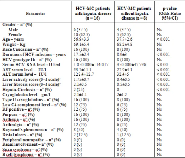
The mean ± SD age was higher in HCV-related type II MC patients with hepatic disease (56.9±3.3 years) than without hepatic disease (37.7±2.6 years). In both groups of patients there was a higher prevalence of female. The mean body weight in both groups was similar (about 69 Kg). All patients were Caucasian. The mean ± SD duration of HCV infection was greater in patients with hepatic disease (17.5±2.4 years) than without hepatic disease (8.9±3 years). The HCV genotype was 1b in all 24 patients and the mean ± SD serum HCV-RNA levels were higher in patients
with hepatic disease (1.050.000±114.017 IU/ml) than without hepatic disease (450.000±37.796 IU/ml). Moreover, serum transaminase levels were abnormally increased in patients with hepatic disease [AST (UI/l) = 93.7±11.1; ALT (UI/l) = 128.4±12.7] while normal in patients without hepatic disease [AST (UI/l) = 27.9±4.1; ALT (UI/l) = 32.4±5]. According to the Metavir criteria, the mean ± SD liver activity (A) and fibrosis (F) score in patients with hepatic disease were A=1.75±0.7 and F=2.1±0.5, while A=0.4±0.5 and F=0.5±0.5 in the patients without hepatic disease. In the first group 2/16 patients were affected by Child-Pugh Class A HC. The type II cryoglobulins were found in all 24 patients and the mean cryoglobulin levels in both groups were similar (about 2 gm/l). Low C4 complement levels (< 0.15 gm/l) and elevated RF levels (> 15 IU/ml) were observed in the most patients: 12/16 with hepatic disease and 6/8
without hepatic disease. Clinical manifestations of MC included purpura (Fig.4-6) and asthenia in 16/16 (100%) and 8/8 (100%), arthralgia in 12/16 (75%) and 6/8 (75%), Raynaud’s phenomenon in 8/16 (50%) and 4/8 (50%), and distal ulcers (Fig.7) in 2/16 (12.5%) and 1/8 (12.5%) patients with and without hepatic disease, respectively. Other clinical manifestations of MC such as peripheral neuropathy, renal involvement, sicca syndrome, and B cell lymphoma were not observed in our patients.
Table 6. Baseline (week 0) characteristics of all patients and significant predictors of a complete clinical response evaluated by multiple logistic regression (MLR) analyses (n = 24)
Abbreviations. HCV-MC =hepatitis C virus–associated mixed cryoglobulinemia; ALT: Alanine Aminotransferase; AST: Aspartate Aminotransferase; RF: Rheumatoid Factor; GI: gastrointestinal RF levels > 15 IU/ml were considered positive
C4 complement levels < 0.15 gm/l were considered low Except where indicated otherwise, values are the mean ±SD.
† Liver activity and fibrosis were graded according to the Metavir scoring system. All liver specimens were assessed by a local pathologist for histology status and reviewed by one expert pathologist, blinded about specimens group origin. P was calculated by multiple logistic regression analysis. ANOVA was also performed inside each group.
Fig.4
Fig.6
Treatment-related data and outcomes. The main
treatment-related data are summarized in Tables 7-8.
Virological response.
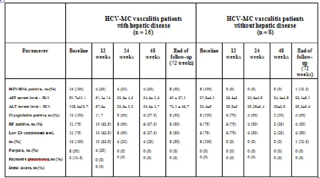
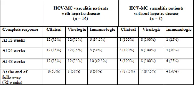
At 12 weeks, 4/16 (25%) patients with hepatic disease (2 with CH and 2 with Child-Pugh Class A HC), were non-responders to therapy and stopped it. Twenty remaining patients (12 with CH, 8 without hepatic disease) early virological responders (EVR) continued therapy until to 48 weeks. At the end of treatment (EOT), HCV-RNA levels became undetectable in 12/16 (75%) and 8/8 (100%) patients with and without hepatic disease, respectively, and they were classified as ETR (end of treatment responders). At the end of follow-up (EFU), 8 patients out of 16 in the first group (50%) achieved a SVR, 4/16 (25%) non-responders and 4/16 (25%) relapsers, while 7 patients out of 8 in
the second group (87.5%) achieved a SVR and 1/8 (12.5%) relapsers. In conclusion, at the end of follow-up, we observed a higher rate of SVR (87.5% vs 50%) in patients without hepatic disease compared with patients with hepatic disease (p<0.01) (Fig.8).
Clinical response.
At the EOT, a complete clinical response was found in 12/16 (75%) and 8/8 (100%) patients with and without hepatic disease, respectively, and they were classified as complete responders. At the EFU, 8 patients out of 16 in the first group (50%) were complete responders, 4/16 (25%) non-responders and 4/16 (25%) relapsers, while 7 patients out of 8 in the second group (87.5%) were complete responders and 1/8 (12.5%) relapsers. In conclusion, at the end of follow-up, we observed a strict association between the eradication of HCV and a complete clinical response with a higher rate of complete clinical response (87.5% vs 50%) in patients without hepatic disease compared with patients with hepatic disease (p<0.01) (Fig.9-14).
Fig.10
Fig.12
Immunological response.
At the EOT, normalization of serum RF levels and disappearance of circulating cryoglobulins were observed in 10/16 (62.5%) and 6/8 (75%) patients with and without hepatic disease, respectively, and they were classified as complete responders. At the EFU, 8 patients out of 16 in the first group (50%) were complete responders, 6/16 (37.5%) non-responders and 2/16 (12.5%) relapsers, while 4 patients out of 8 in the second group (50%) were complete responders, 2/8 (25%) non-responders and 2/8 (25%) relapsers. In conclusion, at the end of follow-up, we observed the same rate of complete immunological response (50%) in patients with and without hepatic disease (Fig.15).
Table 7. The main treatment-related data
Abbreviations. HCV-MC =hepatitis C virus–associated mixed cryoglobulinemia; ALT: Alanine Aminotransferase; AST: Aspartate Aminotransferase; Rheumatoid Factor (RF) levels > 15 IU/ml wereconsidered positive
C4 complement levels < 0.15 gm/l were considered Cryoglobulin level > 0,05gm/l was considered positive Except where indicated otherwise, values are the mean ±SD.
Table 8. Chronologic response to antiviral therapy*
* Values are the number (%) of patients.
Abbreviations. HCV-MC =hepatitis C virus–associated mixed cryoglobulinemia
P values were calculated using Fisher’s exact test. Two-tailed p value <0.01 was considered statistically significant
Tolerance of therapy.
Four patients out of 16 in the first group (25%) were non-responders to therapy and stopped it at 12 weeks. The side effects in the remaining 20 patients [12/16 (75%) with hepatic disease and 8/8 (100%) without hepatic disease)] included fever, asthenia,
loss of concentration, insomnia, anxiety, mild depression, mild anemia, mild leukopenia and thrombocytopenia. The fever was successfully treated with administration of paracetamol. Asthenia,
loss of concentration, insomnia, anxiety, mild depression were successfully controlled by Psychological Support Programmes or, sometimes, administration of antidepressant drugs. No severe psychiatric side effects, such as major depression or psychosis have been reported during the antiviral treatment administration as well as in the follow-up. Mild anemia, mild leukopenia and
thrombocytopenia didn’t require adjuvant treatments [70]. No dosage reductions or interruptions of therapy were needed. No cases of death occurred during the study.
MLR analyses.
Significant predictors of a complete clinical response are presented in Table 6. In our regression model, factors significantly associated with a complete clinical response were: age < 40 years old (p < 0.001), duration of HCV infection <12 years (p < 0.001), serum HCV-RNA levels < 500000 IU/l (p < 0.001), normal transaminases levels (p < 0.001), liver activity and fibrosis scores < 1 (p < 0.001).
DISCUSSION
HCV is the most frequent cause of MC and it is primarily associated with type II MC and less frequently, with type III MC [28;33;36]. All HCV genotypes have been found in MC. The MC prevalence in HCV+ patients ranges widely from 10-70%. Female gender is slight prevalent [32;71]. MC is more frequent in Southern Europe than in Northern Europe and North America. In our study, we observed type II MC and genotype 1b HCV in all patients, the MC prevalence was 24/235 (10.2%) patients, F/M ratio was 15/9 and all patients were Caucasian (all Italians).
HCV is a hepatotropic virus that can lead to CH, HC and HCC [4-5], but, in some cases, it cannot be associated with
hepatic disease. HCV+ patients without hepatic disease have not clear clinical, biochemical and histological signs of liver involvement. In our study, we presented 24 patients affected by genotype 1b HCV-related type II MC: 16 with hepatic disease (14 with CH, 2 with Child-Pugh Class A HC) and 8 without hepatic disease.
The main clinical features of MC are a typical clinical triad – purpura, arthralgias, and weakness – and frequent multiple organ involvement (Table 3). A lot of MC patients are asymptomatic. In our patients, we observed purpura and asthenia in 100% of cases, arthralgia, Raynaud’s phenomenon and distal ulcers in 75%, 50% and 12.5% of cases, respectively.
Circulating cryoglobulins are frequently detected in HCV+ patients (60–80%) whereas cryoglobulinemic vasculitis develops
in only 5–10% of the cases [44]. Serum RF, which is elevated in 16-70% of HCV+ patients [30;45], is usually increased in HCV-related MC patients (>70%), and levels of complement, particularly C4, may be profoundly decreased. Skin histology typically reveals a non-specific immune-complex-mediated leukocytoclastic vasculitis, with deposition of IgM RF, IgG, C3, and neutrophils in the vessel wall. A necrotizing vasculitis, with fibrinoid necrosis of vessel wall and endovascular thrombi, may also occur.
We made the diagnosis of MC according to serological, pathological and clinical criteria proposed in 1989 from the Italian Group for the Study of Cryoglobulinemia (GISC) (Table 4) [28;36].
The treatment of MC in HCV+ patients includes several drugs such as steroids [72], cyclosporins [73], colchicines [74], plasmapheresis [75] or others [76], but given the documented association with HCV virus, the treatment of choice in patients with detectable HCV-RNA levels seems to be the antiviral therapy used for CCH (Table 5) [77-80]. According to current guidelines, the standard treatment of CCH is based on the combination of peg-IFN plus RBV for 48 weeks [59-63]. In our study, all patients affected by genotype 1b HCV-related type II MC, with and without hepatic disease, underwent treatment with standard dose of peg-IFN alpha-2a 180 mcg once weekly and WBV 1000–1200 mg/day for 48 weeks.
At 12 weeks, 4/16 (25%) patients with hepatic disease (2 with CH and 2 with Child-Pugh Class A HC) were non-responders to
therapy and stopped it. Twenty remaining patients (12 with CH and 8 without hepatic disease) EVR continued therapy until to 48 weeks with complete clinical and virologic response in 12/16 (75%) and 8/8 (100%) patients with and without hepatic disease, respectively. At the end of follow-up (72 weeks), we observed a complete clinical and virologic response in 8/16 (50%) and 7/8 (87.5%) patients with and without hepatic disease, respectively.
Our results show a higher rate of complete clinical and virologic responses in patients without hepatic disease (87.5%) compared with patients with hepatic disease (50%). Clinical and virologic responses were closely correlated. HCV-RNA relapse is associated with recurrence of MC symptoms [81-82]. Factors associated with a better response to peg-INF alpha-2a and RBV therapy in patients without hepatic disease compared with those
with hepatic disease were low levels of viremia before treatment (< 500000 IU/l), young age (< 40 years old), short duration of HCV infection (<12 years), normal transaminases levels and a low grade of liver activity and fibrosis (<1). Clearance of cryoglobulins and normalization of serum RF levels were noted in 50% of patients with and without hepatic disease. The persistence of cryoglobulins and RF after viral eradication supports the hypothesis of a continued HCV replication below the limit of detection or long-term persistence of viral antigen in the absence of replicating virus. Taking into account that IFNs are endowed with anti-viral and anti-proliferative properties (inhibition of lymphocytes B clone activity), it is likely that a longer treatment period, perhaps 18 or 24 months, could be necessary to get a complete immunological outcome [83].
CONCLUSIONS
In conclusion, important advances have been obtained in the understanding of the epidemiology, etiology, and pathogenesis of mixed type II cryoglobulinemia. The recognition of the major role played by HCV infection as a trigger and maintenance factor of the disease has allowed effective antiviral treatment for this condition. Our data suggest that the therapy with standard dose of peg-IFN alpha-2a in combination with oral WBV seems safe and useful in patients with genotype 1b HCV-related type II MC. Moreover, it seems that the antiviral therapy is more efficacy in patients without hepatic involvement than in those with hepatic disease, although more data are necessary to confirm these results. However, an higher response rates could be obtained with
different treatment schedules, such as higher drug dosages or longer treatment periods. New drugs, such as anti-CD20 monoclonal antibody (rituximab) or new immunosuppressive agents, should be considered for the future, hoping these new approaches will offer a better understanding of this disease and significant advantages for its therapy.
REFERENCES
1. Choo QL, Richman KH, Han JH, et al. Genetic organization
and diversity of the hepatitis C virus. Proc Natl Acad Sci USA 1991;88:2451–5.
2. Major ME, Feinstone SM. The molecular virology of
hepatitis C. Hepatology 1997; 25:1527–1538.
3. Cohen J. The scientific challenge of hepatitis C. Science
1999;285:26–30.
4. Strader DB, Wright T, Thomas DL et al. American
Association for the Study of Liver Diseases. Diagnosis, management, and treatment of hepatitis C. Hepatology 2004;38(4):1147-71.
5. Hoofnagle JH. Course and outcome of hepatitis C.
6. Dore MP, Fattovich G, Sepulveda AR, et al.
Cryoglobulinemia related to Hepatitis C Virus Infection. Dig Dis Sci 2007;52:897–907.
7. Sansonno D, Dammacco F. Hepatitis C virus,
cryoglobulinaemia, and vasculitis: immune complex relations. Lancet Infect Dis 2005;5:227–36.
8. Manns MP, Rambusch EG. Autoimmunity and extrahepatic
manifestations in hepatitis C virus infection. J Hepatol 1999;31(Suppl 1):39–42.
9. Lenzi M, Johson PJ, McFarlane IG, et al. Antibodies to
hepatitis C virus in auto-immune liver disease: evidence for geographical heterogeneity. Lancet 1991;338:277–280.
10. Bianchi FB. Auto-immune hepatitis: the lesson of the
discovery of hepatitis C virus. J Hepatol 1993;18:273–275.
11. Zauli D, Ghetti S, Grassi A, et al. Anti-neutrophil
cytoplasmic antibodies in type 1 and 2 autoimmune hepatitis. Hepatology 1997:25:1105–1107.
12. Haddad J, Deny P, Munz-Gotheil C, et al. Lymphocytic
sialadenitis of Sjogren’s syndrome associated with chronic hepatitis C virus liver disease. Lancet 1992;339:321–323.
13. Divano MC, Parodi A, Rebora A. Lichen planus, liver kidney
microsomal (LKM-1) antibodies and hepatitis C virus antibodies. Dermatology 1992;185:132–133.
14. Huang MJ, Tsai SL, Huang BY, et al. Prevalence and
hepatitis C virus infection: a prospective controlled study. Clin Endocrinol (Oxf) 1999;50:503–509.
15. Custro N, Montalto G, Scafidi V, et al. Prospective study on
thyroid autoimmunity and dysfunction related to chronic hepatitis C and interferon therapy. J Endocrinol Invest 1997;25:938–946.
16. Johnson RJ, Gretch DR, Yamabe H, et al.
Membranoproliferative glomerulonephritis associated with hepatitis C virus infection. N Engl J Med 1993;328:465–470.
17. Cacoub P, Lunel-Fabiani F, Du LT. Polyarteritis nodosa and
hepatitis C virus infection. Ann Intern Med 1992;7:605–606.
18. Lunel F. Hepatitis C virus and autoimmunity: fortuitous
19. Pileri P, Uematsu Y, Campagnoli S, et al. Binding of
hepatitis C virus to CD81. Science 1998;282:938–94.
20. Ichiki Y, He XS, Shimoda S, et al. T cell immunity in
hepatitis B and hepatitis C virus infection: implications for autoimmunity. Autoimmun Rev 2005;4:82–95.
21. Freni MA, Artuso D, Gerken G, et al. Focal lymphocytic
aggregates in chronic hepatitis C: occurrence, immunohistochemical characterization, and relations to markers of autoimmunity. Hepatology 1995;22:389–394.
22. Mosnier JF, Degott C, Marcellin P, et al. The intraportal
lymphoid nodule and its environment in chronic active hepatitis C: an immunohistochemical study. Hepatology 1993;17:366–371.
23. Racanelli V, Sansonno D, Piccoli C, et al. Molecular
characterization of B cell clonal expansions in the liver of chronically hepatitis C virus–infected patients. J Immunol 2001;167:21–29.
24. Bertoletti A, DıElios MM, Boni C, et al. Different cytokine
profiles of intrahepatic T cells in chronic hepatitis B and hepatitis C virus infections. Gastroenterology 1997;112:193–199.
25. Gumber SC, Chopra S. Hepatitis C: a multifaceted disease.
Review of extrahepatic manifestations. Ann Intern Med 1995;123:615–620.
26. Lauer GM, Walker BD. Hepatitis C virus infection. N Engl J
27. Ferri C, Antonelli A, Mascia MT, et al. B-cells and mixed
cryoglobulinemia. Autoimmun Rev 2007;7(2):114-20.
28. Ferri C, Zignego AL, Pileri SA. Cryoglobulins. J Clin Pathol
2002;55:4-13.
29. Gorevic PD, Frangione B. Mixed cryoglobulinemia
cross-reactive idiotypes: implications for the relationship of MC to rheumatic and lymphoproliferative diseases. Semin Hematol 1991;28:79 94.
30. Meltzer M, Franklin EC, Elias K, McCluskey RT, Cooper N.
Cryoglobulinemia. A clinical and laboratory study. II. Cryoglobulins with rheumatoid factor activity. Am J Med 1966;40:837–56.
31. Brouet JC, Clauvel JP, Danon F, Klein M, Seligmann M.
Biologic and clinical significance of cryoglobulins. Am J Med 1974;57:775–88.
32. Cacoub P, Poynard T, Ghillani P, et al. Extrahepatic
manifestations of chronic hepatitis C. MULTIVIRC Group. Multidepartment Virus C. Arthritis Rheum 1999;42:2204–2212.
33. Dammacco F, Sansonno D, Piccoli C, Tucci FA, Racanelli V.
The cryoglobulins: an overview. Eur J Clin Invest 2001;31:628– 638.
34. Dammacco F, Lauletta G, Montrone M, Sansonno D. Mixed
cryoglobulinemia: a model of virus-related disease in internal medicine. Dig Liver Dis 2007 Sep;38 Suppl 1:S8-S12.
35. Charles ED, Dustin LB. Hepatitis C virus-induced
cryoglobulinemia. Kidney Int 2009;76(8):818-24.
36. Ferri C, Sebastiani M, Guggioli D, et al. Mixed
cryoglobulinemia: demographic, clinical, and serologic features and survival in 231 patients. Semin Arthritis Rheum 2004;33:355-374.
37. Agnello V, Chung RT, Kaplan LM. A role for hepatitis C
virus infection in type II cryoglobulinemia. N Engl J Med 1992;327:1490–1495.
38. Mascia MT, Ferrari D, Campioli D, et al. Non HCV-related
39. Sansonno D, De Vita S, Iacobelli AR, et al. Clonal analysis
of intrahepatic B cells from HCV-infected patients with and without mixed cryoglobulinemia. J Immunol 1998;160:3594-601.
40. Sansonno D, IacobelliAR, CornacchiuloV, et al. Detection of
hepatitis C virus (HCV) proteins by immunofluorescence and HCV RNA genomic sequences by non-isotopic in situ hybridization in bone marrow and peripheral blood mononuclear cells of chronically HCV-infected patients. Clin Exp Immunol 1996;103:414–21.
41. Zignego AL, et al, for the Italian Association of the Study of
Liver (A.I.S.F.) Commission on Extrahepatic Manifestations of HCV infection. Extrahepatic manifestations of Hepatitis C Virus infection: A general overview and guidelines for a clinical approach. Dig Liver Dis 2007;38:2–17.
42. Zignego AL, Macchia D, Monti M, Thiers V, Mazzetti M,
Foschi M, et al. Infection of peripheral mononuclear blood cells by hepatitis C virus. J Hepatol 1992;15:382–6.
43. Ferri C,Monti M, LaCivita L, LongombardoG,Greco F,
PaseroG, et al. Infection of peripheral bloodmononuclear cells by hepatitis C virus in mixed cryoglobulinemia. Blood 1993;82:3701–4.
44. Koike K. Hepatocarcinogenesis in hepatitis viral infection:
lessons fromtransgenic mouse studies. J Gastroenterol 2002;37(Suppl 13): 55–64.
45. Ferri C, Mascia MT. Cryoglobulinemic vasculitis. Curr Opin
46. Sansonno D, Carbone A, De Re V, Dammacco F. Hepatitis C
virus infection, cryoglobulinaemia, and beyond. Rheumatology (Oxford) 2007;46:572–8.
47. Ferri C, Antonelli A, Mascia MT, Sebastiani M, Fallahi P,
Ferrari D, et al. HCV-related autoimmune and neoplastic disorders: the HCV syndrome. Dig Liver Dis 2007;38(Suppl 1):S13–21.
48. Antonelli A, Ferri C, Ferrari SM, Colaci M, Fallahi P.
Immunopathogenesis of HCV-related endocrine manifestations in chronic hepatitis and mixed cryoglobulinemia. Autoimmun Rev 2008 Oct;8(1):18-23.
49. Saadoun D, Landau DA, Calabrese LH, Cacoub PP. Hepatitis
autoimmunity and lymphoproliferation. Rheumatology (Oxford). 2007 Aug;46(8):1234-42. Epub 2007 Jun 12.
50. Zignego AL, Ferri C, Giannelli F, et al. Prevalence of Bcl-2
rearrangement in patients with hepatitis C virus-related mixed cryoglobulinemia with or without B-cell lymphomas. Ann Intern Med 2002;137:571–80.
51. Machida K, Cheng KT, Sung VM, et al. Hepatitis C virus
induces a mutator phenotype: enhanced mutations of immunoglobulin and protooncogenes. Proc Natl Acad Sci U S A 2004;23(101):4262–7.
52. Luppi M, Longo G, Ferrari MG, et al. Additional neoplasms
and HCV infection in low-grade lymphoma of MALT type. Br J Haematol 1996;94:373–375
53. Morra E. Cryoglobulinemia. Hematology 2005;368-372.
54. Misiani R, Bellavita P, Fenili D, et al. Interferon alfa-2a
therapy in cryoglobulinemia associated with hepatitis C virus. N Engl J Med 1994;330:751–756.
55. Dammacco F, Sansonno D, Han JH, et al. Natural
interferon-alpha versus its combination with 6-methyl-prednisolone therapy of type II mixed cryoglobulinemia: a long-term, randomized, controlled study. Blood 1994;84:3336–3343.
56. Davis GL, Esteban-Mur R, Rustgi V, et al. Interferon alfa-2b
alone or in combination with ribavirin for the treatment of relapse of chronic hepatitis C. N Engl J Med 1998;339:1493–9.
57. Bruchfeld A, Lindahl K, Stahle L, et al. Interferon and
disease and renal insufficiency. Nephrol Dial Transplant 2003;18:1573–80.
58. Cacoub P, Saadoun D, Limal N, et al. PEGylated Interferon
alfa-2b and ribavirin treatment in patients with hepatitis C virus-related systemic vasculitis. Arthritis Rheum 2005;52:911–9.
59. Manns MP, McHutchison JG, Gordon SC et al. Peginterferon
alpha-2b plus ribavirin compared with interferon alpha-2b plus ribavirin for initial treatment of chronic hepatitis C: a randomised trial. Lancet 2001;358:958-956.
60. Fried MW, Shiffman ML, Reddy KR et al. Peginterferon
alpha-2a plus ribavirin for chronic hepatitis C virus infection. N Engl J Med 2002;347:975-982.
61. Hadziyannis SJ, Sette HJr, Morgan TR et al. PEGASYS
International Study Group. Peginterferon-alpha2a and ribavirin combination therapy in chronic hepatitis C: a randomized study of treatment duration and ribavirin dose. Ann Intern Med 2004; 140:346-355.
62. Shiffman ML, Di Bisceglie AM, Lindsay KL et al. Hepatitis
C Antiviral Long-Term Treatment Against Cirrhosis Trial Group. Peginterferon alpha-2a and ribavirin in patients with chronic hepatitis C who have failed prior treatment. Gastroenterology 2004;126(4):1015-23.
63. Leiner S. Peginterferon and ribavirin for hepatitis C. N Engl J
Med 2007;356(12):1270
64. Kawai S, Yokosuka O, Imazeki F et al. Evaluation of the
assay (ver2.0): Comparison with AMPLICOR HCV MONITOR assay (ver1.0) and HCV core protein level. J Med Virol 2002;68(3):343-51
65. Simmonds P. Variability of hepatitis C virus. Hepatology
1995;21:570-83.
66. Simmonds P, Alberti A, Halter HJ et al. A proposed system
for the nomenclature of hepatitis C viral genotypes. Hepatology 1995;22(2):418-25.
67. Lee S, McHutchinson J, Francis B et al. Improved detection
of antibodies to hepatitis C virus using a second generation ELISA. Adv Exp Med Biol 1992;312:183-9.
68. Bayer PM, Fabian B, Hübl W. Immunofluorescence assays
autoimmune disease diagnostics--technique, benefits, limitations and applications. Scand J Clin Lab Invest Suppl 2001;235:68-76.
69. Mikosch P, Gallowitsch HJ, Kresnik E et al. Influence of
human anti-mouse antibodies on thyrotropin in-vitro analysis: a comparison of 6 thyrotropin IRMA kits. Eur J Clin Chem Clin Biochem. 1997;35(11):881-3.
70. Bertino G, Ardiri A, Boemi PM, Calvagno GS, et al. Epoetin alpha improves the response to the antiviral treatment in hcv-related chronic hepatitis. Eur J Clin Pharmacol 2010;66(10):1055-63.
71. Cicardi M, Cesana B, Del Ninno E, et al. Prevalence and risk
factors for the presence of serumcryoglobulins in patients with chronic hepatitis C. J Viral Hepat 2000;7:138–143.
72. Vacca A, Dammacco F. Deflazacort versus prednisone in the
treatment of EMC: a controlled clinical study. Int Arch Allergy Appl Immunol 1992;99:306–313.
73. Ballare` M, Bobbio F, Poggi S, Bordin G, Bertoncelli MC,
Catania E, et al. A pilot study on the effectiveness of cyclosporine in type II mixed cryoglobulinemia. Clin Exp Rheumatol 1995;13:201–203.
74. Monti G, Saccardo F, Rinaldi G, Petrozzino MR, Gomitoni
A, Invernizzi F. Colchicine in the treatment of mixed cryoglobulinemia. Clin Exp Rheumatol 1995;13:197–199.
75. Ferri C, Gremignani G, Bombardieri S, Moriconi L,
Pontrandolfo A, Vitali C, et al. Plasma exchange in mixed cryoglobulinemia: the effect on renal, liver and neurologic
involvement. La Ricerca in Clinica e in Laboratorio 1986;16:403– 411.
76. Tavoni A, Mosca M, Ferri C, Moriconi L, La Civita L,
Lombardini F, et al. Guidelines for the management of essential mixed cryoglobulinemia. Clin Exp Rheumatol 1995;13:191–195.
77. Willson RA. The benefit of long-term interferon alfa therapy
for symptomatic mixed cryoglobulinemia (cutaneous vasculitis/membranoproliferative glomerulonephritis) associated with chronic hepatitis C. J Clin Gastroenterol 2001;33:137–140.
78. De Rosa FG, Di Lullo L, Coviello R, Donanno S, Lagana` B,
Casato M. Interferon-alpha treatment of hepatitis C virus-associated mixed cryoglobulinemia [letter]. J Hepatol 1998;28:335.
79. Mazzaro C, Lacchin T, Moretti M, Tulissi P, Manazzone O,
Colle R, et al. Effects of two different alpha-interferon regimens on clinical and virological findings in mixed cryoglobulinemia. Clin Exp Rheumatol 1995;13:180–185.
80. Mazzaro C, Carniello GS, Doretto P, Mazzi G, Crovatto M,
Santini GF, et al. Interferon therapy in HCV-positive mixed cryoglobulinemia: viral and host factors contributing to efficacy of the therapy. It J Gastroenterol Hepatol 1997;29:343–347.
81. Levine JW, Gota C, Fessler BJ, et al. Persistent
cryoglobulinemic vasculitis following successful treatment of hepatitis C virus. J Rheumatol 2005;32:1164–1167.
82. Landau DA, Saadoun D, Halfon P, et al. Relapse of hepatitis
C virus-associated mixed cryoglobulinemia vasculitis in patients with sustained viral response. Arthritis Rheum 2008;58:604–611.
83. Mazzano C, Zorat F, Caizzi M, Donada C, et al. Treatment with peg-interferon alfa-2b and ribavirin of hepatitis C virus-associated mixed cryoglobulinemia: a pilot study. J Hepatol 2005;42:632–638.