Accuracy of Cystosonography without Voiding Phase in the Diagnosis of
Vesicoureteral Reflux in Children: A Variant of the Original Technique
Carmela Visalli1*, Nunziella Sicilia1, Ignazio Salamone1, Roberto Chimenz2, Carmelo Fede2 and Stefania Mondello31Department of Biomedical and Dental Sciences and Morphofunctional Imaging, Division of Radiology, University of Messina, Messina, Italy
2Department of Human Pathology of the Adult and Children and Adolescents, Division of Pediatric Nephrology and Dialysis, University of Messina, Messina, Italy 3Department of Biomedical and Dental Sciences and Morphofunctional Imaging, University of Messina, Messina, Italy
*Corresponding author: Carmela Visalli, Department Biomedical Sciences and of Morphologic and Functional Imaging, Division of Radiology, University of Messina, Messina, Italy, Tel: +39 0902212941; Fax: +39 090 2937427; E-mail: [email protected]
Received date: February 08, 2016; Accepted date: March 21, 2016; Published date: March 24, 2016
Copyright: © 2016 Visalli C, et al. This is an open-access article distributed under the terms of the Creative Commons Attribution License, which permits unrestricted use, distribution, and reproduction in any medium, provided the original author and source are credited.
Abstract
Background: Voiding urosonography (VUS) can assess vesicoureteral reflux (VUR) in children. However, during
examination, the voiding phase may create management problems for the operator as well as psychological disorders for the patient.
Objective: To assess diagnostic accuracy of cystosonography with the Valsalva maneuver
(Valsalva-cystosonography) instead of voiding phases in the evaluation of VUR in children and compares this new technique with traditional VUS.
Materials and methods: A total of 41 kidney-ureter-units (KUU) were assessed in 22 children (11 girls and 11
boys, mean age 5.6 years old, range 4 to 9 years) who were referred to our Institution for evaluation of VUR. Clinical indications included urinary tract infection without pelvicalyceal and ureter dilatation, pelvicalyceal and ureter dilatation without urinary tract infection or both. Children were eligible if they were able to carry out the Valsalva maneuver. All patients were informed on how to perform the Valsalva maneuver before the exam was performed. Valsalva-cystosonography and VUS were consecutively carried out in the same patient. The voiding phase was performed by another examiner in all patients.
Results: VUR was detected in 16 (39%) of the 41 KUUs. In 15 KUUs (36.6%), reflux was detected with both
methods. In 1 patient, grade II reflux was detected by VUS only. The agreement between VUS and Valsalva-cystosonography was 0.96 (p < 0.0001), which indicates an “almost perfect agreement”. Taking the VUS as the reference standard, Valsalva-cystosonography had a sensitivity of 93.75% (95% CI 69.77% - 99.84%) and a specificity of 100% (95% CI 86.28% - 100%). The grade of VUR detected with Valsalva-cystosonography also showed moderate agreement with grading by voiding VUS [κ = 0.51, p = 0.002].
Conclusion: Our results demonstrate that the Valsalva-cystosonography is an effective approach for assessing
VUR and support its potential use as an alternative to the traditional voiding phase.
Keywords: Ultrasound; Echo-enhancer; Vesicoureteral reflux; Voiding urosonography; Valsalva maneuver; Valsalva-cystosonography
Introduction
Voiding urosonography (VUS) with second generation contrast agent is a widely accepted and acknowledged method for vesicoureteral reflux (VUR) diagnosis and follow-up in children. Combined use with tissue harmonic imaging that has a high sensitivity for contrast agent [1,2] has improved the diagnostic accuracy of this method, which is nearly 100% in the most recent case studies [3]. VUS, as the standardized technique, is used to acquire information during the bladder filling and voiding phase [4-7]. However, the voiding phase, especially in older children, creates both psychological and management problems. Hence, we have recently carried out a technical change that introduces the use of the Valsalva maneuver in collaborating patients instead of voiding phase. We hypothesized that this maneuver producing an increased intra-abdominal and
intravesical pressure that mimics the voiding effort would represent an effective approach to demonstrate the presence of VUR. The aim of the present study was to assess the diagnostic accuracy of cystosonography with the Valsalva maneuver compared to the traditional voiding urosonography (VUS) in the evaluation of pediatric VUR.
Materials and Method
Subjects
Approval of this study was obtained from the local ethics committee. Twenty-two children (11 girls and 11 boys, with a mean age of 5.6 years [SD 1.4, range: 4-9 years]) referred to our Institution for evaluation of VUR between February 2013 and June 2015 were recruited. Indications for VUS included urinary tract infection without pelvicalyceal and ureter dilatation, pelvicalyceal and ureter dilatation without urinary tract infection or both. Children were eligible if they were able to carry out the Valsalva maneuver. All patients were
OMICS Journal of Radiology
Visalli et al., OMICS J Radiol 2016, 5:2http://dx.doi.org/10.4172/2167-7964.1000219
Research Article Open Access
OMICS J Radiol
ISSN:2167-7964 Roa an open access journal Volume 5 • Issue 2 • 1000219
OM ICS J
ournal of Radio
logy ISSN: 2167-7964
informed on how to perform the Valsalva maneuver before the exam was performed, with multiple attempts and palpation verification of the abdominal wall to test the efficacy of the muscle contraction. Valsalva cystosonography and Voiding urosonography (VUS) were carried out in the same patient by different examiners. Examiners were blinded to the results of the other’s findings and images. Each operator has expressed independently his judgment on the absence / presence of reflux and its degree. Written informed consent was obtained from parents of all children included in the study.
Urosonography modalities
An Esaote Mylab 70 XVG scanner was used for the study with contrast specific second harmonic software (CnTI) dedicated for second generation of contrast media, characterized by eliminating signals from stationary tissue, based on subtractive imaging techniques and low acoustic pressure (35 Kpa). A low frequency (3 MHz - 5 MHz), wide-band convex phased-array probe and a high frequency (7 MHz - 13 MHz) linear phased-array probe were used.
For the cystosonography, a second generation contrast agent was used with sulphur hexafluoride microbubbles stabilized with phospholipid membranes, at a dose of 0.5 ml diluted in 100 ml saline solution. All patients were catheterized before the exam. A preliminary study was carried out in basal conditions, initially with a convex probe for a panoramic analysis of the kidneys and urinary tract and, after, with a high frequency linear phased-array probe for a detailed analysis of the renal parenchyma. The contrastographic phase was carried-out with a convex probe after specific contrast software was activated, and after administration of previously prepared contrast solution, by intravesical infusion. Bladder filling was followed in continuous real time, taking care also to evaluate the kidney sites to identify any appearance of early VUR, already at partial filling. After maximum filling, the patient was invited to carry out the Valsalva maneuver repeatedly (two per side), while kidneys were reassessed. As the resistance of children to maintain the phase of stress with the Valsalva can be limited, the choice of the two Valsalva is dictated by the following:
1. The phase of stress that is realized with the Valsalva can have a shorter duration than the voiding phase that has a longer time. 2. With the first Valsalva we aim to identify the reflux; with the
second Valsalva we aim to see if the degree of reflux is increased with the persistence of the stress.
Subsequently the voiding phase was performed by another examiner in all patients. Valsalva-cystosonography and VUS alternatively arranged for the exploration of bladder, ureteral course and kidneys with rapid passages in both sides. Due to high sensitivity of contrast-specific imaging and high persistence of contrast, the assessment of reflux is not invalidated. The switch from one kidney to contralateral is extremely fast and does not happen to lose reflux. The entire examination was recorded on video-clip, for later evaluation.
Statistical analysis
All statistical analyses were performed using SAS (SAS version [9.0] of the SAS System. Copyright © 2002-2008 by SAS Institute Inc., Cary, NC, USA) and Sigmaplot v.11.0 (Systat Software, Inc., Chicago, IL). Continuous variables are presented as mean with standard deviation (SD) and range. Categorical variables were presented as frequencies and percentages. Agreement between Valsalva-cystosonography and VUS was estimated using Cohen’s Kappa index (κ) and compared
using McNemar test. Sensitivity and specificity with 95% confidence intervals for Valsalva voiding were calculated. The significance level was set at 5% and all hypothesis tests conducted were 2-tailed.
Results
During baseline ultrasound examination 8 patients were diagnosed with pelvicalyceal and ureter dilatation; 3 had congenital kidney atrophy; and 11 were believed to have no evidence of kidney disease. Therefore, a total of 41 KUUs were examined. At contrast phase, VUR was detected in 16 (39%) of the 41 KUUs. Of the 8 patients diagnosed with pelvicalyceal and ureter dilatation, 4 had bilateral VUR, 3 had isolated left-sided VUR, and 1 had no VUR. In 15 KUUs (36.6%), the reflux was detected by both methods. An illustrative case of concordant results by VUS and Valsalva-cystosonography is shown in Figure 1. In one patient, grade II reflux was detected by VUS only. The agreement between VUS and Valsalva-cystosonography was 0.96 (96%) (p < 0.0001), which indicates an “almost perfect agreement” [8]. Differences in the detection rate of reflux between VUS and Valsalva-cystosonography did not prove to be statistically significant (p = 0.32).
Figure 1: US images in a 6-year old boy with urinary tract infection without pelvicalyceal and ureter dilatation. (a) Double image on the kidney and pelvicalyceal cavities in b-mode (left) and contrast-specific software (right); (b) voiding phase. Both modalities demonstrate a grade IV VUR.
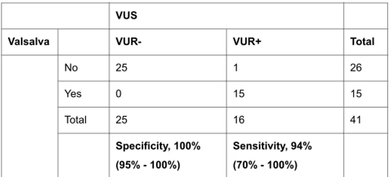
Taking the VUS as the reference standard, Valsalva-cystosonography had a sensitivity of 93.75% (95% CI 69.77% - 99.84%) and a specificity of 100% (95% CI 86.28% - 100%) (Table 1). The specificity of 100% indicates that Valsalva-cystosonography allows reliable identification of patients who had no evidence of VUR.
VUS
Valsalva VUR- VUR+ Total
No 25 1 26 Yes 0 15 15 Total 25 16 41 Specificity, 100% (95% - 100%) Sensitivity, 94% (70% - 100%)
Table 1: Contingency table of diagnoses of VUR measured by VUS and Valsalva-cystosonography.
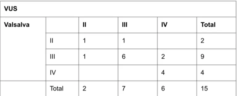
Table 2 shows results concerning the grade of VUR detected by voiding VUS and Valsalva-cystosonography. The grade of VUR detected with Valsalva-cystosonography showed also moderate agreement with grading by VUS [κ = 0.51, p = 0.002]. The reflux grade was rated III or IV in more than four-fifths of the patients (13 of 15). Citation: Visalli C, Sicilia N, Salamone I, Chimenz R, Fede C, et al. (2016) Accuracy of Cystosonography without Voiding Phase in the Diagnosis
of Vesicoureteral Reflux in Children: A Variant of the Original Technique. OMICS J Radiol 5: 219. doi:10.4172/2167-7964.1000219
Page 2 of 4
OMICS J Radiol
VUS showed more lesions rated as grade IV than did Valsalva-cystosonography.
VUS
Valsalva II III IV Total
II 1 1 2
III 1 6 2 9
IV 4 4
Total 2 7 6 15
Table 2: KUUs according to the grade of VUR detected by both VUS and Valsalva-cystosonography.
Discussion
Voiding urosonography with second generation contrast agent and contrast-specific and tissue harmonic imaging has, over the years, obtained increasingly clear recognition as a safe and reliable test to the diagnosis and follow-up of patients with VUR [9-13]. The VUS has advantages even if compared with the conventional fluoroscopic cystography. As the VUR is a real-time radiation free technique and the switch from one kidney to contralateral is extremely rapid, the continuous exploration without any pauses captures a higher number of refluxes in both sides. Many authors consider that the VUS with a second-generation contrast agent is superior to conventional fluoroscopic cystography in the detection and grading of VUR [3,5,13]. In addition, the absence of major side effects and low incidence of minor side effects such as dysuria, - not related to the contrast agent, but rather to bladder catheterization- are definite advantages [14]. However, a shortcoming, in common with traditional retrograde cystography, is catheter placement, which cannot be avoided. This ultrasound method is well-accepted by the patient up to the voiding phase, when the subject can experience some difficulties in voiding the bladder in the presence of health-care personnel. As a consequence, often the patient may even refuse to perform the act negatively affecting the exam both in terms of diagnostic performance and duration of the exam. The introduction of the Valsalva maneuver to carry out the stress phase aims at overcoming this obstacle.
The child must inspire with force with a closed glottis, to obtain the desired effect. This maneuver is relatively easy to explain, well-tolerated and thus accomplished with success. The number of Valsalva in our work has been set at two, but it may be increased depending on the diagnostic confidence that the operator gets on the diagnosis. Definitely two is the minimum number, because the first maneuver ensures the evidence of reflux, the second allows to express a more reliable judgment on the degree of reflux. The increase of the number of Valsalva maneuver, in our opinion, should not change the reproducibility of the technique. Another substantial advantage of Valsalva technique over traditional VUS is that each side can be examined multiple times with the same single bladder filling, precluding potential need for refilling bladder as may be the case for VUS. In our opinion the refilling must be avoided because it increases the intolerance of the children. Then our Valsalva technique may represent another strong point. Our study demonstrated high rates of sensitivity and specificity for Valsalva-cystosonography as compared to VUS. Patients presenting with VUR had no false-positives on Valsalva-cystosonography and only 1 false-negative. In particular, this subject
had a bilateral reflux. The left grade III was assessed both by Valsalva maneuver and voiding phase, while the right grade II was seen only during urination.
The discrepancy between the 2 methodologies may be attributable to the incomplete patient collaboration in a slightly overweight subject who struggled to maintain adequate contraction of the abdominal wall during the second Valsalva maneuver as well as the prolonged procedure due to the child's condition. Noteworthy, this fact did not change the management of the patient, as the Valsalva had demonstrated a higher VUR in one of the two KUU. With regard grade of reflux, the two methods showed moderate agreement (κ = 0.51, p = 0.002) (Table 2). Two VUR were a grade higher at VUS (IV grade vs. III at Valsalva-cystosonography), and one VUR was a grade higher at Valsalva cystosonography (III grade vs. II at VUS) (Figures 2 and 3). Also in these cases, patient management was not changed because, in any case, a protocol based on an antibiotic therapy, clinical follow-up and cystosonography was established. Furthermore as the latter case shows, the reflux detected at VUS not necessarily will be to higher degree.
Figure 2: US images in 8-year-old girl with urinary tract infection. Valsalva cystosonography (a) shows a grade III VUR that becomes a grade IV during the voiding phase (b). Arrow heads: dilated pelvicalyceal cavities; arrows: ureteral dilatation.
Figure 3: US images in 5-year old girl with mild pelvicalyceal dilatation without urinary infection. (a) Valsalva cystosonography shows a grade III VUR; (b) at voiding phase the reflux is a grade II.
Our study demonstrated that the Valsalva maneuver may be a valid alternative to the voiding phase in compliant children. This technique is intended for a pediatric population who are old enough to collaborate. The youngest child in our study was 4 years old. Excellent collaboration was obtained from all young enrolled patients with a single exception. However, with suitable communication techniques Citation: Visalli C, Sicilia N, Salamone I, Chimenz R, Fede C, et al. (2016) Accuracy of Cystosonography without Voiding Phase in the Diagnosis
of Vesicoureteral Reflux in Children: A Variant of the Original Technique. OMICS J Radiol 5: 219. doi:10.4172/2167-7964.1000219
Page 3 of 4
OMICS J Radiol
the diagnostic exam was transformed into a game, and well received by the children.
Conclusion
Notwithstanding the small number of patients, our results are encouraging and provide evidence that the Valsalva maneuver might be an easy, rapid, and effective alternative technique to the voiding phase. Further studies are warranted.
References
1. Novljan G, Levart TK, Kljucevsek D, Kenig A, Kenda RB (2010) Ultrasound detection of vesicoureteral reflux in children. J Urol 184: 319-324.
2. Cosgrove D (2006) Ultrasound contrast agents: an overview. Eur J Radiol 60: 324-330.
3. Wong LS, Tse KS, Fan TW, Kwok KY, Tsang TK, et al. (2014) Voiding urosonography with second-generation ultrasound contrast versus micturating cystourethrography in the diagnosis of vesicoureteric reflux. Eur J Pediatr 173: 1095-1101.
4. Darge K, Troeger J (2002) Vesicoureteral reflux grading in contrast-enhanced voiding urosonography. Eur J Radiol 43: 122-128.
5. Papadopoulou F, Anthopoulou A, Siomou E, Efremidis S, Tsamboulas C, et al. (2009) Harmonic voiding urosonography with a second-generation contrast agent for the diagnosis of vesicoureteral reflux. Pediatr Radiol 39: 239-244.
6. Ascenti G, Zimbaro G, Mazziotti S, Chimenz R, Fede C, et al. (2004) Harmonic US imaging of vesicoureteric reflux in children: usefulness of a second generation US contrast agent. Pediatr Radiol 34: 481-487.
7. Zimbaro G, Ascenti G, Visalli C, Bottari A, Zimbaro F, et al. (2007) Contrast-enhanced ultrasonography (voiding urosonography) of vesicoureteral reflux: state of the art. Radiol Med 112: 1211-1224. 8. Landis JR, Koch GG (1977) An application of hierarchical kappa-type
statistics in the assessment of majority agreement among multiple observers. Biometrics 33: 363-374.
9. Berrocal T, Gayá F, Arjonilla A, Lonergan GJ (2001) Vesicoureteral reflux: diagnosis and grading with echo-enhanced cystosonography versus voiding cystourethrography. Radiology 221: 359-365.
10. Mentzel HJ, Vogt S, Patzer L, Schubert R, John U, et al. (1999) Contrast-enhanced sonography of vesicoureterorenal reflux in children: preliminary results. AJR Am J Roentgenol 173: 737-740.
11. Darge K, Riedmiller H (2004) Current status of vesicoureteral reflux diagnosis. World J Urol 22: 88-95.
12. Radmayr C, Klauser A, Pallwein L, Zurnedden D, Bartsch G, et al. (2002) Contrast enhanced reflux sonography in children: a comparison to standard radiological imaging. J Urol 167: 1428-1430.
13. Kis E, Nyitrai A, Várkonyi I, Máttyus I, Cseprekál O, et al. (2010) Voiding urosonography with second-generation contrast agent versus voiding cystourethrography. Pediatr Nephrol 25: 2289-2293.
14. Papadopoulou F, Ntoulia A, Siomou E, Darge K (2014) Contrast-enhanced voiding urosonography with intravesical administration of a second-generation ultrasound contrast agent for diagnosis of vesicoureteral reflux: prospective evaluation of contrast safety in ,010 children. Pediatr Radiol 44: 719-728.
Citation: Visalli C, Sicilia N, Salamone I, Chimenz R, Fede C, et al. (2016) Accuracy of Cystosonography without Voiding Phase in the Diagnosis of Vesicoureteral Reflux in Children: A Variant of the Original Technique. OMICS J Radiol 5: 219. doi:10.4172/2167-7964.1000219
Page 4 of 4
OMICS J Radiol