S
APIENZAU
NIVERSITY OFR
OMED
EPARTMENT OFP
HYSIOLOGY ANDP
HARMACOLOGYA
DISSERTATION IN FULFILLMENT OF THE REQUIREMENTS FORTHE DEGREE OF
D
OCTOR OFP
HILOSOPHY INT
OXICOLOGYMODULATION OF MEMORY FOR STRESSFUL
EXPERIENCES: REVEALING HYPERMNESIA
Paola Colucci
Director of the PhD Program
Prof. Maura Palmery
Thesis committee
Prof. Viviana Trezza
Prof. Pietro Marini
Supervisor
Prof. Luca Romanelli
Prof. Patrizia Campolongo
Rome February, 13th 2020
If in the twilight of memory we should meet once more, we shall speak again together and you shall sing to me a deeper song. And if our hands should meet in another dream, we shall build another tower in the sky.
CONTENTS
G
ENERAL INTRODUCTIONC
HAPTER1:
Enhanced brain activity associated with memory access inhighly superior autobiographical memory.
C
HAPTER2:
Enhancing endocannabinoid neurotransmission augments theefficacy of extinction training and ameliorates traumatic stress-induced behavioral alterations in rats.
C
HAPTER3:
Predicting susceptibility and resilience in an animal model ofpost-traumatic stress disorder (PTSD).
C
HAPTER4:
Anandamide modulation of circadian- and stress-dependenteffects on rat short-term memory.
C
HAPTER5:
Amphetamine modulation of long-term object recognitionmemory in rats: Influence of stress.
C
HAPTER6:
Amphetamine and the smart drug3,4-methylenedioxypyrovalerone (MDPV) induce generalization of fear memory in rats.
C
HAPTER7:
General discussion and conclusion.C
URRICULUMV
ITAEA
PPENDIX1
G
ENERAL INTRODUCTIONThe American Psychological Association defines Hypermnesia as a condition characterized by an extreme degree of memory retention and retrieval (American Psychiatric Association, 2013).
In the field of memory research, substantial attention has always been given to the completely opposite situation: memory loss. Memory loss has long been recognized as the principal characteristic of several mental disorders (e.g. Alzheimer Disease) and it is principally related to impairments in memory retrieval and more generally, to hypofunction of memory processes (Jahn, 2013). On the other hand, memory hyperfunction has to be considered an equally serious memory alteration which can lead, at the same way of memory loss, to many psychiatric disorders such as the Post-Traumatic Stress Disorder (PTSD) characterized by intrusive thoughts and flashbacks due to an excessive and involuntary memory retrieval of a negative past experience (McNally, 2006, Brewin, 2011, Parsons and Ressler, 2013).
Recently, some individuals with extraordinary autobiographical memory abilities have been identified. These subjects have a superior ability to recall specific details of autobiographical events and this memory phenomenon is called Highly Superior Autobiographical Memory (HSAM) (LePort et al., 2012). HSAM subjects have the natural ability to recall past autobiographical events independently from the emotional charge linked to the experience lived. Therefore, investigating the neurobiological mechanisms sustaining HSAM may expand our basic knowledge on maladaptive memory processes in psychiatric conditions such as PTSD, conducting to new scenario in the research for innovative therapeutic targets.
2 Currently, there is no effective drug treatment for PTSD cure (Hoskins et al., 2015) and the most practiced therapeutic approach is represented by the cognitive behavioral therapy (Milad and Quirk, 2012, Furini et al., 2014). The cognitive behavioral therapy is based on the repeated exposure of patients to trauma-related reminders, in a safe situation, thus lacking of negative consequences for the subject (Abramowitz, 2013). In the recent years, the research in the field of PTSD is more and more moving toward the identification of pharmacological approaches aimed at treating not only the symptoms but rather the causes of the pathology, which combined with the cognitive behavioral therapy, may improve efficacy (de Quervain et al., 2011, Trezza and Campolongo, 2013, Singewald et al., 2015). Considering the features of PTSD, a good drug should be able to interfere with the excessive retrieval and with the impaired extinction of the traumatic memory, and a great attention, in this regards, is nowadays given to drugs potentiating the endocannabinoid system (Campolongo et al., 2013, Morena and Campolongo, 2014, Rabinak and Phan, 2014). The endocannabinoid system is composed by endogenous lipidic retrograde neurotransmitters, called endocannabinoids, and by their receptors: the cannabinoid receptor type 1 (CB1) and type 2 (CB2). The two principal endocannabinoids are represented by N-arachidonoyl ethanolamine (anandamide, AEA) and 2-N-arachidonoyl glycerol (2-AG), which are metabolized by the enzymes fatty-acid amide hydrolase (FAAH) and monoacylglycerol lipase (MAGL), respectively (Di Marzo, 2018). It has been shown in rodents that drugs acting as cannabinoid agonists enhance the extinction (Bowles et al., 2012, Gunduz-Cinar et al., 2013, Bitencourt et al., 2014) and attenuate the retrieval of aversive memory (Atsak et al., 2012, Morena and Campolongo, 2014, Morena et al., 2015), thus highlighting their positive potential role in the treatment of PTSD.
An important boundary to overcome, toward the identification of an optimal therapeutic approach for PTSD treatment, is represented by the susceptibility
3 issue. Most PTSD animal models do not take in consideration the individual differences in trauma response. The whole population is considered as belonging to a unique trauma response phenotype and only the anxiety outcomes of this pathology are studied, without considering the cognitive alterations related to PTSD. On the contrary, literature data demonstrate that among a human population exposed to a traumatic event, only a subset of people will ultimately develop PTSD, the others fully recover and mostly without any specific intervention (Javidi and Yadollahie, 2012). This evidence underlines the existence of different trauma response phenotypes among a population, and that genetic, epigenetic and environmental factors can play a crucial role in the susceptibility to PTSD development determining how the stress response to the traumatic experience can lead to aberrant and enduring alteration of memory processes (Skelton et al., 2012, Yehuda et al., 2015, Bolsinger et al., 2018). It is widely known that normally, emotionally arousing experiences are well remembered over time with respect to no emotional events (McGaugh and Roozendaal, 2002, McGaugh, 2006, Roozendaal and McGaugh, 2011). Both positive and negative stressful situations can activate several physiological responses which end with stress mediators release (Roozendaal and McGaugh, 2011). Hundreds of preclinical studies have been conducted and indicate the key role of stress mediators, such as glucocorticoids and noradrenaline, in the modulation of memory processes (Barsegyan et al., 2014, Morena et al., 2015, Barsegyan et al., 2019). Particularly, it has been observed that enhanced levels of noradrenaline in specific brain areas are linked with improved memory performances (Barsegyan et al., 2014, Ferry et al., 2015).
Noradrenaline levels can be increased through psychostimulant drugs, such as amphetamine (Berridge, 2006, Faraone, 2018). Extensive research has proven the potent arousal-enhancing effect due to the noradrenergic modulation exerted by amphetamine (Berridge et al., 1999, Berridge and Morris, 2000), and, several
4 studies have shown the amphetamine enhancing effects on learning and memory processes (Martinez et al., 1980a, Martinez et al., 1980b).
Notwithstanding, the effect of amphetamine in the enhancement of memory is well-established, currently, its effect on memory generalization is poor investigated. An excessive memory retrieval in a situational reminder condition, may lead to memory generalization. From an ethological perspective, memory generalization may be both adaptive and maladaptive (Asok et al., 2018). For example, the excessive memory retrieval of a fear related situation in a context with high predatory risk, is a common adaptive response in animals, but this excessive memory retrieval in an environment lacking of an imminent threat is maladaptive and represents the core of fear memory generalization (Asok et al., 2018). The overgeneralization of a contextual fear memory is one of the most important features of trauma related disorders such as PTSD (Bian et al., 2019).
Outline
As mentioned above, recently individuals with a remarkable memory ability to recall accurately autobiographical information over time, were found. The study on HSAM provides a great opportunity to investigate the neurological underpinnings of hypermnesia. Under the supervision of Prof. Campolongo, during my PhD I have screened more than 200 subjects and identified the first European population of rare individuals with HSAM. This HSAM cohort is one of only two established HSAM populations in the world, the first study was conducted in USA by Prof. J. McGaugh (LePort et al., 2012), and up to today, 80 HSAM subjects in the USA have been identified. In Chapter 1, we carried out an fMRI investigation of a group of subjects identified in Italy as having HSAM. During fMRI scanning, subjects with HSAM and control subjects (subjects with normal autobiographical memory) were asked to retrieve autobiographical memories as well as non- autobiographical memories, in order
5 to elucidate the basic mechanisms and the brain networks which sustain hypermnesia in humans.
The excessive memory retention and retrieval (as seen in HSAM) are cardinal features of hypermnesia-related psychiatric disorders such as PTSD. As previously said, nowadays there is no specific treatment for PTSD, but the endocannabinoid system, with its peculiar role in the modulation of memory retention and retrieval, seems to be a promising therapeutic target. In Chapter 2 we closely investigated the effects of a pharmacological manipulation of the endocannabinoid system in a preclinical model of PTSD. We recently proposed a footshock-based paradigm able to mimic both the enduring cognitive and emotional facets of PTSD pathology useful to model the extinction-based exposure therapy combined with pharmacological treatment (Berardi et al., 2014). Here, we evaluated whether activation of the endocannabinoid system, by facilitating extinction and dampening the retrieval of fear memory, could ameliorate the efficacy of extinction-based exposure therapy, leading to improvements that remain stable long after trauma exposure and pharmacological treatment termination.
Even if the endocannabinoid system looks as a promising target for the treatment of PTSD, it is also important to take in consideration the susceptibility aspect of this debilitating psychopathology. Animal models are a useful tool to investigate this issue (Musazzi et al., 2018). In Chapter 3 we aimed at the development of an animal model of PTSD able to predict the susceptibility and the resilience phenotype, with a high translational value to human PTSD, considering both cognitive and emotional alteration long after trauma. For this purpose, we outstretched our previously validated animal model (Berardi et al., 2014) in order to identify before the extinction sessions and drug treatment, susceptible and
6 resilient rats in terms of over-consolidation, impaired extinction and social behavior alterations.
As previously said, not all people will respond at the same way to a traumatic experience and, moreover, among those who will develop the pathology, there are people that do not respond to the currently available treatments or relapse after the end of treatment. For this reason, it is tentative to hypothesized that there are also individual differences in PTSD’s treatment success. Literature data indicate the close relationship between endocannabinoids and stress mediators, such as glucocorticoids (Morena et al., 2016, Harris et al., 2019, Siller-Perez et al., 2019). On the basis of these data the endocannabinoid system can be considered as an emotional buffer which modulates memory function differentially, depending on the level of stress and arousal lived by the subject and associated with the experimental context (Campolongo et al., 2013, Morena and Campolongo, 2014, Morena et al., 2015). Both endocannabinoids and glucocorticoids release are influenced by circadian rhythm (Vaughn et al., 2010, Spencer et al., 2018). In Chapter 4 we investigated, in an object recognition task, how the exogenous augmentation of AEA levels at different times of the day, could influence the effect of different stress intensities on rat short-term memory.
Normally, only emotional arousing experiences can create memories that last a lifetime. Since it is well established the crucial role of stress in making some experiences unforgettable (McGaugh and Roozendaal, 2002) and that psychostimulants can enhance memory as well (Spencer et al., 2015), in Chapter 5 we investigated in more details at preclinical level, how different stress intensities modulate the amphetamine effects on long term memory consolidation.
7 Memory generalization could be one of the consequences of excessive memory related to an emotional arousing experience. Psychostimulant outcomes on memory enhancement are known from many years and there are clinical data showing also their memory generalization effects (Easton and Bauer, 1997). However, the mechanisms through which psychostimulants affect memory quality is still poor investigated. In Chapter 6 we explored the memory generalization effects induced by amphetamine and a new psychostimulant: the 3,4-methylenedioxypyrovalerone (MDPV), also called “Bath salt”. Both the psychostimulants share a similar, yet not identical mechanism of action augmenting noradrenaline and dopamine levels in the synaptic cleft. Thus, in a second experiment we aimed at evaluating the different involvement of the noradrenergic and dopaminergic system in the effects on memory enhancement, induced only by amphetamine, and on memory generalization, induced by both the two drugs.
Chapter 7 summarizes and discusses the findings of this thesis and provides conclusions and future perspectives.
References
Abramowitz JS (2013) The practice of exposure therapy: relevance of cognitive-behavioral theory and extinction theory. Behavior therapy 44:548-558. Asok A, Kandel ER, Rayman JB (2018) The Neurobiology of Fear Generalization.
Frontiers in behavioral neuroscience 12:329.
Atsak P, Hauer D, Campolongo P, Schelling G, McGaugh JL, Roozendaal B (2012) Glucocorticoids interact with the hippocampal endocannabinoid system in impairing retrieval of contextual fear memory. Proceedings of the National Academy of Sciences of the United States of America 109:3504-3509. Barsegyan A, McGaugh JL, Roozendaal B (2014) Noradrenergic activation of the
basolateral amygdala modulates the consolidation of object-in-context recognition memory. Frontiers in behavioral neuroscience 8:160.
Barsegyan A, Mirone G, Ronzoni G, Guo C, Song Q, van Kuppeveld D, Schut EHS, Atsak P, Teurlings S, McGaugh JL, Schubert D, Roozendaal B (2019) Glucocorticoid enhancement of recognition memory via basolateral amygdala-driven
8
facilitation of prelimbic cortex interactions. Proceedings of the National Academy of Sciences of the United States of America 116:7077-7082. Berardi A, Trezza V, Palmery M, Trabace L, Cuomo V, Campolongo P (2014) An updated
animal model capturing both the cognitive and emotional features of post-traumatic stress disorder (PTSD). Frontiers in behavioral neuroscience 8:142. Berridge CW (2006) Neural substrates of psychostimulant-induced arousal.
Neuropsychopharmacology : official publication of the American College of Neuropsychopharmacology 31:2332-2340.
Berridge CW, Morris MF (2000) Amphetamine-induced activation of forebrain EEG is prevented by noradrenergic beta-receptor blockade in the halothane-anesthetized rat. Psychopharmacology 148:307-313.
Berridge CW, O'Neil J, Wifler K (1999) Amphetamine acts within the medial basal forebrain to initiate and maintain alert waking. Neuroscience 93:885-896. Bian XL, Qin C, Cai CY, Zhou Y, Tao Y, Lin YH, Wu HY, Chang L, Luo CX, Zhu DY
(2019) Anterior Cingulate Cortex to Ventral Hippocampus Circuit Mediates Contextual Fear Generalization. The Journal of neuroscience : the official journal of the Society for Neuroscience 39:5728-5739.
Bitencourt RM, Pamplona FA, Takahashi RN (2014) Corticosteroid-endocannabinoid loop supports decrease of fear-conditioned response in rats. European neuropsychopharmacology : the journal of the European College of Neuropsychopharmacology 24:1091-1102.
Bolsinger J, Seifritz E, Kleim B, Manoliu A (2018) Neuroimaging Correlates of Resilience to Traumatic Events-A Comprehensive Review. Frontiers in psychiatry 9:693.
Bowles NP, Hill MN, Bhagat SM, Karatsoreos IN, Hillard CJ, McEwen BS (2012) Chronic, noninvasive glucocorticoid administration suppresses limbic endocannabinoid signaling in mice. Neuroscience 204:83-89.
Brewin CR (2011) The nature and significance of memory disturbance in posttraumatic stress disorder. Annual review of clinical psychology 7:203-227.
Campolongo P, Morena M, Scaccianoce S, Trezza V, Chiarotti F, Schelling G, Cuomo V, Roozendaal B (2013) Novelty-induced emotional arousal modulates cannabinoid effects on recognition memory and adrenocortical activity. Neuropsychopharmacology : official publication of the American College of Neuropsychopharmacology 38:1276-1286.
de Quervain DJ, Bentz D, Michael T, Bolt OC, Wiederhold BK, Margraf J, Wilhelm FH (2011) Glucocorticoids enhance extinction-based psychotherapy. Proceedings of the National Academy of Sciences of the United States of America 108:6621-6625.
Di Marzo V (2018) New approaches and challenges to targeting the endocannabinoid system. Nature reviews Drug discovery 17:623-639.
Easton CJ, Bauer LO (1997) Beneficial effects of thiamine on recognition memory and P300 in abstinent cocaine-dependent patients. Psychiatry research 70:165-174. Faraone SV (2018) The pharmacology of amphetamine and methylphenidate: Relevance to the neurobiology of attention-deficit/hyperactivity disorder and other psychiatric comorbidities. Neuroscience and biobehavioral reviews 87:255-270.
9
Ferry B, Parrot S, Marien M, Lazarus C, Cassel JC, McGaugh JL (2015) Noradrenergic influences in the basolateral amygdala on inhibitory avoidance memory are mediated by an action on alpha2-adrenoceptors. Psychoneuroendocrinology 51:68-79.
Furini C, Myskiw J, Izquierdo I (2014) The learning of fear extinction. Neuroscience and biobehavioral reviews 47:670-683.
Gunduz-Cinar O, MacPherson KP, Cinar R, Gamble-George J, Sugden K, Williams B, Godlewski G, Ramikie TS, Gorka AX, Alapafuja SO, Nikas SP, Makriyannis A, Poulton R, Patel S, Hariri AR, Caspi A, Moffitt TE, Kunos G, Holmes A (2013) Convergent translational evidence of a role for anandamide in amygdala-mediated fear extinction, threat processing and stress-reactivity. Molecular psychiatry 18:813-823.
Harris C, Weiss GL, Di S, Tasker JG (2019) Cell signaling dependence of rapid
glucocorticoid-induced endocannabinoid synthesis in hypothalamic
neuroendocrine cells. Neurobiology of stress 10:100158.
Hoskins M, Pearce J, Bethell A, Dankova L, Barbui C, Tol WA, van Ommeren M, de Jong J, Seedat S, Chen H, Bisson JI (2015) Pharmacotherapy for post-traumatic stress disorder: systematic review and meta-analysis. The British journal of psychiatry : the journal of mental science 206:93-100.
Jahn H (2013) Memory loss in Alzheimer's disease. Dialogues in clinical neuroscience 15:445-454.
Javidi H, Yadollahie M (2012) Post-traumatic Stress Disorder. The international journal of occupational and environmental medicine 3:2-9.
LePort AK, Mattfeld AT, Dickinson-Anson H, Fallon JH, Stark CE, Kruggel F, Cahill L, McGaugh JL (2012) Behavioral and neuroanatomical investigation of Highly Superior Autobiographical Memory (HSAM). Neurobiology of learning and memory 98:78-92.
Martinez JL, Jr., Jensen RA, Messing RB, Vasquez BJ, Soumireu-Mourat B, Geddes D, Liang KC, McGaugh JL (1980a) Central and peripheral actions of amphetamine on memory storage. Brain research 182:157-166.
Martinez JL, Jr., Vasquez BJ, Rigter H, Messing RB, Jensen RA, Liang KC, McGaugh JL (1980b) Attenuation of amphetamine-induced enhancement of learning by adrenal demedullation. Brain research 195:433-443.
McGaugh JL (2006) Make mild moments memorable: add a little arousal. Trends in cognitive sciences 10:345-347.
McGaugh JL, Roozendaal B (2002) Role of adrenal stress hormones in forming lasting memories in the brain. Current opinion in neurobiology 12:205-210.
McNally RJ (2006) Cognitive abnormalities in post-traumatic stress disorder. Trends in cognitive sciences 10:271-277.
Milad MR, Quirk GJ (2012) Fear extinction as a model for translational neuroscience: ten years of progress. Annual review of psychology 63:129-151.
Morena M, Campolongo P (2014) The endocannabinoid system: an emotional buffer in the modulation of memory function. Neurobiology of learning and memory 112:30-43.
Morena M, De Castro V, Gray JM, Palmery M, Trezza V, Roozendaal B, Hill MN, Campolongo P (2015) Training-Associated Emotional Arousal Shapes Endocannabinoid Modulation of Spatial Memory Retrieval in Rats. The Journal
10
of neuroscience : the official journal of the Society for Neuroscience 35:13962-13974.
Morena M, Patel S, Bains JS, Hill MN (2016) Neurobiological Interactions Between Stress and the Endocannabinoid System. Neuropsychopharmacology : official publication of the American College of Neuropsychopharmacology 41:80-102. Musazzi L, Tornese P, Sala N, Popoli M (2018) What Acute Stress Protocols Can Tell Us About PTSD and Stress-Related Neuropsychiatric Disorders. Frontiers in pharmacology 9:758.
Parsons RG, Ressler KJ (2013) Implications of memory modulation for post-traumatic stress and fear disorders. Nature neuroscience 16:146-153.
Rabinak CA, Phan KL (2014) Cannabinoid modulation of fear extinction brain circuits: a novel target to advance anxiety treatment. Current pharmaceutical design 20:2212-2217.
Roozendaal B, McGaugh JL (2011) Memory modulation. Behavioral neuroscience 125:797-824.
Siller-Perez C, Fuentes-Ibanez A, Sotelo-Barrera EL, Serafin N, Prado-Alcala RA, Campolongo P, Roozendaal B, Quirarte GL (2019) Glucocorticoid interactions with the dorsal striatal endocannabinoid system in regulating inhibitory avoidance memory. Psychoneuroendocrinology 99:97-103.
Singewald N, Schmuckermair C, Whittle N, Holmes A, Ressler KJ (2015) Pharmacology of cognitive enhancers for exposure-based therapy of fear, anxiety and trauma-related disorders. Pharmacology & therapeutics 149:150-190.
Skelton K, Ressler KJ, Norrholm SD, Jovanovic T, Bradley-Davino B (2012) PTSD and gene variants: new pathways and new thinking. Neuropharmacology 62:628-637.
Spencer RC, Devilbiss DM, Berridge CW (2015) The cognition-enhancing effects of psychostimulants involve direct action in the prefrontal cortex. Biological psychiatry 77:940-950.
Spencer RL, Chun LE, Hartsock MJ, Woodruff ER (2018) Glucocorticoid hormones are both a major circadian signal and major stress signal: How this shared signal contributes to a dynamic relationship between the circadian and stress systems. Frontiers in neuroendocrinology 49:52-71.
Trezza V, Campolongo P (2013) The endocannabinoid system as a possible target to treat both the cognitive and emotional features of post-traumatic stress disorder (PTSD). Frontiers in behavioral neuroscience 7:100.
Vaughn LK, Denning G, Stuhr KL, de Wit H, Hill MN, Hillard CJ (2010) Endocannabinoid signalling: has it got rhythm? British journal of pharmacology 160:530-543.
Yehuda R, Hoge CW, McFarlane AC, Vermetten E, Lanius RA, Nievergelt CM, Hobfoll SE, Koenen KC, Neylan TC, Hyman SE (2015) Post-traumatic stress disorder. Nature reviews Disease primers 1:15057.
11
C
HAPTER1
FROM ORDINARY TO EXTRAORDINARY MEMORY: ENHANCED BRAIN ACTIVITY RELATED TO MEMORY ACCESS IN HIGHLY
SUPERIOR AUTOBIOGRAPHICAL MEMORY
Valerio Santangelo1,2, Clarissa Cavallina1,2, Paola Colucci3, Alessia Santori3,
Simone Macrì4, James L. McGaugh5, Patrizia Campolongo3,6
1 Dept. of Philosophy, Social Sciences & Education, University of Perugia, Piazza G. Ermini 1, 06123 Perugia, Italy; 2 Neuroimaging Laboratory, Santa Lucia Foundation, Via Ardeatina 306, 00179 Rome, Italy; 3 Department of Physiology and Pharmacology, Sapienza University of Rome, P.le A. Moro 5, 00185 Rome, Italy; 4 Centre for Behavioural Sciences and Mental Health, Istituto Superiore di Sanità, Viale Regina Elena, 299 00161 Rome, Italy; 5 Center for the Neurobiology of Learning and Memory, Department of Neurobiology and Behavior, University of California, Irvine, California, 92697-3800; 6 Neurobiology of Behavior Lab, Santa Lucia Foundation, Via del Fosso di Fiorano 64, 00143 Rome, Italy
13 Abstract
Brain systems underlying human memory function have been classically investigated studying patients with selective memory impairments. The discovery of rare individuals who have highly superior autobiographical memory (HSAM) provides, instead, an opportunity to investigate the brain systems underlying enhanced memory. Here we carried out the first fMRI investigation of a group of subjects identified as having HSAM. During fMRI scanning, 8 HSAM and 21 control subjects were asked to retrieve autobiographical memories as well as non-autobiographic memories (e.g., examples of animals). Subjects were instructed to signal the “access” to the autobiographical memory (AM) by a key press and continue “reliving” it immediately after. Compared to controls, HSAM individuals provided a richer AM recollection and were faster in accessing AMs. The access to AMs was associated with enhanced prefrontal/hippocampal functional connectivity. AM access also induced increased activity in the left temporoparietal junction and enhanced functional coupling with sensory cortices in HSAM subjects as compared to controls. In contrast, HSAM subjects did not differ from controls in functional activity during the reliving phase. These findings, based on fMRI assessment, provide novel evidence of interaction of brain systems engaged in memory retrieval and suggests that enhanced activity of these systems is selectively involved in enabling more efficient access to past experiences in HSAM.
14 Significance
Recent research has identified human subjects who have highly superior autobiographical memory (HSAM). Here we investigated, using fMRI, the neural activation induced by retrieval of autobiographical and semantic memories in HSAM and control subjects. While their brains were being scanned, subjects had to retrieve autobiographical as well as non-autobiographic memories (e.g. examples of animals). The HSAM subjects displayed superior ability to retrieve details of autobiographical memories, supported by enhanced activation of several brain regions, including the medial prefrontal cortex and temporoparietal junction, as well as increased connectivity of the prefrontal cortex with the hippocampus, a region well known to be involved in memory representation. These findings suggest that activation of these systems may play a critical role in enabling HSAM.
15 Introduction
The ability to remember personal experiences (i.e., autobiographical memory, AM) is essential for survival (Allen et al., 2013). Brain systems underlying human AM function have been classically investigated studying patients with selective memory impairments (Greenberg et al., 2003). The discovery of extremely rare individuals who spontaneously show highly superior autobiographical memory (HSAM) (Parker et al., 2006; LePort et al., 2012) provides, instead, an opportunity to investigate the brain processes underlying enhanced AM. HSAM individuals demonstrate an extraordinary ability to recall vividly and accurately many remote autobiographical events, irrespective of their emotional saliency, and without the explicit use of mnemonic strategies. In contrast, their performance is generally comparable to that of control subjects in performance assessed by laboratory memory tests (Parker et al., 2006; LePort et al., 2016; LePort et al., 2017). Prior MRI assessment of HSAM revealed that several brain regions differ in size and shape (e.g., parahippocampal gyrus, posterior insula, IPS; putamen and caudate) as well as in coherence of fiber tracts (e.g., uncinate fasciculus) when compared to those of control subjects (LePort et al., 2016). The present study investigated brain activity induced by AM using functional MRI (fMRI).
Prior evidence of detailed re-experiencing in HSAM subjects (Parker et al., 2006; LePort et al., 2016; LePort et al., 2017) suggests that HSAM subjects may express increased neural activity underlying memory “reliving”. Previous fMRI investigations of normal (i.e., not superior) AM retrieval assessed memory “access” and memory “reliving” by asking participants to confirm elicitation of an AM through a response button (access phase) and then continue to elaborate on the retrieved event (reliving phase) in as much detail as possible for the remaining part of the trial (Daselaar et a., 2008). Previous studies using this
16 approach have reported activity in prefrontal/medial temporal regions related to access and activity in sensory cortex related to reliving (Cabeza et al., 2007). Retrieval by HSAM subjects may therefore involve enhanced activity in sensory cortex associated with detailed reliving of re-activated experiences. Alternatively, HSAM might entail enhanced prefrontal/medial temporal resources devoted to AM access. This interpretation is consistent with recent findings showing a selective decrease of neural activity in the medial prefrontal cortex as well as a reduced hippocampal volume in individuals who have impaired AM retrieval (Palombo et al., 2015). These findings may suggest that HSAM individuals show hyper-functioning of prefrontal/hippocampal regions. In the present study we addressed this question by performing the first fMRI examination of a group of HSAM subjects.
During fMRI, HSAM subjects and controls were asked to mentally retrieve “easy” AMs, thus to guarantee good performance in controls. Subjects were presented with memory-cues pointing to specific spatiotemporal coordinates that emphasize the difference between very old and more recent AMs (e.g., “The first time you drove a car” or “The last time you went to a restaurant”). Participants confirmed the appearance of the AM through a response button (access phase) and then continue to relive the retrieved event in as much detail as possible (reliving phase; Fig. 1a) (Daselaar et al., 2008). The specificity of AM activations was controlled by subtracting neural activity induced by accessing and generating examples of specific semantic categories (e.g., “example of vegetables” or “example of animals”; i.e., a semantic memory, SM, task). In comparison with controls, the HSAM subjects had faster access and more detailed retrieval of AMs. Memory access was associated with increased prefrontal/hippocampal functional connectivity and increased connectivity between the temporieparietal junction and sensory cortices. The prefrontal and hippocampal regions were found to be particularly involved with access to
17 remote AMs. In contrast, HSAM subjects did not differ from controls in brain activity during the reliving phase.
Methods
Participants
Eight HSAM individuals (5 males; mean age: 32.5 y.o.; range: 24-37 y.o.) recruited in accordance with the previous literature (LePort et al., 2012) (SI Appendix) and twenty-one control subjects (10 male; mean age: 32.5 y.o., range: 24-39 y.o.) participated in the study. All participants gave written consent to the study, which was approved by the independent Ethics Committee of the Santa Lucia Foundation (CE/PROG.540). Before conducting the experiment, we performed a power analysis, which estimated a reliable statistical power of 84% for our sample size (8 HSAM subjects plus a minimum of 20 controls) based on an effect size of 0.5 in line with those reported by the previous literature on HSAM (LePort et al., 2016)- and a significance level of 0.05.
Task and stimuli
During scanning, participants were asked to retrieve autobiographical and non-autobiographical – i.e., semantic – memories (AMs and SMs, respectively) (Fig. 1a and SI Appendix). The experiment included 3 functional runs, each including 18 memory cues: 12 AM (6 “first” and 6 “last” time events) and 6 SM trials, and a variable inter-trial interval (2-3 sec, uniformly distributed). Post-scanning, participants were asked to provide details about memories retrieved during the experiment (SI Appendix).
Magnetic Resonance Imaging and fMRI data analysis
18 3T and equipped for echo-planar imaging (EPI) was used to acquire the functional magnetic resonance images (SI Appendix). We used SPM12 (Wellcome Department of Cognitive Neurology) implemented in MATLAB 7.4 (The MathWorks Inc., Natick, MA) for data pre-processing (SI Appendix) and statistical analyses. Each participant underwent three fMRI-runs, each comprising 477 volumes. Statistical inference was based on a two-steps random effects approach (see SI Appendix). Briefly, the first-level models included separated Access and Reliving regressors (Daselaar et al., 2008) for each of the three trial types: “first AM”, “last AM”, SM. For each subject we estimated contrast images that removed the activity associated with access to and reliving of SM (control condition) to the main AM conditions. For the second-level group-analysis, the single-subjects contrast images of parameter estimates were entered into a mixed design ANOVA with group (HSAM vs. control) as between-subjects variable and phase (access vs. reliving) and AM type (first vs. last) as within-subjects variables. First of all, we highlighted the regions involved with AM in HSAM vs. control group (and vice-versa), irrespective of phase and AM type. As an additional constraint, we considered only voxels showing an overall activation across all conditions and groups (T-contrast, p-unc. = 0.001), ensuring that we selected only regions activated by AM retrieval (see, e.g., Buchel et al., 1998). The statistical threshold was set to p-FWE-corrected < 0.05 at the voxel level, considering the whole brain as the volume of interest. This comparison allowed us to highlight different circuits recruited by AM retrieval in the HSAM and control groups (Table 1 and Fig. 2a). The resulting activations were used to define regions of interest (ROIs) that were then used to test for condition-specific effects in interaction with the group variable, i.e., the two-way phase X group and AM type X group interactions, and the three-way phase X AM type X group interaction. For this, we considered spheres (radius, 8 mm; matching the FWHM of smoothing filter) centred on the regions activated by
19 AM retrieval in the two groups (Table 1) as the volume of interest (small volume correction) (Worsley et al., 1996).
Functional connectivity analysis
The procedure described above allowed us to identify three regions selectively involved in AM access in HSAM subjects (i.e., the significant group X phase interaction), namely the left VMPFc, the left DMPFc, and the left TPJ (Fig. 2b). Given that these seed-regions are related HSAM we did not expect any increased functional connectivity in the control group. The main goal of this analysis was to understand whether additional neural resources supported access to memory in HSAM individuals (SI Appendix).
ROI correlations with memory access latencies and obssessiveness scores in HSAM subjects
We further exploratory analyses using multiple regression models to investigate whether the brain activity related to the access to remote or recent memories covariated as a function of the individual latency to access memories or as a function of obsessive-compulsive traits in HSAM subjects (SI Appendix).
20 Results
Behavioral data
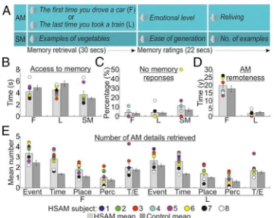
Behavioral results are illustrated in Figure 1. First, we assessed whether HSAM subjects were faster than controls to access their AMs. We performed a Group (HSAM vs. control) by Trial type (first AM, last AM, SM) ANOVA on the response latencies, defining the timing of access to the specific autobiographical or semantic memories (Fig. 1b). This analysis revealed a main effect of trial type [F(2, 54) = 21.2, p < .001; η2 = .440], indicating faster response latencies to
access semantic (M = 3.368 secs) than first (4.545 secs) or last (5.059 secs) AMs. This effect was further qualified by the significant group x trial type interaction [F(2, 54) = 5.8, p = .005; η2 = .177], indicating that HSAM subjects had faster
access to autobiographical material than control subjects (first AM: 4.196 vs. 4.894 secs, p = .029; last AM: 4.504 vs. 5.614 secs, p = .013), but not to semantic information (3.681 vs. 3.056 secs, p = .074). The main effect of group was not significant [F(1, 27) < 1, n.s.]. A similar 2x3 ANOVA on the “no memory response” data (Fig. 1c) revealed a main effect of trial type [F(2, 54) = 3.7, p = .032; η2 = .120], indicating that subjects failed more to retrieve semantic (9%)
than first time (3.7%) and last (3.9%) autobiographical events. This effect was not further modulated by the group factor, as indicated by the absence of both main effect of group and group x type of trial interaction (both Fs < 1). No differences were observed between the groups in the self-evaluation ratings of the emotional level and reliving of AMs and easiness of generation and number of generated example for SMs during scanning, as assessed by 2-tailed independent sample t-tests (all Ts ranging between -1.4 and 0.9; all ps > .181; SI Appendix, Fig. S1).
After the fMRI scanning, participants were presented again with the memory cues and asked to provide a verbal account of their memories. We analyzed the
21 temporal distribution (Fig. 1d) of the retrieved AMs using a Group by Remoteness of AM (“first” or “last” AM event) ANOVA. This analysis revealed a main effect of remoteness [F(1, 27) = 188.4, p < .001; η2 = .875], indicating
that first AMs were older than last AMs, 18.76 and 2.16 years, respectively. The two groups did not differ in the remoteness of AMs reported as indicated by the absence of both main effect of group and group x age of AM interaction (both Fs < 1.3; ps > .257). Finally, we compared how detailed were the AMs reported by the two groups, using a Group by Remoteness of AM by Type of detail (event, time, place, perceptual, emotional/though; (Levine et al., 2002). ANOVA. HSAM subjects retrieved a greater number of details than controls (2.0 vs. 1.4), as evidenced by the main effect of group [F(1, 27) = 10.1, p = .004; η2 = .272].
This was particularly true for the remote events, as indicated by the three way interaction [F(4, 108) = 2.5, p 0 .047; η2 = .085], showing more details reported
in the event (p = .005), time (p < .001), and place (p = .048) categories relatively to remote AMs, but only more details in the time category (p < .001) relatively to recent AMs (Fig. 1e). HSAM subjects also provided higher vivid descriptions of their AMs when assessed qualitatively (all Ts ranging between 5.0 and 18.2; all ps < .001; SI Appendix, Fig. S2).
Overall these findings indicate that, when compared to controls, HSAM subjects had faster and more vivid access to AM, especially for the most remote AMs, but had normal SM.
22
Figure 1. Task and behavioural results. a) Sequence of events in an example trial, involving 30 seconds to access and elaborate (“first”, F, and “last”, L) AMs or SMs, and 22 seconds to provided memory ratings; b) Time to access AMs and SMs in seconds; c) Percentages of no memory trials; d) Remoteness of reported AMs (in years); e) Mean number of details according to Levine et al.’s (2002) categories (event, time, place, perceptual, thought/emotion, T/E) and AM remoteness. In all graphs, for HSAM subjects are plotted individual scores. The error bars represent the standard error of the mean.
Assessment of Obsessive/Compulsive traits in HSAM individuals
The findings of previous studies suggest that HSAM subjects tend to have symptoms of obsessiveness/compulsiveness (LePort et al., 2017). To assess whether HSAM individual participating in the current study experienced an obsessive/compulsive symptomatology they were administered the Personality Assessment Inventory (PAI), which included – among others – the “Obsessive-Compulsive” (ARD-0) sub-scale. The average score at the obsessive-compulsive sub-scale for HSAM individuals was 67, corresponding to the 92nd percentile in
terms of expressing obsessive/compulsive-related symptoms. We assessed whether faster access to AM (averaging across first and last AM type) and number of retrieved details correlated with obsessive-compulsive traits (averaging across the five categories). However, our analyses failed to reveale
23 any significant correlation (r = .32, p = .444 and r = -.69, p = .58, respectively).
fMRI data
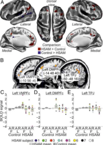
The main aim of the present study was to investigate neural activation associated with AM retrieval in HSAM as compared to control subjects. In the HSAM group AM retrieval recruited a large network of areas extending along the fronto-parietal cortex (see Fig. 2a and Table 1). This activation included dorsomedial regions, such as the left dorsomedial prefrontal cortex (DMPFc; the right DMPFc was marginally significant) and the left and right ventromedial prefrontal cortex (VMPFc); and lateral regions, such as the right dorsolateral prefrontal cortex (DLPFc), the left temporo-parietal junction (TPJ) and the left and right angular gyrus (ANG); plus the right insula. By contrast, we observed in the control group increased activity in few areas, involving the right superior frontal sulcus (SFS) and the posterior cingulate cortex (PCC), plus activation of visual areas, i.e., the left middle occipital gyrus (MOG).
Next, we assessed the contribution of these group-specific activations in the access or reliving phase, i.e., phase x group interaction. Three of the regions activated by AM retrieval in HSAM subjects were found to contribute selectively to memory access, namely, the left VMPFc (peaking at: x, y, z = -8, 58, 0; Z = 3.44; p = .011), the left DMPFc (x, y, z = -14, 42, 40; Z = 3.14; p = .027), and the left TPJ (x, y, z = -54, -44, 20; Z = 3.23; p = .021) (Fig. 2b). Activity in these regions selectively increased for the access vs. reliving phase (Fig. 2c-e: compare bar 5 & 7 vs. bar 6 & 8) in the HSAM group only (compare bar 5 to 8 vs. bar 1 to 4). None of the selected ROIs showed instead a selective involvement with the reliving phase in HSAM subjects. Similarly, none of the regions activated by AM retrieval in the control group was found to contribute selectively to the access or reliving phase. Analogously, no ROIs were found to reveal any AM type x group interaction. However, the left VMPFc (peaking at: x, y, z = -12, 52, 2; Z = 3.51;
24 p = .009) showed a more selective contribution during access to remote AMs (i.e., the three way phase X AM type X group interaction). (The left DMPFc showed a similar trend, despite not statistically significant, peaking at: x, y, z = -10, 44, 44; Z = 2.75; p = .069). Activity in the left VMPFc selectively increased during access to first AMs in HSAM subjects (Fig. 2c).
Figure 2. a) Regions activated by AM in the HSAM (red map) and in the control group (blue map) overlaid on an inflated template (see also Table 1); b) Sagittal sections on a standard MNI template showing the anatomical location of the regions selectively involved with the access phase to autobiographical memory in the HSAM group, i.e., the left VMPFc, the left DMPFc, and the left TPJ; c-e) bar plots summarizing the activity of the left VMPFc, DMPFc and TPJ, respectively, that showed increased activity during access to (vs. reliving of) AMs (compare bar 5 & 7 vs. bar 6 & 8), selectively in the HSAM group (compare bars 5 to 8 vs. bar 1 to 4). In all signal plots, the level of activation is expressed in arbitrary units (a.u., ± 90% confidence interval). For HSAM subjects are plotted the individual scores. For display purposes, all maps are displayed at a threshold of p-uncorrected = .001.
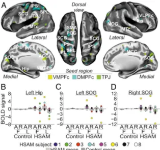
25 We then examined the functional coupling of the regions showing a selective involvement with AM access in HSAM subjects (i.e., the left VMPFc, the left DMPFc, and the left TPJ) with the rest of the brain. Analyses of inter-regional connectivity revealed large networks of areas functionally connected with the seed regions during AM access in HSAM subjects (Fig. 3a & Table 2). Specifically, the left VMPFc resulted connected with the medial temporal lobe, and, in particular with the left hippocampus (Hip) and with the rostral portion of the anterior cingulate cortex (rACC). The VMPFc was also found to be connected with another seed region, namely the left TPJ, and with the left and right sub-central gyrus (SCG). Activity in the left DMPFc was found instead to be synchronized during AM access in HSAM with the left and right intraparietal sulcus (IPS) along the dorsal frontoparietal network, and with prefrontal regions, such as the right ventrolateral prefrontal cortex (VLPFc) extending anteriorly and medially to the left and right VMPFc. The left DMPFc was also functionally connected with the medial portion of the cingulate cortex (MCC), with the pre-central gyrus (PCG), and the middle occipital gyrus (MOG). Finally, the left TPJ showed increased coupling during AM access in HSAM subjects with adjacent regions of the left parietal cortex, such as the superior and inferior parietal lobe (SPL/IPL), plus other posterior regions in the occipital and temporal lobe, such as the left and right superior occipital gyrus (SOG) and in the left and right medial temporal cortex (MTc). The left TPJ also showed increased coupling with the ACC and the supplementary motor area (SMA), bilaterally. Despite a large variability across the individual values (see signal plots in Fig. 3b-d for some representative areas), the general pattern of activity of all of these regions showed increased coupling with the respective seed region during access to (vs. reliving of) autobiographical memories (compare bar 5 & 7 vs. bar 6 & 8), selectively in the HSAM group (compare bars 5 to 8 vs. bar 1 to 4).
26
Figure 3. a) Regions showing functional connectivity with the left VMPFc (yellow map), the left DMPFc (cyan map), and the left TPJ (green map) during access to autobiographical memory in the HSAM group overlaid on an inflated template (see also Table 2). For display purposes, all maps are displayed at a threshold of p-uncorrected = .005, with a minimum cluster size of 20 voxels; b-l) signal plots showing the pattern of functional connectivity for some representative seed regions, namely, the left Hip (b), the left and the right SOG (c & d). All signal plots revealed increased coupling with the respective seed region during access to (vs. reliving of) autobiographical memories (compare bar 5 & 7 vs. bar 6 & 8), selectively in the HSAM group (compare bars 5 to 8 vs. bar 1 to 4). For HSAM subjects are plotted the individual scores. In all signal plots, the level of activation is expressed in arbitrary units (a.u., ± 90% confidence interval).
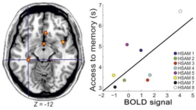
27 Finally, we investigated whether the brain activity related to access remote or recent memories covariated with access latencies or as a function of obsessive-compulsive tendencies in HSAM subjects. We found that in HSAM (p = .026), but not in control subjects, the activity of the left hippocampus increased as a
28 function of individual latencies to access remote memories (Fig. 4). No other effects were observed in the other ROIs or as a function of the individual scores of obsessiveness.
Figure 4. Increased activity in the left hippocampus as a function of increased latency to memory access in HSAM subjects.
Discussion
The HSAM subjects were faster and more efficient in retrieving AMs. In contrast, they did not differ from control subjects in retrieving semantic information. The findings strongly suggest that the shorter latencies in providing AMs reflect HSAM subjects’ superior access to details of past experiences. Additionally, the findings indicate that, in comparison with controls, the HSAM subjects remembered more autobiographical details of their past experiences, consistent with extensive prior investigations of HSAM (McGaugh et al., 2013; McGaugh et al., 2017), especially for the most remote AMs. The findings also confirmed those of previous research indicating that HSAM subjects tend to express obsessive/compulsive symptoms. However, we did not find evidence in our HSAM sample that the individual level of obsessiveness is related to the
29 memory access, neither in response latency or underlying brain activity. While we estimated a reliable statistical power for the current experiment (see Methods), we cannot exclude that smaller effect sizes may be detected with larger sample sizes. Therefore, future experiments shall determine whether the null effects reported in this study reflect a lack of difference or limited statistical power.
The major aim of the present investigation was to determine whether HSAM is associated with enhanced activation of brain systems as assessed by fMRI. The findings provide supporting evidence. Cortical activity increased in several areas, selectively in association with autobiographical remembering, and the increase was greater in HSAM subjects than in controls. During AM retrieval (irrespective to the access or reliving phase), when compared with controls, twice as many brain areas were activated in HSAM subjects. However, while it might be expected that the increased brain activity in HSAM is specifically devoted to memory reliving, given the richness of details provided by HSAM subjects (Parker et al., 2006; LePort et al., 2016; LePort et al., 2017), we did not observe any neural difference between HSAM and control subjects during the reliving phase. In contrast, the findings suggest that the increase in neural activity was specifically involved in accessing AM, recruiting a left-lateralized fronto-parietal network (VMPFc, the DMPFc and the TPJ) in HSAM subjects only during memory access. Additionally, and importantly, during this phase, HSAM subjects exhibit an increased functional connectivity of these regions with brain areas crucial for memory retrieval. These results suggest that HSAM may involve enhanced activation of specific brain areas involved in accessing representations of autobiographical experiences. One may argue that the quicker memory access of HSAM than control subjects might confound the fMRI analysis, requiring a comparison between different amounts of BOLD signals (i.e. less signal for the access phase of HSAM than control subjects). Although the finding of greater
30 brain activity in HSAM subjects during memory access goes against this potential confound, future research will have to solve this possible limitation. The enhanced AM access in HSAM individuals involved increased brain activity within core regions of the frontoparietal cortex, namely the medial prefrontal cortex and TPJ. These areas have been associated with the retrieval of autobiographical material Cabeza et al., 2007; Maguire 2001). AM retrieval is thought to be supported by an extensive network of brain regions – most pronounced on the left hemisphere (see, for a meta-analysis Svoboda et al., 2006) that has typically been interpreted as reflecting the variety of cognitive processes engaged during AM retrieval (Andrews-Hanna et al., 2014; Fossati 2013): executive control and retrieval monitoring (dorsolateral / dorsomedial prefrontal cortex); episodic remembering (hippocampus); emotion-related processes (ventromedial prefrontal cortex and amygdala); self-processing (dorsomedial / ventromedial prefrontal cortex, posterior cingulate); and visuospatial processing (retrosplenial cortex, precuneus, and parietal regions). Whereas the recruitment of the medial prefrontal cortex and TPJ reflects normal functioning of AM retrieval, the current findings provide the novel evidence of an increased activation of these regions associated with enhanced functioning of AM retrieval in HSAM individuals.
The increased activation of medial prefrontal regions might be related to enhanced self-reference processes in HSAM individuals. Recent literature reported consistent activations of both the ventral and the dorsal prefrontal cortex during the engagement of self-referential processes in AM retrieval (Martinelli et al., 2013). Moscovitch and Winocur (2002) suggested that the VMPFc is selectively involved with monitoring the “truthfulness” of AMs during retrieval, providing the feeling of having recollected the correct AM. The enhanced production of confabulation and false memories found in patients with impaired VMPFc provides support for this suggestion (Turner et al., 2008). In contrast,
31 increased activity in the DMPFc may reflect recall of experienced events (Summerfield et al., 2009). The enhanced activation of the medial prefrontal cortex found in HSAM subjects during AM retrieval is in line with evidence of increased propensity of HSAM individuals to express self-referential processes as well as mental rumination of their prior experiences (LePort et al., 2016; McGaugh, 2013; McGaugh 2017).
The present design allowed differentiating brain activity related to retrieval of remote vs. more recent AMs. We found a selective increased activity in the VMPFc during access to remote AMs in HSAM subjects. Bonnici and Maguire (2011) reported evidence that, in normal subjects, the VMPFc is implicated in memory representation of events up to two years of remoteness, but not for more remote events. Here we found instead, an extended temporal window up to twenty years (Fig. 1d) in which the VMPFc contribute to access AMs (and, specifically, the most remote AM details, in line with the behavioral data, Fig. 1E) in HSAM subjects. The current findings also indicate that the VMPFc is functionally connected with the right hippocampus during AM access of HSAM individuals. Extensive findings of both human and animal subjects have suggested that functional coupling between these two regions is essential for episodic/long-term memory retrieval (Sheldon et al., 2018; for recent reviews see Jin et al., 2013; Sheldon et al., 2016). Consistent with that evidence, the present findings suggest that enhanced prefrontal/hippocampal coupling sustains enhanced memory performance in HSAM individuals. This evidence is consistent with findings of a single case HSAM study indicating greater than usual connectivity of the left hippocampus with prefrontal, but also premotor, and retrosplenial cingulate cortex (Brandt et al., 2018).
Importantly, an opposite pattern of results, i.e., decreased neural activity of the VMPFc and the hippocampus, was found in healthy subjects with severely deficient AM (SDAMs): Palombo and colleagues (2015) reported that three
32 SDAMs, who had otherwise preserved cognitive functions, expressed decreased neural activity in the left VMPFc cortex and reduced hippocampal volume. The evidence that the VMPFc and the hippocampus play a key role both in impaired AM subjects (Palombo et al., 2015) and normal subjects (Cabezza et al,.1007; Maguire, 2001; Svoboda et al., 2006; Fossati, 2013) as well as HSAM strongly suggests that the current level of prefrontal/hippocampal activity may play a critical role in determining the hypo- (i.e., SDAM) vs. hyper-functioning (i.e., HSAM) of AM retrieval. Although the increased hippocampal activity in HSAM subjects might potentially reflect task-related encoding activity (Wixted et al., 2017) the fact that the hippocampal activity increased as a function of longer latencies (indicating increased difficulty) to access the most remote (‘first time’) events appears to indicate a selective role of the hippocampus in AM retrieval. This latter finding is in agreement with the hypothesis that AMs might permanently depend on the hippocampal activity (Nadel et al., 1997) Together with the VMPFc, the hippocampus therefore appears to enable HSAM subjects to have faster and more detailed remote memory access (Bonnici et al 2018). In addition to enhanced prefrontal/hippocampal functional connectivity, memory access in HSAM subjects was further supported by increased activity of the ventral parietal cortex (left TPJ) during AM retrieval. A growing body of evidence indicated that TPJ lesions entail dysfunctions related to self vs. other distinctions (Eddy, 2016). The increased activation of TPJ in HSAM might therefore be linked to an increased capability of HSAM subjects in selecting the correct AMs, better distinguishing between facts experienced by self or others. However, the findings also suggest a more parsimonious interpretation (Cabeza et al., 2015; Cabeza et al., 2008). TPJ activity during AM access in HSAM subjects might reflect internal attentional capture driven by information re-activated from long-term memory by the search mechanisms (i.e., the prefrontal/hippocampal cortex). The functional coupling between the left TPJ
33 and the visual and auditory sensory cortex is consistent with this “attentional account”. Recent findings revealed the causal role played by the TPJ in the modulation of sensory representations (Beauchamp et al., 2012; Fiori et el., 2015), as well as in mental imagery (Grol et al., 2017). Accordingly, after internal focusing on re-activated memories, the TPJ might contribute to activate and maintain sensory representations in visual and auditory cortex, triggering visual/auditory imagery (Huijbers et al., 2011; Kraemer et al., 2005). These TPJ-centred mechanisms might contribute to the HSAM individuals’ enhanced memory performance, allowing these subjects to check, as early as during AM access, the validity of recollected AMs through visual/auditory imagery. This possibility is consistent with the prevailing view that episodic memories are based (at least in part) on the re-activation on the sensory representation developed at encoding (Folkerts et al., 2018, Grol et al., 2017). However, previous findings suggest that imagery-related activity in sensory cortex occurs after full access to the memory trace in normal subjects, progressively increasing during explicit reliving of memory details (Cabeza et al., 2007; Rubin et al., 2007). In contrast, the present findings indicate that in HSAM individuals the recruitment of neural resources possibly devoted to visual or auditory imagery (i.e., the visual and auditory sensory cortices; (Huijbers et al., 2011; Kraemer et al., 2005) are anticipated in the access phase, thus contributing to their enhanced memory performance.
The findings have identified brain activation that differs in HSAM and control subjects and suggest that the differential activation may play a role in enabling a more efficient access, with a subsequent enhanced retrieval, to autobiographical imformation. These findings provide novel targets for brain stimulation and/or therapeutic interventions to enhance memory retrieval in conditions related to altered memory functioning.
34 References
Allen TA, Fortin NJ (2013) The evolution of episodic memory. Proc Natl Acad Sci U S A 110:10379-10386.
Greenberg DL, Rubin DC (2003) The neuropsychology of autobiographical memory. Cortex 39:687-728.
Parker ES, Cahill L, McGaugh JL (2006) A case of unusual autobiographical remembering. Neurocase 12:35-49.
LePort AK, Mattfeld AT, Dickinson-Anson H, Fallon JH, Stark CE, Kruggel F, et al. (2012) Behavioral and neuroanatomical investigation of Highly Superior Autobiographical Memory (HSAM). Neurobiol Learn Mem 98:78-92. LePort AK, Stark SM, McGaugh JL, Stark CE (2017) A cognitive assessment of highly superior autobiographical memory. Memory 16:1-13.
Daselaar SM, Rice HJ, Greenberg DL, Cabeza R, LaBar KS, Rubin DC (2008) The spatiotemporal dynamics of autobiographical memory: neural correlates of recall, emotional intensity, and reliving. Cereb Cortex 18:217-229.
Cabeza R, St Jacques P (2007) Functional neuroimaging of autobiographical memory. Trends Cogn Sci 11:219-227.
Palombo DJ, Alain C, Söderlund H, Khuu W, Levine B (2015) Severely deficient autobiographical memory (SDAM) in healthy adults: a new mnemonic syndrome. Neuropsychologia 72:105-118.
Levine B, Svoboda E, Hay JF, Winocur G, Moscovitch M (2002) Aging and autobiographical memory: dissociating episodic from semantic retrieval. Psychol Aging 17:677-689.
LePort AK, Stark SM, McGaugh JL, Stark CE (2016) Highly superior autobiographical memory: quality and quantity of retention over time. Front Psychol 6:2017. McGaugh JL (2013) Making lasting memories: remembering the significant. Proc Natl
Acad Sci USA 110:10402-10407.
McGaugh JL (2017) Highly superior autobiographical memory. Learning and memory: a comprehensive reference (second edition), ed Byrne JH (Academic Press, Oxford), pp 137-145.
Maguire EA (2001) Neuroimaging studies of autobiographical event memory. Philos Trans R Soc Lond B Biol Sci 356:1441-1451.
Svoboda E, McKinnon MC, Levine B (2006) The functional neuroanatomy of autobiographical memory: A meta-analysis. Neuropsychologia 44:2189-2208. Fossati P (2013) Imaging autobiographical memory. Dialogues Clin Neurosci
15:487-490.
Andrews-Hanna JR, Saxe R, Yarkoni T (2014) Contributions of episodic retrieval and mentalizing to autobiographical thought: evidence from functional neuroimaging, resting-state connectivity, and fMRI meta-analyses. Neuroimage 91:324-335.
Martinelli P, Sperduti M, Piolino P (2013) Neural substrates of the self-memory system: new insights from a meta-analysis. Hum Brain Mapp 34:1515-1529.
35
Moscovitch M, Winocur G (2002) The frontal cortex and working with memory. Principles of Frontal Lobe Function, eds Stuss DTE, Knight RTE (Oxford University Press, New York, NY). pp 188-209.
Turner MS, Cipolotti L, Yousry TA, Shallice T (2008) Confabulation: damage to a specific inferior medial prefrontal system. Cortex 44:637-648.
Summerfield JJ, Hassabis D, Maguire EA (2009) Cortical midline involvement in autobiographical memory. Neuroimage 44:1188-200.
Bonnici HM, Maguire EA (2018) Two years later – Revisiting autobiographical memory representations in vmPFC and hippocampus. Neuropsychologia 110:159-169. Sheldon S, Levine B (2018) The medial temporal lobe functional connectivity patterns
associated with forming different mental representations. Hippocampus 28:269-280.
Jin J, Maren S (2015) Prefrontal-hippocampal interactions in memory and emotion. Front Syst Neurosci 9:170.
Sheldon S, Levine B (2016) The role of the hippocampus in memory and mental construction. Ann N Y Acad Sci 1369:76-92.
Brandt J, Bakker A (2018) Neuropsychological investigation of "the amazing memory man". Neuropsychology 32:304-316.
Wixted JT, Goldinger SD, Squire LR, Kuhn JR, Papesh MH, Smith KA, et al. (2018). Coding of episodic memory in the human hippocampus. Proc Natl Acad Sci USA 115:1093-1098.
Nadel L, Moscovitch M (1997) Memory consolidation, retrograde amnesia and the hippocampal complex. Curr Opin Neurobiol 7:217-227.
Eddy CM (2016) The junction between self and other? Temporo-parietal dysfunction in neuropsychiatry. Neuropsychologia 89:465-477.
Cabeza R, Ciaramelli E, Moscovitch M (2012) Cognitive contributions of the ventral parietal cortex: an integrative theoretical account. Trends Cogn Sci 6:338-352. Cabeza R, Ciaramelli E, Olson IR, Moscovitch M (2008) The parietal cortex and episodic
memory: an attentional account. Nat Rev Neurosci 9:613-625.
Beauchamp MS, Sun P, Baum SH, Tolias AS, Yoshor D (2012) Electrocorticography links human temporoparietal junction to visual perception. Nat Neurosci 15:957-959.
Fiori F, Candidi M, Acciarino A, David N, Aglioti SM (2015) The right temporoparietal junction plays a causal role in maintaining the internal representation of verticality. J Neurophysiol 114:2983-2990.
Grol M, Vingerhoets G, De Raedt R (2017) Mental imagery of positive and neutral memories: a fMRI study comparing field perspective imagery to observer perspective imagery. Brain Cogn 111:13-24.
Huijbers W, Pennartz CM, Rubin DC, Daselaar SM (2011) Imagery and retrieval of auditory and visual information: neural correlates of successful and unsuccessful performance. Neuropsychologia 49:1730-1740.
Kraemer DJ, Macrae CN, Green AE, Kelley WM (2005) Musical imagery: sound of silence activates auditory cortex. Nature 434:158.
Folkerts S, Rutishauser U, Howard MW (2018) Human episodic memory retrieval is accompanied by a neural contiguity effect. J Neurosci 38:4200-4211.
Xue G (2018) The neural representations underlying human episodic memory. Trends Cogn Sci Apr 3. pii: S1364-6613(18)30065-2.
36
Rubin DC (2007) The basic-systems model of episodic memory. Perspect Psychol Sci 1:277-311.
Buchel C, Holmes AP, Rees G, Friston KJ (1998) Characterizing stimulus-response functions using nonlinear regressors in parametric fMRI experiments. Neuroimage 8:140-148.
Worsley KJ, Marrett S, Neelin P, Vandal AC, Friston KJ, Evans AC (1996) A unified statistical approach for determining significant signals in images of cerebral activation. Hum Brain Mapp 4:58-73.
37
C
HAPTER2
ENHANCING ENDOCANNABINOID NEUROTRANSMISSION AUGMENTS THE EFFICACY OF EXTINCTION TRAINING AND AMELIORATES TRAUMATIC STRESS-INDUCED BEHAVIORAL
ALTERATIONS IN RATS
Maria Morena PharmD, PhD1, #, +, Andrea Berardi PhD1, #, Paola Colucci
PharmD1, Maura Palmery PhD1, Viviana Trezza PharmD, PhD 3, Matthew N.
Hill PhD 2, Patrizia Campolongo PharmD, PhD1,*
1Dept. of Physiology and Pharmacology, Sapienza University of Rome, 00185 Rome, Italy; 2Hotchkiss Brain Institute, depts. of Cell Biology & Anatomy and Psychiatry, University of Calgary, T2N 4N1 Calgary, Canada; 3Dept. of Science, Section of Biomedical Sciences and Technologies, University Roma Tre, 00146 Rome, Italy
39 Abstract
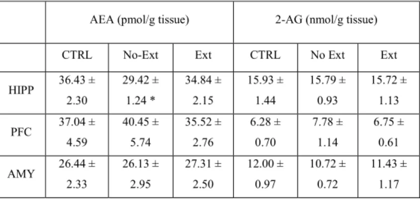
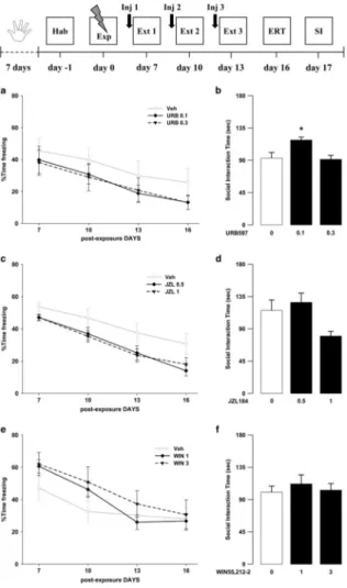
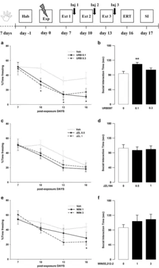
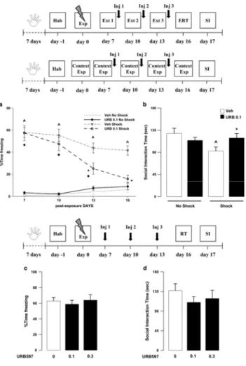
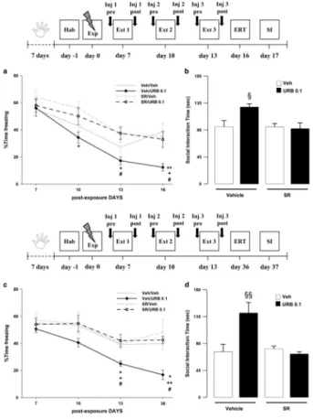
Exposure to a traumatic event may result in the development of Post-Traumatic Stress Disorder (PTSD). Endocannabinoids are crucial modulators of the stress response, interfere with excessive retrieval and facilitate the extinction of traumatic memories. Exposure therapy, combined with pharmacotherapy, represents a promising tool for PTSD treatment. We investigated whether pharmacological manipulations of the endocannabinoid system during extinction learning ameliorates the behavioral changes induced by trauma exposure. Rats were exposed to inescapable footshocks paired with social isolation, a risk factor for PTSD. One week after trauma, rats were subjected to three spaced extinction sessions, mimicking human exposure therapy. The anandamide hydrolysis inhibitor URB597, the 2-arachidonoylglycerol hydrolysis inhibitor JZL184 or the cannabinoid agonist WIN55,212-2 were administered before or after the extinction sessions. Rats were tested for extinction retention 16 or 36 days after trauma and 24-h later for social interaction. Extinction training alone reduced fear of the trauma-associated context but did not restore normal social interaction. Traumatized animals not exposed to extinction sessions exhibited reductions in hippocampal anandamide content with respect to home-cage controls. Noteworthy, all drugs exerted beneficial effects, but URB597 (0.1 mg/kg) induced the best improvements by enhancing extinction consolidation and restoring normal social behavior in traumatized rats through indirect activation of CB1 receptors. The ameliorating effects remained stable long after treatment and trauma exposure. Our findings suggest that drugs potentiating endocannabinoid neurotransmission may represent promising tools when combined to exposure-based psychotherapies in the treatment of PTSD.