R E S E A R C H
Open Access
Differences in cardiac phenotype and
natural history of laminopathies with and
without neuromuscular onset
Raffaello Ditaranto
1, Giuseppe Boriani
2, Mauro Biffi
1, Massimiliano Lorenzini
1,3, Maddalena Graziosi
1,
Matteo Ziacchi
1, Ferdinando Pasquale
1, Giovanni Vitale
1, Alessandra Berardini
1, Rita Rinaldi
4, Giovanna Lattanzi
5,
Luciano Potena
1, Sofia Martin Suarez
1, Maria Letizia Bacchi Reggiani
1, Claudio Rapezzi
1and Elena Biagini
1*Abstract
Objective: To investigate differences in cardiac manifestations of patients affected by laminopathy, according to the presence or absence of neuromuscular involvement at presentation.
Methods: We prospectively analyzed 40 consecutive patients with a diagnosis of laminopathy followed at a single centre between 1998 and 2017. Additionally, reports of clinical evaluations and tests prior to referral at our centre were retrospectively evaluated.
Results: Clinical onset was cardiac in 26 cases and neuromuscular in 14. Patients with neuromuscular presentation experienced first symptoms earlier in life (11 vs 39 years; p < 0.0001) and developed atrial fibrillation/flutter (AF) and required pacemaker implantation at a younger age (28 vs 41 years [p = 0.013] and 30 vs 44 years [p = 0.086] respectively), despite a similar overall prevalence of AF (57% vs 65%; p = 0.735) and atrio-ventricular (A-V) block (50% vs 65%; p = 0.500). Those with a neuromuscular presentation developed a cardiomyopathy less frequently (43% vs 73%; p = 0.089) and had a lower rate of sustained ventricular tachyarrhythmias (7% vs 23%; p = 0.387). In patients with neuromuscular onset rhythm disturbances occurred usually before evidence of cardiomyopathy. Despite these differences, the need for heart transplantation and median age at intervention were similar in the two groups (29% vs 23% [p = 0.717] and 43 vs 46 years [p = 0.593] respectively).
Conclusions: In patients with laminopathy, the type of disease onset was a marker for a different natural history. Specifically, patients with neuromuscular presentation had an earlier cardiac involvement, characterized by a linear and progressive evolution from rhythm disorders (AF and/or A-V block) to cardiomyopathy. Keywords: Lamin, Emerin, Neuromuscular disorders, Atrial fibrillation, Bradyarrhythmias, Ventricular tachycardias, Familial cardiomyopathies
Introduction
Laminopathies are a group of inherited conditions due to mutations in the LMNA gene, that encodes the nuclear envelope proteins lamin A and C, via alternate splicing [1]. Laminopathies are characterized by a high phenotypic heterogeneity including heart disease, neuromuscular disorders, premature aging and metabolic disorders [2–5]. LMNA associated cardiac and skeletal muscle disease
-often coexisting in the same patient - are the most frequent clinical manifestations. The spectrum of cardiac involve-ment ranges from supraventricular tachyarrhythmias and/ or conduction system disease to dilated cardiomyopathy (DCM) and ventricular tachyarrhythmias [6–12]. Sudden cardiac death may occur due to bradyarrhythmias or to malignant ventricular arrhythmias [13], even in the pres-ence of mild left ventricle systolic dysfunction. Similarly, LMNA-related neuromuscular disorders are characterized by a wide heterogeneity in clinical manifestations, Emery Dreifuss muscular dystrophy (EDMD) being the most common phenotype. EDMD is typically characterized by
© The Author(s). 2019 Open Access This article is distributed under the terms of the Creative Commons Attribution 4.0 International License (http://creativecommons.org/licenses/by/4.0/), which permits unrestricted use, distribution, and reproduction in any medium, provided you give appropriate credit to the original author(s) and the source, provide a link to the Creative Commons license, and indicate if changes were made. The Creative Commons Public Domain Dedication waiver (http://creativecommons.org/publicdomain/zero/1.0/) applies to the data made available in this article, unless otherwise stated. * Correspondence:[email protected]
1Cardiology Unit, Cardio-Thoracic-Vascular Department, Sant’Orsola-Malpighi
Hospital, University of Bologna, Via G. Massarenti 9, 40138 Bologna, Italy Full list of author information is available at the end of the article
early onset joint contractures with slowly progressive scapulo-humero-peroneal muscle weakness, and can be caused by mutations in genes other than LMNA, mainly EMDthat encodes emerin.
Although clinical manifestation in patients with LMNA mutations have been extensively described, the exact time course of cardiological and neuromuscular disease and their relation remain unclear. Specifically, it is not known if a neuromuscular onset is associated with a different cardiac phenotype or cardiac disease progression. The aim of this study was therefore to investigate differences in cardiac phenotype and natural history in relation to the presence of neuromuscular involvement at presentation, in patients with a diagnosis of laminopathy. Furthermore, in order to test the hypothesis that neuromuscular presen-tation (phenotype) per se might be associated with a spe-cific cardiac natural history, irrespective of genetics, we compared patients with neuromuscular presentation and LMNA or EMD mutations.
Methods
In this observational study, we prospectively evaluated all
LMNA mutation carriers from a single Italian centre
(S.Orsola-Malpighi University Hospital, Bologna), between December 1998 and November 2017. We also retrospect-ively examined clinical, ECG and echocardiographic reports available prior to evaluation at our centre. According to the presence of signs and/or symptoms of skeletal myopathy at clinical presentation (not necessarily in our centre), patients were divided into two groups:“with neuromuscular onset” and“without neuromuscular onset”. Data of 7 patients with EMD-related disease were also recorded. All patients underwent periodical clinical, electrocardiographic and echocardiographic monitoring.
DCM was defined as the presence of left ventricular (LV) dilation and systolic dysfunction in the absence of abnormal loading conditions (hypertension or valve disease) or coron-ary artery disease sufficient to cause global systolic
impair-ment [14]. “Hypokinetic non-dilated cardiomyopathy”
(HNDC) was defined as LV ejection fraction (EF) < 45% or biventricular global systolic dysfunction in absence of dilata-tion [15]. Restrictive cardiomyopathy was defined as a non-dilated LV with normal wall thickness and EF, and severe diastolic dysfunction with restrictive filling pattern, elevated filling pressures and dilated atria [14]. Sustained ventricular tachyarrhythmias (SVT) were defined as ventricular tachyar-rhythmias with a rate≥ 120/min, lasting > 30 s.
A neurological involvement was investigated on the basis of personal history and/or symptoms including joint contractures, muscle weakness or wasting, orthopaedic surgery, and exercise tolerance. Electromyography (EMG), muscle imaging or muscle biopsy were performed in se-lected cases. Patients underwent periodical neurological evaluation, even when no skeletal muscle involvement was
recorded at first evaluation. Elevation of serum creatine kinase (CK) in isolation was not considered diagnostic of neuromuscular involvement in absence of clinical, EMG, imaging or histological evidence of skeletal myopathy.
A detailed family history was collected for each pro-band in order to identify other potentially affected family members. A genetic diagnosis was made by DNA se-quencing from peripheral blood and mutations were considered pathogenic if previously described in litera-ture, in the presence of co-segregation or based on the site and the type of mutation. After mutation identifica-tion, cascade genetic screening was performed in family members, following informed consent.
Continuous data distribution was assessed with the Shapiro-Wilk test and expressed as median and interquar-tile range (IQR). Data were compared by the Fisher’s exact test for proportions, and Mann Whitney test for continu-ous variables. Clinical events were reported as counts and percentages (i.e. events/total number of patients × 100). Data were collected following informed consent.
Results
Of the 41 LMNA mutation carriers, 40 were clinically af-fected. Table 1 reports clinical, ECG and echocardio-graphic characteristics at first evaluation at our centre.
Fourteen patients were assigned to the group “with
neuromuscular onset” and 26 to the group “without neuromuscular onset”. A single LMNA mutation carrier, who did not have any cardiac or neuromuscular pheno-typic expression at baseline or during follow up, was ex-cluded from the analysis.
Patients with neuromuscular onset
Among the 14 patients in this group males and females were equally represented. LMNA mutations were: 12 missense, 1 splice site, and 1 deletion (Table 2). Median age at first evaluation at our Centre was 31 years (IQR 20–44). The diagnosis of laminopathy occurred follow-ing familial screenfollow-ing in 2 patients. At time of genetic diagnosis, overt neuromuscular involvement was present in all cases whereas none had cardiac involvement. Eleven patients (79%) were diagnosed with EDMD in the first or second decade of life and 3 (21%) were diagnosed with a non-specific myopathy in the third decade; me-dian age at diagnosis was 11 years (IQR 8–30).
Nine patients (64%) developed cardiac involvement prior to referral to our centre (Fig.1a summarizes the spectrum of cardiac phenotypic expression). All had rhythm distur-bances: 2 (14%) had atrial fibrillation (AF), 1 (7%) atrio-ventricular (A-V) block, 1 (7%) sinus node dysfunction, and 5 (36%) had a combination of brady- and atrial tachyar-rhythmias (Fig. 2). Cardiomyopathy was also present in 5 patients (36%): 2 DCM, 2 HNDC and 1 isolated right ventricular cardiomyopathy mimicking arrhythmogenic
right ventricular cardiomyopathy. Patients with LV cardio-myopathy had a severe LV dysfunction (LVEF≤35%) and 3 had biventricular involvement.
Two patients (14%) had previously undergone pace-maker (PM) implantation for A-V block, while a single patient had a primary prevention implantable cardiac resynchronization therapy defibrillator (CRT-D). Serum CK levels were raised in 65% of the patients, with a mean abnormal value of 631 UI/L.
Patients without neuromuscular onset
Fifteen (58%) of the 26 patients were males, median age at first evaluation at our centre was 43 years (IQR 36–49). Diagnosis was made due to rhythm disturbances or heart failure symptoms in most cases (n = 19, 73%). Seven pa-tients (27%) were identified after family screening. LMNA
mutations were: 16 missense, 7 splice site, 2 deletion and 1 frameshift (Table3).
At first clinical evaluation at our centre, 19 patients (73%) had a cardiomyopathy, in isolation (n = 3, 11%) or associated with arrhythmias (AF n = 5, 19%; A-V block n= 6, 23%; both n = 5, 19%; Fig.3). Seven patients (27%) had arrhythmias in absence of cardiomyopathy: 1 (4%) AF, 5 (19%) A-V block and 1 (4%) a combination of sinus node and A-V node dysfunction. Overall, AF was present in 11 patients (42%). Figure1b summarizes the spectrum of cardiac phenotypic expression. Six patients (23%) had previously undergone PM implantation for A-V block and 7 patients (27%) had received a primary prevention im-plantable cardioverter defibrillator (ICD).
Cardiomyopathy phenotype included: 11 DCM, 6 HNDC and 2 restrictive cardiomyopathy. Among the
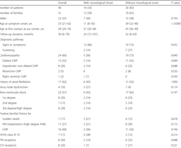
Table 1 Characteristics at first clinical evaluation at our centre of patients with LMNA mutations with and without neuromuscular onset
Overall With neurological Onset Without neurological onset P value
Number of patients 40 14 (35) 26 (65)
Number of families 31 12 (39) 19 (61)
Males 22 (55) 7 (50) 15 (58) 0.744
Age at symptom onset, yrs 33 (21–42) 11 (8–30) 39 (32–46) < 0.0001
Age at first contact at our center, yrs 39 (29–74) 31 (20–44) 43 (36–49)
Follow-up duration, months 30 (6–70) 24 (12–101) 32 (8–63)
Diagnostic pathway
Signs or symptoms 12 (86) 19 (73) 0.452
Screening 2 (14) 7 (27)
Cardiomyopathy 24 (60) 5 (36) 19 (73) 0.040
Dilated CMP 13 (32) 2 (14) 11 (42) 0.089
Hypokinetic non dilated CMP 8 (20) 2 (14) 6 (23) 0.688
Restrictive CMP 2 (5) 0 2 (8) 0.533
Right ventricle CMP 1 (2) 1 (7) 0 0.350
History of atrial fibrillation 17 (42) 6 (43) 11 (42) 1.000
Sinus node dysfunction 4 (10) 3 (21) 1 (4) 0.114
Atrio-ventricular block 23 (57) 6 (43) 17 (65) 0.197
1st degree 8 (20) 2 (14) 6 (23)
2nd degree 7 (17) 2 (14) 5 (19)
3rd degree/high degree 8 (20) 2 (14) 6 (23)
Positive familial history for
Sudden death 7 (17) 3 (21) 4 (15) 0.678
PM implantation (high degree AVB) 11 (27) 3 (21) 8 (30) 0.715
CMP 16 (40) 5 (36) 11 (42) 0.746
NYHA class III- IV 7 (17) 4 (28) 3 (12) 0.214
PM recipients 8 (20) 2 (14) 6 (23) 0.688
ICD recipients 8 (20) 1 (7) 7 (27) 0.221
Values are expressed as N, N (%) or median (interquartile range)
LMNA, EMD gene codifying for lamin A/C and emerin, respectively, ICD implantable cardioverter defibrillator, PM pacemaker, AVB atrio-ventricular block, NYHA New York Heart Association
17 patients with LV systolic dysfunction, 7 (41%) had a severe impairment (LVEF≤35%), 6 (35%) had biventri-cular involvement and 3 (17%) had increased LV trabecu-lations. One 54-year old patient with a history of complete A-V block and AF had a biventricular cardiomyopathy with multiple aneurysms in the diaphragmatic and free wall of the right ventricle. Coronary arteries were unob-structed at angiography. Cardiac magnetic resonance showed a severely dilated left ventricle (indexed end dia-stolic volume: 146 mL/m2) with sydia-stolic dysfunction (EF 40%) and mildly dilated right ventricle (indexed end dia-stolic volume: 111 ml/m2) with reduced EF (40%) and confirmed the wall motion abnormalities (Fig. 4 a-b). Tissue characterization (Fig. 4 c-d) revealed multiple areas of fibro-fatty replacement. In this case, possible phenocopies including desmosomal-related cardiomy-opathy and sarcoidosis were excluded by genetic ana-lysis, positron emission tomography, lung CT and, endomyocardial biopsy.
No patient had evidence of neuromuscular involvement. Serum CK levels were raised in 47% of the patients with a mean abnormal value of 217 UI/L.
Follow-up of patients with neuromuscular onset
Median follow up was 24 months (IQR 12–101). New onset AF was recorded in 2 patients, therefore 57% experienced atrial tachyarrhythmias by the end of the follow-up. In one patient, the atrial conduction disease progressed to atrial paralysis. Three patients underwent PM implantation for A-V block (n = 1), sinus node disease (n = 1) or both (n = 1). A primary prevention CRT-D was implanted in a patient with new onset HNDC due to positive family history for sudden death, high inducibility of VT on electrophysio-logical study and moderate LV dysfunction. The patient affected by right ventricular cardiomyopathy received a primary prevention ICD, due to severe right ventricular dysfunction, non-sustained ventricular tachycardias and the need of a pacing for sinus and A-V node dysfunction.
Table 2 Genetics of LMNA mutated patients with neurological onset (N = 14)
Gene Location Nucleotide Change Protein Change Predicted Effect
Family 1
F; 16 yo LMNA Exon 4 c.746G > A p.Arg249Gln Missense
Family 2
F; 50 yo LMNA Exon 9 c.1580G > C p.Arg.527.Pro Missense
Family 3
M; 38 yo LMNA Exon 11 c.1930C > T p.Arg644Cys Missense
M; 38 yo LMNA Exon 11 c.1930C > T p.Arg644Cys Missense
Family 4 F; 46 yo LMNA Exon 3 Exon 4 c.569G > A; c. 746G > A p.Arg190Gln p.Arg249Gln Missense Missense Family 5
F; 34 yo LMNA Exon 1 c.203_208 (delAGGTGG) p.Glu68_Val69 del Deletion
Family 6
M; 52 yo LMNA Exon 4 c.746G > A p.Arg249Gln Missense
Family 7
M; 46 yo LMNA Exon 9 c.1567G > A p.Gly523Arg Missense
Family 8
M; 17 yo LMNA Exon 4 c.775 T > G p.Tyr259Asp Missense
Family 9
M; 19 yo LMNA Exon 4 c.746 G > A p.Arg249Gln Missense
Family 10
F; 29 yo LMNA Intron 9 c.1608 + 1G > T – Splice site
Family 11
F; 22 yo LMNA Exon 1 c.188 T > A p.Ile63Asn Missense
F; 19 yo LMNA Exon 1 c.188 T > A p.Ile63Asn Missense
Family 12
M; 27 yo LMNA Exon 4 c.746G > A p.Arg249Gln Missense
Thereafter he experienced an appropriate ICD activation and a progression towards severe biventricular involvement. No sudden death occurred. Five patients with cardiomyop-athy had hospital admissions due to heart failure during follow-up and 4 of them subsequently underwent heart transplantation (median age 43 [IQR 34–48]).
Follow-up of patients without neuromuscular onset
New onset AF was reported in 6 patients (23%) during a median follow up of 32 months (IQR 8–63); 65% of patients had atrial tachyarrhythmias at the end of follow up. With the exception of 2 patients with atrial flutter – who were treated successfully with cavo-tricuspid isthmus ablation – the attempts of rhythm control with electrical or pharmacological cardioversion were ineffective. No pa-tients underwent pulmonary vein isolation. Atrial paralysis was documented in a single patient. One patient under-went PM implantation due to A-V block. A primary pre-vention ICD was implanted in 7 patients (4 new implants and 3 device upgrades) and 1 ICD was implanted for secondary prevention. Four of the ICD recipients (50%) received a CRT-D device. During follow up 6 patients (23%) experienced appropriate shocks and/or antitachycar-dia pacing for ventricular arrhythmias, with an arrhythmic storm in 3 cases. Six (23%) patients underwent cardiac transplantation (median age 46 [IQR 34–53]) due to end stage heart failure (5/6) or to recurrent ventricular arrhyth-mias (1/6). One patient developed a mild neuromuscular involvement, with muscle atrophy involving the shoulder girdle. Table 4 compares clinical events reported during follow-up in the two groups.
Differences in clinical manifestations between patients with and without neuromuscular onset
Patients with neuromuscular onset had an earlier pres-entation, during infancy or adolescence in most of the cases (median age 11 years), mainly as EDMD, followed by the first evidence of cardiac disease by a median age of 13 years (IQR 10–15) (maximum timelag 38 years). In patients without neuromuscular onset, first cardiac symptoms occurred later in life, at a median age of 39
Fig. 1 a Cardiac phenotype spectrum at first clinical evaluation at our centre of patients with LMNA mutations and neurological onset (N = 14). SSS: sick sinus syndrome; AVB: atrio-ventricular block; CMP: cardiomyopathy; AF atrial fibrillation/flutter. b Cardiac phenotype spectrum at first clinical evaluation at our centre of patients with LMNA mutations without neurological onset (N = 26). SSS: sick sinus syndrome; AVB: atrio-ventricular block; CMP: cardiomyopathy; AF atrial fibrillation/flutter
Fig. 2 Second degree sino-atrial block with 2:1 conduction ratio in a 22-year old female, with Emery Dreifuss muscular dystrophy due to p.Ile63Asn missense LMNA mutation
Table 3 Genetics and cardiac manifestations of LMNA mutated patients without neurological onset (N = 26)
Gene Location Nucleotide Change Protein Change Predicted Effect
Family 1
M; 53 yo LMNA Exon 6 c.1004G > A p.Arg335Gln Missense
M; 26 yo LMNA Exon 6 c.1004G > A p.Arg335Gln Missense
Family 2
F; 47 yo LMNA Exon 7 1370delA p.Lys457SerfsX2 Deletion
Family 3
M; 19 yo LMNA Exon 6 c.1003G > A p.Arg335Glu Missense
Family 4
F; 55 yo LMNA Exon 11 c.1912G > A p.Gly638Arg Missense
Family 5
M; 54 yo LMNA Exon 7 c.1202G > A p.Arg401His Missense
Family 6
M; 42 yo LMNA Exon 1 n/a p.Arg72Alafs*24 Frame shift
Family 7
F; 51 yo LMNA Exon 4 c.752A > C p.Gln251Pro Missense
Family 8
M; 46 yo LMNA Exon 9 c.1517 A > C p.His506Pro Missense
Family 9
F; 38 yo LMNA Intron 9 c1608 + 1G > T – Splice site
M; 35 yo LMNA Intron 9 c1608 + 1G > T – Splice site
Family 10
F; 29 yo LMNA Exon 3 c.548 T > C p.Leu183Pro Missense
Family 11
F; 41 yo LMNA Exon 6 c.1007G > A p.Arg336Gln Missense
Family 12 M; 60 yo LMNA Intron 1 c.357-1G > A IVS1-1G > A – Splice site F; 46 yo LMNA Intron 1 c.357-1G > A IVS1-1G > A – Splice site F; 49 yo LMNA Intron 1 c.357-1G > A IVS1-1G > A – Splice site F; 49 yo LMNA Intron 1 c.357-1G > A IVS1-1G > A – Splice site M; 21 yo LMNA Intron 1 c.357-1G > A IVS1-1G > A – Splice site Family 13
M; 38 yo LMNA Exon 6 c.1129C > T p.Arg377Cys Missense
Family 14
M; 50 yo LMNA Exon 2 c.481G > A p.Glu161Lys Missense
Family 15
M; 46 yo LMNA Exon 2 c.466C > A p.Arg156Ser Missense
M; 34 yo LMNA Exon 2 c.466C > A p.Arg156Ser Missense
Family 16
M; 44 yo LMNA Exon 4 c.671 C > T p.Thr224Ile Missense
Family 17
years (p < 0.0001). Regarding arrhythmias, at the end of the follow-up A-V block (of any degree) and AF had a similar prevalence between the two groups (50% vs 65%, p= 0.500 and 57% vs 65%; p = 0.735 respectively). Sinus node dysfunction was more frequent in patients with skel-etal myopathy (21% vs 4%; p = 0.114), whereas atrial par-alysis was reported in one patient for each group. Patients with neuromuscular presentation (Fig. 5) experienced earlier AF (age 28 vs 41, p = 0.013) and PM implantation (age 30 vs 44; p = 0.086). The percentage of patients requiring permanent pacing (including PM recipients and those who received an ICD due to a concomitant indica-tion for prevenindica-tion of ventricular arrhythmias) was equal in the two groups (42% vs 42%; p = 1.000).
Patients without neuromuscular presentation had a higher prevalence of cardiomyopathy (73% vs 43%, p = 0.089) and were older at diagnosis (age 42 vs 35, p = 0.259). DCM was the dominant phenotype in this group (58% of
all cardiomyopathies), whereas DCM and HNDC where equally represented in the other group. The higher preva-lence of heart muscle involvement in patients without neuromuscular onset was associated with a higher number of implanted ICDs (58% vs 21%, p = 0.045) and a higher burden of SVT (23% vs 7%, p = 0.387). Despite this, no sig-nificant differences were reported in the prevalence of heart transplantation (23% vs 29%; p = 0.717) or in the median recipient age (43 vs 46; p = 0.593).
All patients with neuromuscular presentation who received a diagnosis of cardiomyopathy had a previous history of rhythm disturbance with the exception of 2 cases, where the diagnosis was concomitant. On the con-trary, no pattern of progression from rhythm disturb-ance to cardiomyopathy was present in those without a neuromuscular presentations: AF and A-V block could precede the diagnosis of cardiomyopathy, be diagnosed at the same time or later. Figure 6 shows the different
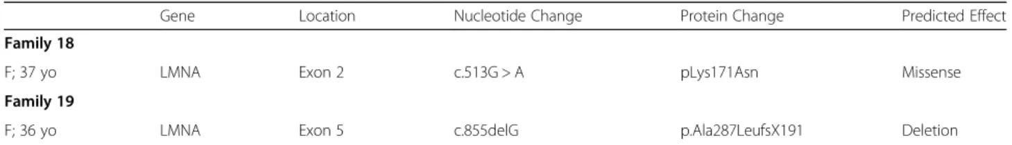
Table 3 Genetics and cardiac manifestations of LMNA mutated patients without neurological onset (N = 26) (Continued)
Gene Location Nucleotide Change Protein Change Predicted Effect
Family 18
F; 37 yo LMNA Exon 2 c.513G > A pLys171Asn Missense
Family 19
F; 36 yo LMNA Exon 5 c.855delG p.Ala287LeufsX191 Deletion
M male, F female, yo years old. The age reported refers to first contact at our centre. n/a not available
Fig. 3 43-year old male with a LMNA frameshift mutation without neuromuscular presentation. a V1 lead ECG showing first degree atrio-ventricular block, b-c cardiac magnetic resonance showing midwall late gadolinium enhancement in the basal interatrio-ventricular septum. Suggestive for myocardial fibrosis
overall prevalence of clinical events between the two populations.
Clinical characteristics and follow up of patients with emerinopathy
Seven male patients (4 families) affected by X-linked EDMD were referred to our Centre at a median age of 26 years (IQR 14–30) and followed for a median time of 108 months (IQR 72–172). All had neuromuscular symptoms as first evaluation, with a median age of 6 years (IQR 5–8). At last follow up 6 patients (85%) had
cardiac involvement. All developed AF at a median age of 27 [IQR 23–37]) and 5 required PM implantation at a median age of 23 [IQR 22–24] due to A-V block (n = 1), sinus node dysfunction (n = 1) or both (n = 3). A single patient developed cardiomyopathy with mild systolic dysfunction; none had ventricular arrhythmias. The time interval from neurological to cardiac disease onset was of 14,5 years (IQR 14–15). Compared to patients with LMNAmutations and a neurological onset, patients with emerinopathy presented a higher burden of AF (85% vs 57%; p = 0.337) that occurred an earlier age (27 vs 31
Fig. 4 CMR of a 54-year male, carrier of p.Arg401His missense LMNA mutation, affected by DCM. (a-b) Four chamber and RV long axis SSFP images show biventricular dilation, bulging of the RV free wall (white arrow, panel a-b) and diaphragmatic wall (white arrow, panel b). (c) Two chamber T1-weighted and (d) fat suppressed T1-weighted slices showing LV fatty replacement of mid lateral wall (white arrow). (e) IR LGE slice showing fibrosis in the infero-lateral wall (with focal transmural pattern) and in the interventricular septum (image quality was due to respiratory artifacts and to the presence of pacemaker [*]). CMR: cardiac magnetic resonance. DCM: dilated cardiomyopathy. SSFP: steady-state free precession. LV-RV: left-right ventricle. IR LGE: inversion recovery late gadolinium enhancement
Table 4 Clinical events reported during follow-up in LMNA mutated patients
Patients with neurological onset (N = 14) Patients without neurological onset (N = 26) P value
New onset of atrial fibrillation (any form) 2 (14) 6 (23) 0.688
PM implantation 3 (21) 1 (4) 0.114
ICD implantation 2 (14) 8 (31) 0.445
SVT/arrhythmic storm 1 (7) 6 (23) 0.387
Admission for heart failure 5 (36) 11 (42) 0.746
Heart transplantation 4 (29) 6 (23) 0.717
Thromboembolic events 0 0
p= 0.746), and a higher rate of PM implantation (71 vs 36%; p = 0.182) at an earlier age (23 vs 28; p = 0.461). Differently, heart muscle involvement was rare (a single case of cardiomyopathy) and no SVT was documented.
Discussion
The main findings of our study are: 1) neuromuscular on-set is a marker for a specific natural history in laminopa-thy patients. Specifically, these patients have a linear and predictable progression over time, from muscular dys-trophy to rhythm disturbances and finally cardiomyop-athy. 2) With the exception of sinus node dysfunction, that was more frequent in EDMD patients, the prevalence of A-V block and AF was similar between the two groups,
but patients with a neuromuscular presentation had earl-ier arrhythmias. 3) Prevalence of cardiomyopathy (particu-larly DCM) and SVT was higher among patients without neuromuscular onset, although the two groups had a simi-lar rate and age of cardiac transplantation.
Most of the patients with neuromuscular presentation (64%) developed cardiac involvement later in life: the delay between neuromuscular and cardiac symptom onset was variable and sometimes very long. These findings suggest that serial reassessment of cardiac status in patients with a diagnosis of EDMD is mandatory. On the other hand, patients without neuromuscular onset did not develop an overt skeletal myopathy (with a single exception). The pos-sibility that these patients could develop neuromuscular
Fig. 5 Box and whiskers plot showing age distribution of different clinical events in LMNA patients with (white) and without (gray) neuromuscular onset. Middle horizontal line inside box indicates median. Bottom and top of the box are 25th and 75th percentiles, the whiskers indicate the lowest and highest value. PM: pacemaker. ICD: implantable cardioverter defibrillator
Fig. 6 Different overall prevalence at the end of follow-up of clinical events in LMNA patients with (white) and without (gray) neuromuscular presentation. AF: atrial fibrillation; PM: pacemaker; CMP: cardiomyopathy; SVT: sustained ventricular tachycardias
involvement in the future cannot however be entirely ruled out due to the limited observation time. Our results are in line with the phenotypic clustering reported by Benedetti et al. [16] in a cohort of patients with LMNA mutations, where those with childhood onset had an almost exclusively skeletal muscle involvement (predominantly EDMD), while patients with adult onset developed cardiac disorders or muscle weakness with a limb-girdle distribution. With the limitation of the small size of the screened families in our study population and the limited numbers of relatives who carried the mutation, all the affected relatives of probands with neuromuscular presentation had a skeletal muscle involvement as clinical onset. At the same time, all the af-fected relatives of patients with an exclusive cardiac pheno-type had an isolated cardiological involvement. Differently from our findings, Bonne et al. and Brodsky et al. [17, 18] have previously described the possibility of the coexistence of both phenotypes as clinical onset within the same family. Our data confirm the high frequency of AF in lamino-pathies, as well as advanced A-V block, requiring PM implantation at a young age. Although the prevalence of high degree A-V block and AF was similar, irrespective of clinical presentation, patients with neuromuscular on-set experienced arrhythmia earlier in life (on average AF and PM implantation occurred more than 10 years earl-ier). On the other hand, sinus node dysfunction was more frequent in patients with EDMD (21% vs 4%). Re-garding heart muscle involvement, patients without neuromuscular onset had a prevalence of cardiomyop-athy that was almost twice that of the other group (42% vs 73%; p = 0.089), mostly DCM. On the contrary DCM and HNDC were equally distributed in patients with neuromuscular presentation. A progression from HNDC to DCM was not observed in this study, suggesting that they could be the expression of two different pathophys-iologic models; however, a limited follow-up duration (median 41 months) and heart failure therapy could have masked this progression. We described two cases, 1 in each group, with a cardiac phenotype that mimicked arrhythmogenic cardiomyopathy. LMNA carriers have
been described with clinical, morphological and histo-logical phenotypes overlapping with arrhythmogenic car-diomyopathy [19], in the absence of desmosomal gene mutations, with conduction disease being the sole ‘red flag’ for the correct diagnosis. These findings may justify the need to exclude LMNA mutations in patients with suspected arrhythmogenic cardiomyopathy, particularly when conduction disease is present.
This study confirms the malignant nature of laminopa-thies in terms of ventricular arrhythmias and progression to advanced heart failure. In our series sustained SVTs during the follow-up were more frequent in patients without neuromuscular involvement (23% vs 7%) with just 1 EDMD patient experiencing SVT. The low incidence of events in patients with skeletal myopathy differs from previous re-ports. Van Rijsingen et al. [10] reported that in patients with LMNAmutations, the diagnosis of muscular dystrophy or a positive family history of muscular dystrophy was not asso-ciated with a different incidence of ventricular arrhythmias. The rate of SVT reported by the Authors was of 17% in EDMD patients and 19% in non-EDMD patients. More than 20% of the whole population of this study required cardiac transplantation during follow-up and this is consist-ent with previous reports. Hasselberg et al. [11] reported that 19% of genotyped LMNA patients underwent heart transplantation during a follow up of 8 years (median age 46 years). The need for heart transplantation in our series was independent from the involvement of the skeletal muscle and occurred at a median age of 45 years. Differ-ently from what observed for other clinical events, neuro-muscular involvement did not lead to an anticipation in the timeline and heart transplant was performed at a similar age in the two groups (median 43 vs 46 years).
In this series, patients with a neuromuscular presenta-tion had a linear predictable progression over time (Fig. 7). Specifically, skeletal myopathy developed first, followed by arrhythmias (A-V block, sick sinus syn-drome and AF in various combinations) and eventually, cardiomyopathy. This pattern of progression was not ob-served in the other patients.
The cardiac involvement in X-linked EDMD patients of our study, compared to patients with LMNA muta-tions and a neurological onset, was characterized by higher burden of supraventricular tachyarrhyrhmias and bradyarrhythmias that occurred at a younger age and a lower frequency of cardiomyopathy.
Limitations
Referral bias cannot be excluded since this is a study from a single tertiary centre with a cardiac transplant program and expertise for management of complex ven-tricular tachycardias.
Conclusion
In patients with laminopathy, the type of disease onset was a marker for a different natural history. Specifically, patients with neuromuscular presentation had an earlier cardiac involvement, characterized by a linear and progressive evolution from rhythm disorders (AF and/or V block) to cardiomyopathy. Prevalence of AF and A-V block was similar, regardless of clinical onset, whereas sinus node dysfunction was more frequent in EDMD pa-tients. Patients with neuromuscular onset had a lower prevalence of cardiomyopathy and ventricular arrhyth-mias, but a similar prevalence of heart transplantation at a similar age.
Abbreviations
AF:Atrial fibrillation/flutter; AVB: Atrio-ventricular block; CK: Creatine kinase; CRT-D: Implantable cardiac resynchronization therapy defibrillator; DCM: Dilated cardiomyopathy; EDMD: Emery Dreifuss muscular dystrophy; EMG: Electromyography; HNDC: Hypokinetic non dilated cardiomyopathy; HT: Heart transplantation; ICD: Implantable cardioverter defibrillator; IQR: Interquartile range; LV: Left ventricle; LVEF: Left ventricle ejection fraction; NYHA: New York Heart Association; PM: Pacemaker; SVT: Sustained ventricular tachyarrhythmias
Acknowledgements None.
Authors’ contributions
EB, RD and CR wrote the initial manuscript. EB, CR, MB, GB, RD and ML provided critical discussion of research. All Authors participated in clinical data collection, read and approved the final manuscript.
Funding
This work was supported by the University of Bologna and“Fondazione Luisa Fanti Melloni”.
Availability of data and materials
The datasets used and analysed during the current study are available from the corresponding Author on reasonable request.
Ethics approval and consent to participate
Informed consent was obtained from patients for collection of anonymised clinical data.
Consent for publication
All participants gave consent for publication of anonymised individual personal data.
Competing interests
The authors declare that they have no competing interests.
Author details
1Cardiology Unit, Cardio-Thoracic-Vascular Department, Sant’Orsola-Malpighi
Hospital, University of Bologna, Via G. Massarenti 9, 40138 Bologna, Italy.
2
Cardiology Division, Department of Biomedical, Metabolic and Neural Sciences, University of Modena and Reggio Emilia, Policlinico di Modena, Modena, Italy.3University College London Institute for Cardiovascular Science
and Barts Heart Centre, St. Bartholomew’s Hospital, London, UK.4Neurology
Unit, Sant’Orsola-Malpighi University Hospital, Bologna, Italy.5Italian National Research Council (CNR), Institute of Molecular Genetics IGM Bologna, Bologna, Italy.
Received: 21 May 2019 Accepted: 29 October 2019
References
1. Mounkes L, Kozlov S, Burke B, Stewart CL. The laminopathies: nuclear structure meets disease. Curr Opin Genet Dev. 2003;13:223–30. 2. Shackleton S, et al. LMNA, encoding Lamin a/C, is mutated in partial
lipodystrophy. Nat Genet. 2000;24:153–6.
3. Boriani G, et al. Clinical relevance of atrial fibrillation/flutter, stroke, pacemaker implant, and heart failure in Emery-Dreifuss muscular dystrophy: a long-term longitudinal study. Stroke. 2003;34:901–8.
4. Eriksson M, et al. Recurrent de novo point mutations in Lamin a cause Hutchinson-Gilford progeria syndrome. Nature. 2003;423:293–8.
5. Bonne G, et al. Mutations in the gene encoding Lamin a/C cause autosomal dominant Emery-Dreifuss muscular dystrophy. Nat Genet. 1999;21:285–8. 6. Arbustini E, et al. Autosomal dominant dilated cardiomyopathy with
atrioventricular block: a Lamin a/C defect-related disease. J Am Coll Cardiol. 2002;39:981–90.
7. Kumar S, et al. Long-term arrhythmic and nonarrhythmic outcomes of Lamin a/C mutation carriers. J Am Coll Cardiol. 2016;68:2299–307. 8. Pasotti M, et al. Long-term outcome and risk stratification in dilated
cardiolaminopathies. J Am Coll Cardiol. 2008;52:1250–60.
9. Taylor MR, et al. Natural history of dilated cardiomyopathy due to Lamin a/ C gene mutations. J Am Coll Cardiol. 2003;41:771–80.
10. van Rijsingen IA, et al. Risk factors for malignant ventricular arrhythmias in Lamin a/c mutation carriers a European cohort study. J Am Coll Cardiol. 2012;59:493–500.
11. Hasselberg NE, et al. Lamin a/C cardiomyopathy: young onset, high penetrance, and frequent need for heart transplantation. Eur Heart J. 2018; 39:853–60.
12. Captur G, et al. Lamin and the heart. Heart. 2018;104:468–79.
13. Wahbi K, et al. Development and validation of a new risk prediction score for life-threatening ventricular Tachyarrhythmias in Laminopathies. Circulation. 2019;140:293–302.
14. Elliott P, et al. Classification of the cardiomyopathies: a position statement from the European Society of Cardiology working group on myocardial and pericardial diseases. Eur Heart J. 2008;29:270–6.
15. Pinto YM, et al. Proposal for a revised definition of dilated cardiomyopathy, hypokinetic non-dilated cardiomyopathy, and its implications for clinical practice: a position statement of the ESC working group on myocardial and pericardial diseases. Eur Heart J. 2016;37:1850–8.
16. Benedetti S, et al. Phenotypic clustering of Lamin a/C mutations in neuromuscular patients. Neurology. 2007;69:1285–92.
17. Bonne G, et al. Clinical and molecular genetic spectrum of autosomal dominant Emery-Dreifuss muscular dystrophy due to mutations of the Lamin a/C gene. Ann Neurol. 2000;48:170–80.
18. Brodsky GL, et al. Lamin a/C gene mutation associated with dilated cardiomyopathy with variable skeletal muscle involvement. Circulation. 2000;101:473–6.
19. Quarta G, et al. Mutations in the Lamin a/C gene mimic arrhythmogenic right ventricular cardiomyopathy. Eur Heart J. 2012;33:1128–36.
Publisher’s Note
Springer Nature remains neutral with regard to jurisdictional claims in published maps and institutional affiliations.