1 DIPARTIMENTO PER LA INNOVAZIONE NEI SISTEMI BIOLOGICI,
AGROALIMENTARI E FORESTALI
Corso di Dottorato di Ricerca in
BIOTECNOLOGIE DEGLI ALIMENTI - XXVI Ciclo
Metabolic and structural effects of dehydration and ozone
postharvest treatments on wine grape
s.s.d. AGR/15
Tesi di dottorato di:
Dott.ssa Federica De Sanctis
Coordinatore del corso Tutore
Prof. Marco Esti Prof. Fabio Mencarelli
Firma ……….. Firma ………
Co-tutore
Dott. Rinaldo Botondi Firma………
2 Ringraziamenti
Ringrazio tutto il team di ricerca dell’ex LAPO per il supporto fisico ed intellettuale. Ringrazio inoltre il prof. John M. Labavitch, Joseph L. Smilanick P.h.D, Kent Fjeld, il prof. James Kennedy e tutte le altre persone che hanno contribuito, non solo attivamente, al lavoro svolto in California.
Ringrazio infine la mia famiglia e mio marito per la pazienza mostratami ed il supporto ricevuto.
3 Ricerca scientifica parzialmente finanziata dalla PC Engineering srl,
4 Index
Summary 1
Abstract- English version 1
Abstract- Italian version 2
1-Introduction 3
2- Ozone 5
2.1 General aspects and chemical property 5
2.1.1 Brief History of Ozone Use for Water and Food Products 6 2.2 Microbiological Aspects of Ozone
2.3 Interaction between ozone and organic matrix 18
3. Grape Cell wall and Postharvest dehydration 23
3.1 Cell Wall composition 23
3.2 Cell Wall architecture 28
3.3 Cell Wall enzymes 29
3.4 Postharvest dehydration of wine grape 33
3.5 General dehydration techniques 34
3.6 Water loss: Important changes on wine grape 36
Aim of the study 37
4.Materiali e Metodi 38
4.1 Effects of ozone on grape cell wall, phenolic content and
volatile organic compounds (VOCs) 38
5
4.3 Chemical analysis 41
4.4 Cell wall analysis 43
4.5 Enzymes activity 45
5-Results and discussion 47
5.1 Effects of ozone on wine grape: var Sauvignon blanc 47 5.2 Effects of ozone on wine grape: var Cabernet sauvignon 53 5.3 Effects of ozone during postharvest dehydration of Pignola grapes 60
6-Conclusions 70
7-Bibliography 73
6 Summary
Abstract- English version
This PhD thesis is aimed to study metabolic, biochemical and structural changes on white wine grape, red wine grape and raisins using ozone post-harvest treatments without addiction of sulfites.
In wine production, sulfur dioxide is currently used for its positive effects on wine stabilization but it’s even known its negative effects on grape and human health(Bush, 1986).
Ozone was approved in 2001 by FDA ( Food and Drug Administration) as GRAS (generally recognized as safe) for food sanitation and it started to be used in many food industry as natural alternative to chemicals disinfection process. The use of ozone in the food industry represent an important step forward towards technological innovations.
To evaluate the effect of ozone on wine grape was studied first to what happen when O3 is applied on white and red wine grape and then on the modification caused by its application during postharvest dehydration.
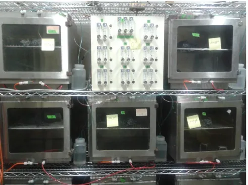
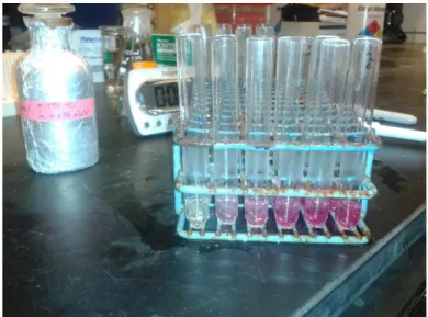
Grape clusters variety Sauvignon Blanc (Castello de "La Sala" winery,Orvieto, Italy), Cabernet Sauvingon (Fresno State University, California) and Pignola (Cantine Di Villa, Valtellina, Italy ) were carefully hand harvested, selected and placed in a single layer into perforated crates (60 x 40 x 15 cm) until 6 kilograms Kg (±500 g) each, and divided into thermohygrometric controlled rooms (12 m3) at 10 (±1)°C and UR 70%.
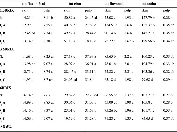
The O3 exposure effects on Cabernet S. was studied on three different stages of grape ripening, 21,3°BRIX (unripe stage), 23,6°BRIX (ripe stage), 26°BRIX (overripe stage) and each one was treated with 300, 800 and 1300 ppb of ozone.
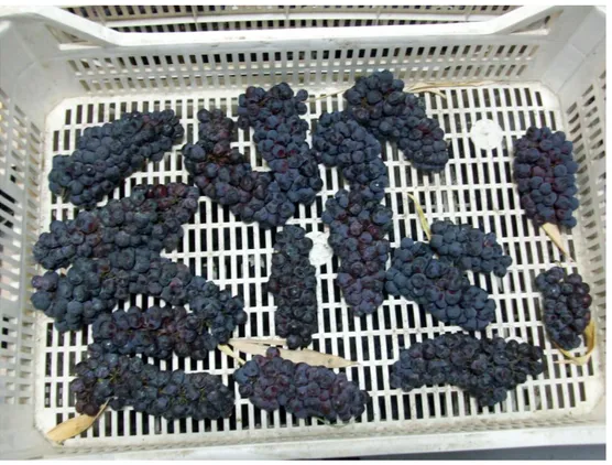
Sauvignon blanc sampling was performed at the beginning and then after 4, 8 and 16 hours of ozone treatment (European pending patent 12704901.3-1357 PC Engineering srl, Uggiate Trevano, Italy). Pignola grapes, after a primary sampling, was subsequently exposed to three different treatments: O3 for 18 hours followed by dehydration; O3 for 18 hours followed by dehydration with 0.5g/h of O3 for 4 hours every day; and not treated grapes (CK), dehydrated until 20% and 35% of weight loss. Sampling was performed at the beginning and then at 20 and 35% weight loss(wl).
Color parameters, weight loss, total SSC, CO2 production and TA remained quite constant right after ozone treatment. On the contrary, important differences have been found on phenolic content.
Some fraction as cinnamates and flavonols resulted to be positively affected by the treatment whereas catechins and epicatechin are resulted to be more susceptible.
Anthocyanins content are resulted to be less sensitive to the treatments, indeed, an increase was shown on unripe grape exposed to medium ozone concentration, while others samples remain quite stable after the exposure.
Glycosilated volatiles organic compounds (VOCs) increased significantly in Sauvignon blanc treated samples expecially benzyl alcol, methoxyeugenol, Coniferol 2, 7-OH a-terpineol and Isogeraniol, while free VOCs, whom are perceptible directly from the berry, as expected after ozone treatment fallen.
High O3 concentration for long term intensified the oxidant strength of ozone on carotenoids content especially when it’s used during postharvest dehydration.
Cell wall enzymes activity increased on ozonated grapes especially during drying and cell wall composition is modified by the exposure that caused a release of neutral sugars and a decrease of cellulose and uronic acid.
7 Abstract- Italian version
Lo scopo principale di questa tesi di dottorato è stato quello di studiare i cambiamenti metabolici, biochimici e strutturali di uve da vino bianche, rosse e disidratate, causati dall’utilizzo di ozono come trattamento post raccolta, senza addizioni di solfiti.
Nei processi di vinificazione l’anidrite solforosa è correntemente utilizzata per i suoi effetti positivi sulla stabilizzazione del vino ma nel contempo sono anche conosciuti i suoi effetti negativi sull’uva e sulla salute umana (Bush, 1986).
L’ozono, approvato e riconosciuto come sicuro (GRAS) per la sanificazione dei prodotti alimentari dalla Food and Drug Administation nel 2001 ha iniziato ad essere utilizzato in molte industrie alimentari come valida alternativa ai classici processi di disinfezione chimica. Il suo utilizzo nell’industria alimentare rappresenta un’importante passo in avanti nelle tecnologie innovative.
Per valutare gli effetti dell’ozono sulle uve da vino sono state studiate le modificazioni a carico di uve da vino bianche e rosse ed in seguito gli effetti su uve poste in disidratazione post raccolta.
Grappoli di uva varietà Sauvignon Blanc (Castello de "La Sala",Orvieto, Italia), Cabernet Sauvingon (Fresno State University, California) e Pignola (Cantine Di Villa, Valtellina, Italia) sono stati accuratamente raccolti a mano, selezionati e posti su singolo strato in cassette perforate(60 x 40 x 15 cm) di circa 6 kg (±500 g) ognuna, e divise in celle termo igrometriche (12m3) a 10 (±1)°C e UR 70%. Gli effetti dell’esposizione all’ozono su uve Cabernet S. sono stati studiati su tre differenti gradi di maturazione delle uve, 21,3°BRIX (immature), 23,6°BRIX (mature), 26°BRIX (surmature) utilizzando 300, 800 and 1300 ppb di ozono.
Il campionamento del Sauvignon blanc è avvenuto dopo 4, 8 e 16 ore di trattamento con ozono (European pending patent 12704901.3-1357 PC Engineering srl, Uggiate Trevano, Italy).
Le uve Pignola, in seguito ad un campionamento iniziale, sono state esposte a tre differenti trattamenti: O3 per 18h seguito da disidratazione, O3 per 18h seguito da disidratazione con 0,5 g/h di O3 per 4h al di e uve di controllo disidratate fino al 20% e 35% di calo peso. Il campionamento è stato effettuato all’inizio e al 20 e 35% di calo peso (wl).
Colore, calo peso, contenuto in solidi solubili, produzione di CO2 e acidità sono rimasti piuttosto costanti in seguito al trattamento con ozono. Al contrario, sono state trovate importanti differenze sul contenuto fenolico. Alcune frazioni come cinnamati e flavonoli sono stati positivamente influenzati dai trattamenti mentre catechine e epicatechine sono risultate essere le più suscettibili.
Gli antociani sono risultati essere poco influenzati dai trattamenti, infatti, un incremento è stato riscontrato in uve immature esposte a medie concentrazioni di ozono mentre gli altri campioni sono rimasti piuttosto stabili in seguito all’esposizione.
I composti organici volatili glicosilati (VOCs) sono incrementati significativamente nei campioni trattati di Sauvignon blanc, specialmente l’alcol benzilico, il metossieugenolo, il coniferolo 2, 7-OH a-terpineolo e l’isogeraniolo mentre i VOCs liberi, i quali sono percettibili direttamente nelle bacche, come atteso, in seguito all’esposizione con ozono sono diminuiti drasticamente.
Alte concentrazioni di O3 per lunghi tempi ne intensificano il potere ossidante sui carotenoidi specialmente quando l’esposizione avviene durante la disidratazione post raccolta.
Un incremento dell’attività degli enzimi di parete è stato riscontrato su campioni esposti al gas specialmente durante la disidratazione mentre la parete cellulare è risultata modificata dal trattamento causando un rilascio di zuccheri neutri e un decremento del contenuto in cellulosa e acido uronico.
8 Introduction
In wine production, sulfur dioxide is currently used for its positive effects on wine stabilization but it’s even known its negative effects on grape and human health(Bush, 1986). Recognized from US EPA (Environmental Protection Agency) as a safe agent for food contact, ozone was approved in 2001 by FDA ( Food and Drug Administration) as GRAS (generally recognized as safe) for food sanitation.
It is fungistatic, effective to control decay, although it’s dose dependent, and high concentrations (above 5000 ppm h−1) can be phytotoxic. Many cold storage facilities in California have installed equipment that generates a constant low dose of ozone (100 ppb/day and 300 ppb/night cycle) allowing for the reduction of the spread of gray mold and to prolong the storage of grapes for several weeks (Smilanick et al., 2010).
The risk of injury to table grapes from ozone has not been completely evaluated. There are no reports indicating that ozone harms grape berries themselves; when injuries have been reported, the rachis was harmed. Constant low concentrations of ozone (0.3 ppm) caused no harm to the rachis (Palou et al., 2002; Smilanick et al., 2010), while rachis injuries developed in some tests after treatments of 30 min with very high concentrations (5000 ppm) of ozone (Mlikota Gabler et al., 2010).
Ozone reacts with apoplast constituents to form reactive oxygen species (ROS), including hydroxyl radicals (OH•), superoxide anions (O2–) and hydrogen peroxide (H2O2) (Mehlhorn et al., 1990).
The first reaction in detoxification of superoxide anion is its conversion (a dismutation reaction) to H2O2 by the enzyme superoxide dismutase. Hydrogen is further reduced to H2O
by catalases in peroxisomes and by ascorbate peroxidase in the chloroplasts and cytosols. The ultimate scavenging of H2O2 involves at least two antioxidants, ascorbate and reduced
glutathione (Srivastava, 1999). Unfortunately, the inability of ascorbate to totally block O3
molecules has been confirmed by Jakob and Heber in 1998 using fluorescence technique on spinach leaves.
Postharvest ozone treatment enhances synthesis of resveratrol and other bioactive phenolics in grapes (González- Barrio et al., 2006; Artés-Hernández et al., 2007; Cayuela et al., 2009), confirming earlier work on this subject (Sarig et al., 1996). Biosynthesis of phenilpropanoids, flavonoids and phytoalexins is elicited by ozone treatment, causing accumulation of different
9 types of secondary metabolites as lignin and phenols (Saleem et al., 2001; Caban´e et al., 2004). Production of peroxidase, catalase, superoxide dismutase as well as polyamide and glutathione increased after ozone exposure (Srivastava 1999, Sharma and Davis, 1997). In the last decades ozone (O3) has been studied for its effects on fruit rheological properties.
Cell wall represents the first defense line against ozone. The plant cell wall is a highly organized composite of cellulose, water, cross-linking glycans (often called hemicelluloses), pectins, structural proteins and aromatic substances (Carpita and McCann, 2000). Of these constituents, the aromatic compounds, lignin and proteins are most vulnerable to oxidative modification (Wiese and Pell, 2003). Differences in cell wall morphology and composition could explain the varietal differences observed in the easiness of anthocyanin extraction from skin to must during winemaking ( Romero-Cascales et al., 2005).
Despite grape pulp tissue represent the 75% of fresh weight of berries, CW from grape skin tissue is threefold higher than in the pulp (Vidal et al., 2001), ascribing the differences on the wall thickness or cell volume of pulp and skin tissue.
As sugar, pH, color and phenolic composition change during grape berries development, differences in cell wall composition have been reported. The amount of cell wall isolated (gram of fresh weight) decreased steadily throughout development. The CW protein content increased during ripening (Nunan et al.,1998) while cellulose and xyloglucan content did not significantly change. An increase in water-soluble polysaccharides has been reported in grape as well as for other fruit (Gross and Wallner, 1979; Ahmed and Labavitch,1980).
Plant exposure to O3 could induced the activity of cell wall-associated enzymes that could
then modify the cell wall compounds.
In tomato, Aguayo et al. (2006) analyzed the effect of cyclic exposures to ozone (4 μL/L for 30 min every 3 h) in minimally processed fruit, in which softening delay and high levels of sugars and organic acids was promoted.
In wine process the addition of sulfites is a great advantage in term of wine stabilization but it is toxic for human being. So alternatives to the use of sulfites in wine process are continuously under research. Recently it has been proposed the Purovino technology (EU pending patent 12704901.3-1357) to produce wine without addition of sulfites.
In this PhD research I present the results of different experimental studies conducted using ozone as postharvest treatment on different types of grape even during grape dehydration to evaluate its effect on metabolites formation and texture changes.
10 Ozone
General aspects and chemical properties
Ozone is an important constituent of the atmosphere, although present in trace amounts (Iriti and Faoro 2003). Actually, two different pools of O3 exists, the beneficial and the detrimental one. In the stratosphere (the higher atmosphere, ranging approximately from 15 to 40 km in altitude), the ozone layer absorbs the harmful UV-B and UV-C radiations, thus saving the living organisms (Dutsch 1978; Kerr and McElroy 1993). In the past decades, emission of ozone-depleting chemicals led to the reduction of the ozone shield against UV radiation, worsening its harmful effects on animals and plants (Platt and Honninger 2003). Otherwise, in the troposphere (the lower part of the atmosphere, approximately from the earth surface to 10–12km in altitude), that is to say the layer where the climatic conditions originate, and temperature decreases with elevation, ozone is regarded as a pollutant (Logan 1985).
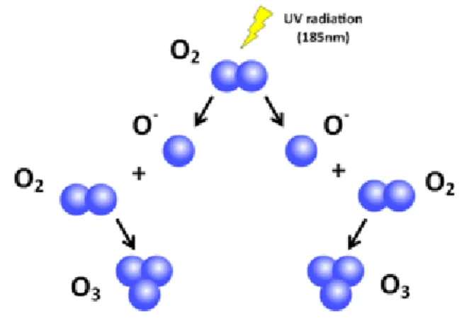
In nature, ozone is formed by UV irradiation (185nm) from the sun and during lightening discharge (Suslow 1998).
Fig. 1.1Ozone formation in nature
Commercially, UV-based generators pass ambient air (20% O2) across an UV light source, typically less than 210nm.
11
These systems have a lower cost but also have a more limited output than corona discharge systems. Corona discharge generators pass dry O2 enriched air across a high electric voltage (>5,000 V) or corona; similar to a spark plug. Excess O3 not dispersed in water must be captured and destroyed to prevent corrosion and personal injury. One method of destruction is by UV light at a longer wavelength, 254nm, combined with the use of a catalytic agent.
In the 1990s, member utilities of the Electric Power Research Institute (EPRI) encouraged EPRI to conduct research into new technologies that enhance food safety. One of the technologies studied was the use of aqueous and gaseous ozone as a contact antimicrobial agent. Through the efforts of EPRI and other interested parties, ozone was recognized and allowed as an antimicrobial food additive by the US Food and Drug Administration (FDA) in 2001. The chemical ozone (CAS Number 10027-15-6) has been recognized for more than 100 years and has been used extensively in water purification and other sanitizer and fumigant functions in several countries since the early 1900s. In response to a petition filed with the FDA by the American Bottled Water Association (now the International Bottled Water Association), ozone was classified GRAS for use in bottled water in 1982.
The FDA reaffirmed this GRAS classification in 1995.
In order to win ozone clearance for use in food processing, EPRI in 1996 convened an Expert Panel of independent food scientists to evaluate the history and safety of ozone use in food processing.
An Expert Panel Report, “Evaluation of the History and Safety of Ozone in Processing Foods for Human Consumption” (TRStudies 108026, volumes 1-3) was published in 1997 by EPRI. Based on its critical evaluation of available information, the panel concluded that: “The available information supports the safety of ozone when used as a food sanitizer or disinfectant, and further that the
available information supports a Generally Recognized as Safe (GRAS) classification of ozone as a sanitizer or disinfectant for foods when used at levels and by methods of application consistent with Good Manufacturing Practices”. ( Sopher C.D., Graham D.M., Rice R.G., Strasser J.H..2002)
Brief History of Ozone Use for Water and Food Products
• 1906 – Ozone used to provide safe drinking water in Nice, France • 1910 – First use of ozone in a German meat packing plant
12 • 1936 – Ozone used to treat shellfish in France
• 1942 – Ozone used in egg storage rooms and in cheese storage facilities in the United States
• 1972 – Ozone used to purify process water in Germany
• 1977 – Ozone used to reduce Salmonella in shell eggs in Russia
• 1982 – Ozone declared GRAS (Generally Recognized as Safe) for bottled water in the United States – Reaffirmed Gras in 1995
• 1997 – Expert Panel convened by EPRI declared ozone GRAS in food processing in the United States
• 2000 – Food Additive Petition filed with the FDA, August 15, 2000
• 2001 – FDA recognizes ozone as a secondary direct food additive. (Federal Register, Vol. 66, no. 123, Tuesday, June 26, 2000. Rules and Regulations) • 2001 – FSIS determines the use of ozone on meat and poultry products is
Ozone, a molecule consisting of three oxygen atoms, was first discovered in the 1830s by the German scientist Christian Schönbein (Guzel-Seydima et al. 2001) . It was first used commercially in 1907 in municipal watersupply treatment in Nice and in 1910 in St. Petersburg (Kogelschatz, 1988).
Major physical properties of pure ozone were given in Table 1. Ozone is the second most powerful common oxidizing agent.
Table 1.1 Oxidizing agent s and their oxidation potential (mV) (Manley and Niegowski,1967)
Ozone is formed in the stratosphere, in photochemical smog and by UV sterilization lamps, high voltage electric arcs, and gamma radiation plants (Mustafa, 1990). At room temperature, ozone decomposes rapidly and, thus, does not accumulate substantially without continual ozone generation ( Peleg, 1976; Milleret al., 1978). At room temperature, ozone is a nearly colorless gas. Ozone has a pungent, characteristic odor described as similar to ‘‘fresh air after a thunderstorm’’ (Coke, 1993). It is readily detectable at 0.01–0.05 ppm level (Milleret al.,
Fluorine 3.06 Ozone 2.07 Permanganate 1.67 Chlorine dioxide 1.50 Hypochlorus acid 1.49 Chlorine gas 1.36
13 1978; Mustafa, 1990; Mehlman & Borek, 1987). It is found in low concentration in nature. Ozone has a longer half-life in the gaseous state than in aqueous solution (Rice, 1986). Ozone in pure water rather quickly degrades to oxygen, and even more rapidly in impure solutions (Hill and Rice, 1982). Ozone solubility in water is 13 times that of oxygen at 0–30C and it is progressively more soluble in colder water (Rice, 1986). Ozone decomposition is faster at higher temperatures (Rice et al., 1981).
Table1.2Temperature and solubility relationship of ozone in water (Rice et al.,1981)
Ozone is a blue gas at ordinary temperature, but at concentrations at which it is normally produced the color is not noticeable. At 112C, ozone condenses to a dark blue liquid. Liquid ozone is easily exploded if greater than 20% ozone to oxygen mixtures occur.
Explosions may be detonated by electrical sparks or by sudden changes in temperature or pressure. However, in practical usage explosions of ozone are extremely rare.
The three atoms of oxygen in the ozone molecule are arranged at an obtuse angle whereby a central oxygen atom is attached to two equidistant oxygen atoms; the included angle is approximately 116490 and the bond length is 1.278A (Oehlschlaeger, 1978).
Although in low concentrations it is not an extremely toxic gas, at high concentration ozone may be fatal to humans. After1–2 h exposure to ozone (0.65 ppm) dogs exhibited rapid breathing whereas long-term (4–6 weeks) ozone exposure (0.2 ppm) to young rats exhibited lung distension (Barlett, Faulkner, & Cook, 1974). It was found that 0.2 ppm and higher concentrations of ozone can cause varying degrees of damage to the respiratory tract, depending on exposure length (Schwartz, Dungworth, Tarkington, & Tyler, 1976) bronchi (Castleman, Dungworth, & Tyler, 1973), and alveoli.
Ozone is almost insoluble in water (0.00003g/100mL at 20°C and effective dispersal is essential for antimicrobial activity (Suslow, 1998) ANCHE QUESTO.. Ozone’s disinfectant activity is inefficient at a water pH from 6 to 8.5. Ozone is highly corrosive to equipment and lethal to humans with prolonged exposure at concentrations above 4 ppm. Ozone is readily detectable by human smell at 0.01 to 0.04 ppm. OSHA limits of exposure specify a 0.1ppm threshold for continuous exposure during an 8-hr period and 0.3ppm for a 15-min period. At 1 ppm ozone has a pungent disagreeable odor and is irritating to eyes and throat.
14 Purity and pH of water greatly affect the rate of ozone solubilization.
Fig.1.3 Decomposition cycle of O3 in water. ( J. N. B. Bell,Michael Treshow, 2002)
J-G Kim (1998) bubbled gaseous ozone (1mM) into double distilled, deionized or tap (from two sources) water. Ozone gas dissolved faster in deionized and distilled water than in tap water. Higher maximum ozone concentration was also obtained in the water from the former two sources. The pH values, measured before ozonation, were 5.6 and 5.9 for deionized and distilled water, respectively, and 8.23 and 8.39 for tap water from the two sources. The high pH of tap water may have destabilized ozone, and thus the apparent rate of solubilization decreased. In addition, tap water may contain organic matter that consumes ozone.
Presence of minerals in water may also catalyze ozone decomposition (Hoigné and Bader 1985). Therefore, solubility of ozone increases when purity of water increases.
15
Fig.1.4 Effect of pH on ozone decay (T = 15 °C)( Lenntech)
Fig.1.5 Mechanism of O3 action (www.purfresh.com)
Ozone and microorganisms
Ozone is 1.5 stronger than chlorine (Xu, 1999) and 3000 times higher than hypochlorous acid (HOCL). The O3 contact time is generally 4-5 times less than chlorine (Khadre et al., 2001).
One of the most important aspect of the ozone application is the absence of residuals and byproducts as trihalomethane (like chloroform) or Hydrocarbon that usually occurs when chlorine is applied.
Several studies recognize the importance of air humidity for the antiseptic action of ozone, finding that its action on bacteria in a dry air treatment is considerably lower respect to a high humidity treatment.
Attribute Hypochlorite Ozone
Microbial potency
Kills plant pathogens and microbial saprophytes effectively. Some humanpathogenic, spore-forming protozoa resistant. Maximum allowable rates under regulatory control
Kills plant pathogens and microbial saprophytes effectively, including spore-forming protozoa. Maximum rate limited by ozone solubility, difficult to exceed about 10 μg/ml
Cost Chemical cost low. Repeated
delivery
required, sometimes pH and concentration controller systems needed, minor maintenance and energy costs, chlorine storage issues
Variable: no chemical cost, but high
initial capital cost for generator, usually needs filtration system when
water re-used some are complex, modest maintenance and energy costs
Influence of pH Efficacy diminishes as pH
increases, above pH 8, pH
Potency not influenced very much by
16
adjustment may be needed. Chlorine gas released at very low pH (4 or less)
pH, but ozone decomposition increases at high pH
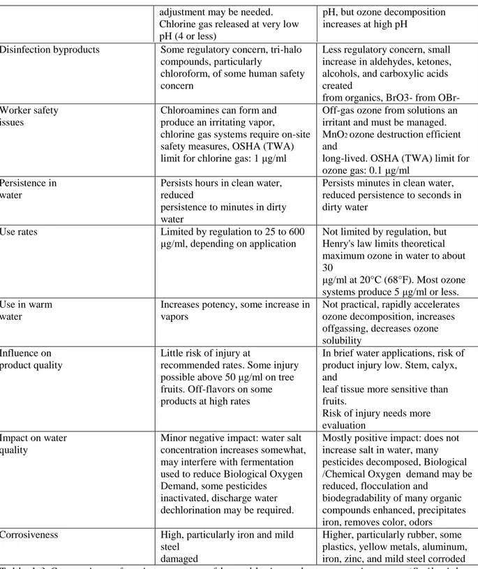
Table 1.3 Comparison of various aspects of hypochlorite and ozone us e in water. (Smilanick et al., 1999)
Sarig et al. (1996) found ozone concentrations fell rapidly upon contact with organic matter and the amount which reacted with grape berries and the microflora on their surface was about 0.1 mg/g, when supplied at a rate of 8 mg/ min for 20 min. The number of colony forming units (cfu) of fungi, yeasts and bacteria naturally present on the berry surface was considerably reduced by a 20 min exposure to ozone. Positive effect of ozone on gray mold
Disinfection byproducts Some regulatory concern, tri-halo compounds, particularly
chloroform, of some human safety concern
Less regulatory concern, small increase in aldehydes, ketones, alcohols, and carboxylic acids created
from organics, BrO3- from OBr- Worker safety
issues
Chloroamines can form and produce an irritating vapor,
chlorine gas systems require on-site safety measures, OSHA (TWA) limit for chlorine gas: 1 μg/ml
Off-gas ozone from solutions an irritant and must be managed. MnO2 ozone destruction efficient
and
long-lived. OSHA (TWA) limit for ozone gas: 0.1 μg/ml
Persistence in water
Persists hours in clean water, reduced
persistence to minutes in dirty water
Persists minutes in clean water, reduced persistence to seconds in dirty water
Use rates Limited by regulation to 25 to 600
μg/ml, depending on application
Not limited by regulation, but Henry's law limits theoretical maximum ozone in water to about 30
μg/ml at 20°C (68°F). Most ozone systems produce 5 μg/ml or less. Use in warm
water
Increases potency, some increase in vapors
Not practical, rapidly accelerates ozone decomposition, increases offgassing, decreases ozone solubility
Influence on product quality
Little risk of injury at
recommended rates. Some injury possible above 50 μg/ml on tree fruits. Off-flavors on some products at high rates
In brief water applications, risk of product injury low. Stem, calyx, and
leaf tissue more sensitive than fruits.
Risk of injury needs more evaluation
Impact on water quality
Minor negative impact: water salt concentration increases somewhat, may interfere with fermentation used to reduce Biological Oxygen Demand, some pesticides
inactivated, discharge water dechlorination may be required.
Mostly positive impact: does not increase salt in water, many pesticides decomposed, Biological /Chemical Oxygen demand may be reduced, flocculation and
biodegradability of many organic compounds enhanced, precipitates iron, removes color, odors
Corrosiveness High, particularly iron and mild
steel damaged
Higher, particularly rubber, some plastics, yellow metals, aluminum, iron, zinc, and mild steel corroded
17 nesting on ‘Thompson Seedless’ table grapes was found by Palou et al., (2002): 0.3 ppm ozone on table grapes stored for 7 weeks at 5 °C completely inhibited the gray mold.
O3 use in citrus storage rooms is now common in California to retard the production of conidia on decaying fruit infected with Penicillium digitatum or Penicillium italicum (Palou et al., 2001). These authors observed that, although spread of decay by the growth of aerial mycelia was effectively inhibited by 0.3µLL−1 O3, conidia on berries in this atmosphere could
germinate and infect the fruit, which indicated higher concentrations of O3 gas were needed to inactivate conidia.
Early work by Spalding (1966, 1968) showed that ozone did not significantly retard the growth of both M. fructicola and R. stolonifer on artificially inoculated peaches until a concentration of at least 0.5 ppm was used; ozone at this concentration and higher inhibited the fungal surface growth. In contrast, Ridley and Sims (1967) found reductions in decay incidence and severity on peaches inoculated with M. fructicola or Rhizopus sp. and stored under 0.5 ppm ozone. High atmospheric ozone levels (0.1 ppm) during the growing season increased postharvest weight loss in plums, but did not affect internal fruit quality or the incidence of internal breakdown (Crisosto et al., 1993). Ozone at 0.3 ppm for two 6 h exposure periods significantly inhibited in vitro sporulation and germination of B. cinerea (Krause and Weidensaul, 1977).
Treatment with 5000 ppm h−1 ozone in a commercial chamber of organically grown ‘Autumn Seedless’ and ‘Black Seedless’ table grape bunches reduced gray mold incidence from natural inoculum by about 50% after 6 weeks storage at 0 ◦C and on ‘Redglobe’ grapes decay reduction was 65% (Mlikota Gabler et al., 2010). Many cold storage facilities in California have installed equipment that generates a constant low dose of ozone (100 ppb day and 300 ppb night cycle) and it reduces the spread of gray mold and prolongs the storage of grapes for several weeks (Smilanick et al., 2010)
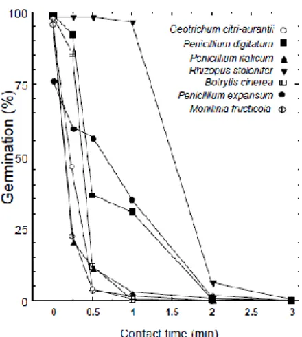
Fig. 1.6 Germination of spores of various postharvest
pathogenic fungi after exposure to 1.5 μg/ml ozone in water at 16.5°C (62°F) and pH 6.4. (Smilanick et al. 1999)
18 A number of commercial fruit juice processors in the USA have started to employ ozone to meet the recent FDA mandatory 5 log reduction of the most resistant pathogens in their finished products (Cullen et al., 2009)
In winery, ozone has been generally accepted and documented to be effective for barrel cleaning and sanitation, tank cleaning and sanitation, clean-in-place systems, and for general surface sanitation (Hampson, 2000 ) but apparently no research has been reported on the use of ozone on wine grape to produce wine without addition of sulfites.
Ozone is a strong, broad-spectrum antimicrobial agent that is active against bacteria, fungi, viruses, protozoa, and bacterial and fungal spores. Strain of the microorganism, age of the culture, density of the treated population, presence of ozone-demanding medium components, method of applying ozone (that is, gas bubbles, or uniform aqueous solution), accuracy of ozone measuring procedures and devices, and method of measuring antimicrobial efficacy are some of the confounding factors that make comparison among different studies unfeasible. Kim and Yousef in 2000 estimated nz (number of ozone molecules sufficient to inactivate a single bacterial cell) for Leuconostoc mesenteroides at 109. Finch and others (1988) found that 3 × 10⁸ molecules of ozone were used to inactivate each cell of E. coli.
The rate of microbial inactivation {D(log10 CFU/mL)/(D time)} is calculated using the linear plot or the steepest slope on the survivor curve. The negative reciprocal of this inactivation rate, known as decimal reduction time or D-value, is a useful term in comparing resistance to ozone of different microorganisms or of the same microorganism under different conditions (Khadre et al., 2001).
Usually heat and other physical factors are applied constantly during the course of the treatment, while ozone can be applied in a single dose at the beginning of the treatment or using low concentration throughout the treatment. Ozone reacts with microorganisms rapidly, and a nonlethal threshold concentration is reached quickly in a batch treatment.
Inactivation of bacteria by ozone is a complex process because ozone attacks numerous cellular constituents including proteins, unsaturated lipids and respiratory enzymes in cell membranes, peptidoglycans in cell envelopes, enzymes and nucleic acids in the cytoplasm, and proteins and peptidoglycans in spore coats and virus capsids. Some authors concluded that molecular ozone is the main inactivator of microorganisms, while others emphasize the antimicrobial activity of the reactive by-products of ozone decomposition such as ~OH, ~O2
19 –, and HO~3 (Chang 1971; Harakeh and Butler 1985; Glaze and Kang 1989; Bablon and others 1991b; Hunt and Marinas 1997).
Organism Concentration Exposition time
E. Coli, Legionella, Mycobacterium fecal, Streptococcus
0,23-2,2ppm < 20 min
Poliovirus type-1, uman Rotevirus, Enteric virus
0,2-4,1ppm < 20 min
A. Niger, Penicillum, Cladosporium 2ppm 60 min Candida Parapsilosis, Candida Tropicalis 0,02-0,26ppm < 1,67 min Acarus Siro, Tyrophagus Casei,
Tyrophagus Putrescientiae
1,5-2ppm 30 min
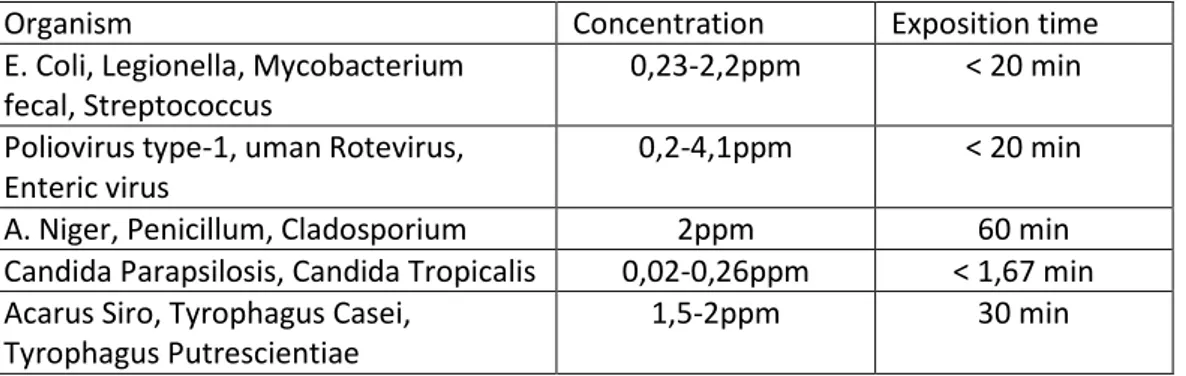
Table 1.4 Inactivation of bacteria, virus, fungi, mould and bugs after ozonization (Edelstein et al., 1982; Joret et al., 1982; Farooq and Akhlaque,1983; Harakeh and Butle, 1985; Kawamuram et al. 1986)
Ozone and Cell membranes. Ozone may oxidize various components of cell envelope including polyunsaturated fatty acids, membrane-bound enzymes, glycoproteins and glycolipids leading to leakage of cell contents and eventually causing lysis (Scott and Lesher 1963; Murray and others 1965). When the double bonds of unsaturated lipids and the sulfhydryl groups of enzymes are oxidized by ozone, disruption of normal cellular activity including cell permeability and rapid death ensues. Komanapalli and Lau (1996) found that short-term exposures of E. coli K-12 to ozone gas compromised the membrane permeability but did not affect viability, which progressively decreased with longer exposure (Khadre et al. 2001).
Ozone andf Bacterial spore coats. Foegeding (1985) found that Bacillus cereus spores with coat proteins removed were rapidly inactivated by ozone, compared to intact spores. The researcher concluded that the spore coat is a primary protective barrier against ozone. Recently, Khadre and Yousef (2001b) found that spores of Bacillus subtilis treated with aqueous ozone showed heavily disrupted outer spore coats (Khadre et al. 2001).
Ozone and Enzymes. Several authors referred to enzyme inactivation as an important mechanism by which ozone kills cells. Sykes (1965) reported that chlorine selectively destroyed certain enzymes, whereas ozone acted as a general protoplasmic oxidant.
20 Ingram and Haines (1949), in view of their finding general destruction of the dehydrogenating enzyme systems in the cell, proposed that ozone kills E. coli by interfering with the respiratory system. Takamoto and others (1992) observed that ozone decreased enzyme activity in E. coli at a greater degree in case of cytoplasmic â-galactosidase than in case of the periplasmic alkaline phosphatase (Khadre et al. 2001). Inactivation of enzymes by ozone is probably due to oxidation of sulfhydryl groups in Cysteine residues (Chang 1971).
Ozone and Nucleic material. Reaction of aqueous ozone with nucleic acids in vitro supports the notion that it may damage nucleic material inside the cell (Khadre et al. 2001). Ozone modified nucleic acids in vitro , with thymine being more sensitive than cytosine and uracil (Scott 1975; Ishizaki and others 1981). In another study, ozone opened the circular plasmid DNA and reduced its transforming ability, produced single- and double-strand breaks in plasmid DNA (Hamelin 1985), and decreased transcription activity (Mura and Chung 1990). Studying E. coli, l’Herault and Chung (1984) found that ozone may induce mutations. However, other investigators did not detect any mutagenic effect of ozone on Salmonella spp. (Victorin and Stahlberg 1988). Compared to other known mutagens, ozone was found to be a weak mutagen on Saccharomyces cerevisiae (Dubeau and Chung 1982).
Ozone and Viruses. Sproul and Kim (1980) and CK Kim and others (1980) found that aqueous ozone inactivated both f2 and T4 bacteriophages by attacking capsid protein, with liberation and inactivation of the nucleic acid. The RNA from f2 bacteriophage was partially inactivated prior to release from the capsid. They suggested that ozone breaks the protein capsid into subunits liberating RNA and disrupting virus adsorption to the host pili, and that the RNA may be secondarily inactivated (Khadre et al. 2001).
The DNA released from T4 bacteriophage was rapidly inactivated by ozone at about the same rate as that in the intact phage. CK Kim and others (1984) confirmed the results of Sproul and Kim (1980) on bacteriophage T4; they found that ozone randomly destroyed the head, collar, contractile sheath, end plate, and tail fibers and liberated the DNA from the head.
Yoshizaki and others (1988) found that aqueous ozone caused the coat proteins subunits of tobacco mosaic virus (TMV) to aggregate with each other and cross-link with the viral RNA. Despite their observation of a good correlation between loss of infectivity and decrease of recovery of viral RNA, Yoshizaki and others (1988) and Shriniki and others (1988) concluded that the major cause of TMV inactivation by ozone was the inability of the treated virus to
21 uncoat. Roy and others (1981) found that ozone altered two of the four polypeptide chains in the poliovirus protein coat. They, however, attributed the inactivation of the virus to the damage in its RNA by ozone. The observation by Herbold and others (1989) that 0.38 mg/mL aqueous ozone was needed for complete inactivation of hepatitis A virus (HAV) and only 0.13 mg/mL for complete inactivation of poliovirus may support the hypothesis that damage to viral envelopes is the main cause of inactivation of viruses by ozone. Enveloped viruses such as HAV are expected to be much more resistant to ozone compared to nonenveloped viruses such as poliomyelitis.
Efficacy of ozone. Efficacy of ozone is demonstrated more readily when targeted microorganisms are suspended and treated in pure water or simple buffers (with low ozone demand) than in complex systems such as food. The simplicity of low-ozone demand aqueous environment makes it possible to compare ozone efficacy against microorganisms within the same study, and occasionally among different studies. Ozone also may be compared with other sanitizers when experiments are done in the simple treatment environments just indicated, but differences in experimental designs, treatment conditions, and microbial strains tested should be considered (Khadre et al. 2001).
Inactivation spectrum
Bacteria. Studies shown that from 0.12 to 3.8 mg/mL aqueous ozone inactivated gram-positive bacteria by 1 to 7 log10 CFU/mL. When gram-negative bacteria were treated with 0.004 to 6.5 mg/mL aqueous ozone, their populations decreased 0.5 to 6.5 log10 CFU/mL (Khadre et al. 2001).
Sobsey (1989) reviewed studies to inactivate health-related microorganisms in water by several disinfectants and concluded that gram-positive bacteria, including S. aureus and Bacillus spp., and the Mycobacteria were more resistant than were gram-negatives.
Lee and Deniniger (2000) observed the dominance of grampositive bacteria among the surviving microorganisms in ozonated drinking water. When positive and gram-negative bacteria were compared in side-by-side experiments, however, variable results were obtained. Restaino and others (1995) studying a group of food-related microorganisms, observed that gram-negative bacteria were substantially more sensitive to ozone in pure water than were the gram-positive ones including L. monocytogenes. Kim and Yousef (2000) and
J-22 G Kim and others (1999b) treated foodborne spoilage and pathogenic bacteria with ozone under identical conditions and found results inconsistent with the previous conclusion.
Resistance of bacteria tested in this study followed this descending order: Escherichia coli O157:H7, Pseudomonas fluorescens, Leuconostoc mesenteroides, and Listeria monocytogenes. Ozone is generally more effective against vegetative bacterial cells than bacterial and fungal spores.
J-G Kim and others (2001) studied inactivation kinetics of different microorganisms that commonly spoil fruit juices. Results of this study show that Alicyclobacillus acidocaldarius vegetative cells and Zygosaccharomyces bailii ascospores were inactivated rapidly with aqueous ozone.
Spores of A. acidocaldarius were the most resistant to ozone, and survivor’s plot exhibited both a shoulder and a tail. Mold spores (Neosartorya fischeri) were intermediate in resistance to ozone, and tailing of survivor plots was apparent.
Khadre and Yousef (2001b) measured ozone efficacy against spores of 8 Bacillus spp. B. stearothermophilus, which is known for high resistance to heat, also possessed the highest resistance to ozone among the species tested.
Viruses. A limited number of studies on inactivation of viruses with ozone have been published. Researchers tested ozone concentrations in the range of 0.1 to 15.9 mg/mL against 8 different viruses; the treatment caused destruction of 0 to 7 log10-units. This may indicate that viruses are comparable to bacteria in sensitivity to ozone. Sobsey (1989), however, concluded that viruses are generally more resistant than vegetative bacteria and that bacteriophages are the most sensitive to ozone among the viruses tested. Other researchers (CK Kim and others 1980; Hall and Sobsey 1993) also reported the sensitivity of the bacteriophages MS2, and f2 to ozone. Based on the limited studies in Table3, it may be concluded that bacteriophages are the least resistant to ozone, followed by polioviruses, whereas human rotavirus was the most resistant to the sanitizer. This conclusion is in agreement with those reports by Herbold and others (1989) and Hall and Sobsey (1993).
23 Interaction between ozone and organic matrix
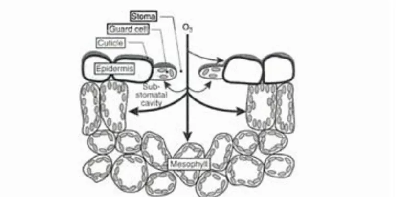
A number of environmental stresses, including pathogen attack, drought, and exposure to ultraviolet-irradiation, heavy metals and air pollutants such as sulfur dioxide and ozone (O3 ) , are thought to affect plants by causing the excess accumulation of active oxygen species (ROS).
Once O3, a very strong oxidant, enters the intercellular leaf space (apoplast) through stomates, it is converted into ROS such as O2. -, HO· and H2O2 (Apel and Hirt, 2004). These
ROS can react with membrane lipids to generate lipid peroxides that can initiate a series of reactions producing damaging reactive oxygen intermediates. These damaging free radicals and their products react with proteins, DNA and membrane lipids to cause the reduced photosynthesis, electrolyte leakage and accelerated senescence usually associated with O3 exposure. Plants respond to O3-induced oxidative stress by activating a number of
antioxidative stress-related defense mechanisms ( Sharma and Davis,1997) .
Fig. 1.7 Interaction between O3 and cuticle. Fonte Air pollution and plant life ( J. N. B. Bell,Michael Treshow, 2002)
Some molecules, like phenolic compounds in the cell wall, could accelerate the formation of radical since considerable enhancement of OH. production has been observed when ferulic or caffeic acid was added to ozonated water solution with pH 4.5-7.8 (Grimes et al. 1983). Plants produce O2 - and H2O2 as a result of their normal aerobic metabolism in chloroplasts
and mitochondria during photosynthesis and respiration. During evolution, plants have acquired a number of distinct biochemical mechanisms to efficiently and rapidly remove damaging ROS from different cellular compartments.
Non-enzymatic scavengers of ROS include a number of compounds with high reducing potentials such as vitamins C (ascorbic acid) and E, b-carotenes, polyamines and the tripeptide, glutathione.
24 Glutathione and ascorbic acid work in concert with enzymes such as ascorbate peroxidase (APX) , dehydroascorbate reductase (DHAR) and glutathione reductase (GR) to modulate the oxidation state of the cell. In the ascorbate-glutathione cycle (Fig.1.8 ) , H2O2 generated from
ROS metabolism is converted to H2O at the expense of NADPH. In addition to the
ascorbate-glutathione cycle, there are other well studied antioxidant enzymes that play crucial roles in removing ROS.
These include multiple isoforms of superoxide dismutase (SOD) that convert O2 i0 into H2O2
and catalases and peroxidases that further metabolize H2O2 to H2O.While cat is primarily
localized in peroxisomes, isoforms of peroxidase and SOD are distributed throughout the cell and can be found in cytosolic, mitochondrial and chloroplastic compartments.
Fig.1.8 O3 Ascorbate -glutathione cycle ( J. N. B. Bell,M. Treshow, 2002)
The direct reaction between ozone and ascorbate in cell wall 0.3-0.5 µm thick and at an ascorbate concentration of 0.5mM is able to detoxify 50-70% of the O3 that impinges on the wall surface (Moldau,1998). The inability of ascorbate to totally stop O3 has been confirmed by Jakob and Heber in 1998 by foliar fluorescence technique.
When in contact with ozone, lipids are subjected to “lyses”, the rupture of carbon double bond and the resulting reorganization of the molecule, forming the “Ozonide”, with three oxygen between the two carbons.
The light and dark reactions of photosynthesis might be impaired by ROS, lipid peroxidation and changes in membrane permeability (Andersen, 2003). As a consequence, plants have evolved physiological and biochemicalmechanisms, including increases in the activities of enzymes associated with general stress tolerance and increasing antioxidant concentration, which can avoid the effects of this pollutant (Conklin and Barth, 2004)
25 Stimulation of apoplatic SOD activity has been reported in few instances (Padu et al., 2006), while activation of apoplastic POD under ozone load is a well-documented phenomenon ( Ranieri et al., 1999).
Cell wall polysaccharides were increased after 7 days and 2 month of ozone stress, while water-soluble pectins were elevated after 7 days but similar or lower after 2 month. Sod activity was lower after both treatment while POD activity was significantly higher in ozone-exposed leaves of Korona strawberries (Keutgen and Pawelzik, 2008).
Catechin, an allelochemical and antioxidant, is strongly induced by ozone in spruce and pine. The elevated levels of catechins and stilbenes persist over several months and may be related to the memory effect of ozone in conifers(Langebartels, C. et al., 1998). In addition to the furanocoumarin phytoalexins of parsley plants, ozone can also induce the genes, enzymes and metabolites of the UV-B-induced flavone malonyl-glycoside pathway13, which is known not to respond to fungal elicitor. The ability of ozone to mimic other stresses has been termed cross-induction (Eckey-Kaltenbach, H. et al.1994). This process can lead to cross-tolerance against pathogens as well as numerous abiotic stresses that are proposed to act via activated oxygen species (Schraudner et al.,1997)
Ozone induces the lignin biosynthesis enzyme, cinnamyl alcohol dehydrogenase, at the protein and transcript levels (Sandermann 1996), but the derived lignan- or lignin-like product has so far not been identified.
The oxidative stress caused by ozone appears to have two opposing effects: it is generally held responsible for the detrimental effects of ozone (Heath and Taylor 1997) and may serve as the initial signal for programmed cell death and HR.
This signalling may involve ethylene (Greenberg 1997), which was specifically induced in the ozone-hypersensitive biomonitoring plant tobacco Bel W3. The detrimental effect of activated oxygen species is well illustrated by an ascorbic acid-deficient Arabidopsis mutant that is hypersensitive to ozone, as well as to sulphur dioxide (SO2) and UV-B (Conklin, 1996). Hydrogen peroxide and the superoxide anion radical are potential primary signalling species, only hydrogen peroxide being able to permeate over longer distances (Lamb 1997; Schraudner 1997; Heath and Taylor 1997).
Ozone, and to a lesser extent SO2 and UV-B, cause an increase in transcript levels of the
antioxidative enzymes catalase and glutathione peroxidase. There are small and delayed effects on transcript levels of several superoxide dismutase isoforms and ascorbate peroxidase (Willekens et al. 1994). In another study with tobacco, the cytosolic ascorbate peroxidase was
26 shown to be up-regulated by high-level ozone exposure as well as by methyljasmonate (Örvar 1997). Somewhat similar results were obtained for ozone in Arabidopsis (Kubo et al. 1995), where transcripts for the antioxidative enzymes Cu/Zn-superoxide dismutase, ascorbate peroxidase and GST-1, but not catalase, were induced by ozone (Sharma and Davis 1994; Sharma et al. 1996).
Only certain members of the antioxidative gene families seem to respond to ozone. Furthermore, compartmentation of antioxidative systems (e.g. apoplast, cytosol and chloroplast) has been shown to be of ey importance for ozone sensitivity and for generating ozone tolerance by gene transfer (Sandermann 1996; Schraudner 1997; Pell 1997).
Biosynthesis of phenilpropanoids, flavonoids and phytoalexins is elicited by ozone treatment, causing accumulation of different types of secondary metabolites as lignin and phenols (Saleem et al., 2001; Caban´e et al., 2004). In the last decades ozone (O3) has been studied for
its effects on fruit rheological properties. In tomato, Aguayo et al. analyzed the effect of cyclic exposures to ozone (4 μL/L for 30 min every 3 h) in minimally processed fruit, in which softening delay and high levels of sugars and organic acids was promoted
In 1996 Sarig et al found the phytoalexins resveratrol and pterostilbene were elicited at levels similar to those produced by uv-c irradiation. Resveratrol accumulated in greater quantities than pterostilbene.
In plants, phenylpropanoid metabolism is induced as a general response to stress.
Therefore, enhancement of key enzyme activities and accumulation of secondary metabolites are events that occur in order to improve the resistance against pathogen attack and/or tolerance to adverse environmental conditions and pollutants. PAL is an extremely sensitive indicator of stress conditions, and commonly considered as a biochemical marker indicating the activation of plant defenses which include the synthesis of both structural and protective compounds.
27
Fig.1.9 Influence of different stresses on plant metabolism. (Iriti and Faoro 2003)
In particular, ozone exposure elevates the level of flux through the phenylpropanoid pathway, thereby supplying carbon skeletons for secondary metabolites (Toumainen et al. 1996)
In Arabidopsis PAL mRNA is rapidly and transiently induced within 3h of ozone treatment (300 nL L−1 daily for 6 h), reaching a 3-fold higher levels than control plants (Sharma and Davis 1994). In parsley plants, a similar trend has been reported in which ozone treatment (200 nL L−1 for 10 h) induced an early 3-fold and 1.2-fold increase of PAL and CHS activity, respectively, followed by a 2-fold increase of total leaf furanocoumarins and flavone glycosides (Eckey-Kaltenbach et al. 1994).
28 Grape Cell wall and Postharvest dehydration
Cell wall composition
The shape of a plant cell is defined largely by its cell wall.
The plant cell wall is a dynamic compartment that changes throughout the life of the cell. The primary cell wall is born in the cell plate during cell division and rapidly increases in surface area during cell expansion, in some cases by more than a hundred-fold. The middle lamella forms the interface between the primary walls of neighboring cells. At the end many cells elaborate within the primary wall a secondary cell wall building complex structures uniquely suited to the cell’s function.
The plant cell wall is a highly organized composite of many different polysaccharides, proteins, and aromatic substances. Some structural molecules act as fibers, others as a cross-linked matrix, analogous to the glassfibers- and-plastic matrix in fiberglass (Carpita and McCann, 2000).
The wall provides support and shape for the plant, allowing it to stand upright. The plant cell wall also provides a barrier against the environment and potentially pathogenic organisms. However, in spite of the strong, rigid, and seemingly impenetrable properties of cell walls, they are metabolically active, allowing exchange of material and signals between cells, and are capable of expanding (Scheller and Ulvskov, 2010).
Cell wall Sugars
Polymers of sugar, polysaccharides are the principal components of the cell wall and form its main structural framework. Polysaccharides are consider like long chains of sugar molecules covalently linked at various positions, some being decorated with side chains of various lengths. Almost all cell wall sugars are aldoses.
29 Sugars in polymers are always locked in pyranose or furanose rings. During sugar
polymerization, the anomeric carbon of one sugar molecule is joined to the hydroxyl group of another sugar, a sugar alcohol, a hydroxylamino acid, or a phenylpropanoid compound in a glycosidic linkage.
Sugars are such important molecules for their ability to form linkages at multiple positions. It has been found 11 different sugars in plant cell wall with 4 different linkage positions, 2 configurations with respect to the oxygen atom, the transformation of pentasaccharide structure exceed 5 billion, just think about that glucose can form about 15.000 different pantametric structures.
Cellulose
Cellulose is the most abundant organic chemical on the face of the earth.
Cellulose is the most abundant plant polysaccharide, accounting for 15% to 30% of the dry mass of all primary cell walls and an even larger percentage of secondary walls (Carpita and McCann, 2000).It is a glucan polymer of D-glucopyranose units, which are linked together by β-(1 → 4)-glucosidic bonds. In plants, on average, each microfibril is 36 individual chains thick in crosssection, but microfibrils of algae can form either large, round cables or flattened ribbons of several hundred chains. Microfibrils of angiosperms have been measured to be between 5 and 12 nm wide in the electron microscope.
30
Fig.3.1.2 Cellulose in plants cell wall
(http://bio1151.nicerweb.com/Locked/media/ch05/cellulose.html)
Each (1→4)β-D-glucan chain may be just several thousand units (about 2 to 3 μm long), but individual chains begin and end at different places within the microfibril to allow a microfibril to reach lengths of hundreds of micrometers and to contain thousands of individual glucan chains. This structure is analogous to a spool of thread that consists of thousands of individual cotton fibers, each about 2 to 3 cm long.
Most cross-linking glycans are often called “hemicelluloses” (Carpita and McCann, 2000). Hemicelluloses are polysaccharides in plant cell walls that have β-(1→4)-linked backbones with an equatorial configuration. Hemicelluloses include xyloglucans, xylans, mannans and glucomannans, and β-(1→3,1→4)-glucans. These types of hemicelluloses are present in the cell walls of all terrestrial plants, except for β-(1→3,1→4)-glucans, which are restricted to Poales and a few other groups (Scheller and Ulvskov, 2010).
The xyloglucans consist of linear chains of (1→4)β-D-glucan and is the most abundant hemicellulose in primary walls of spermatophytes except for grasses
Pectins
The pectic polysaccharides comprise a class of D-galacturonic acid-containing polysaccharides that are abundant in the plant cell wall; comprising as much as 30% of dicot, gymnosperm, and non-Poales monocot walls (Caffall and Mohnen, 2009).
31 Two fundamental constituents of pectins are homogalacturonan (HGA) and rhamnogalacturonan I (RG I).
HGAs are polymers of a-1,4-linked-D-galacturonic acid that can account for greater than 60% of pectins in the plant cell wall (Ridley et al., 2001). There are two kinds of structurally modified HGAs, xylogalacturonan and rhamnogalacturonan II (RG II).
The unmethylated C-6 of HGA GalA residues is negatively charged and may ionically interact with Ca2+ to form a stable gel with other pectic molecules if >10 consecutive unmethyl-esterified GalA residues are coordinated (Liners et al., 1989). The hypothesized in vivo structure of the HG–calcium complex is sometimes referred to as the egg-box model. The egg-box model describes the close packing of HG that occurs upon Ca2+-induced gelling, which accounts for ∼70% of the pectic gel in the cell walls of plants (Jarvis and Apperley, 1995).
Other polysaccharides, composed mostly of neutral sugars—such as arabinans, galactans, and highly branched type I arabinogalactans (AGs) of various configurations and sizes—are attached to the O-4 of many of the Rha residues of RG I. In general, about half of the Rha units of RG I have side chains, but this ratio can vary with cell type and physiological state (Carpita and McCann, 2000).
HG, RG-I, and RG-II are structurally diverse polysaccharides that contribute to primary wall function with regard to cell strength, cell adhesion, stomatal function, and defense response (Caffall and Mohnen, 2009).
Calcium crosslinking of HG contributes to wall strength by bringing blocks of unmethylesterified HG chains into a tightly packed conformation that is dependent on three characteristics: the intramolecular conformation of HG, the charge separation between two GalA molecules in a HG chain, and the efficiency with which HG chains pack together (Stolle-Smits et al., 1999).
32
Fig3.13. Pectic polysaccharides of plants(Carpita and McCann, 2000).
Structural proteins
The structural proteins of the wall make up 2–10% of wall dry weight and comprise a variety of wall-associated proteins (Caffall and Mohnen, 2009).
There are four major classes of structural proteins, three of them named for their uniquely enriched amino acid: the hydroxyproline- rich glycoproteins (HRGPs), the proline-rich proteins (PRPs), and the glycine-rich proteins (GRPs) (Carpita and McCann, 2000).
33 The arabinogalactan proteins (AGPs), proline-rich proteins (PRPs), glycine-rich proteins (GRPs), and wall-associated kinases (WAKs) are wall-associated proteins and are hypothesized to aid in the wall structural reinforcement and regulatory pathways (Carpita, 1989; Hengel and Roberts 2003).
Extensin, encoded by a multigene family, is one of the best-studied HRGPs of plants and are important for secondary and tertiary structure.
Cell wall architecture
The primary cell wall is made up of two, sometimes three, structurally independent but interacting networks. The fundamental framework of cellulose and crosslinking glycans lies embedded in a second network of matrix pectic polysaccharides.
Fig.3.2.1 (A) A three-dimensional molecular model of the Type I wall shows the molecular interactions between cellulose, XyG, pectins, and wall proteins. A three -dimensional molecular model of the Type II wall sho ws the molecular interactions between cellulose, GAX, pectins, and aromatic substances (Carpita and McCann, 2000).
The third independent network consists of the structural proteins or a phenylpropanoid network. Evidence for these networks comes partially from direct imaging of walls.
34 The walls of most dicotyledons and the noncommelinoid monocotyledons contain about equal amounts of XyGs and cellulose. These kinds of wall we denote as Type I walls.
In Type I walls, the cellulose-XyG framework is embedded in a pectin matrix that controls, among other physiological properties, wall porosity. Type II walls of commelinoid monocots contain cellulose microfibrils similar to those of the Type I wall; instead of XyG, however, the principal polymers that interlock the microfibrils are GAXs (Carpita and McCann, 2000).
Cell wall enzymes
Cell walls contain numerous enzymes capable of hydrolyzing the major components of the wall matrix, and it is attractive to think that some of these enzymes might function to loosen the wall by breaking load-bearing links between cellulose microfibrils, thereby allowing the wall to extend
Alternatively, wall hydrolytic enzymes might stimulate wall extension indirectly: by reducing the size and viscosity of matrix polymers, such enzymes could act synergistically to enhance the action of primary wall-loosening agents, such as expansin. A third alternative is that such hydrolytic enzymes have functions unrelated to wall loosening, e.g. in defense, in signaling, or in polysaccharide processing or breakdown to serve the cell’s other metabolic or energy needs (Cosgrove, 1999).
The most copious RNAm transcript, after cell wall modification, are resulted to be the one that encoded for polygalacturonases and pectin methylesterases, and others for α and β galactosidase , pectinliase and cellulase (Nunan et al. 2001).
Pectin metilesterasi (PME, E.C. 3.1.1.11)
Pectin methylesterases (PME) catalyse the demethylesterification of cell wall polygalacturonans. In dicot plants, these ubiquitous cell wall enzymes are involved in important developmental processes including cellular adhesion and stem elongation(Micheli 2001).
PME is also produced by several types of fungi, most of all pathogen as B. cinerea (Lee et al., 1979).
PME acts on structural polysaccharides of cell wall catalyzing the hydrolysis of methyl groups in position six of galacturonic acid chains, releasing free methanol.
35
Fig.3.3.1 PME hydrolysis of methyl groups.
Once polymerized by PME, pectin could be attacked by polygalacturonase (PG) causing depolymerization and consequently the loss of consistence during fruits ripening.
it has been shown that the several PME isoforms detected in cell walls are encoded by a multigene family (Micheli, 2001). The study of this several PME isoforms has shown that they differed each other by molecular mass, optimal pH, optimal temperature and isoelectric point.It has also demonstrated that most of PME isoforms acts on neutral or alkaline pH (Bordenave, 1996) even though some of them seems to prefer acid pH (Ren & Kermode, 2000).
The systematic sequencing of the Arabidopsis genome has revealed the presence of the 67 PME-related genes in this species( Micheli,2001). The PMEgenes encode pre-pro-proteins that have peptide motifs considered to be signatures of PMEs. The pre-region or signal peptide is required for protein targeting to the endoplasmic reticulum. The pro-PME is secreted to the apoplasm via the cis, medial and trans Golgi cisternae, and the trans Golgi network, and only the mature part of the PME (without the pro region) is found in the cell wall.
PMEgenes can be divided into two classes, according to the systematic sequencing of the Arabidopsis genom. Genes in the first class contain only two or three introns and a long pro region, and genes in the other class contain five or six introns and a short or nonexistent pro region; these two classes have been called type I and type II, respectively (L. Richard, pers. commun.). The type II sequences have a structure close to that of the PMEs identified in phytopathogenic organisms (bacteria, fungi) and are involved in cell wall soaking during plant infection. Such data force us to consider the role and development of the PME pro region. According to Markovic & Kohn (1984) this enzymes seems to have different types of action mechanisms, and it depends on the target, if they are acting on plant or on microorganisms. In the first scenario, the alkaline isoforms of the enzyme operated linearly on the homogalacturonans causing the increase of free carboxyl groups that can interact with cations, especially with calcium.
Because acidic PMEs were thought to be essentially confined to fungi, the simplest hypothesis was that random demethylesterification depended on acidic PMEs, whereas linear
36 demethylesterification depended on alkaline PMEs. However, more recent studies have shown that PME activity also depends on pH and the initial degree of methylesterification of the pectins. Some isoforms can act randomly at acidic pH but linearly at alkaline pH. And, at a given pH, some isoforms are more effective than others on highly methylesterified pectins (Catoire, et al. 1998; Denès, et al. 2000).
The activity of this enzyme has shown to be influenced by cations concentration, which can influence the affinity for the substrate. Also, trivalent cations has revealed to have more efficacy than bivalents and monovalents cation (Micheli,2001). Therefore organic and inorganic cations content can affect PME activity, stability and its natural tendency to induce pectin aggregations.
Some studies (Lee et al., 1979; Barnavon et al., 2001) demonstrated that PME pH optimum is included between 7 and 7.5, it has also been shown how the enzyme activity rapidly raise up on Concorde grape variety(Lee et al., 1979) when pH increased from 5 to 7.5 and it decreases when pH goes further up.
Same trend was also confirmed on different species of tomato and citrus (Kertesz, 1938; Guyer et al., 1956).
In grape, Lecas and Brillouet in 1994 found that homogalacturonans methylesterification degree can change from 40 to 80% and it depends on cultivarripe stage and specific tissue; Vitis Vinifera species present lower activity when it is compared with Vitis Lambruscana and usually red grapes have higher activity than white grapes. As a consequence, methanol content in white wines is lower than red ones since PME and pectins are mainly located in grape skins (Lee et al., 1975).
Polygalacturonase (PG, EC 3.2.1.15)
Polygalacturonase (PG) are a class of enzymes involved on degradation of cell wall.
They usually catalyzed the hydrolysis of the α-(1,4) glycosidic bond of not-esterified galacturonic acids chains. Indeed, PG acts after methyl esterification of PME on pectins; releasing single unit of galacturonic acid molecules.