University of Messina
Department of Clinical and Experimental Medicine, Phd course in “Medical and Surgical Biotechnologies”
XXXIII Cycle
Additional functional activities
of Plasminogen Binding Surface Protein (PbsP),
a cell wall protein of Group B Streptococcus
PhD Student:
Dott. Giuseppe Valerio De Gaetano Supervisor:
Prof.ssa Concetta Beninati
Academic Year 2019/2020
Index
Introduction
Vitronectin in microbial pathogenesis……….………….3
Group B Streptococcus and its adhesins………7
Aim of the thesis………17
Results PbsP involvement in the direct binding to epithelial cells………..18
Recombinant PbsP binds to Vtn………..22
PbsP mediates GBS binding to Vtn……….25
The αv integrin subunit promotes Vtn-dependent invasion of epithelial cells by GBS………28
Discussion………..32
Matherial and Methods ………..36
Bacterial strains and Streptococcal recombinant proteins………36
Reagents……… 36
Bacterial adhesion and invasions assays………..37
Adherence of microspheres coated with PbsP and PbsP fragments…………..38
PbsP binding to ECM components……….39
Adhesion of bacteria to immobilized Vtn………..39
Statistical analysis……….40 References……….41
Introduction
Vitronectin in microbial pathogenesis
Bacterial pathogens have evolved various strategies to colonize the host (Pizarro-Cerdá and Cossart, 2006; Paulsson M. and Riesbeck K., 2018) and to survive against immune responses (Singh et al., 2010; Hallstrom et al., 2016). In these processes, a major role is played by interactions between microbes and components of the extracellular matrix (ECM) of the host. The ECM consists of a dynamic network of proteoglycans (e.g. heparan sulfate, perlecan and agrin), glycoproteins (vitronectin and fibronectin) and fibrous proteins (collagen, elastin, laminin) surrounding cells in mammalian tissues (Frantz C et al., 2010). It has been recently demonstrated that many microorganisms are able to bind to the multifunctional glycoprotein vitronectin, in addition to other ECM components, and that these interactions promote pathogenesis during infectious diseases (Singh et al., 2010).
Vitronectin (Vnt) is an important component of the ECM, by virtue of its ability to influence a wide range of cell functions, as well as activation of components of the coagulation cascade. This protein is synthesized in the liver and is secreted into plasma. In addition, Vtn can also be found in different tissues, including duodenal, tonsil and lung tissues, as well as in some malignant tumors (Berglund et al., 2008).
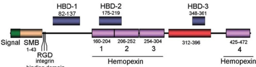
Fig. 1 shows a schematic representation of the Vtn molecule. A somatomedine
B (SMB) domain is present at the N-terminus of the protein, followed by an Arg-Gly-Asp (RGD) integrin-binding motif (Lossner et al., 2009), and by four haemopexin-like sequences. Vnt also displays three heparing-binding domains (HBD) called HBD-1, HBD-2 and HBD-3 (Liang et al., 1997).
Adapted from Singh et al. 2010
Fig. 1. Vitronectin structure
SMB, somatomedin B domain; RGD, Arg-Gly-Asp motif binding to integrin receptors; Hemopexin, haemopexin-like domain; HBD, heparin-binding domain.
Physiologically, vitronectin plays an important role in different processes,
including cell adhesion, cell migration, angiogenesis, tissue remodeling, wound healing and, particularly, in the regulation of the innate immune response mediated by the terminal pathway of complement activation (Milis et al., 1993; Preissner and Seiffert, 1998, D’Mello et al., 2009; Smith and Marshall, 2010).
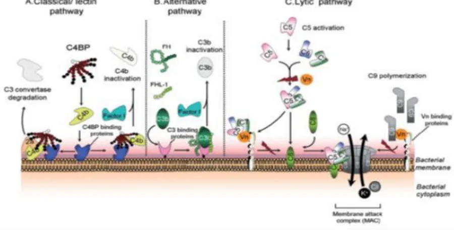
As shown in Fig. 2, there are 3 main distinct pathways of complement activation that can be triggered on microbial surfaces: i) the classical pathway, involving binding of C1q to the Fc portion of surface-bound antibodies or, less often, directly to microbial components; ii) the lectin-mediated pathway, triggered by binding of the serum protein mannan-binding lectin to mannosecontaining bacterial components; iii) the alternative pathway, initiated by the spontaneous, covalent binding of C3b to the bacterial surface (Blom et al., 2009). All three pathways commonly end with the formation of C5 convertase that promotes the assembly of the cell membrane attack complex by recruiting other complement system proteins, from C5b to C9. In this
manner, the bacterial cell membrane integrity can be disrupted in “serum sensitive” gram negative bacteria (Moffitt and Frank, 1994).
To avoid an excessive immune response, which can result in host tissues damage, the complement system is tightly regulated by soluble and membrane-bound proteins, such as factor-I, factor H, C4b-binding protein (C4BP) and also Vnt. It has been proposed that Vnt contains distinct binding sites for interaction with the C5b-7 complex and C9, with the function of inhibiting pore formation by the membrane attack complex (Milis et al., 1993).
Singh et al. 2010
Fig. 2. Role of Vn as a regulatory molecule for the complement lytic pathway
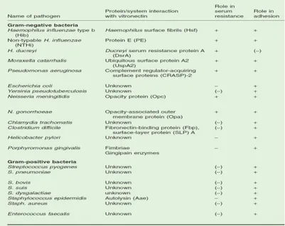
Recent data have highlighted how numerous pathogenic microorganisms
are able to take advantage of Vtn in acquiring serum resistance, tissue-adhesive properties and invasiveness (Singh et al., 2010) (Table 1).
The mechanisms underlying vitronectin-dependent serum resistance have recently attracted much interest in bacterial pathogenesis. H. influenzae type b (Hib) displays a trimeric auto-transporter surface fibril (Hsf), which binds to the heparin binding domains of the C-terminal Vnt region thereby acquiring serum resistance (Hallström et al., 2006). Moreover, M. catarrhalis surface protein (Usp) A2 directly interacts with Vnt and Usp A2-deleted mutants acquire serum-sensitivity (Singh et al., 2010b). Similarly, Pseudomonas
aeruginosa serum resistance is mediated by the direct binding of complement regulator-acquiring surface proteins (CRASP-2) to Vnt (Hallström et al., 2010). N. gonorrhoeae strains causing disseminated gonococcal infection (DGI) also use similar mechanisms (Arko et al., 1991).
A crucial step in bacterial pathogenesis is the adhesion of pathogens to
mucosal surfaces by virtue of pili and other surface-exposed membrane proteins, called adhesins (Boyle and Finlay, 2003). In this process, ECM molecules exposed on the surface of epithelial cells or in sub-epithelial tissues represent a fundamental target for pathogenic microorganisms. Moreover, interactions between adhesins and ECM components can occur in the blood, since several ECM molecules are also plasma components, and in connective tissues following penetration of tissue barriers (Pizarro-Cerdá and Cossart, 2006). Vnt has been described as an important matrix component used by pathogens to efficiently adhere to epithelial tissues such as in the case, for example, of Escherichia coli and Staphylococcus aureus infections (Chhatwal et al., 1987). Moreover, the surface-layer proteins (SLPs) of Clostridium difficile can interact with Vnt under in vivo and in vitro conditions (Calabi et al., 2002; Cerquetti et al., 2002), while sialic acid-containing haemagglutinins are used by the gastrointestinal pathogen Helicobacter pylori to firmly bind to Vnt in adherence processes (Ringnér et al., 1992; 1994). Several studies have been carried about S. pneumoniae interactions with Vnt. In particular, it has been shown that S. pneumoniae, during adhesion, has a great affinity to a multimeric form of Vnt, mainly exposed on epithelial cells and characterized by ample accessibility of its heparin binding sites (Zhuang et al., 1997; Sa E Cunha et al., 2010).
Vnt binding by pathogens can lead to the engagement of integrins, which
responses, including ligand internalization. The signaling mechanisms activated by integrins involve activation of tyrosine kinases (PTKs), various adaptor molecules and small Rho GTPases (Brunton et al.,2004). This can lead to actin remodelling and intracellular uptake of bacteria adhering to nonphagocytic cells through different internalization mechanisms (Isberg et al., 2000; Wang et al., 2006).
Singh et al. 2010
Table 1. Bacterial pathogens and interactions with vitronectin regarding serum resistance and
adhesion.
The capacity of several pathogens, including S. pneumoniae, to express
adhesins capable of interacting with Vnt has been investigated. However, little is known of the ability of Group B streptococcus, a frequent human pathogen, to interact with this ECM component.
Group B Streptococcus and its adhesins
Streptococcus agalactiae (also referred to as group B Streptococcus, GBS) is a β-haemolitic Gram-positive encapsulated bacterium, which colonizes the human gastrointestinal system and is found in the genital tract of 15–30% of healthy women (Campbell et al., 2000; Edwards et al., 2006 Le Doare and
Heath, 2013). However, GBS can also cause serious infections in neonates and pregnant women. Moreover, this organism is a common agent of a variety of infections in non-pregnant adults, particularly those with predisposing pathologies such as diabetes, cirrhosis or tumors, as well as in the elderly (Farley et al., 1993). Neonatal GBS infections are normally classified in two clinical syndromes, the early-onset disease (EOD) and the late-onset disease (LOD). While EOD occurs during the first week of life, LOD can occur from 8 days up to three months after birth. EOD generally involves vertical transmission of GBS by inhalation of contaminated amniotic or vaginal fluid during parturition, which is followed by bacterial translocation across the respiratory epithelium of the neonate and subsequent sepsis (Edwards and Baker, 2005). The transmission route of GBS during LOD is less clear, but several studies suggest the occurrence of early colonization of the neonatal intestinal tract followed by bacterial translocation across the intestinal epithelium and bloodstream invasion (Weindling et al., 1981; Hansen et al., 2004; Tazi A et al. 2019). Preventive strategies based on intrapartum antibiotic prophylaxis (IAP) have decreased the incidence of EOD by 80%, but have left unaffected the incidence of LOD (Tazi A et al. 2019).
Multi-locus sequence typing (MLST) has established that most of the GBS
clinical isolates can be classified to a small number of sequence types (ST) or clonal complexes (CCs), namely CC17, CC23, CC19, CC1 and CC10. CC17 (associated with a serotype III capsular polysaccharide) is frequently associated with LOD and meningitis and is therefore often referred to as the “hypervirulent” clone (Musser et al., 1989; Lamy et al., 2006; Lin et al., 2006; Phares et al., 2008; Poyart et al., 2008). GBS is capable of expressing many virulence factors including a large number of adhesins, which endow the pathogen with the ability to colonize mucosal surfaces and to invade, under
certain circumstances, host tissues. Several adhesins also enable GBS to evade immune responses and to enhance its pathogenic potential (Armistead et al., 2019). Each adhesin binds to at least one, and more often to multiple, specific receptors. Like in the case of other bacteria, the majority of GBS adhesins are cell-wall anchored proteins which can recognize specific host receptors (Tazi et al., 2010; Buscetta et al., 2014; Wang et al., 2014; Mu et al., 2014; Six et al., 2015).
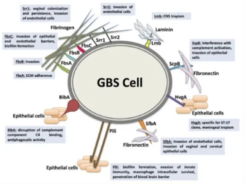
Fig. 3 shows some of the most important adhesins, which enable GBS to adhere
to the ECM, including the fibrinogen binding proteins (Fbs), laminin-binding protein (Lmb), streptococcal fibronectin-binding protein A (SfbA) and group B streptococcal C5a peptidase (ScpB).
Shabayek et al. 2018 Fig.3 Representation of some of the major GBS adhesins with their cognate ligands
The Fbs that have been identified in GBS are FbsA (Schubert et al. 2004), FbsB
(Gutekunst et al. 2004), Srr1, Srr2 (Seo et al. 2012; Six et al. 2015) and FbsC (Buscetta et al. 2014). FbsA is a surface fibrinogen-binding protein found in
almost all serotypes, which displays a variable number of amino acid repeats in different strains. Each of these repeats can bind to fibrinogen. The protein plays a protective role against opsonophagocytosis (Schubert et al. 2002) and elicits platelet aggregation and thrombus formation (Pietrocola et al. 2005). While FbsA is mainly involved in bacterial adherence to human brain microvascular endothelial cells (hBMEC) and lung epithelial cells (A549) (Schubert et al. 2004), FbsB appears to be preferentially involved in cell invasion (Gutekunst et al. 2004). The serine-rich repeat proteins Srr1 and 2 are both able to bind to fibrinogen, but Srr2, which is selectively expressed in CC17 strains, also binds plasminogen and thereby can promote bacterial dissemination and tissue invasion after conversion of surface-bound plasminogen to plasmin (Six et al 2015). In in vivo studies, absence of srr1 results in reduced bacterial invasion of the central nervous system, kidneys and spleen (Seo et al. 2013). FbsC is expressed on the surface of almost all GBS clinical strains with the exception of CC17 strains and has higher affinity for the fibrinogen β-chain compared with FbsB, but lower affinity compared with FbsA. Mutant strains lacking fbsC (ΔfbsC strains) are impaired in their ability to bind to fibrinogen as well as to adhere to and invade epithelial cells. Moreover pretreating epithelial cells with anti-fibrinogen antibodies inhibits bacterial adherence, suggesting that fibrinogen might function as a receptor for GBS on the surface of epithelial cells (Buscetta et al. 2014). In addition, GBS is also able to bind to laminin in damaged tissues by means of the Lmb protein thereby promoting its dissemination in the host, as demonstrated in in vivo studies using lmb-deleted mutants (Tenembaum et al., 2007; Ragunathan P et al. 2009). Lmb expression is higher in GBS strains associated with meningitis in comparison to other GBS strains (Al Safadi et al. 2011). The GBS ScpB protein interferes with complement effector mechanisms by cleaving the
chemoattractant C5a (Cheng et al. 2002). Moreover the protein also binds to insoluble fibronectin. SfbA is highly conserved among GBS strains and is directly involved in fibronectin binding and invasion of brain endothelial cells. Strains lacking the sfbA gene show reduced ability to cause meningitis, while recombinant expression of SfbA on non-pathogenic bacteria significantly improves their binding to fibronectin (Mu et al. 2014).
The shift of GBS from a commensal to a pathogenic life stile may be triggered
by changes in micro-environmental conditions, leading to reprogramming of gene transcription. In this regulation process, target genes may include those encoding for cell wall proteins, enzymes involved in cell growth and metabolism and, above all, virulence factors such as adhesins (Armistead et al.; 2019). The main signaling molecules used by GBS and other bacteria to respond to changing environments are represented by specialized two-component regulatory systems (TCS). Genomic analysis has identified a relatively large number of TCS in GBS (more than 20 TCS), compared with other streptococcal pathogens, such as S. pneumoniae and S. pyogenes. This might reflect a considerable ability of GBS to sense environmental stimuli and to adapt to them (Poyart et al., 2001; Glaser et al., 2002; Spellerberg et al., 2002; Tettelin et al., 2002; Tettelin et al., 2005; Thomas and Cook, 2020).
Canonically a TCS is composed of a histidine kinase (HK) and a response regulator (RR). The HK is a cell-membrane associated protein that, after having sensed extracellular, intra-membrane or intracellular signals, is autophosphorylated at the His residue placed in its cytoplasmatic domain. Subsequently, the receiver domain of the intracellular RR is phosphorylated in a conserved Asp amino acidic residue, which induces a conformational change in the RR C-terminal domain allowing it to bind to DNA regulatory sequences (Stock et al., 2000; Galperin et al. 2006; Mitrophanov et al., 2008; Mascher et al,
2014, Thomas and Cook, 2020). The main GBS TCS have been classified into three main groups on the basis of their role in the course of GBS infections: 1) pathogenesis and colonization of the host (CovRS, BgrRS, HssRs, LtdRS); 2) colonization and adhesion to host mucosal tissues (RgfAC, FspSR); 3) resistance
to antimicrobial peptides (DltRS, LiaSR, BceRS, CiaRH) (Thomas and CooK 2020)(Fig.4).
Thomas and Cook 2020
Fig.4 Main TCS in Group B Streptococci
The most studied TCS is the CovR/S system which is highly conserved among GBS strains and is able to regulate a large number of virulenceassociated genes, by activating or repressing them. Therefore it has been suggested that regulation by this TCS may allow GBS to behave differently in various pathogenetic steps, such as colonization and invasion (Lamy et al., 2004; Jiang et al., 2005; Jiang et al., 2008; Lembo et al. 2010; Park et al. 2012). CovR has been shown to be able to down-regulate more than 70 genes and to activate more than 60 genes in GBS suggesting a major role as a master
regulator in strains belonging to different CC. Mutants with reduced CovRS activity, exhibit a hyper-hemolityc phenotype in both NEM316 (serotype III) and 2603V/R (serotype V) strains, with transcriptional activation of the cyl operon and reduced production of CAMP factor (Lamy et al 2004). Moreover, theGBS A909 covR-deleted strain showed a reduced capacity to invade human brain microvascular endothelial cells (hBMEC), but increased adherence to these cells. Strikingly, the same mutant displayed an opposite behavior using human vaginal epithelial cells (HVEC) (Lembo et al 2010; Patras et al. 2013). CovR/S was shown to play an important role also in in vivo experimental models, such as murine urinary tract infection (UTI), murine vaginal colonization and sepsis in neonatal rats (Lamy et al 2004; Sullivan et al 2017).
A TCS homologous to the S. aureus SaeR/S system has been recently identified in GBS. This system appears to be important for the in vivo upregulation of essential genes involved in vaginal colonization, as shown in a mouse model. Moreover, this GBS SaeR/S regulatory system appears to be activated by an unknown molecule present in vaginal lavage fluid, supporting the hypothesis that this TCS is primarily triggered in vivo and is sensitive to changing environmental conditions (Cook L. et al. 2018).
In S. aureus the sae operon consists of four genes (saeP, saeQ, saeR, and saeS) and two promoters (P1 and P3). While the P3 promoter is placed inside saeQ and contributes to the basal expression of saeR and saeS, the P1 promoter is located upstream saeP and is highly inducible, resulting in increased expression of all the four sae genes after activation (Steinhuber et al. 2003; Jeong et al. 2011). The S. aureus sensor histidine kinase SaeS displays two transmembrane domains and can perform both kinase and phosphatase activity. The linkerpeptide located between the two transmembrane domains is so short that the SaeS might be viewed as an intramembrane-sensing HK (Mascher 2006;
Jeong et al. 2015). Amino acid substitutions in the protein sequence of the linker peptide and transmembrane helices have provided important informations on the mechanisms underlying upregulation of SaeS kinase activity inresponse to the human neutrophil peptide 1 (HNP1) and other neutrophil products (Geiger et al. 2008) (Fig.5).
Liu et al. 2016; Mlynek et al 2018
Fig.5. Organization, function and regulation of the sae operon in S. aureus. Green boxes represent phospho-SaeR binding sites.
SaeP and SaeQ are proteins involved in up-regulating the phosphatase activity of the cytoplasmatic tail of SaeS, by forming a ternary complex (Steinhuber et al. 2003; Mainiero et al. 2003). SaeR is a response regulator belonging to the OmpR family. It contains at the N-terminus the receiver domain with a phosphorylation site and, at the C-terminus, the DNA binding domain, which is unable to bind target sequences in the unphoshorylated state (Sun et al 2010). In S. aureus, the saeR binding DNA sequence (SBS) is GTTAAN6GTTAA, where the nucleotidic sequence TTAA seems to be mainly involved in saeR-mediated transcriptional activation. The number of SBSs and sensitivity for phosphorylated saeR (SaeR-P) varies among the promoters of various staphylococcal virulence genes (Sun et al. 2010; Nygaard et al. 2010). In light of this, two different classes of promoters were established in S. aureus: class I
promoters (e.g., coa, fnbA, eap), which have a low binding affinity for SaeR-P and require high amounts of this response regulator to be upregulated; class II promoters (hla and hlb), which are responsive to small quantities of saeR-P and are active also under under un-induced conditions (Mainiero et al. 2003; Cho et al. 2012).
Little information is available about the recently discovered SaeR/S system
in GBS. The amino acid residues of the SaeR DNA binding site are almost entirely conserved as compared to the S. aureus SaeR. However, the SaeR sequences flanking the DNA binding site are quite divergent, raising the possibility that the two orthologs recognize different DNA motifs. Alignment of the sae operons in S. aureus and GBS indicates the presence of a cell wall protein gene (pbsP) in place of saeP and saeQ. The pbsP gene was found to encode for a protein that was identified by mass spectroscopy in the GBS exoproteome, characterized as a plasminogen binding surface protein and named PbsP (Buscetta et al. 2014).
Adapted from Cook et al. 2018
The gene pbsP gene is well conserved among GBS strains and is finely
regulated both in vitro an in vivo (Buscetta et al., 2016; Lentini et al., 2018; Cook et al. 2018). PbsP protein is similar similar to Plasminogen and fibronectin binding protein B (PfbB, also called PavB) of Streptococcus pneumoniae (Papasergi et al., 2010), because of the presence of two Streptococcal SUrface REpeats (SSURE). In addition, PbsP has a methionine and lysine-rich (MK-rich) region, an N-terminal signal peptide with a YSIRK sorting motif (Carlsson et al., 2006; Brega et al., 2013) and a C-terminal cell wall-anchoring LPXTG motif. Recents studies have highlighted the ability of PbsP to bind plasminogen through both the MK-rich and SSURE domains (Fig.7). Furthermore, GBS can take advantage of PbsP-mediated binding of plasminogen on the bacterial surface to acquire proteolytic activity after conversion of plasminogen into plasmin by virtue of endogenous plasminogen activators. PbsP was found to be required for invasiveness of the blood brain barrier in both in vivo and in vitro models (Buscetta et al. 2016; Lentini et al. 2018).
Pietrocola et al. 2018
Fig.7 Schematic representation of PbsP and its main domains
Plasminogen binding by PbsP has been mainly investigated in GBS strains
belonging to the CC23 and CC17 clonal complexes, in which PbsP is primarily involved in hematogenous dissemination of the bacteria during experimental
infection (Buscetta et al. 2016; Lentini et al. 2018). In addition, in the context of the prototype CC7 A909 strain, PbsP appears to be involved in the ability of GBS to colonize the genital tract, as shown in a mouse vaginal colonization model (Cook et al 2018). In the prototype CC23 NEM316 strain, pbsP was shown to be moderately upregulated in the absence of covR suggesting a regulatory role by the covRS system, likely through an indirect mechanism, in the context of this particular CC (Lentini et al. 2018).
Aim of the thesis
The aim of the studies presented in this thesis is to investigate whether PbsP interacts with other components of the ECM, in addition to plasminogen. Indeed, several bacterial adhesins appear to be multifunctional, in the sense that they bind to more than one receptor. While there was an indication in the literature that PbsP might promote adherence to vaginal cells (Cook et al 2018), the molecular basis for this function was unclear. Data presented here indicate that PbsP, and particularly its SSURE domains, interact with vitronectin to promote GBS adherence to respiratory and intestinal epithelial cells.
Results
Involvement of PbsP in GBS binding to epithelial cells
Several epidemiological studies prompted clinicians and researchers to
classify two forms of GBS-linked neonatal disease, called, as mentioned above, early onset (EOD) and late onset disease (LOD). In EOD, GBS is believed to be predominantly inhaled from contaminated amniotic fluid by the fetus and to adhere to the respiratory epithelium, causing pneumonia (Nizet et al. 2000; Melin et al. 2011). On the contrary, in LOD, the route of transmission is still uncertain, but it is likely to involve the ability of GBS to colonize the intestinal tract, followed by invasion of the gut epithelial barrier, bloodstream invasion and hematogenous dissemination to distant organs (Tazi et al. 2010; Tazi et al. 2019).
Given the importance of the lung and of the gut in GBS pathogenesis, it was
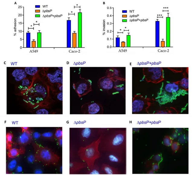
of interest to study the potential role of PbsP in GBS adherence and invasion to epithelial cells, by using A549 cells and Caco-2 cells, which are type II lung alveolar cells and colon epithelial cells, respectively. At first, we evaluated the role of PbsP in mediating adhesion to epithelial cells by comparing the numbers of cell-associated CFU using wild type BM110 GBS strain, belonging to CC17 clonal complex, and a mutant of this strain bearing a deletion for the pbsP gene (ΔPbsP). The absence of pbsP significantly reduced GBS adhesive and invasive capacities using either A549 or Caco-2 cells. These phenotypes were reversed by genetic complementation, as shown in Fig.1 A-B. Fluorescence microscopy confirmed the role of PbsP in GBS adhesion to Caco-2 (Fig.1 C-DE) and A549 epithelial cells (Fig.1 F-G-H).
Fig. 1. PbsP is involved in interactions between GBS and epithelial cells:
Adherence to and invasion of Caco-2 (colon epithelial) and A549 (alveolar epithelial) cells by GBS strain BM110 (WT), its pbsP deletion mutant (ΔpbsP) and a complemented strain (ΔpbsP+pbsP). A and B, GBS adherence and invasion was measured by CFU counting and expressed as percentages of cell-associated bacteria relative to the total number of bacteria added to the monolayers. Shown are means ± standard deviations of three independent experiments conducted in triplicate. *p < 0.05; ***p < 0.001; by one-way ANOVA and Bonferroni test. C, D and E, bacterial adherence to Caco-2 cells; F, G and H, adherence to A549 cells. Fluorescence microscopy images show actin filaments (red), GBS (green), cell nuclei and bacterial nucleoids (blue).
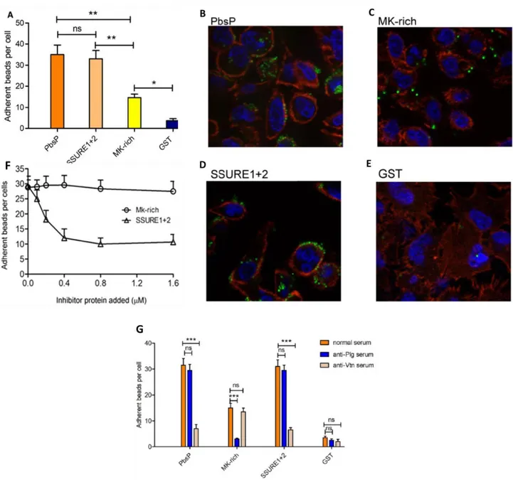
Subsequently, to investigate if PbsP can directly bind to epithelial cells, we
recombinantly produced the whole PbsP molecule or PbsP fragments, as fusions with glutathione-S-transferase (GST). Then the recombinant proteins were adsorbed to fluorescent beads to observe their attachment to respiratory
epithelial cells. Fluorescence beads coated with whole PbsP adhered to A549 cells considerably more efficiently compared to GST-coated beads, used as negative control. The adhesive pattern of beads coated with the two SSURE domains (SSURE 1+2) was similar to that of beads coated with whole PbsP, while MK-rich fragment-coated beads adhered to a lower extent (Fig. 2 A to E). Next, we assessed the ability of soluble PbsP fragments to inhibit binding of beads coated with whole PbsP. Fig.2 F shows the SSURE 1+2, but not the
MK-rich, fragment produced dose-dependent inhibition of binding to epithelial cells of whole PbsP-coated beads. Next, we hypothesized that the moderate
binding of the MK-rich fragment to epithelial cells could be due to its previously demonstrated capacity of binding to Plg (Buscetta et al. 2016), since plasminogen can be also found on the surface of cells. Fig. 2 G shows that this
was indeed the case, since pretreatment of epithelial cells with antiplasminogen antibodies inhibited binding of MK-rich-coated, but not SSURE 1+2-coated, beads.
Collectively, this first set of data demonstrated a significant role played by SSURE domains in allowing PbsP to directly bind to epithelial cells. The ability of PbsP to bind plasminogen does not apparently provide a major contribution to the epithelial cell-adhering properties of the protein.
Fig. 2. Adherence to epithelial cells of beads coated with isolated recombinant PbsP and its
fragments.
A. Number of adherent beads per cell expressed as means ± standard deviations of three independent experiments performed in triplicate.*p < 0.05; **p < 0.01; ns, non-significant by one-way ANOVA and Bonferroni test.
B, C, D and E. Representative images obtained by fluorescent microscopy showing actin filaments (red), latex microspheres (green) and cell nuclei (blue). Alveolar epithelial cells (A549) grown on coverslips were incubated with fluorescent latex microspheres (1μ in diameter) covalently bound with PbsP or PbsP fragments (SSURE 1+2, MK-rich). Latex micropheres covalently bound with glutathione-S-transferase (GST) were used as a negative control.
F. A549 monolayers were mixed with fluorescent beads coated with PbsP in the presence of soluble SSURE1+2 or MK-rich fragments at the indicated concentrations. Binding of PbsP was assessed by
counting the numbers of adherent beads per cell. Shown are means ± standard deviations of three independent experiments performed in duplicate
G. A549 monolayers were treated with anti-Vtn, anti-plasminogen or normal IgG before the addition of beads coated with PbsP, SSURE1+2 or MK-rich recombinant proteins. Beads coated with GST were used as controls. Binding was assessed by counting the numbers of adherent beads per cell. Shown are means ± standard deviations of three independent experiments performed in duplicate. ***, p<0.001; ns, non significant by Bonferroni test and one-way ANOVA
Recombinant PbsP binds to Vtn
Recombinant PbsP binding to ten host components (immobilized human plasminogen, collagen, fibronectin, fibrinogen, C-reactive protein, complement components factor H, factor I, factor B, C1q and C3) has been previously tested, demonstrating preferential binding to plasminogen (Buscetta et al., 2016). In order to identify the receptor responsible for plasminogen-independent PbsP binding to the surface of epithelial cells, we tested additional host components, including thrombospondin (Binsker et al., 2015) and vitronectin (Vtn) (Singh et al., 2010) in ELISA experiments in which Plg was used as a positive control. Recombinant PbsP bound as efficiently to Vtn as to plasminogen, while no significant binding to fibronectin, thrombospondin or collagen was observed (Fig. 3A). Binding of PbsP to Vtn is specific since Vtn did not interact with two other recombinant GBS cell wall adhesins, such as FbsA and FbsC (Fig. 3B). Experiments involving recombinant PbsP fragments demonstrated that the SSURE1+2 domains of PbsP, but not the MK-rich region, bind Vtn (Fig. 3B). ELISA and dot blot experiments showed a similar dose-dependent and saturable interaction between PbsP or SSURE1+2 domains and Vtn (Fig. 3C and D). Cell pretreatment with anti-Vtn antibodies inhibited binding of PbsP or of the SSURE1+2 domain to the epithelial cells surface, while binding of the MK-rich domain involved in plasminogen
binding was unaffected (Fig. 2 G). Dissociation constants (KD) measured by surface plasmon resonance using immobilized Vtn on the sensor chip and different concentrations of soluble ligands were 289 and 219 nM for PbsP and SSURE1+2, respectively (Fig. 3E).
Since Vtn-binding proteins from different bacterial species are known to preferentially target heparin-binding sites in the Vtn molecule (Liang et al., 1997; Hallstrom et al., 2016), we next investigated whether PbsP also interacts with these sites. To this end, immobilized Vtn was pretreated with heparin or chondroitin sulfate (used as a negative control) before adding PbsP. Under these conditions, heparin pretreatment prevented PbsP binding to Vtn in a dose-dependent fashion (Fig. 3 G). The heparin effect was specific, since chondroitin sulfate pretreatment was not inhibitory.
Collectively, these data suggest that the GBS cell wall protein PbsP efficiently recognizes heparin-binding sites on the human Vtn molecule and that this property is mediated by the SSURE domains of PbsP, but not by its MK-rich region.
Fig. 3. PbsP selectively binds to immobilized human Vtn.
A. Binding of recombinant PbsP to immobilized extracellular matrix components using plates coated with fibronectin, vitronectin, collagen, plasminogen or thrombospondin; binding of the GST-PbsP
fusion protein was revealed by ELISA using anti-GST antibodies. Shown are the results of three experiments performed in duplicate. ***p < 0.001; ns, non-significant by one-way ANOVA and Bonferroni test.
B. Binding of PbsP to Vtn in comparison with other recombinant cell wall proteins (FbsA and FbsC) and with PbsP fragments (SSURE 1+2 and MK-rich). Plates were coated with human Vtn or with bovine serum albumin (BSA, used as a negative control) and binding of recombinant proteins fused to GST was revealed by ELISA using anti-GST antibodies.
C. Dose-dependent binding of PbsP and its SSURE1+2 region to immobilized Vtn; binding of increasing concentrations of recombinant proteins fused to GST was revealed by ELISA using antiGST antibodies. Shown are the results of three experiments performed in duplicate.
D. Dot blot analysis of PbsP-Vtn interactions; increasing concentrations of Vtn were spotted onto the nitrocellulose membranes and probed using 5 μg of recombinant proteins (PbsP, SSURE1+2 or the negative control GST), which were detected using anti-GST serum. Shown are data from one representative experiment of two producing similar results.
E. PbsP and SSURE 1+2 interaction with Vnt analysed by surface plasmon resonance. PbsP or SSURE 1+2 were captured on a Vnt coated BIAcore sensor chip. Each sensorgram is representative of three indipendent experiments conducted in duplicate; binding affinity was obtained by plotting the Response Units (RU) values against different concentrations of soluble PbsP or SSURE 1+2.
F. Binding of PbsP (fused to GST) to immobilized Vtn was assessed by ELISA, using anti-GST antibodies, in the presence of soluble heparin or chondroitin sulfate (used as a negative control) at the indicated concentrations. Binding of PbsP to Vtn in the absence of inhibitors was set to 100%. Shown are means ± standard deviations of three independent experiments performed in duplicate.
PbsP mediates GBS binding to Vtn
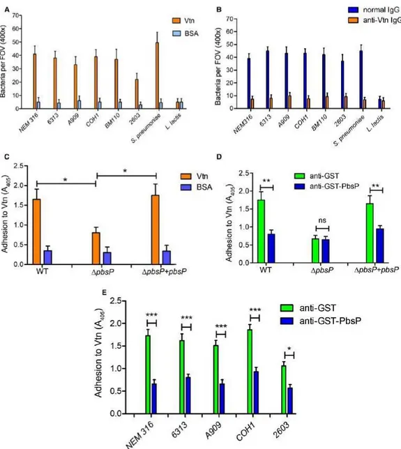
Clinical GBS strains do not have an identical set of adhesins, a diversity associated to the emergence of hypervirulent clones (Brochet et al., 2006, Tazi et al., 2012). Therefore, it was of interest to ascertain whether unrelated GBS clones bind Vtn. GBS strains belonging to the major pathogenic clonal complexes adhered to Vtn immobilized on glass coverslips but not to bovine albumin, used as a negative control (Fig. 4A). GBS binding to Vtn is similar to
that of the control species Streptococcus pneumoniae, a Gram-positive pathogen known to adhere efficiently to Vtn (Voss et al., 2013). In contrast, the nonpathogenic species Lactococcus lactis was unable to bind Vtn (Fig. 4A). GBS adherence to Vtn-coated coverslips was abrogated by pre-incubating coverslips with anti-Vtn antibodies, confirming specific bacterial binding to Vtn (Fig. 4B). Comparative binding of the parental strain BM110 and its pbsP deletion mutant (ΔpbsP) to immobilized Vtn was measured using a sensitive ELISA assay in which GBS binding to Vtn-coated plates was revealed by antiGBS polyclonal antibodies. As shown in Fig. 4C, deletion of pbsP significantly reduced, but did not completely abrogate, GBS binding to Vtn and this phenotype was reversed by genetic complementation. To confirm the role of PbsP in GBS-Vtn interactions, we next assessed whether antibodies raised against a recombinant GST-PbsP fusion protein inhibited GBS binding. Pretreatment with anti-GST-PbsP, but not anti-GST serum, significantly reduced Vtn binding of GBS strains belonging to different clonal complexes (Fig. 4D and 4E).
Overall, our results demonstrate that GBS strains interact with Vtn, that this interaction is dependent on the conserved adhesion PbsP through its SSURE domain, and that at least one additional adhesin is necessary for optimal GBS binding to Vtn.
Fig. 4. Binding of GBS to immobilized Vtn.
A. Silanized coverslips were sensitized with Vtn or BSA (used as a negative control) and incubated with the indicated GBS strains (NEM316, 6313, A909, COH1, BM110 and 2603l), S. pneumoniae (unencapsulated R6 strain) or L. lactis (subsp. Cremoris, MG1363 strain). Bacterial binding was detected after Gram staining of coverslips. Results were expressed as bacteria per field of vision (FOV) at the indicated magnification and represent means ± standard deviations of three independent experiments.
B. Inhibition of bacterial binding to Vtn by anti-Vtn antibodies. Silanized coverslips were sensitized with Vtn and treated with the indicated IgG preparation. Bacterial binding was assessed, and results expressed, as described in A.
C. The BM110 strain (WT) was compared with its pbsP deletion mutant (ΔpbsP) or with the complemented strain (ΔpbsP+pbsP) for its ability to adhere to immobilized Vtn or BSA, used as a control.
D. The BM110 strain (WT), its pbsP deletion mutant (ΔpbsP) or the complemented strain (ΔpbsP+pbsP) were treated with anti-PbsP or anti-GST mouse serum, used as a control, before assessing bacterial adherence to immobilized Vtn by ELISA using an anti-GBS serum.
E. Inhibition of adherence to Vtn of different GBS strains (NEM316, 6313, A909, COH1 and 2603) by anti-PbsP or anti-GST mouse serum, used as a control; bacterial binding was assessed by ELISA using an anti-GBS serum. *p < 0.05; ***p < 0.001, by one-way ANOVA and Bonferroni test
The αv integrin subunit promotes Vtn-dependent invasion of epithelial cells by GBS
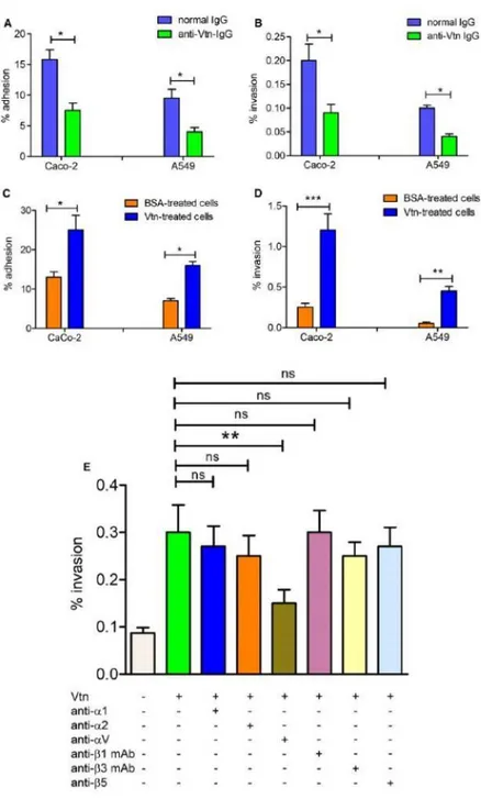
Vtn is expressed at the surface of various cell types, including A549 cells, where it can behave as an adherence or internalization substrate for pathogens (Leroy-Dudal et al., 2004; Singh et al., 2010; Su et al., 2016). In preliminary experiments, the presence of Vtn on the surface of Caco-2 cells was demonstrated by immunofluorence microscopy. To test if Vtn plays a significant role in GBS-host cell interactions, we pretreated A549 or Caco-2 cells with anti-Vtn antibodies before infection with the BM110 GBS strain. Vtn masking by antibodies significantly decreased the number of adhering or invading bacteria (Fig. 5A and B). Conversely, pre-incubation of either epithelial cell lines with purified human Vtn (5 μg ml–1) enhanced interactions of GBS with these cells (Fig. 5C and D). The enhancing effect of Vtn pretreatment was particularly evident in the case of bacterial invasion, as evidenced by an approximately 5-fold increase in the number of internalized GBS. The marked invasion phenotypes suggest that GBS might exploit Vtn to enter cells. Vtn is anchored to the cell surface through interactions with specific transmembrane proteins of the integrin family, displayed as α and β chain heterodimers. To gain entry into host cells, bacterial pathogens can use Vtn as a molecular bridge, thereby triggering integrin-dependent internalization responses (Grashoff et al., 2004). To assess whether GBS can exploit Vtn-specific
integrins to invade epithelial cells, we used a panel of blocking antibodies directed against either integrin subunit. In these experiments, monolayers of A549 respiratory cells were pretreated with Vtn in the presence of the various anti-integrin antibodies before infection with GBS. Under these conditions, anti-αv, but not anti-α1/α2 polyclonal antibodies or anti-β1/β3/β5 monoclonal antibodies, significantly decreased cell invasion by GBS (Fig. 5E). These data suggest that the Vtn-specific αv integrin subunit might function as a receptor for GBS entry into respiratory cells.
Fig. 5. The Vtn/αv integrin axis is involved in interactions between GBS and epithelial cells.
A–D. Effects of pretreating epithelial cells with anti-Vtn IgG (A and B) or Vtn (C and D) on adhesion or invasion of GBS strain BM110 to intestinal (Caco-2) or respiratory (A549) epithelial cells. Adherence (A and C) and invasion (B and D) were measured by CFU counts and expressed as the percentages of cell-associated bacteria relative to the total number bacteria added to the monolayers. E. monolayers of A549 respiratory cells were pretreated with Vtn in the presence of various blocking polyclonal or monoclonal anti-integrin antibodies before the addition of GBS. Bacterial invasion was assessed by CFU counts and expressed as the percentage of cell-associated bacteria relative to the total number bacteria added to the monolayers. Shown are the means ± SD of results from 3
independent experiments performed in triplicate (A–D) or in duplicate (E); *p < 0.05; **p < 0.01; ***p < 0.001 by Bonferroni test and one-way ANOVA.
Discussion
In this study, we demonstrated that GBS interacts with human Vtn through the SSURE domains of the PbsP adhesin to promote bacterial adherence and invasion of epithelial cells. Vtn is an ubiquitous glycoprotein involved in blood coagulation and inhibition of the assembly of the terminal complex of complement, among other functions (Preissner and Jenne, 1991). The Vtn function is dependent upon its conformation and multimerization, which regulate its interaction with different ligands. The folded monomeric form is largely predominant in plasma, where the protein is present at high concentrations (100-400 mg l–1), while Vtn is multimeric in the ECM. The unfolded multimeric state exposes cryptic epitopes and acts as an anchor point for integrins expressed on the cell surface, thereby promoting cell adherence to the ECM (Schvartz et al., 1999). Invasive pathogens have independently evolved mechanism to bind Vtn in order to evade the cytolytic or proinflammatory activities of the terminal complement complex (Singh et al., 2010; Hallstrom et al., 2016). Moreover, Vtn is expressed at the surface of different cell types as a result of endogenous secretion or of absorption from extracellular fluids and is upregulated during inflammation and injury (Hallstrom et al., 2016; Aulakh, 2018). Consequently, microbial pathogens might target Vtn for host invasion. Extracellular bacterial pathogens, in particular, interact with Vtn to efficiently adhere to host tissues and cross cell barriers, as shown for S. pneumoniae, Streptococcus pyogenes, Enterococcus faecalis, Staphylococcus aureus, Staphylococcus epidermidis, Pseudomonas aeruginosa and Haemophylus influenzae (Chhatwal et al., 1987; Kostrzynska et al., 1992; Liang et al., 1995; Virkola et al., 2000; Li et al., 2001; Heilmann et al., 2003; Styriak and
Ljungh, 2003; Leroy-Dudal et al., 2004; Bergmann et al., 2009; Singh et al., 2014; Hallstrom et al., 2016). Bacteria-host interactions may involve Vtnmediated bridging with integrins, particularly αvβ3 or αvβ5 integrins, followed by intracellular signaling, activation and bacterial uptake (Dehio et al., 1998; Spreghini et al., 1999; Leroy-Dudal et al., 2004; Bergmann et al., 2009; Singh et al., 2014). Our results suggest a similar mechanism in Vtn-dependent GBS internalization by epithelial cells, which was found to involve αv subunitcontaining integrins. This mechanism does not exclude the participation of other integrins/integrin receptors in cell invasion mediated by GBS. For example, GBS can adhere to surface-immobilized fibronectin (Beckmann et al., 2002) and invade lung epithelial cells (Cheng et al., 2002) through the cell wall protein C5a peptidase, which suggests the potential involvement of fibronectin-binding integrins, such as the α5-β1 integrin. Moreover, the α-C protein of GBS can directly bind to α1-β1 integrins (Bolduc and Madoff, 2007). It will be of interest to assess in future studies the relative importance of various integrins in GBS invasion of different cell types in order to better understand the tissue tropism of these bacteria.
Our data show that the PbsP cell wall protein, which was previously found to mediate plasmin-dependent invasion of endothelial barriers (Buscetta et al., 2016), also promotes Vtn-dependent bacterial adhesion and invasion of epithelial cells. This indicates that PbsP is a multi-ligand protein capable of hijacking, either at the same or at different times during pathogenesis, at least two major components of the host ECM. Interactions with Vtn may contribute to the initial colonization process (Cook et al., 2018), enabling GBS to attach to the mucosal surface, whereas the uptake of plasminogen, after invasion of deeper tissues, may be indispensable to acquire surface proteolytic activity and cross the blood brain barrier (Magalhaes et al., 2013; Buscetta et al., 2016).
Interestingly, the plasminogen- and Vtn-binding abilities of PbsP apparently reside on different domains. The SSURE1+2 domains binds Vtn while the Cterminal MK-rich region of the molecule (so-called for the presence of several lysine and methionine residues) is unable to bind Vtn. At the opposite, plasminogen binding on PbsP predominantly involved the MK-rich region (Buscetta et al., 2016). The 150 amino acids long repeated domain, designated as SSURE, is conserved in a family of cell wall proteins widespread in streptococci. In pneumococci, the PfbP protein containing 6 repetitions of the SSURE domain is involved in adhesion to respiratory epithelial cells (Papasergi et al., 2010). Moreover, fragments containing single SSURE domains were shown to bind to the surface of epithelial cells, as well as to plasminogen and fibronectin immobilized on inert substrates (Papasergi et al., 2010). Binsker et al. later found that fragments of PfbP (referred to as PavB), containing multiple SSURE domains also bind thrombospondin (Binsker et al., 2015). We show here that, instead of fibronectin or thrombospondin, the GBS SSURE region binds Vtn. Functional differences between the S. pneumoniae and S. agalactiae SSURE domains might be explained by structural differences, since their amino acid sequences cluster apart from each other (Buscetta et al., 2016) (Fig 6).
Fig.6 Sequence alignments of SSURE domains from group B streptococci and pneumococci.
Spn1_R6, Spn3_R6 and Spn6_R6 are amino acid sequences from the R6 strain that are representative of, respectively, the first, core and last SSURE domains of Streptococcus pneumoniae. SGB1_BM110 and SGB2_BM110, amino acid sequences from the BM110 strain that are representative of, respectively, the first and second SSURE domains of Streptococcus agalactiae. Residues that are
homologous in all SSURE domains are highlighted in black. Species specific residues are highlighted in yellow (S. pneumoniae) or blue (S. agalactiae).
Some bacterial pathogens, such as S. pneumoniae (Bergmann et al., 2009) or Neisseria meningitidis (Cunha et al., 2010) preferentially bind to the multimeric, unfolded form of Vtn. In our study, we measured binding to Vtn after its adsorption to a solid phase, using an initially monomeric form of the molecule. Once bound to a solid surface, monomeric Vtn can be recognized by a large array of ligands, including mAbs binding to cryptic epitopes exposed only on the unfolded, multimeric form (Seiffert and Smith, 1997; Underwood et al., 2002). Thus, it cannot be inferred from our data whether PbsP binds preferentially to any particular form of Vtn and further studies are necessary to decipher the in vivo contribution of Vtn in cellular adhesion or in the manipulation of the terminal complement complex by GBS.
Elucidation of the functional role of PbsP as a multifaceted virulence factor highlights the various strategies used by GBS to interact with the host. The PbsP adhesinis conserved in GBS strains belonging to different clonal complexes. Its expression was recently demonstrated to be regulated at the transcriptional level by the SaeRS two-component system in a murine model in which PbsP is necessary for vaginal colonization (Cook et al., 2018).
In conclusion, PbsP is a multi-ligand adhesion playing important roles in different steps of the pathogenesis of GBS disease. Its conservation in all human isolates and its strong in vivo upregulation make it an interesting vaccine candidate.
Matherial and Methods
Bacterial strains and Streptococcal recombinant proteins
The following GBS reference strains were used: NEM316 (serotype III, CC23); 6313 (serotype III, CC23); BM110 (serotype III, CC17); COH1 (serotype III, CC17); A909 (serotype Ia, CC7); 2603V/R (serotype V, CC19) (Glaser et al., 2002; Tettelin et al., 2005; Da Cunha et al., 2014). In some experiments, an unencapsulated type 2 pneumococcal R6 strain, (Alexander Tomasz,
Rockefeller Institute, New York, N.Y.) and the Lactococcus lactis subsp. Cremoris MG1363 strain (Mancuso et al., 2009) have been also used. The mutant ΔpbsP strain used here was obtained from GBS strain BM110, as previously described (Buscetta et al., 2016). GBS strains growth was conducted at 37°C in ToddHewitt broth or Todd-Hewitt agar (both from Difco Laboratories). Cloning, production and purification of recombinant PbsP and of the PbsP fragments MK-rich and SSURE1+2 fused to GST (Buscetta et al., 2016) and recombinant FbsA and FbsC were obtained as previously described (Buscetta et al., 2014).
Reagents
In this study, recombinant, monomeric, human Vtn, purchased from Abcam (ab 94369), was used. Bovine serum albumin (BSA), heparin, chondroitin sulfate and thrombospondin were obtained from Sigma-Aldrich. Collagen type II was purified from bovine nasal septum as reported previously (Reese and Mayne, 1981). Human fibronectin was prepared as previously described (Pietrocola et al., 2017). Polyclonal anti-Vtn (ab20091) and anti-plasminogen (ab7336) rabbit IgG were purchased from Abcam. Polyclonal anti-GST goat IgG and normal rabbit IgG were from Sigma-Aldrich. Rabbit anti-α1, anti-α2, and anti-αv IgG and mouse monoclonal anti-β1 (BV7) and anti-β3 antibodies (B212)
were a generous gift from Dr. G. Tarone (University of Turin, Italy). Rabbit anti-β5 IgG (Ab 15459) was purchased from Abcam. Rabbit anti-GBS antibody was prepared as previously described (Papasergi et al., 2013). Rabbit anti-mouse or goat anti-rabbit horseradish peroxidase (HRP)-conjugated secondary antibodies were purchased from DakoCytomation (Glostrup, Denmark).
Bacterial adhesion and invasions assays
The human epithelial cell lines A549 (ATCC CCL-185; lung carcinoma) and Caco-2 (ATCC HTB-37; colorectal adenocarcinoma) were used throughout this study. Cells were cultured in 24-well plates at a density of 1 × 10⁵ cells per well in Dulbecco’s modified Eagle medium (DMEM) supplemented with 10% of fetal bovine serum (FBS). At 24 h before the adherence or invasion assays, the medium was removed and replaced with serum-free medium to reduce the influence of ECM components in serum. The adherence and invasion assays were performed as described (Buscetta et al., 2014). Briefly, bacteria were grown to the mid-log phase and added to sub-confluent monolayers at a multiplicity of infection (MOI) of 30 for 1 h. To determine bacterial adhesion, the infected cells were washed three times with Dulbecco’s phosphate buffered saline, lysed, and plated on Todd Hewitt agar for CFU counts. To kill extracellular bacteria and to enumerate internalized bacteria, the monolayers were incubated for an additional 1 h before cell lysis in medium supplemented with penicillin and streptomycin (200 U ml–1 and 200 μg ml–1, respectively). Bacterial adherence and invasion were calculated as: recovered CFU/initial inoculum CFU × 100.
In some experiments, cells were pretreated for 30 min with various reagents, including Vtn (5μg ml–1), anti-integrin, anti-plasminogen or anti-Vtn antibodies (all at a concentration of 5 μg ml–1) before the addition of bacteria.
In further experiments, bacteria were pre-incubated for 30 min with mouse serum (1:100) directed against recombinant PbsP fused to GST or against GST, as previously described (Buscetta et al., 2016). In selected experiments adherent bacteria were visualized by immunofluorence microscopy, as described
(Papasergi et al., 2010).
Adherence of microspheres coated with PbsP and PbsP fragments
Fluorescent beads (Fluoresbrite YG 1.00 μm microspheres, Polysciences) were conjugated with recombinant PbsP, PbsP fragments or with GST at equimolar concentrations (10 μM) by previously described procedures (Papasergi et al., 2010). The amount of protein coupled on beads was calculated by subtracting the amount of protein present in the supernatant after adsorption. Adhesion of microspheres to A549 cells was performed as previously described (Papasergi et al., 2010). Briefly, protein-coupled beads were added to cells at a concentration of 108 beads ml–1. After 30 min of incubation at 37°C (in the presence of 1 μM GST to avoid non-specific binding) monolayers were washed, fixed with 3.7 % formaldehyde and permeabilized with Triton 0.1% in phosphate buffered saline (PBS), as described (Papasergi et al., 2010). After labeling actin and nuclei with, respectively, phalloidin-iFluor 555 (ABCAM ab 176756) and DAPI, attached beads were counted using a fluorescent microscope equipped with structured illumination (Apotome, Zeiss), as previously described (Papasergi et al., 2010). In binding inhibition assays, cell monolayers were pre-incubated with increasing concentrations of soluble SSURE1+2, MK-rich, anti-Vtn or anti-plasminogen antibodies for 30 min before performing the assay with PbsP-coated microspheres.
PbsP binding to ECM components
Vitronectin, fibronectin, collagen or thrombospondin were coated at the concentration of 10 μg ml–1 onto microtiter wells overnight at 4°C in 0.05 M carbonate buffer (pH 9.0). After washing with PBS supplemented with 0.05% Tween 20, the wells were blocked with PBS supplemented with 0.01% Tween 20 and 1% non fat dry milk for 2 h at 25°C. After incubation with 5 μg ml–1 of recombinant PbsP MK-rich, SSURE1+2, FbsA or FbsC (all fused to GST) for 1 h, the binding was detected with goat anti-GST (1: 4,000; GE Healthcare), followed by the addition of alkaline phosphatase-conjugated rabbit anti-goat IgG (1: 5,000; Sigma-Aldrich). For dot blot analysis, increasing concentrations of Vtn were spotted through circular templates directly onto the nitrocellulose membranes and probed using 5 μg of recombinant proteins. The latter were detected using anti-GST serum as described above. The effect of heparin and chondroitin sulfate on PbsP/Vtn interaction was examined incubating Vtn immobilized onto microtiter plates with 0.5 μg of recombinant PbsP in fusion with GST in the presence of increasing concentrations (0–2 μM) of heparin or chondroitin sulfate. PbsP bound to Vtn was detected by addition of goat antiGST antibodies followed by HRP-conjugated anti-goat IgG.
Adhesion of bacteria to immobilized Vtn
For microscopic assessment of bacterial adhesion to immobilized Vtn, silanetreated 18-mm2 glass coverslips were incubated overnight at 4°C with Vtn or BSA (both at 10 μg ml–1 in PBS), blocked with 2% casein for 1 h at 20°C, and exposed to bacteria, as previously described (Papasergi et al., 2010). Briefly, bacteria were grown to the late log phase (A560 = 0.8), washed, resuspended in PBS and applied to the coverslips at a concentration of approximately 1 x 10⁵ CFU ml–1. Slides were then Gram stained and observed under a bright field microscope. Results were expressed as numbers of bacteria per field of vision
(FOV) at the indicated magnification. At least 20 different fields per slide were counted. Adherence of bacteria to immobilized Vtn was also assessed by ELISA. After sensitizing microtiter plates overnight at 4°C with 10μg ml–1 of Vtn or BSA in PBS, plates were blocked with 2 % casein for 1 h at 20°C. Bacteria grown to the late log phase were resuspended in PBS to a concentration of approximately 1 × 10⁵ CFU ml–1 and incubated at 37°C for 1 h. After washing, anti-GBS rabbit serum (1: 10,000) was added and incubated for 1 h at 37°C followed by the addition of alkaline phosphatase-conjugated rabbit antigoat IgG (1: 5,000; Sigma-Aldrich).
Statistical analysis
One way analysis of variance (ANOVA) followed by Bonferroni correction was used to assess statistical significance of differences between the numbers of adhering or invading bacteria and between ELISA test absorbance values.
References
Al Safadi , Souheila Amor, Geneviève Hery-Arnaud, Barbara Spellerberg, Philippe Lanotte, Laurent Mereghetti, François Gannier, Roland Quentin, Agnès Rosenau (2011). Enhanced expression of lmb gene encoding laminin-binding protein in Streptococcus agalactiae strains harboring IS1548 in scpB-lmb intergenic region. Plos one. 5(5): e10794
Armistead B, Oler E, Adams Waldorf K, Rajagopal L. (2019) The Double Life of Group B Streptococcus: Asymptomatic Colonizer and Potent Pathogen. J Mol Biol. 431(16):2914-2931.
Arko, R.J., Chen, C.Y., Schalla, W.O., Sarafian, S.K., Taylor, C.L., Knapp, J.S., and Morse, S.A. (1991) Binding of S protein by Neisseria gonorrhoeae and potential role in invasion. J Clin Microbiol 29: 70–75
Aulakh, G.K. (2018) Neutrophils in the lung: "the first responders". Cell Tissue Res 371: 577–588
Bergmann, S., Lang, A., Rohde, M., Agarwal, V., Rennemeier, C., Grashoff, C., et al. (2009) Integrin-linked kinase is required for vitronectin-mediated internalization of Streptococcus pneumoniae by host cells. J Cell Sci 122: 256–267. Binsker, U., Kohler, T.P., Krauel, K., Kohler, S., Schwertz, H. and Hammerschmidt, S. (2015) Pneumococcal Adhesins PavB and PspC Are Important for the Interplay with Human Thrombospondin-1. J Biol Chem 290: 14542–14555
Boyle, E.C., and Finlay, B.B. (2003) Bacterial pathogenesis: exploiting cellular adherence. Curr Opin Cell Biol 15: 633–639.
Berglund, L., Bjorling, E., Oksvold, P., Fagerberg, L., Asplund, A., Szigyarto, C.A., et al. (2008) A genecentric Human Protein Atlas for expression profiles based on antibodies. Mol Cell Proteomics 7: 2019–2027
Blom, A.M., Hallström, T., and Riesbeck, K. Complement evasion strategies of pathogens – acquisition of inhibitors and beyond. Mol Immunol, 2009, 46: 2808– 2817.
Bolduc, G.R. and Madoff, L.C. (2007) The group B streptococcal alpha C protein binds alpha1beta1-integrin through a novel KTD motif that promotes internalization of GBS within human epithelial cells. Microbiology 153:4039– 4049.
Brega, S., Caliot, E., Trieu-Cuot, P., and Dramsi, S. (2013) SecA localization and SecA-dependent secretion occurs at new division septa in group B Streptococcus. PloS One 8: e65832.
Brunton, V.G., MacPherson, I.R., and Frame, M.C. (2004) Cell adhesion receptors, tyrosine kinases and actin modulators: a complex three-way circuitry. Biochim Biophys Acta 1692: 121–144.
Buscetta, M., Firon, A., Pietrocola, G., Biondo, C., Mancuso, G., Midiri, A., et al. (2016) PbsP, a cell wall-anchored protein that binds plasminogen to promote hematogenous dissemination of group B Streptococcus. Mol Microbiol 101: 27– 41.
Buscetta, M., Papasergi, S., Firon, A., Pietrocola, G., Biondo, C., Mancuso, G., et al. (2014) FbsC, a novel fibrinogen-binding protein, promotes Streptococcus agalactiae-host cell interactions. J Biol Chem 289: 21003–21015
Calabi, E., Calabi, F., Phillips, A.D., and Fairweather, N.F. (2002) Binding of Clostridium difficile surface layer proteins to gastrointestinal tissues. Infect Immun 70: 5770–5778
Campbell, J.R., S.L. Hillier, M.A. Krohn, P. Ferrieri, D.F. Zaleznik, and C.J Baker. 2000. Group B streptococcal colonization and serotype-specific immunity in pregnant women at delivery. Obstet. Gynecol. 96:498–503 Carlsson, F., Stalhammar-Carlemalm, M., Flardh, K., Sandin, C., Carlemalm,
E., and Lindahl, G. (2006) Signal sequence directs localized secretion of bacterial surface proteins. Nature 442: 943–946
Cerquetti, M., Serafino, A., Sebastianelli, A., and Mastrantonio, P. (2002) Binding of Clostridium difficile to Caco-2 epithelial cell line and to extracellular matrix proteins. FEMS Immunol Med Microbiol 18: 211–218
Chhatwal, G.S., Preissner, K.T., Muller-Berghaus, G., and Blobel, H. (1987) Specific binding of the human S protein (vitronectin) to streptococci, Staphylococcus aureus, and Escherichia coli. Infect Immun 55: 1878–1883
Chauhan, A.K., and Moore, T.L. (2006) Presence of plasma complement regulatory proteins clusterin (Apo J) and vitronectin (S40) on circulating immune complexes (CIC). Clin Exp Immunol 145: 398–406
Cheng, Q., Stafslien, D., Purushothaman, S.S. and Cleary, P. (2002) The group B streptococcal C5a peptidase is both a specific protease and an invasin. Infect Immun 70: 3309–3309
Cho, H.; Jeong, D.W.; Li, C.; Bae, T. Organizational requirements of the SaeS binding sites for a functional P1 promoter of the sae operon in Staphylococcus aureus. J. Bacteriol. 2012, 194, 2865–2876
Cook, L.C., Hu, H., Maienschein-Cline, M. and Federle, M.J. (2018) A vaginal tract signal detected by the GBS SaeRS system elicits transcriptomic changes and enhances murine colonization. Infect immun. 86(4). pii: e00762-17.
Cunha, C.S.E., Griffiths, N.J. and Virji, M. (2010) Neisseria meningitidis Opc Invasin Binds to the Sulphated Tyrosines of Activated Vitronectin to Attach to and Invade Human Brain Endothelial Cells. PLoS pathogens 6: e1000911 Dehio, M., Gomez-Duarte, O.G., Dehio, C. and Meyer, T.F. (1998) Vitronectindependent invasion of epithelial cells by Neisseria gonorrhoeae involves alpha(V) integrin receptors. Febs Lett 424: 84–88.
D’Mello, V., Singh, S., Wu, Y., and Birge, R.B. (2009) The urokinase plasminogen activator receptor promotes efferocytosis of apoptotic cells. J Biol Chem 284: 17030–17038.
Edwards MS, Baker CJ. (2005) Group B streptococcal infections in elderly adults. Clin Infect Dis. 15;41(6):839-47. Review. PMID: 16107984
Edwards, M.S., V. Nizet, and C.J. Baker. (2006). Group B streptococcal infections. In: JS Remington, JO Klein, C Wilson and CJ Baker (eds.), Infectious Diseases of the Fetus and Newborn Infant. Saunders Elsevier, Philadelphia, PA, pp. 403–464
Farley MM1, Harvey RC, Stull T, Smith JD, Schuchat A, Wenger JD, Stephens DS. (1993) A population-based assessment of invasive disease due to group B Streptococcus in nonpregnant adults. N Engl J Med.;328(25):1807-11
Frantz C, Stewart KM, Weaver VM (2010)The extracellular matrix at a glance. J Cell Sci. ;123:4195-200
Galperin MY. 2006. Structural classification of bacterial response regulators: diversity of output domains and domain combinations. J Bacteriol 188:4169-82. Geiger, T.; Goerke, C.; Mainiero, M.; Kraus, D.; Wolz, C. The virulence regulator sae of Staphylococcus aureus: Promoter activities and response to phagocytosis-related signals. J. Bacteriol. 2008, 190, 3419–3428
Glaser, P., Rusniok, C., Buchrieser, C., Chevalier, F., Frangeul, L., Msadek, T. et al. (2002) Genome sequence of Streptococcus agalactiae, a pathogen causing invasive neonatal disease. Mol Microbiol 45: 1499–1513
Gutekunst, H., Eikmanns, B. J., and Reinscheid, D. J. (2004). The novel fibrinogen binding protein FbsB promotes Streptococcus agalactiae invasion into epithelial cells. Infect. Immun. 72, 3495–3504.
Heckman KL, Pease LR (2007) Gene splicing and mutagenesis by PCR-driven overlap extension. Nat Protoc 2: 924–932
Hallström, T., Trajkovska, E., Forsgren, A., and Riesbeck, K. (2006) Haemophilus influenzae surface fibrils contribute to serum resistance by interacting with vitronectin. J Immunol 177: 430–436
Hallström, T., Uhde, M., Mörgelin, M., Skerka, C., and Zipfel, P.F. (2010) Pseudomonas aeruginosa CRASP-2 is a surface protein that uses the human terminal complement regulator vitronectin for protection against complement mediated attack. Mol Immunol 46: 2835
Hallstrom, T., Singh, B., Kraiczy, P., Hammerschmidt, S., Skerka, C., Zipfel, P.F. and Riesbeck, K. (2016) Conserved patterns of microbial immune escape: pathogenic microbes of diverse origin target the human terminal complement inhibitor vitronectin via a single common motif. PloS one 11: e0147709.
Hansen, S.M., N. Uldbjerg, M. Kilian, and U.B. Sørensen. (2004). Dynamics of Streptococcus agalactiae colonization in women during and after pregnancy and in their infants. J. Clin. Microbiol. 42:83–89.
Hauck, C.R., and Meyer, T.F. (2003) ‘Small’ talk: Opa proteins as mediators of Neisseria–host-cell communication. Curr Opin Microbiol 6: 43–49
Heilmann, C., Thumm, G., Chhatwal, G.S., Hartleib, J., Uekotter, A. and Peters, G. (2003) Identification and characterization of a novel autolysin (Aae) with adhesive properties from Staphylococcus epidermidis. Microbiology 149: 2769– 2778
Kostrzynska, M., Paulsson, M., Schmidt, K.H. and Wadstrom, T. (1992) Comparative-studies on binding of vitronectin and fibronectin to Group-a and Group-CStreptococci. Microbios 71: 179–192.
Jeong, D.W.; Cho, H.; Lee, H.; Li, C.; Garza, J.; Fried, M.; Bae, T. Identification of P3 promoter and distinct roles of the two promoters of the SaeRS twocomponent system in Staphylococcus aureus. J. Bacteriol. 2011, 193, 4672– 4684 Jeong, D.W.; Cho, H.; Jones, M.B.; Shatzkes, K.; Sun, F.; Ji, Q.; Liu, Q.;