Università degli Studi di Messina
PhD in "Surgical and Medical Biotechnology"
XXX Ciclo
Role of endosomal
Role of endosomal
Role of endosomal
Role of endosomal
Toll
Toll
Toll
Toll
in pneumococcal pneumonitis
in pneumococcal pneumonitis
in pneumococcal pneumonitis
in pneumococcal pneumonitis
Ph student
Famà Agata
Università degli Studi di Messina
PhD in "Surgical and Medical Biotechnology"
XXX Ciclo - Academic year 2016-2017
Role of endosomal
Role of endosomal
Role of endosomal
Role of endosomal
Toll
Toll
Toll
Toll----like receptors
like receptors
like receptors
like receptors
in pneumococcal pneumonitis
in pneumococcal pneumonitis
in pneumococcal pneumonitis
in pneumococcal pneumonitis
Supervisor
Prof. Teti Giuseppe
PhD in "Surgical and Medical Biotechnology"
2017
Role of endosomal
Role of endosomal
Role of endosomal
Role of endosomal
like receptors
like receptors
like receptors
like receptors
in pneumococcal pneumonitis
in pneumococcal pneumonitis
in pneumococcal pneumonitis
in pneumococcal pneumonitis
Supervisor
1
Index
Pg1. Introduction ... 3
1.1 TLRs structure... 5
1.2 Positioning and trafficking of the TLRs... 7
1.3 PAMPs recognized by TLRs... 11 1.4 TLRs signaling pathway... 13 2. Endosomal TLRs... 17 2.1 TLR3... 17 2.2 TLR7/8... 19 2.3 TLR9... 21 2.4 TLR11 and TLR13... 22 3. Streptococcus pneumoniae ... 26 3.1 Epidemiology... 27 3.2 Microbiology... 30 3.3 Colonization... 32 3.4 Pneumococcal vaccines... 33 4. TLRs in S. pnuemoniae infections... 36
5. Materials and methods... 40
5.1 Mice... 40
5.2 Genotiping TLR7/9/13-/- mice... 41
5.3 S. pneumoniae strain and murine infection models... 43
5.4 Cytokine measurement... 44
5.5 Bone-marrow derived cells... 44
2
5.7 Peritoneal macrophages... 47
5.8 IFN-β mRNA analysis by quantitative real-time PCR... 48
5.9 Data expression and statistical significance... 49
6. Results... 50
6.1 MyD88-/- mice are extremely susceptible to S. pneumoniae infections... 50
6.2 The combined abscence of TLR7/9/13 recapitulates severe phenotype of 3D mice... 54
6.3 Reduced cytokine/chemokine levels in 3D mice... 60
6.4 Differental effects of endosomal TLR in resident vs elicited MØ... 64 7. Discussions... 67 8. Bibliography... 71
3
1. Introduction
1. Introduction
1. Introduction
1. Introduction
One of the most fundamental questions in immunology concerns self-recognition, which for the most part means microbes. As we initially realize we have been infected with the microbes, and how is the immune response turned on?1 Host defense against invading microbial pathogens is caused by the immune system, which consists of two components: innate
immunity and acquired immunity.
In acquired immunity, B and T lymphocytes use antigen receptors such as immunoglobulins and T cell receptors to recognize non-self. Acquired immunity is characterized by specificity and is developed by clonal selection from a vast repertoire of lymphocytes that carry specific antigen receptors generated through a mechanism generally known as gene rearrangement2.
The innate immune response is not completely nonspecific, as was originally thought, but is able to discriminate between self and a variety of pathogens. The innate immune system recognizes microorganisms through a limited number of germline-encoded pattern-recognition receptors (PRRs). This is in contrast to the great repertoire of reconstituted receptors used by the acquired system. PRRs possess common features. First, PRRs
4
recognize microbial components, known as "pathogen associated molecular patterns (PAMPs)" essential for the survival of the microorganism and are therefore difficult to alter the microorganism. Secondly, PRRs are expressively expressed in host and detect pathogens regardless of their stage of life. Third, PRRs are germline encoded, nonclonal, expressed on all cells of a given type, and independent of the immune memory. Several PRR respond with PAMP specifications, show distinct expression patterns, trigger specific signaling pathways, and lead to distinctive anti-pathogen responses. The basic machinery underlying innate immune recognition is highly conserved among species, plants and fruit flies to mammals3. PRRs include Toll-like receptors (TLRs), retinoid acid-inducible gene (RIG)-like receptors (RLRs) and nucleotide oligomerization domain (NOD)-like receptors (NLRs)4-6.
At the end of the twentieth century, Toll has been shown to be an essential receptor for host defense against fungal infection in Drosophila, which has only innate immunity7. A year later, a Toll receptor mammalian homologue (now defined as TLR4) has been shown to induce the expression of genes involved in inflammatory reactions8. Ten TLRs have been identified in humans so far, and twelve in mice. Both humans and mice express TLR1-9. In addition humans but no mice express TLR10, mice also have TLR11, 12 and 13 that human beings are missing.
5
1.1. TLRs structure
1.1. TLRs structure
1.1. TLRs structure
1.1. TLRs structure
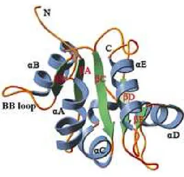
TLRs are expressed in innate immune cells such as dendritic cells (DCs) and macrophages, as well as non-immune cells such as fibroblast cells and epithelial cells. TLR are found on cell surfaces or within endosomes and have important roles in the host defense against pathogenic organisms throughout the animal kingdom. The TLRs are type I integral membrane receptors, each having an N-terminal ligand recognition domain, a single transmembrane helix, and a C-terminal cytoplasmic signaling domain9. TLRs signaling domains are known as Toll IL-1 Receptor (TIR) domains because they share homology with the signaling domains of IL-1R family members10. TIR domains are also found in many adaptive proteins that interact homologously with the TIR domains of TLRs and IL-1 receptors as the first step in the signaling cascade. Significantly, homologues of TIR domains are also found in some plant proteins that confer resistance to pathogens11, suggesting that TIR domain are a very ancient motif that served an immune function before the divergence of plants and animals. TIR domains have a common fold with a β-sheet (βA-βE) parallel to five central wires that is surrounded by a total of five α-helixes (αA-αE) on both sides (Fig. 1)12, 13. The BB loop, connecting strand βB and helix αB, containing three highly conserved residues, arginine BB3, aspartic acid BB4, and glycine BB8, plays a major role in TIR dimerization and/or adaptor recruitment14-15.
6
Fig. 1:Schematic drawing of the structure of the TIR domain of human TLR2. The common fold of TIR contains a central five-stranded parallel β-sheet (green) surrounded by a total of five α-helices
(blue) on both sides linked by loops (gold).
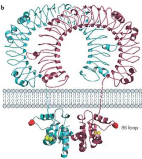
The TLR transmembrane domains each contain a typical trait of approximately 20 non-loaded, predominantly hydrophobic residues. TLR N-terminal ectodomains (ECD) are glycoproteins with 550-800 residues of amino acids9 and meet and recognize the molecules released by invasion of pathogens. All TLR ECDs are constructed of tandem copies of a motif known as the leucine-rich repeat (LRR), which is typically 22–29 residues in length and contains hydrophobic residues spaced at distinctive intervals. LRRs that do not form a complete circle, but form a “horseshoe” structure (Figure 2).
7
Fig. 2: The complete structure of a TLR, in which the "horseshoe" structure of the leucine-rich motif is well
visible.
1.2 Positioning and trafficking of the
1.2 Positioning and trafficking of the
1.2 Positioning and trafficking of the
1.2 Positioning and trafficking of the TLRs.
TLRs.
TLRs.
TLRs.
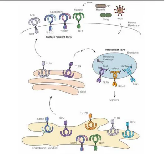
All TLRs are synthesized in ER, traffic to Golgi and recruited to cell surfaces or intracellular compartments such as endosomes. The TLRs belonging to the first group are: TLR1, TLR2, TLR4, TLR5, TLR6. Usually these recognize PAMP on extracellular microbes. TLRs belonging to the second group are: TLR3, TLR7, TLR8, TLR9, TLR11, TLR13 (Fig. 3). These recognize PAMP on intracellular microbes and their nucleic acids after degradation. The multi-pass transmembrane protein UNC93B1 controls intracellular TLR trafficking from the ER to endosomes. TLR trafficking is also controlled by the ER-resident protein PRAT4A, which regulates the output of TLR1, TLR2, TLR4,
8
TLR7, and TLR9 from the ER and their trafficking to the plasma membrane and endososmes16.
Fig. 3: Overview of Toll-like Receptor Trafficking
Unc93b1 was originally identified through a forward genetic screen in mice ("3d mice") where a mutation (H412R) caused a defect in the signaling of nucleic acid sensing TLR3, 7, and 9 but not surface localized TLRs17. Subsequent studies focused on the mechanical details of UNC93B1 function, indicating that this ER-resident protein is needed for a proper
9
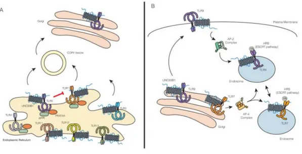
trafficking of multiple TLRs18, 19. UNC93B1 facilitates the incorporation of TLR9 into COPII vesicles, carrying protein loads from the ER to Golgi19 (Figure 4). This finding opens the possibility that active transport mechanisms such as the selection by Sec24 protein loads (part of the COPII machinery) may be involved in TLR export ER regulation. TLRs 3, 7, 11, 12 and 13 also require UNC93B1 to exit the ER19, presumably by loading into the COPII vesicles, even though this mechanism has not been formally demonstrated for these TLRs. Chemical TLR analyzes implicated TLR transmembrane domains as critical for the UNC93B120 association20. In fact, the recent study has demonstrated the importance of acid residues (D812 and E813) superimposed on the TLR9 transmembrane region for the association with UNC93B121. These residues alone are not sufficient to mediate interaction with UNC93B1, but are required for binding and subsequent UNC93B1-dependent trafficking. Similar residues were identified in TLR321. Previous studies have also described roles for other regions within TLRs for a proper trafficking. For example, the linker region, a short sequence of aminoacids between transmembrane and TIR domains, is important for proper TLR3 traffic, while the TLR7 transmembrane domain is predominantly responsible for its proper trafficking22, 23. In addition, TLR9 trafficking has been shown to depend on both its transmembrane and cytosolic regions24, 25, 26. However, these studies did not examine in detail whether these regions still have a
10
functional relationship with UNC93B1. A number of additional factors regulate TLR output from the ER as: Glycoprotein 96 (gp96)27, and Protein associated with TLR4 A (PRAT4A), also known as CNPY316. Gp96 and CNPY3 seem to work together to coordinate the folding of all TLRs28. After export from ER, TLR nucleic acid sensors pass through Golgi before being ordered into endosomal compartments29, 30, 31. Surprisingly, theUNC93B1 remains associated with TLRs after ER output and plays a role in these subsequent traffick events18, 19. Additionally, the hepatocyte growth factor-regulated tyrosine kinase substrate (HRS) has been identified as a factor key in TLR7 and TLR9 trafficking32 (Fig 4).
Fig. 4: Trafficking of TLRs from ER to Golgi (A) and from Golgi to endosome (B).
UNC93B1 mediates TLR7 and TLR9 differential traffic, potentially at multiple levels. Infact, point mutation (D34A) in terminal N of UNC93B1
11
enhanced TLR7 responses, while overlapping TLR9 responses simultaneously33. Further examination of the cells expressing UNC93B1 D34A revealed an increase in interaction with TLR7 and simultaneous decrease with TLR9 resulting in increased TLR7 endosomal and decreased TLR9. Therefore, the N-terminal portion of UNC93B1 seems to be involved in the selectivity of this chaperone for distinct TLR family members. Additional, TLR9 uses UNC93B1 to interact with AP-2, TLR7 trafficking is unexpectedly independent of this pathway. Instead, TLR7 appears to recruit directly AP-4, a distinct sorting system known to mediate direct transport between the Golgi's network and endosomes19.
1.3
1.3
1.3
1.3 PAMPs recognized by
PAMPs recognized by
PAMPs recognized by TLRs
PAMPs recognized by
TLRs
TLRs
TLRs....
Each TLR extracellular domains is responsible for recognizing specific
PAMPs (Table 1)20.
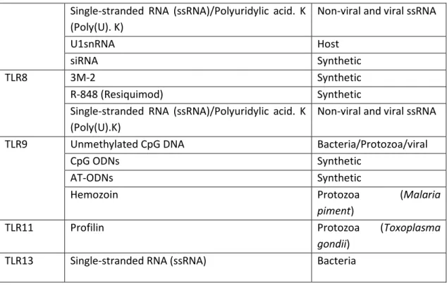
TLR Ligands Origin
TLR1–TLR2
Lipoproteins/triacylated lipopeptides: Pam3CSK4,
JBT3002, OspA
Bacteria, Mycobacteria
Soluble Lipoproteins Neisseria meningitides
TLR2
Bacterial Lipoproteins (BLPs) Bacteria
Lipoarabinomannan (LAM) Mycobacteria
MALP-2 (Mycoplasma, Macrophage-activating Lipopeptide-2)
Mycoplasma
Glycosylphosphatidylinositol (GPI) Trypanosoma cruzi
Glycolipids Treponema maltophilum
12 TLR2 Zymosan Fungi TLR3
Double-stranded RNA (dsRNA) Viral
Polyinosine-polycytidylic Acid (poly(I:C)) Synthetic
MRNA Host TRNA Host/fungi TLR4
S-Lipopolysaccharides (LPS) (smooth) wild-type (wt), LPS (contains repeated O-polysaccharide units)
Gram-negative bacteria (e.g. E. coli, Salmonella)
R-Lipopolysaccharides (LPS) (rough) mutant (Ra, Rb: extended core-polysaccharide) LPS (S-LPS-like)
Gram-negative bacteria (e.g. E. coli, Salmonella)
R-Lipopolysaccharides (LPS) (rough) mutant (Rc, Rd1, Rd2, Re: short core-polysaccharide) LPS (Lipid A-like)
Gram-negative bacteria (e.g. E. coli, Salmonella)
Flavolipin Flavobacterium
meningosepticum
Paclitaxel (Taxol®) (recognizes mouse TLR4) Plant Acyclic lipid A-like analog (R-112022) Synthetic Type III Repeat Extra Domain A (EDA) Host LMW Oligosaccharides of Hyaluronic acid (sHA) Host
Polysaccharide Fragments of Heparan sulfate Host (only mouse tested)
Fibrinogen Host
Fusion Protein of RSV Respiratory syncytial virus
Envelope Proteins of MMTV Mouse mammary tumor
virus
Glycoinositolphospholipids (GIPLs) Trypanosoma cruzi
Heat Shock Proteins (HSPs) TLR2 vs TLR4
TLR5 Flagellin Gram-negative bacteria
TLR6–TLR2
MALP-2 (Mycoplasmal Macrophage-activating Lipopeptide-2)/Diacylated Macrophage-activating Lipopeptide-2
Mycoplasma
Diacylated Lipopeptide FSL-1 Part of the lipoprotein LP44 of Mycoplasma
salivarium
Diacylated Lipopeptide Pam2CSK4 Synthetic
Soluble tuberculosis factor (STF) Mycobacteria
TLR7
Imiquimod (R-837)/Gardiquimod Synthetic
Resiquimod (R-848) Synthetic
S-27610 Synthetic
Loxoribine/TOG Synthetic
3M-13 Synthetic
13
1.4
1.4
1.4
1.4 TLRs signaling pathway
TLRs signaling pathway
TLRs signaling pathway
TLRs signaling pathway
Individual TLRs recruit members of a set of adapters containing the TIR domain such as MyD88, TRIF, TIRAP/MAL or TRAM. Collectively, depending on the use of the adapter, the TLR signaling is largely divided into two pathways: the MyD88-dependent and TRIF-dependent pathways. MyD88 is used by all TLRs and activates NF-κB and MAPK for the induction of inflammatory cytokine genes. TLRs but also IL-1 and IL-18 proinflammatory cytokine receptors share a common TIR domain in their intracellular region and belong to the TLR/IL1-R superfamily2, 35. MyD88 is critical for signaling
Single-stranded RNA (ssRNA)/Polyuridylic acid. K (Poly(U). K)
Non-viral and viral ssRNA
U1snRNA Host siRNA Synthetic TLR8 3M-2 Synthetic R-848 (Resiquimod) Synthetic
Single-stranded RNA (ssRNA)/Polyuridylic acid. K (Poly(U).K)
Non-viral and viral ssRNA
TLR9
Unmethylated CpG DNA Bacteria/Protozoa/viral
CpG ODNs Synthetic
AT-ODNs Synthetic
Hemozoin Protozoa (Malaria
piment)
TLR11 Profilin Protozoa (Toxoplasma
gondii)
TLR13 Single-stranded RNA (ssRNA) Bacteria
14
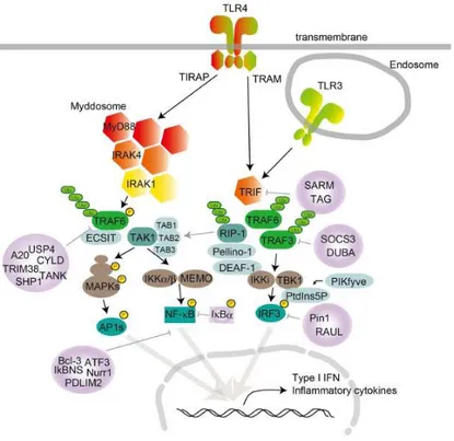
responses of IL-1, IL-18, and all TLRs except TLR33, 10, 36, 37. In addition to its C-terminal TIR domain, MyD88 contains an N-terminal death domain (DD) and a short intermediate domain (ID). Through DD, MyD88 interacts with IRAKs, including IRAK1, IRAK2, IRAK4 and IRAK-M, characterized by an N-terminal DD and a C-N-terminal Ser/Thr kinase or kinase-like domain3, 38, 39. In this way MyD88 forms a complex with members of the IRAK kinase family, called Myddosome40. During Myddosome formation, IRAK4 activates IRAK1, which is then autophosphorylated at several sites41 and released from MyD8842. IRAK1 associates with TRAF6. TRAF6 promotes the polyubiquitination of both TRAF6 and the complex protein kinase complex TAK1. TAK1 is a member of the MAPKKK family and forms a complex with the TAB1, TAB2, and TAB3 regulatory subunits 43, 44. TAK1 then activates two different pathways leading to activation of the IKK complex-NF-κB pathway and -MAPK pathway. The IKK complex is composed of the IKKα and IKKβ catalytic subunits and the NEMO regulatory subunit (also called IKKγ). The IKK complex phosphorylates the NF-κB which translocate into the nucleus to induce proinflammatory gene expression. TAK1 activation also triggers activation of MAPK family members such as ERK1/2, p38 and JNK that mediate the activation of AP-1 transcription factors or mRNA stabilization to regulate inflammatory responses3, 45 (Fig. 5).
15
Fig. 5: TLR signaling: MyD88 and TRIF dependend pathway.
TRIF is recruited to TLR3 and TLR4. TRAM is selectively recruited to TLR4 but not TLR3 to connect between TRIF and TLR4. TLR3, indeed, directly interacts with TRIF. TLR3, in fact, interacts directly with the TRIF. TRIF interacts with TRAF6 and TRAF3. Conversely, TRAF3 recruits the IKK-related kinases TBK1 and IKKi together with NEMO for IRF3 phosphorylation. TRAF6 recovers the RIP-1 kinase, which in turn interacts with and activates the TAK1 complex, leading to the activation of NF-κB and MAPK and the induction of inflammatory cytokines. Subsequently, IRF3 forms a dimer and translocates into the nucleus from the cytoplasm, where it induces the expression of type I IFN genes3, 45. Recently, IRF3
16
activation has been shown to be regulated by an inositol lipid, PtdIns5P. PtdIns5P binds to both IRF3 and TBK1, and thus facilitates complex formation between TBK1 and IRF346.
It should also be added that the induction of inflammatory genes by endosomal TLRs may follow a more detailed mechanism. In fact, nucleic acids meet TLRs in endosomes after uptake from the extracellular milieu. Depending on the delivery method in the cell (eg in an immune complex, such as oligonucleotides or encapsulated in a viral particle), nucleic acids can reach distinct endosomes with unique signaling properties. This possibility has been first suggested by the finding that several classes of synthetic oligodynucleotides (ODNs) produce very different results of downstream signaling in pDCs47, 48, 49, 50. For example, CpG-A ODN, which aggregate to form large complexes, strongly induce type I IFN, while CpG-B ODN, which remain monomeric, instead favor a pro-inflammatory response characterized by TNFα, IL-6 and IL-12p40 production. It is remarkable that this phenomenon is the result of differential ODN traffic in endosomal compartments, where TLR9 signaling leads to the activation of various transcription factors47. CpG-A preferentially traffics to early endosomes where TLR9 signaling recruits the transcription factor IRF7 and leads to a type I IFN response47. In contrast, CpG-B preferentially traffics to late endosomes where TLR9 signaling activates transcription factor NF-κB,
17
resulting in a proinflammatory cytokine response47. Two other compartments capable of type I IFN induction have been described: LROs (lysosome related organelles) induced by AP-3 complex51, and a compartment for DNA-immune complex (DNA-IC) which requires autophagy pathway components through a mechanism termed LC3 associated phagocytosis (LAP)52.
2. Endosomal
2. Endosomal
2. Endosomal
2. Endosomal TLRs
TLRs
TLRs
TLRs
2.1 TLR3
2.1 TLR3
2.1 TLR3
2.1 TLR3
In Homo Sapiens, TLR3 is made up of 903 amino acids and coded by a gene on the chromosome 4. It plays a significant role in the modulation of virus-mediated innate immune response. TLR3 (also known as CD283 or IIAE2) is a nucleic acid-sensing receptor that recognizes the viral replication product (dsRNA) and synthetic ligand polyinosinic–polycytidylic acid [poly(I:C)]53. TLR3 is evolutionarily conserved from insects to vertebrates. TLR3 is expressed on the endosomes of B cells, T cells, macrophages, natural killer (NK) cells, dendritic cells, neurons. The astrocytes and microglia cells express TLR3 on its endosome as well as cell surface54, 55. TLR3 triggers several intracellular signaling pathways leading to the activation of
18
mitogen-activated protein kinases (MAPKs), epidermal keratins, c-Jun, p38, N-terminal kinase, and nuclear factor kappa-light-chain-enhancer of activated B cells (NF-κB), which induces the expression of proinflammatory cytokines and IFN-125. TLR3 recognizes dsRNA that have anti-viral responses as different virus groups are having ds RNA genomes or give rise to dsRNA during replication. TLR3 signals through the TRIF-mediated pathway and induces IFN-α and IFN-β gene expression. To date, 136 SNPs have been identified in TLR3. About seven SNPs are within the protein-coding sequence; however, only four (N284I, Y307D, L412F, and S737T) have led to amino acid substitutions. SNPs, Y307D and S737T, are localized in relatively less conserved residues and do not affect TLR3 activity, whereas N284I and L412F were associated with impaired receptor signaling and functioning in an "in vitro" model. Replacement of amino acids, Leu412Phe, located within a ligand binding surface adjacent to the glycosylated asparagine at position 413, is required for receptor activation56. The crystal structure of TLR-3 identifies histidine 539 and asparagine 541 as critical for dsRNA binding and this may justify the importance of proline 554, very close to the RNA binding site; trasformation into a serine could either prohibit ligand-induced dimerization or prevent conformational modifications required for downstream signaling. Different levels of the TLR3 signaling pathway may be affected (e.g., reduced TLR3 binding sensitivity, down-signal alteration,
19
decreased activation of dendritic cells, which are the link between the innate immunity and adaptive, decreased activity of reactive oxygen species, and diminished production of inflammatory mediators)57 and are responsible for disease progression in viral infections. Several studies have shown that TLR3 is involved in many viral infections such as: HSV-158, 59, HSV-260, Japanese encephalitis virus (JEV)61, Tick borne encephalitis62, 63, 64, Dengue virus65, Chikungunya virus66, EV7167, Influenza A virus68, HIV69, Measles Virus70, HCV71.
2.2 T
2.2 T
2.2 T
2.2 TLR7
LR7
LR7
LR7/
/
/TLR8
/
TLR8
TLR8
TLR8
In Homo Sapiens, TLR7 (1049 AA) and TLR8 (1059 AA) are coded by a gene on the chromosome X. TLR7 and TLR8 are phylogenetically similar72 and both are capable of recognizing single-stranded RNA and short double-stranded RNA (GU-rich)73, hence in sensing different viral pathogens74. TLR8 is known to be primarily expressed in monocytes/macrophages and myeloid dendritic cells (DCs)75, 76, while plasmacytoid dendritic cells constitutively express high levels of TLR7 and are the primary source of interferon alpha (IFN-α) following TLR7 activation77. Monocytes78, eosinophils79, NK cells80, CD8 + T cells81, and CD4 + T cells82 also express TLR7. Activation of TLR7 in leukocytes initiates MyD88-NFκB signaling cascade and robust production of type-1 T helper cell (Th1)-related
20
antiviral and pro-inflammatory cytokines, including IFN-α, tumor necrosis factor alpha (TNF-α), interleukin 12 (IL-12), and IL-683, 84. TLR7 is also expressed by airway epithelial cells and neuronal cells. In addition to viral RNA, Hornung et al.85 showed that some siRNAs activate TLR7, which may complicate the interpretation of studies using siRNA. Endogenous miRNAs such as Let7b86, Let7c and miR2187 also activate TLR7. The ability to distinguish self-versus non-self-RNAs may hinge on endosomal localization of TLR788 as well as structural features of the specific RNA89, 90, 91. The presence of the most frequent TLR-7 polymorphism, TLR-7 Gln11Leu (rs179008), was associated with higher viral loads and accelerated progression to advanced immune suppression in HIV patients92. Conversely, presence of the most frequent TLR-8 polymorphism, TLR-8 1A>G, Met1Val (rs3764880) was shown to confer a significantly protective effect regarding progression of the disease93. These polymorphisms are also related to chronic hepatitis C virus (HCV) infection. Three relatively recent studies suggested that bacterial nucleic acids could induce TLR8 activation. In the first study, TLR8 was upregulated following phagocytosis of Mycobacterium bovis by THP-1 cells94. In the second study, phagocytosis of Helicobacter pylori by THP-1 cells induced TLR8 activation95. Third study shows that phagocytosis of live Borrelia burgdorferi, the spirochetal bacterial agent of Lyme disease, induced transcription of IFN-β29 and that this phenomenon was entirely dependent on the availability of TLR896.
21
Though mouse and human TLR8 are highly related, they have shown differential receptor specificity both to natural/physiological and to synthetic TLR ligands97, 98. Historically, murine TLR8 was thought to be non-functional as it was initially observed that TLR7-/- mice did not respond to the TLR7/8 agonist R84897 or TLR8 RNA ligands99. However, when murine peripheral blood mononuclear cells were treated simultaneously with TLR8 selective imidazoquinoline 3M-002 and polyT oligonucleotides, enhanced TLR8 activation and suppression of TLR7 was observed100.
2.3 TLR9
2.3 TLR9
2.3 TLR9
2.3 TLR9
In Homo Sapiens, TLR9 is made up of 1032 amino acids and coded by a gene on the chromosome 3. TLR-9 is activated by non-methylated CG motifs from nucleotide sequences. Non-methylated CG motifs are common in bacterial and viral genomes, but occur infrequently in mammalian DNA 45, 101. Where CG motifs do occur in mammalian DNA, they tend to be methylated101. The exception is mitochondrial DNA (mtDNA), since mitochondria are evolutionary endosymbionts that were derived from bacteria102. Injury/trauma releases mitochondrial DNA – i.e., methylated CG motifs – into the circulation. Consequently, the non-methylated CG motifs cause a strong immune response, which is mediated primarily by TLR-9101-105. TLR-9 is located in the endoplasmic reticulum,
22
primarily in plasmacytoid DC (pDC), where the functional role of this receptor has been proven. TLR-9 is also expressed on the majority of innate and adaptive (CD4+, CD8+, NKT and T) effector cells106, where its function is less clear, and in B cells86, 107, 108. Following this binding, TLR-9 engages with MyD88, which triggers a distinct signaling pathway107, 109. Transduction molecules activated in this cascade include interleukin-1R-associated kinases and tumor necrosis factor receptor-interleukin-1R-associated factor 6110. In turn, this stimulates both the nuclear factor kappa B and IFN regulatory factor- 7 pathways, resulting in production of pro-inflammatory cytokines and type I IFN, respectively 45, 110. TLR-9 has also been shown to be associated with certain other tumors111, 112. As a consequence, TLR-9 has become a target of investigation for various malignancies113, 114. In this regard, development of synthetic ODN containing non-methylated CG motifs has been the cornerstone of research into TLR-9 agonists for tumor immunotherapy113-122.
2.4 TLR11
2.4 TLR11
2.4 TLR11
2.4 TLR11 and TLR13
and TLR13
and TLR13
and TLR13
TLR11 is known to recognize Toxoplasma gondii (T. gondii) profilin (TPRF)124 and flagellin (FliC) from E. coli and Salmonella typhimurium125. In particular, TLR11 recognizes the unconventional apicomplexan actin-binding protein profilin125, which regulates parasite motility and host cell
23
invasion126. TLR11 is not expressed in humans127, but its investigation informs our understanding of the human immune response against bacterial TLR11 regulates immune responses which are shared with other TLRs123, 124, 127, 128.
TLR13 is expressed in mice and recognizes a highly conserved bacterial 23S rRNA sequence containing the sequence 5 0-CGGAAAGACC-3 0129, 130. Compared to other nucleic acid-sensing TLRs, TLR13 recognizes RNA in a highly sequence-specific manner. TLR13 is expressed in myeloid dendritic cells (mDCs) and bone marrow–derived macrophages (BMDM) in mice and recognizes a highly conserved bacterial 23S rRNA sequence containing the sequence 5′-CGGAAAGACC-3′129, 130. Compared to other nucleic acid-sensing TLRs, TLR13 recognizes RNA in a highly sequence-specific manner. Binding of the conserved 10-nucleotide pathogenic bacterial rRNA to the ecto-LRR domain of TLR13 induces LRR domain dimerization, which may result in dimerization of the cytoplasmic TIR domain. The TIR domain then recruits MyD88 for its signaling cascade. TLR13 does not adopt a canonical horseshoe-shaped structure as observed for other TLRs, but assumes an almost closed oval-shaped structure (Fig. 6).
24 Fig. 6: TLR13 structure
Specific recognition of the ssRNA is through the splayed-out nucleotides that mainly contact the concave surface of TLR13. Hydrogen bonds between TLR13 and the ssRNA appear to be important for their interaction. RNA-specific interactions are important for TLR13 to distinguish RNA from DNA. Structural and biochemical data indicate that ssRNA-induced TLR13 dimerization is mediated by both RNA–protein and protein–protein interactions. The TLR13-bound ssRNA forms a stem-loop-like structure that is completely different from that in the bacterial ribosome. This structure is important for TLR13 recognition. For these reasons we can say that disruption of the secondary structure of 23S rRNA in the ribosome is required prior to its recognition by TLR13131. The evolutionary loss of TLR13 in several mammalian species, including humans, has been explained in various ways. Oldenburg et al. favored the hypothesis that widespread ancient antibiotic resistance129, 132 may have
25
obviated the need for TLR13-mediated antibacterial defenses. This hypothesis appears, however, to contrast with evidence that clinical isolates predating the antibiotic era are highly susceptible to antibiotics133. Li and Chen suggested that TLR13 was lost in humans to avoid the autoimmune threats posed by two mRNAs that display exactly the same sequence as the immunostimulatory sequence in bacterial rRNA134. Signorino G. et al. suppose that TLR13 (and perhaps other TLR11 family receptors, such as TLR11 and TLR12) might have been lost because other TLRs can compensate for its function and abrogate any significant selective pressure for conservation of this receptor135. This thesis is also supported by other studies, which see in the TLR8 the human homologue of the TLR13. Kruger et al. have shown that human TLR8 recognizes not only SA19 (typical TLR13 agonist) but also variants of it including mitochondrial (mt) 16S rRNA136 . Nishibayashi et al. have shown that the recognition of SsRNA of EC-12 (Enterococcus faecalis) is mediated by: TLR8 in humans and TLR7 and TLR13 in mice137. Eigenbrod et al. have shown that TLR8 is the receptor for bacterial RNA in human monocytes138.
26
3.
3.
3.
3. Streptococcus p
Streptococcus p
Streptococcus pneumoniae
Streptococcus p
neumoniae
neumoniae
neumoniae
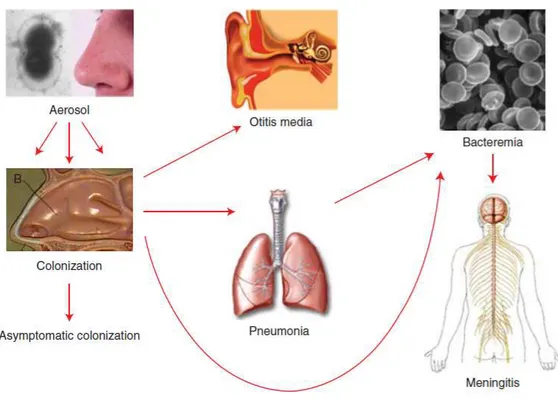
Streptococcus pneumoniae is a Gram-positive, encapsulated diplococcus. The polysaccharide capsule of this bacterium is an essential virulence factor and there are determined >90 distinct types of pneumococcal serotypes based on differences in the composition of this capsule. Generally, the immune system after infection is specific to the serotype, but cross-protection may occur between related serotypes. Pneumococcal diseases range from mild respiratory tract infections such as otitis media and sinusitis to more serious illnesses such as pneumonia, septicemia and meningitis. Although pneumococcus can cause lethal diseases, it is more commonly a quiescent colonizer of the upper respiratory tract139. Pneumococcal infections are thought to spread from person to person thought droplets/aerosols and nasopharyngeal colonization is a prerequisite for pneumococcal disease. The bacteria enter the nasal cavity and stick to the nasopharyngeal epithelial cells and may therefore remain as a colonizer or further spread to other organs, such as the ears, sinuses, or the bronchi to the lungs and then potentially penetrate the mucous barrier to enter the bloodstream and/or cross the blood–brain barrier to cause meningitis (Figure 7). In these last cases we talk about IPD (Invasive Pneumococcal Disease). IPD is commonly defined as morbidity associated
27
with the isolation of pneumococci from a normally sterile body site, such as the bloodstream, or secondary blood flow diffusion, e.g. meningitis or septic arthritis; it does not include sites such as the middle ear that are infected by contiguous diffusion from nasopharynx.
Fig. 7: Colonization of S. pnuemoniae
3.1
3.1
3.1
3.1 Epidemiology
Epidemiology
Epidemiology
Epidemiology
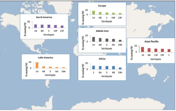
The distribution of serotypes that cause disease varies by age, disease syndrome, disease severity, geographic region, and over time. In young children and immunoompromised hosts, typically less immunogenic serotypes (e.g. 6, 14, 19 and 23) predominate. More invasive serotypes
28
(e.g. 1, 5, and 7) tend to infect individuals without co-morbidities140. Globally, seven serotypes (1, 5, 6A, 6B, 14, 19F and 23F) are the predominant strains causing IPD in most countries141, 142 (Figure 8), particularly in developing nations.
Fig. 8: Distribution of S. pneumoniae serotypes.
Pneumococcal infection is a major cause of morbidity and mortality worldwide. In 2005, WHO estimated that 1.6 million deaths were caused by this agent annually; this estimate included the deaths of 0.7–1 million children aged under 5 years. Disease rates and mortality are higher in developing than in industrialized settings, with the majority of deaths
29
occurring in Africa and Asia. It interests above all children >2 years. Case fatality rates (CFR) can be high for IPD, ranging up to 20% for septicaemia and 50% for meningitis in developing countries. Even in industrialized countries, the overall CFR for pneumococcal bacteraemia may reach 15%– 20% among adults and 30%–40% among elderly patients, despite appropriate antibiotic therapy and intensive care. Among meningitis survivors, long-term neurological sequelae such as hearing loss, mental retardation, motor abnormalities and seizures have been observed in frequencies as high as 58% of cases. In Europe and the USA, S. pneumoniae is the most common cause of community-acquired bacterial pneumonia in adults. In these regions, the annual incidence of invasive pneumococcal disease ranges from 10 to 100 cases per 100000 population. In Europe and the United States, S. pneumoniae is estimated to cause approximately 30– 50% of communityacquired pneumonia (CAP) requiring hospitalization in adults. Some of the differences could be explained by differences in case ascertainment and surveillance sensitivity, though incidence in Africa did appear to be generally higher than in Europe or North America. Pneumococcal middle-ear infection and sinusitis are less severe, but they are considerably more common health problems worldwide.
30
3.2
3.2
3.2
3.2 MICROBIOLOGY
MICROBIOLOGY
MICROBIOLOGY
MICROBIOLOGY
3.2 3.2 3.2
3.2.1 CAPSULE.1 CAPSULE.1 CAPSULE.1 CAPSULE
The pneumococcal surface is covered by a polysaccharide capsule that overlays the cell wall comprised of peptidoglycan and teichoic acid. Although the peptidoglycan has the classical gram-positive structure of N-acetylglucosamine, N-acetylmuramicacid, and a lysine-containing stem peptide, the teichoic acid is unusual in containing a ribitol phosphate backbone and covalently attached phosphorylcholine (PCho). The polysaccharide capsule is antiphagocytic and sterically hinders the access of leukocytes to complement fixed on the underlying cell wall.
3 3 3
3.2.2.2.2.2 PILI.2 PILI.2 PILI.2 PILI
Pili are multimeric filamentous surface structures composed of subunit proteins with LPxTG motifs recognized by sortases and attached to the cell wall. Two pathogenicity islets encoding pili, PI-1 and PI-2, are involved in adhesion143, 144. In particular, PI-1, encoded by the rlrA accessory region, has been shown to influence colonization, virulence, and the inflammatory response in mouse challenge models. This pilus is composed of three covalently attached structural proteins: RrgA, RrgB and RrgC.
31
3.2 3.2 3.2
3.2.3 SURFACE PROTEIN.3 SURFACE PROTEIN.3 SURFACE PROTEIN.3 SURFACE PROTEIN
Surface-exposed pneumococcal proteins are marked by one of three sequence motifs: the LPxTG cell wall anchor, a choline-binding domain, or a lipoprotein domain. Among the most important surface proteins we remember choline binding protein (Cbp) which play important roles in adherence. Between these, pneumococcal surface protein A (PspA) inhibits complement deposition and activation, binds lactoferrin, and interferes with uptake into phagocytes.
3.2 3.2 3.2
3.2.4.4.4.4 GENOMEGENOMEGENOME GENOME
The complete annotated 2.16 Mbps genome of S. pneumoniae encoding 2236 predicted proteins was first published in 2001145. Onethird of the predicted coding regions have no known function and a third of the genome varies sufficiently between strains to be considered to encode non core functions. With the ability to naturally take up DNA and undergo transformation, the genome is exceptionally highly variable. Besides, next generation sequencing has shown that pneumococci carried in the nasopharynx progressively accumulate recombination events indicative of the sequential transfer of DNA in vivo146.
32
3.3 COLONIZATION
3.3 COLONIZATION
3.3 COLONIZATION
3.3 COLONIZATION
When the bacteria enter the nasal cavity, the negatively charged capsule protects the bacteria from being trapped in the mucous and the bacteria can reach the epithelial surface. Many surface proteins affect nasopharyngeal colonization. A prominent example is CbpA that binds to the polymeric immunoglobulin receptor (pIgR) and promotes adherence and uptake of bacteria into nasopharyngeal cells147, 148, 149. Other adhesins include CbpG150, CbpD151, and LPxTG proteins such as the pilus proteins and IgA protease152. The formation of a biofilm is promoted by PsrP which mediates bacterial aggregation and intraspecies adherence. Enzymes, including NanA (neuraminidase), BgaA (b-glucosidase), and SrtH (b-N-glucosaminidase), affect colonization by cleaving terminal sugars from human glycoconjugates, thereby potentially exposing receptors for adherence. Many respiratory pathogens have converged on a common strategy to advance mucosal disease to bacteremia. This process is termed “innate invasion” and is counteracted by the very early participants induced in innate immunity153, 154. Control of bacterial multiplication in the lung requires innate immune recognition of the pneumococccal pathogen-associated molecular patterns (PAMPs) that initiate recruitment of neutrophils and the accumulation of hemorrhagic debris as the hallmark of pulmonary consolidation. Pneumococcal peptidoglycan components
33
induce signaling by TLR2 and LPS binding protein155, 156, PCho-bearing teichoic acid binds to PAFr157, and small intracellular peptidoglycan fragments are recognized by Nod2158, 159. Pneumolysin, a potent cytotoxin which plays an important role in the respiratory tract, lysing host cells but also by inhibiting the mucociliary beat of respiratory cells and separating epithelial cell tight junction, is detected byTLR4160. DNA is sensed by TLR9161. NF-κB signaling leads to the release of cytokines, particularly IL-6 and IL-1β, which then induces neutrophil recruitment and macrophage activation in the lung. Although required for clearance of bacteria, the intense inflammation induced by neutrophils contributes to lung damage; neutropenia (although not a complete absence of neutrophils) may attenuate damage and improve outcome162.
3.
3.
3.
3.4 PNEUMOCOCCAL VACCINES
4 PNEUMOCOCCAL VACCINES
4 PNEUMOCOCCAL VACCINES
4 PNEUMOCOCCAL VACCINES
A 23-valent pneumococcal polysaccharide vaccine (PPSV23) was developed later, in 1983, to provide protection against 80 to 90% of the pneumococcal capsular serotypes causing disease. The current PPSV23 formulation contains the following capsular serotypes: 1, 2, 3, 4, 5, 6B, 7F, 8, 9N, 9V, 10A, 11A, 12F, 14, 15B, 17F, 18C, 19F, 19A, 20, 22F, 23F, and 33F. This vaccine’s efficacy against pneumococcal infections with serotypes
34
contained in the vaccine in immunocompetent persons was 65%. Although the rate of invasive pneumococcal disease was reduced in PPSV23-immunized adults, the overall pneumococcal carriage rate was not reduced163. Unfortunately, the vaccine did not generate an immune response in the group with the highest rate of pneumococcal disease burden, children younger than 2 years of age. The antibody response generated by PPSV23 is T-cell independent due to the fact that the repeating subunits of the capsular polysaccharide can stimulate an immune response in B-cells independent of T-cell help164. The theory is that their inability to mount a T-cell–independent immune response until this age prevented the vaccine from generating protective antibodies165. In order to elicit a protective immune response in children under the age of 2, vaccines with the capsular polysaccharide conjugated to diphtheria toxin were developed. These conjugated antigens generated a T-cell–dependent antibody response that was effective in children under 2 years of age. The first 7-valent pneumococcal polysaccharide conjugate vaccine (PCV7) developed in 2002 greatly reduced the rate of infections in children under 2 years of age and in unimmunized individuals in the same community
166-168
. The second conjugate vaccine that was developed, the 13-valent pneumococcal conjugate vaccine (PCV13), contained the 7 serotypes in PCV7, 5 serotypes found in PPSV23, and 1 unique serotype found in neither PPSV23 nor PCV7, serotype 6A.
35
Despite the many benefits of the new vaccination methodologies, let us not forget that S. pneumonie, as mentioned above, still affects millions today. In fact, approximately 3 million cases of pneumonia per year are estimated in Europe, of which 1 million hospital admissions. In 2013, there were more than 120,000 (9,000 in Italy only) pneumonia deaths (of which 86% in the age range of 55-94 years), four times more than those caused by road accidents. Nevertheless, most people at risk do not know or do not care about how to prevent the disease. Besides, pneumococcal vaccination, however, may be a two-edge sword. The initial benefits in disease reduction may be followed by a steady increase in IPD incidence caused by non-included strains. All of these reasons lead us to stress the importance to study the immune mechanisms involved in the etiopathogenesis of these infections. This is still a key objective of public health and this is what we have set ourselves to focus our attention on the study of the role of endosomal TLR, still not well-known in S. pneuomiae infections.
36
4. TLRs in
4. TLRs in
4. TLRs in
4. TLRs in
S. p
S. p
S. p
S. pnuemoniae
nuemoniae
nuemoniae
nuemoniae
infections
infections
infections
infections
TLR2 involvement in S. pneumoniae infections is known in the literature as it seems to be responsible for the recognition of peptidoglycan components and LPS binding protein. TLR2 is responsible for most CW inflammatory signaling169 and its interaction with S. Pneumoniae results in platelet activation that is likely to contribute to the thrombotic complications of sepsis170. It is so important the function performed by this TLR during this infection that has been shown to be lacking in it results in higher mortality of bacterial meningitis by impaired host resistance171. TLR2 signaling is critical for bacterial clearance and macrophage recruitment during S. pneumoniae infection172.
TLR4 it is known to recognize pneumolysin, a cholesterol-dependent cytolysin released by S. pneumoniae. Indeed, mice lacking functional TLR4 are significantly more susceptible to invasive disease after colonization with a virulent, pneumolysin-producing type-3 strain of S. pneumoniae173. It was shown that induction of endotoxin tolerance, despite reducing cytokine production, improves host defense against infection with a virulent strain of S. pneumoniae. Besides, TLR4 induces CCL2 and CCL5 production174. For these reasons, TLR4 agonist activity can therefore be
37
potentially exploited to provide short-term resistance to infectious challenge. More recently, however, it has been shown that pneumolysin can activate the NLRP3 inflammasome and can promotes proinflammatory cytokines independently of TLR4175 and that, although purified PLN is recognized by TLR4 in vitro, PLN elicits lung inflammation in vivo by mechanisms that may involve multiple TLRs176.
Regarding the role of endosomal TLRs in S. pneumoniae infections, little is known. For TLR3 is known that the released pneumococcal RNA activates TLR3 and TRIF, which subsequently leads to IL-12 expression and secretion177. Furthermore, it has been shown that Poly IC:LC, a potent agonist of TLR3, significantly increases the survival of mice infected with S. pneumoniae178. It was shown also that TLR3 mRNA expression levels are increased in murine neuronal cells during infection with S. pneumoniae179. Almost nothing is known about the role of TLR7 in S. pneumoniae infections. It is known instead of it phagosomal bacteria such as group B streptococcus, potently induced interferon in conventional dendritic cells by a mechanism that required Toll-like receptor 7, the adaptor MyD88 and the transcription factor IRF1, all of which localized together with bacterial products in degradative vacuoles bearing lysosomal markers. Thus, this cell type-specific recognition pathway links lysosomal recognition of bacterial RNA with a robust, host-protective interferon response180. This may lead
38
us to think that a very similar mechanism of bacterial RNA recognition by TLR7 can also be carried out during S. pneumoniae infections.
It is known that S. pneumoniae meningitis led to an enhanced expression of TLR9 mRNA179. Besides, it's shown that TLR9–/– mice are more susceptible to pneumococcal infection and that TLR9 is not involved in the control of nasopharyngeal colonization but is crucial for bacterial clearance from the lower respiratory tract. Furthermore, TLR9 is not required for local cytokine production and immune cell infiltration during pneumococcola infection161. It also seems to cooperate synergistically with TLR2 and TLR4 in response to S. pnuemoniae181. It's known also that if a TLR9 agonist is added to the PCV (polysaccharide conjugate vaccine) of S. Pneumoniae, this greatly improves the immune response. Finally, it appears that TLR9 is responsible for cardiac inflammation during S. Pneumoniae infection and, specifically, as a result of sepsis. ROS produced during the inflammation process would cause mitochondrial DNA damage, these mtDNA fragments would be captured by the TLR9 and trigger the signal transduction pathway182.
TLR13 receptor, is the major expressed TLR in mouse coinfection models (Human metapneumovirus + S. pneumoniae and Influenza A Virus + S. pnemoniae)183. In murine macrophages and DCs, the TLR13 endosomal receptor has recently been identified as a sensor for bacterial RNA that
39
specifically recognizes a conserved region within bacterial 23S rRNA. Recognition by TLR13 can be masked by N6-dimetation at a critical adenosine residue (A2058, E. coli) within the target sequence129. This modification also renders bacteria resistant to macrolide antibiotics184, 185. On the basis of these assumptionst, it was shown that a naturally occurring A2058G point mutation in S. pneumoniae 23S rRNA abolishes TLR13 stimulatory capacity as efficiently as N6-dimetation at this residue. Despite the striking effect of 23S rRNA A2058 modification on immunostimulation in murine cells, there is a clear evidence that these alterations do not affect the activation of human monocyte138. But this function could be done by the human homologue TLR8. Besides, we can say that the role of TLR13 has been more studied in other types of streptococcal infections. Indeed, it was shown that TLR13 is the main macrophage receptor involved in the recognition of streptococci in resident tissue macrophages186. Besides, TLR13 participates in GBS recognition, although blockade of the function of this receptor can be compensated by other endosomal TLRs (in particular TLR2, TLR7 and TLR9)135.
40
5
5
5
5. MATERIALS AND METHODS
. MATERIALS AND METHODS
. MATERIALS AND METHODS
. MATERIALS AND METHODS
5.1
5.1
5.1
5.1 Mice
Mice
Mice
Mice
C57BL/6 wild-type (WT), used as controls, and IL-1R-/- mice were purchased from Charles River Laboratories. Mice lacking MyD88 or TLR2, TLR7 and TLR9 were originally obtained from S. Akira (Osaka University, Japan). Heterozygous TLR13-/+ mice were provided by the KOMP Repository (www.komp.org) and the Mouse Biology Program (www.mousebiology.org) at the University of California Davis. Subsequently, TLR13-/- mice were generated in our laboratoty for carrying out research project189. 3D mutant mice, bearing the H412R mutation of UNC93B1, were obtained from Bruce Beutler (University of Texas Southwestern Medical Center, TX). TLR7, 9, 13−/− triple-knockout mice were generated in our animal facility by crossing TLR9−/− with TLR7−/− and TLR13−/− mice using the genotyping methods described below. All KO mice, bred on a C57BL/6J background, were born and developed normally in the animal facilities of the Department of Human Pathology of the University of Messina. All mice used in the present study were housed under specific pathogen-free conditions in individually ventilated cages at these animal facilities.
41
5.2
5.2
5.2
5.2 Genotiping
Genotiping
Genotiping
Genotiping TLR7/9/13
TLR7/9/13
TLR7/9/13
TLR7/9/13
----////----mice
mice
mice
mice
As already mentioned, TLR7, 9, 13−/− triple-knockout mice were generated in our animal facility by crossing TLR9−/− with TLR7−/− and TLR13−/− mice. To create these triple KO we had to genotypize every single generation of mice. DNA was extracted from mouse tail with "DNeasy Blood & Tissue Kits" (Qiagen).
The protocols and sequences of primers used for genotyping were as follows:
TLR13 WT primer (WT band lenght: 202 bp) PR: TTGTGGCACCGTTTATTTCCCATC PF : AGGAACATTGCATGTGGG KO primer
(KO band lenght 400 bp)
PR: TACACAAGTGCAGTCTCCCATGACC
PF : GCAGCCTCTGTTCCACATACACTTCA
THERMAL PROTOCOL MIX (TOTAL VOLUME: 25 µL)
94 °C 5 min. Buffer (1X)1 2,5 µL 94 ° C 15 sec. 10 cycles dNTPs1 0,2 µL 65 ° C 30 sec. PR 2 1 µL 72 ° C 40 sec. PF 2 1 µL 94 ° C 15 sec. 30 cycles Taq polymerase1 0,5 µL 55 ° C 30 sec. dH2O 17,8 µL 72 ° C 40 sec. DNA 2 µL
42 TLR7 WT primer (WT band lenght: 1200 bp) PF: ACGTGATTGTGGCGGTCAGAGGATAAC Pko : ATCGCCTTCTATCGCCTTCTTGACGAG KO primer
(KO band lenght 1200 bp)
PR: CCAGATACATCGCCTACCTACTAGACC
PKO : ATCGCCTTCTATCGCCTTCTTGACGAG
THERMAL PROTOCOL MIX (TOTAL VOLUME: 25 µL)
94 °C 5 min. Buffer (1X)1 2,5 µL
94 ° C 45 sec.
35 cycles
dNTPs1 0,2 µL
67 ° C 1min and 30 sec. PR
2
0,5 µL
72 ° C 1 min. PF
2
0,5 µL 72° C 10 min. Taq polymerase1 0,5 µL
dH2O 18,8 µL DNA 2 µL TOTAL VOLUME 25 µL TLR9 WT primer (WT band lenght: 1200 bp) PF: GAAGGTTCTGGGCTCAATGGTCATGTG Pko : GCAATGGAAAGGACTGTCCACTTTGTG KO primer
(KO band lenght 1200 bp)
PR: ATCGCCTTCTATCGCCTTCTTGACGAG
PKO : GCAATGGAAAGGACTGTCCACTTTGTG
THERMAL PROTOCOL MIX (TOTAL VOLUME: 25 µL)
94 °C 5 min. Buffer (1X)1 2,5 µL
94 ° C 30 sec.
35 cycles
dNTPs1 0,2 µL
67 ° C 1min and 30 sec. PR
2
43
72 ° C 1 min and 30 sec. PF 2
0,5 µL 72° C 10 min. Taq polymerase1 0,5 µL
dH2O 18,8 µL
DNA 2 µL
TOTAL VOLUME 25 µL
PCR products were then passed on agarose gel at the appropriate percentage.
1. VWR International 2. Thermo Fisher Scientific.
5.3
5.3
5.3
5.3
S.
S.
S. pneumoniae
S.
pneumoniae
pneumoniae
pneumoniae
strain and murine inf
strain and murine infection
strain and murine inf
strain and murine inf
ection
ection model
ection
model
model
modelssss
All studies were performed in strict accordance with the European Union guidelines for the use of laboratory animals. The procedures were approved by the Ethics Committee of the University of Messina (OPBA) and by the Ministero della Salute of Italy.
S. pneumoniae strain D39 was grown to the mid-log phase in Todd-Hewitt broth (Oxoid) with 1% FCS, washed three times in nonpyrogenic PBS [0.01 M phosphate and 0.15 M NaCl (pH 7.2)], and diluted to the appropriate concentration. The number of viable bacteria used in each experiment was carefully determined by plate counting.
Six-weak-old female mice were anesthetized intraperitoneally with zolazepam + tiletamina (0,1 mg/mouse) and xilazina (0,16 mg/mouse) and injected intranasal with 7.5 x 10⁷ CFU of S. pneumoniae. In lethality
44
experiments mice were observed for 10 days every 12 hours. For cytokine measurement mice were sacrificed at different times after infection and organ target have been taken, homogenized in 2 ml sterile PBS and centrifuged. The cytokines were then dosed on the supernatants.
5.4 Cytokine measurement
5.4 Cytokine measurement
5.4 Cytokine measurement
5.4 Cytokine measurement
IL-1β, TNF-α, CXCL1 and CXCL2 levels were determined, according to the manufacturer’s recommendations, using the following ELISA kit: mouse IL-1β/IL-1F2, mouse TNF-α, mouse CXCL1/KC and mouse CXCL2/MIP-2. All kits were purchased from R&D system. The lower detection limits of all these assays were 16 pg/ml.
5.5 Bone
5.5 Bone
5.5 Bone
5.5 Bone----marrow derived cells
marrow derived cells
marrow derived cells
marrow derived cells
Bone marrow-derived cells were obtained as described previously180. Briefly, after flushing murine femurs and tibiae of C57BL/6J and 3D mice, marrow cells were cultured for 6 to 7 days in RPMI 1640 supplemented with 10% heat-inactivated FCS, penicillin (50 IU/ml), and streptomycin (50 μg/ml). Medium was supplemented with either 100 ng/ml macrophage stimulating factor (M-CSF) or 20 ng/ml granulocyte-macrophage
colony-45
stimulating factor (GM-CSF) (both from PeproTech) to obtain bone marrow-derived macrophages (BMDMs) or conventional dendritic cells (BMDCs), respectively. On day 3, 10 ml fresh cytokine-supplemented culture medium was added to each petri dish.
BMDCs and BMDMs were seeded in 96-well plates at a concentration of 5 × 10⁵ cells/well in 200 μl of RPMI 1640 supplemented with 10% heat-inactivated FCS. Cells were then stimulated with different MOI (molteplicity of infection) of S. pneumoniae ranging from 5 to 20 and centrifugated for 10 min at 400 × g in order to facilitate bacterial adherence. After incubation at 37°C in 5% CO2 for 30 min, cells were incubated for 24 h in the presence of
penicillin (250 IU/ml) and streptomycin (250 μg/ml) in order to limit the growth of residual extracellular bacteria. At the end of the incubation, supernatants were then collected and stored at −20°C for ELISAs.
5.6
5.6
5.6
5.6 Neutrophil isolation
Neutrophil isolation
Neutrophil isolation
Neutrophil isolation
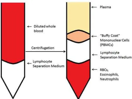
Neutrophils were obtained as described previously190 from the bone marrow of C57BL/6J and 3D mouse using Percoll density gradient centrifugation (Figure 9). Briefly, after removing the femurs and the tibias, a RPMI 1640 containing 10% FBS solution was forced through the bone with a syringe. For neutrophilic isolation, bone marrow cells were layered on the top of 62 and
46
81% at two layer of discontinuous gradient Percoll (GE Healthcare Life Sciences). After centrifugation at 1060 x g for 30 min at room temperature, PMN cells were collected from the two-layer interface, washed extensively in PBS to remove Percoll, and suspended in RPMI containing 10% (v/v) fetal calf serum (FCS). The viability of the PMN cells obtained through this procedure was normally > 95% as evaluated by trypan blue exclusion assay. Isolated cells (5 ± 0.6 × 10⁶ cells/mouse) were stained with May/Grunwald/Giemsa and 94 % of them were morphologically mature neutrophils (bands and segmented). Cells were stimulated with S. pneumoniae strain D39 for 24 and 48 h before supernatans were collected for cytokines determination.
Fig. 9: Neutrophils isolation with Percoll gradient
47
5.7 Peritoneal macrophages
5.7 Peritoneal macrophages
5.7 Peritoneal macrophages
5.7 Peritoneal macrophages
Resident mouse peritoneal macrophages were isolated from the peritoneal cavity by washing with ice PBS as previously described191. Briefly, after centrifugation, cells were resuspended in RPMI 1640 (Euroclone) supplemented with 5% heat-inactivated fetal calf serum, 50 IU of penicillin/ml, and 50 µg/ml of streptomycin. Cells were then seeded in wells of 96-well plates at a density of 5 × 10⁵/well and incubated at 37°C in a 5% humidified CO2 environment. After 24 h, the nonadherent cells were removed
by washing with medium. Adherent cells were stimulated with increasing multiplicities of infection (MOI, 1, 5, and 10 µg/ml) of S. pneumoniae. All infections were performed by centrifuging bacteria on cell monolayers for 10 min to 400 × g to facilitate bacterial adhesion. The number of viable bacteria used in each experiment was carefully determined by the plate count. After incubation at 37°C in 5% CO2 for 25 minutes, monolayers were
incubated for 18 h in the presence of penicillin (250 IU/ml) and streptomycin (250 µg/ml) to limit the growth of extracellular residual bacteria. Cell culture supernatants were collected at 18 h after stimulation to measure cytokine levels.
For elicited macrophages, the isolation procedure is the same, with the only difference that 500 µl of thioglycolated to 4% are administered to the mouse
48
i.p. 3 days before the peritoneal washing to pre-activate peritoneal macrophages.
5.8 IFN
5.8 IFN
5.8 IFN
5.8 IFN----β mRNA analysis by quantitative real
mRNA analysis by quantitative real
mRNA analysis by quantitative real
mRNA analysis by quantitative real----time
time
time
time PCR
PCR
PCR
PCR
IFN-β mRNA was measured in 3D and C57BL/6J lung mice after different time of S. pneumoniae infection (1, 3 and 24 h). The mRNA was extracted with the RNeasy mini kit (Qiagen), following the manufacturer’s instructions, from a little piece of lung (10 ng). For the quantification of IFN-β mRNA, real-time quantitative RT-PCR assays were conducted, in duplicate, with an Applied Biosystems 7500 (Applied Biosystems), as described192. The RNA was retroscribed in cDNA with the cDNA iScript kit (Bio-Rad). PCR conditions were as follows: 50°C, 30 min; 95°C, 10 min; (95°C, 15 s; 60°C, 1 min) X 40 cycles. Real-time PCR data were normalized in each individual sample by the level of β-actin expression. Gene expression was measured by the comparative cycle threshold method (ΔΔCT) and was reported as the n-fold difference relative to the normalized expression of C57BL/6J and 3D. Primers and TaqMan MGB probes for β-actin and IFN-β have been previously described193, 194 and were purchased from Applied Biosystems.
49
5.9
5.9
5.9
5.9 Data expression and statistical significance
Data expression and statistical significance
Data expression and statistical significance
Data expression and statistical significance
Differences in cytokine levels and organ CFUs were assessed by two-way analysis of variance and the Bonferroni test. Survival data were analyzed with Kaplan–Meier survival plots followed by the log rank test (JMP Software; SAS Institute). When p values were lower than 0.05, differences were considered statistically significant.
50
6.
6.
6.
6. Results
Results
Results
Results
6.
6.
6.
6.1111 MyD88
MyD88
MyD88
MyD88
----///----/mice are extremely susceptible to
mice are extremely susceptible to
mice are extremely susceptible to
mice are extremely susceptible to
S.
S.
S.
S.
pneumoniae
pneumoniae
pneumoniae
pneumoniae
infect
infections
infect
infect
ions
ions
ions
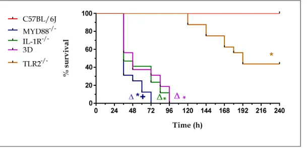
To investigate the role played by TLRs during pneumococcal pneumonia, we initially used a lung infection experimental model. We used genetically-deficient mice for MyD88, IL-1R, TLR-2, and 3D mice. As mentioned before, MyD88 is an adapter that is recruited both by IL-1R and TLRs following recognition of a PAMP. This makes these mice, from a phenotypic point of view, particularly susceptible to infections. In the initial phase of the experiments we tried to find out if Myd88-defective mice were more susceptible to pneumococcal infection and whether this increased sensitivity could be due to the inactivation of the TLR transduction pathway or to the involvement of inflammasoma (and hence of IL-1R route). TLR2-/- mice have been included in this experiment since the literature has already established the involvement of this receptor in S. pneumoniae infections as well as 3D mice that exhibit a mutation at the gene level encoding for UNC93B, a chaperon protein essential for the translocation of endosomal TLR at the endoplasmic reticulum level.
51
The mice were anesthetized intraperitoneally with zolazepam +tiletamina (0,1 mg/mouse) and xilazina (0,16 mg/mouse) and infected intranasally with 7.5x10⁷ CFU of S. pneumoniae, a sublethal dose. Mice were observed for 10 days every 12 hours and lethality was recorded. Two experiments of this type were performed with 8 mice per strain per experiment (Figure 10). The data obtained were then put together and provided the following results:
Fig. 10: Lethality of C57BL/6J, MyD88-/-, TLR2-/-, IL-1R-/- and 3D mice. Mice were infected intranasally with 7.5 x 10⁷ CFU of S. pneumoniae strain D39. Mice were observed twice a day for 10 days.
*, P < 0.05 versus WT mice as determined with Kaplan-Meier survival plots.
+, P < 0.05 versus 3D, IL-1R-/- and TLR2-/- mice as determined with Kaplan-Meier survival plots. ∆, P < 0.05 versus TLR2-/- mice as determined with Kaplan-Meier survival plots
As evidenced by Kaplan-Meier's lethality curves, we have a 100% mortality rate for Myd88-/- mice within 72 h of infection while the same percentage of mortality was observed for 3D and IL-1R mice within 96 h. These results suggest that the severe phenotype of Myd88-/- mice was due not only to