RES EAR CH
Open Access
How to resolve cryptic species of
polypores: an example in Fomes
Ursula Peintner
1*, Regina Kuhnert-Finkernagel
1, Viana Wille
1, Franco Biasioli
2, Anton Shiryaev
3and Claudia Perini
4Abstract
Species that cannot be easily distinguished based on morphology, but which form distinct phylogenetic lineages based on molecular markers, are often referred to as cryptic species. They have been proposed in a number of fungal genera, including the basidiomycete genus Fomes. The main aim of this work was to test new methods for species delimitation in cryptic lineages of polypores, and to define useful characters for species identification. A detailed examination of a number of different Fomes strains that had been collected and isolated from different habitats in Italy and Austria confirmed the presence of distinct lineages in the Fomes fomentarius clade. Our zero hypothesis was that the Mediterranean strains growing on Quercus represent a species which can be delimited based on morphological and physiological characters when they are evaluated in statistically relevant numbers. This hypothesis was tested based on phylogenetic analysis of the rDNA ITS region, morphological characters of
basidiomes and pure cultures, growth rates and optimum growth temperature experiments, mycelial confrontation tests, enzyme activity tests and volatile organic compound (VOC) production. The Mediterranean lineage can unambiguously be delimited from F. fomentarius. A syntype of an obscure and previously synonymized name, Polyporus inzengae, represents the Mediterranean lineage that we recognize as Fomes inzengae, a distinct species. The rDNA ITS region is useful for delimitation of Fomes species. Moreover, also a variety of morphological characters including hymenophore pore size, basidiospore size, and diameter of skeletal hyphae are useful delimiting
characters. The ecology is also very important, because the plant host appears to be a central factor driving speciation. Physiological characters turned also out to be species-specific, e.g. daily mycelial growth rates or the temperature range of pure cultures. The production of VOCs can be considered as a very promising tool for fast and reliable species delimitation in the future.
Keywords: Wood-degrading polypores, Volatile organic compounds, Mycelial growth rates, Chemotaxonomy, Morphological character evaluation
INTRODUCTION
Fomes fomentariussensu lato (s. lat.) is thought to be a polypore taxon with a wide distribution in Europe, Asia, Africa, and North America. It is commonly known as the“tinder fungus”, “hoof fungus”, “tinder conk”, “tinder polypore”, or “Iceman’s fungus”. The 5000-year-old Ice-man probably used this polypore: to make and preserve fire, as a first aid kit, an insect repellent, or for spiritual purposes (Peintner et al. 1998; Pöder & Peintner1999). Besides the widespread and important use as tinder, F.
fomentarius was a valued medicinal polypore in
European traditional medicine. Its use as a styptic per-sisted throughout medieval times and it was prescribed as a remedy against dysmenorrhoea, haemorrhoids, and bladder disorders; the active substance being “fomitin” (Killermann 1938). Grienke et al. (2014) extensively reviewed the applications of F. fomentarius in traditional medicine and the current knowledge on its metabolite profile. Recent phylogenetic analyses based on multiple genetic markers indicated that F. fomentarius possibly contained cryptic species (Pristas et al.2013). Our earlier study also indicated that a European lineage could pos-sibly represent a separate species that could be differen-tiated based on growth characteristics and substrate differences (Dresch et al. 2015). The main aim of this work is to thoroughly investigate multiple vouchers and
© The Author(s). 2019 Open Access This article is distributed under the terms of the Creative Commons Attribution 4.0 International License (http://creativecommons.org/licenses/by/4.0/), which permits unrestricted use, distribution, and reproduction in any medium, provided you give appropriate credit to the original author(s) and the source, provide a link to the Creative Commons license, and indicate if changes were made. The Creative Commons Public Domain Dedication waiver (http://creativecommons.org/publicdomain/zero/1.0/) applies to the data made available in this article, unless otherwise stated. * Correspondence:[email protected]
1University Innsbruck, Institute of Microbiology, Technikerstr. 25, 6020
Innsbruck, Austria
strains of the Fomes fomentarius s. lat. lineage in order to find meaningful and representative characters for the reliable distinction and differentiation of species repre-senting different lineages. Molecular phylogenetic ana-lysis, tests on growth characteristics, enzyme assays, and comparative analysis of volatile compounds, were carried out for this purpose. Moreover, we set high values on morphological characteristics of the basidiomes and of mycelia because they are crucial characters for an easy, fast and correct identification of fungal basidiomes. Our results clarify which methods and characters are most useful for distinguishing otherwise “cryptic” species in polypores.
MATERIALS AND METHODS
Sampling sites and environmental data
Fomes fomentarius s. lat. was sampled in different habi-tats in Austria (Tyrol) and Italy (Tuscany). Voucher num-bers, plant hosts, as well as habitat are given in Table1.
Sampling sites, basidiome morphology, and ecology (substrate) were documented in situ before collecting the basidiomes. Colours were documented based on the
colour code of Cailleux (1986). Basidiomes were
wrapped in greaseproof paper and transported to the la-boratory for isolation. Basidiomes where then dried at 40 °C on a mushroom dryer, and vouchers deposited in the mycological collection in IBF.
Isolation
Sterile techniques were used to obtain cultures from the context tissue of the basidiomes. Small pieces (2.0 mm3) were excised from each basidiome, plated on 2–3% w/v malt extract (MEA) agar plates and incubated for 1 to 3 weeks at 20 °C. Cultures were checked regularly for con-taminants. Mycelial plugs 1–3 mm diam were taken from the edge of the mycelium and transferred to new plates to establish pure cultures and carry out growth experiments.
The tissue cultures and stock cultures are maintained at the Institute of Microbiology, University of Innsbruck, Austria. For cryopreservation, small parts of well-growing cultures were overlaid with 10% skimmed milk and stored at− 80 °C. Isolates were also stored on MEA slants at 4 °C. DNA amplification and sequence analysis
Molecular identification of the fungal isolates was per-formed using the barcoding ITS regions of the ribosomal DNA. DNA amplification was carried out from Fomes pure culture isolates. A direct colony PCR was per-formed on pure cultures that were about 1 week old as previously described (Walch et al. 2016). Alternatively,
total genomic DNA was isolated from 100μg of fungal
matter (one-month-old mycelial cultures) by DNeasy® Plant Mini Kit (QIAGEN, Germany) according to the
manufacturer’s instructions and then eluted in 50 μl of sterile water. ITS-1, 5.8S rDNA and ITS-2 regions were amplified in a 50μl volume reaction containing 1–10 ng of genomic DNA, using the primers pair ITS1 / ITS4, and the LSU was amplified with the primers NL1 / NL4 in a T gradient Thermal Cycler (primus 96; Peqlab, Germany) according to Peintner et al. (2001). PCR products were se-quenced by Microsynth AG (Switzerland) with all primers. Sequences were analysed using the Sequencher® software (version 5.2.3; Gene Codes, Ann Arbor, MI, USA).
As a first step, BLAST searches were conducted in GenBank (http://ncbi.nlm.nih.gov), and closely related sequences downloaded. Only a small part of identical se-quences were downloaded in order to cover geographical range and substrate preferences.
Alignment and phylogenetic analyses were carried out
with MEGA 6.0 (Tamura et al. 2011). The best
Max-imum Likelihood (ML) model was tested before carrying out a ML analysis. The analysis involved 60 nucleotide sequences. All positions with less than 90% site coverage were eliminated. There were 515 positions in the final dataset. Fomes fasciatus was used as outgroup. To evalu-ate branch robustness of trees, parsimony-based boot-strap analyses were applied. Bootboot-strap analyses were conducted Subtree-Pruning-Regrafting (SPR) algorithm level 5 in which the initial trees were obtained by the random addition of sequences (five replicates). For the BP search, all positions with less than 100% site coverage were eliminated.
Bayesian Inference in MrBayes 3.2.6 (Huelsenbeck and Ronquist 2001, Ronquist et al. 2012) was also used to test branch robustness. For prior probability settings, de-faults were kept. For the Markov Chain Monte Carlo (MCMC) analyses, four chains were run for 10 million generations, with trees being sampled every 5000 genera-tions. The analysis was stopped as the convergence diag-nostic (average standard deviation of split frequencies) was below 0.05 after 10 million generations. From the 20, 000 sampled trees (for each of the two runs) 25% were dis-carded as burn-in before summary statistics were calcu-lated (using sump and sumt commands). Diagnostic plots, as well as the convergence diagnostics EES (Estimated Sample Size; min ESS around 10 K) and PSRF (Potential Scale Reduction Factor; 1000 for all parameters), indicated stationarity. Trees were drawn using FigTree 1.4.3. The newly created sequences were submitted to GenBank (Table1).
Microscopical analysis
Vouchers and pure culture isolates (2% MEA) were ex-amined by means of standard microscopic techniques in 3% KOH, water, Melzer’s reagent, Congo red, and Cot-ton blue. Microscopic documentation and measurements were made with a Nikon NS Fi1 camera and the
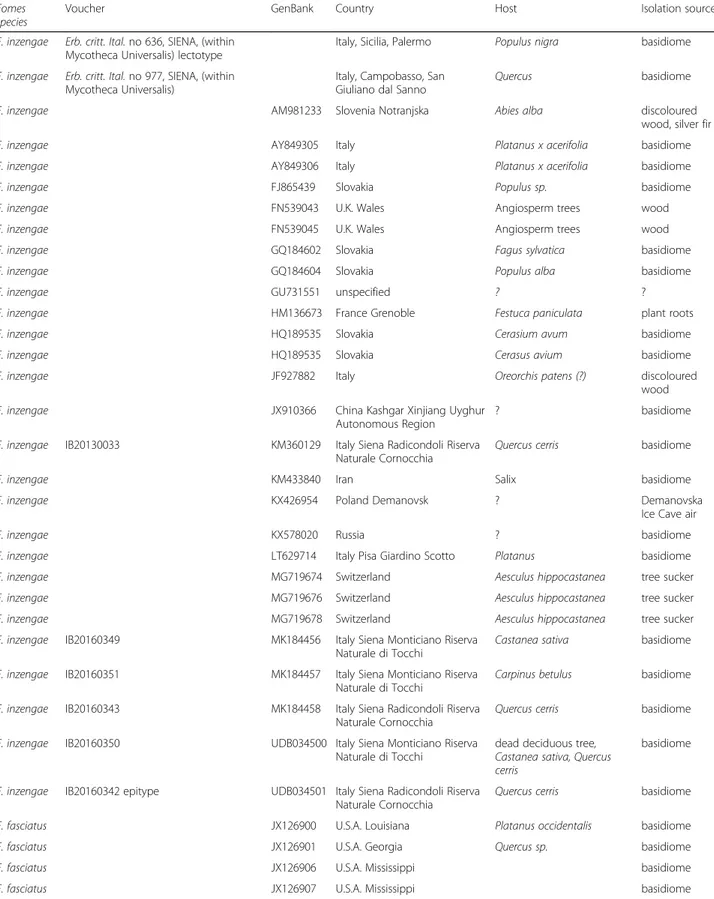
Table 1 Fomes sequences included in the phylogenetic analysis with information on the species identification, the newly
sequenced voucher, the GenBank Accession number, and available information on geographic provenance as well as on host plant and isolation source. Sorted based on GenBank Accession number within clades
Fomes species
Voucher GenBank Country Host Isolation source
F. inzengae Erb. critt. Ital. no 636, SIENA, (within Mycotheca Universalis) lectotype
Italy, Sicilia, Palermo Populus nigra basidiome F. inzengae Erb. critt. Ital. no 977, SIENA, (within
Mycotheca Universalis)
Italy, Campobasso, San Giuliano dal Sanno
Quercus basidiome
F. inzengae AM981233 Slovenia Notranjska Abies alba discoloured
wood, silver fir
F. inzengae AY849305 Italy Platanus x acerifolia basidiome
F. inzengae AY849306 Italy Platanus x acerifolia basidiome
F. inzengae FJ865439 Slovakia Populus sp. basidiome
F. inzengae FN539043 U.K. Wales Angiosperm trees wood
F. inzengae FN539045 U.K. Wales Angiosperm trees wood
F. inzengae GQ184602 Slovakia Fagus sylvatica basidiome
F. inzengae GQ184604 Slovakia Populus alba basidiome
F. inzengae GU731551 unspecified ? ?
F. inzengae HM136673 France Grenoble Festuca paniculata plant roots
F. inzengae HQ189535 Slovakia Cerasium avum basidiome
F. inzengae HQ189535 Slovakia Cerasus avium basidiome
F. inzengae JF927882 Italy Oreorchis patens (?) discoloured
wood F. inzengae JX910366 China Kashgar Xinjiang Uyghur
Autonomous Region
? basidiome
F. inzengae IB20130033 KM360129 Italy Siena Radicondoli Riserva Naturale Cornocchia
Quercus cerris basidiome
F. inzengae KM433840 Iran Salix basidiome
F. inzengae KX426954 Poland Demanovsk ? Demanovska
Ice Cave air
F. inzengae KX578020 Russia ? basidiome
F. inzengae LT629714 Italy Pisa Giardino Scotto Platanus basidiome F. inzengae MG719674 Switzerland Aesculus hippocastanea tree sucker F. inzengae MG719676 Switzerland Aesculus hippocastanea tree sucker F. inzengae MG719678 Switzerland Aesculus hippocastanea tree sucker F. inzengae IB20160349 MK184456 Italy Siena Monticiano Riserva
Naturale di Tocchi
Castanea sativa basidiome F. inzengae IB20160351 MK184457 Italy Siena Monticiano Riserva
Naturale di Tocchi
Carpinus betulus basidiome F. inzengae IB20160343 MK184458 Italy Siena Radicondoli Riserva
Naturale Cornocchia
Quercus cerris basidiome F. inzengae IB20160350 UDB034500 Italy Siena Monticiano Riserva
Naturale di Tocchi
dead deciduous tree, Castanea sativa, Quercus cerris
basidiome
F. inzengae IB20160342 epitype UDB034501 Italy Siena Radicondoli Riserva Naturale Cornocchia
Quercus cerris basidiome F. fasciatus JX126900 U.S.A. Louisiana Platanus occidentalis basidiome
F. fasciatus JX126901 U.S.A. Georgia Quercus sp. basidiome
F. fasciatus JX126906 U.S.A. Mississippi basidiome
Table 1 Fomes sequences included in the phylogenetic analysis with information on the species identification, the newly
sequenced voucher, the GenBank Accession number, and available information on geographic provenance as well as on host plant and isolation source. Sorted based on GenBank Accession number within clades (Continued)
Fomes species
Voucher GenBank Country Host Isolation source
F. fasciatus JX126908 U.S.A. Mississippi basidiome
F. fomentarius
EF155492 Germany Fagus sylvatica wood F.
fomentarius
EF155493 Germany Fagus sylvatica wood F.
fomentarius
EF155495 Germany Fagus sylvatica wood F.
fomentarius
EU162056 Germany wood
F. fomentarius
FJ865440 Slovakia Acer negundo basidiome F.
fomentarius
GQ184603 Slovakia Fagus sylvatica basidiome F.
fomentarius
GU062198 Latvia Alnus incana decayed wood F.
fomentarius
GU203514 unspecified basidiome
F. fomentarius
HQ189534 Slovakia Fagus sylvatica basidiome F.
fomentarius
JF927720 Poland ? ?
F. fomentarius
JQ901965 Russia Moscow region Populus sp. ? F.
fomentarius
JQ901966 Russia Moscow region Betula sp. ? F.
fomentarius
IB20130011 KM360125 Austria Tyrol Innsbruck Picea abies stump basidiome F.
fomentarius
IB20130016 KM360126 Austria Tyrol Innsbruck Picea abies stump basidiome F.
fomentarius
IB20130019 Epitype KM360127 Austria Tyrol Zirl Fagus sylvatica basidiome F.
fomentarius
IB20130022 KM360128 Austria Tyrol Innsbruck Picea abies stump basidiome F.
fomentarius
KM396269 Austria Tyrol Betula sp. basidiome F.
fomentarius
IB20170012 MK184459 Austria Tyrol Achenkirch Fagus sylvatica basidiome F.
fomentarius
IB20140121 MK295658 Italy, Campania, Parco del Cilento
Fagus sylvatica basidiome
F. sp. Asia DQ513402 China ?
F. sp. Asia DQ513402 China Changbai Shan basidiome
F. sp. Asia EU273503 unspecified ?
F. sp. Asia JX290073 unspecified basidiome
F. sp. Asia KJ668550 South Korea Odaesan National Park
?
F. sp. Asia MH114657 China ?
F. sp._Iran MK050587 Iran Darab kola ?
F. fomentarius II
computer program NIS Elements 4.13. All measure-ments were made at 1000 fold magnification. At least 30 spores or hyphal elements were measured for statistical evaluation.
Colony growth temperature experiments
All strains were first cultivated on plates containing 25 mL Malt Extract Agar (3% MEA), in order to ensure the same starting conditions for all strains. After 7 d, four mycelia plugs (5 mm diam.) were taken 1 cm from the leading edge of the colony and transferred to the middle of plates of 9 cm diam containing 25 mL MEA. Plates were randomly placed into a plastic box, and incubated at seven different temperatures (10, 20, 25, 30, 32, 35, and 37 °C). Mean colony diameter (mm), minus the 5 mm plug, was measured after 2, 5, 7 and 10 d. The re-sults are expressed as means ± standard deviations of three parallel cultures.
Drop test for enzymatic activity
Drop tests were used to test for important enzymes of wood decaying fungi, especially for laccases, polyphenol oxidases, and peroxidases. Drop tests were carried out as described in Taylor (1974) with modifications (Gramss et al. 1998). Test solutions were prepared as described by Gramss et al. (1998). Briefly, for the laccase test, 0.1
M α-naphthol was dissolved in 96% denatured ethanol;
with positive laccase reaction, the colour of the fungal tissue changes into blue or violet. For the phenol oxidase test, 2.5% gum guaiac was also dissolved in 96% dena-tured ethanol. When phenol oxidases like catechol oxi-dase, laccase and monophenol monooxygenase are present, the colour changes to very dark green. The per-oxidase test was carried out as pyrogallol(+) or pyrogal-lol(−) test: for the pyrogalpyrogal-lol(−) test, 0.5% pyrogallol diluted in water (w/w) was applied; for the pyrogallol(+) test, pyrogallol was supplemented with a drop of the
0.2% H2O2. Both pyrogallol tests formed a brownish
colour, when reacting with peroxidases. For the drop test, petri dishes containing one pure culture isolate growing for 10 d at 20 °C were used. Petri dishes were divided into four sections, each treated with one test. The colour reactions and their intensities were observed
and documented after 1, 3 h for α-naphthol and gum
guaiac, and after 24 h for pyrogallol. Mycelial confrontation tests
Mycelial confrontation tests were performed based on the heterokaryotic hyphae isolated from Fomes basidiomes. Two mycelial plugs were placed opposite to each other on an agar dishes containing 2% MEA. All possible combina-tions of the two F. fomentarius (IB20130019, IB2013022) and the Mediterranean (subsequently identified as F. inzengae) strains (IB20160349, IB20160351) were tested. Petri dishes were incubated at 25 °C for 6 d. Results of their compatibility were then documented photographic-ally and evaluated in four qualitative categories: very weak, weak, medium, strong interaction.
Analysis of volatile metabolites
Volatile compounds analysis was performed by a Proton Transfer Reaction Time of Flight Mass Spectrometer (PTR-TOF-MS; PTR-TOF 8000, Ionicon Analytik, Inns-bruck, Austria) according to the procedure described in Khomenko et al. (2017). Ensuing spectra were treated and analysed according to Cappellin et al. (2012).
One part of the samples was taken from the air-dried basidiome context in the area of the youngest pore layers. Samples were finely ground by an IKA mill under liquid nitrogen. From the resulting powder, 0.1 g was mixed with 3 mL milli Q water in closed glass vials and left for 6 h at 8 °C. The samples were then incubated at 40 °C for 30 min. and measured for 1 min.
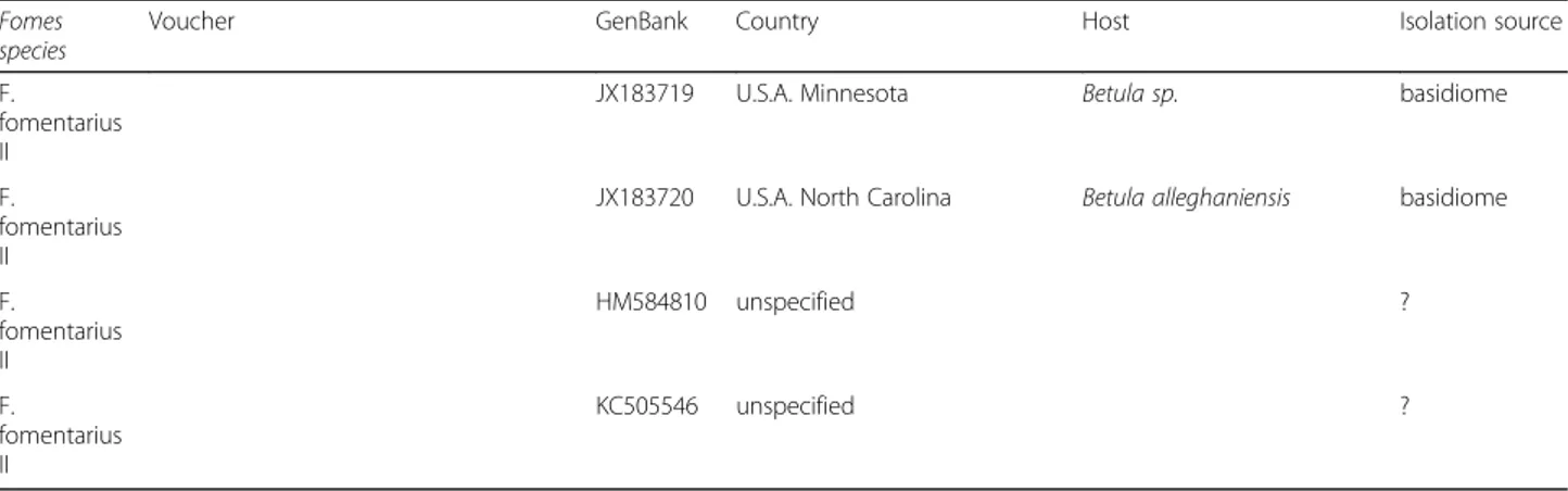
Table 1 Fomes sequences included in the phylogenetic analysis with information on the species identification, the newly
sequenced voucher, the GenBank Accession number, and available information on geographic provenance as well as on host plant and isolation source. Sorted based on GenBank Accession number within clades (Continued)
Fomes species
Voucher GenBank Country Host Isolation source
F. fomentarius II
JX183719 U.S.A. Minnesota Betula sp. basidiome
F. fomentarius II
JX183720 U.S.A. North Carolina Betula alleghaniensis basidiome
F. fomentarius II HM584810 unspecified ? F. fomentarius II KC505546 unspecified ?
Analysis was also performed on freeze-dried mycelial pure cultures grown for 3 wk. on MEA 3% at 25 °C. De-pending on the amount of harvested mycelium, between 7 and 11 mg were used for the analysis. The mycelium was soaked in 1 mL milli Q water in closed glass vials for 6 h at 8 °C. The samples were then incubated at 40 °C for 30 min. and measured for 1 min. This second analysis was carried out to test for a potential influence of the different types of wood substrates of the basidiomes.
Statistics
Data analysis was carried out with Statistica 9.1 (StatSoft 2010) for Windows 10. Data are given as arithmetic means with standard deviations. Variables were tested for normal distribution. Parameters with normal distribution were compared by t-tests (or Mann-Whitney U Test if data show no variance homogeneity). Differences in colony growth development after 5 d by different incubation tem-peratures were tested using the one-way ANOVA and Tukey HSD test. If parameters were not normally distrib-uted, the one-way ANOVA was replaced by the Kruskal-Wallis one-way analysis of variance on ranks. Significance value for all tests was p < 0.05. Unsupervised PCA (Princi-pal Component Analysis) and Kruskal-Wallis one-way analysis of variance on ranks of PTR-TOF-MS data were performed by R (R Core Team 2017).
RESULTS
Phylogenetic analysis
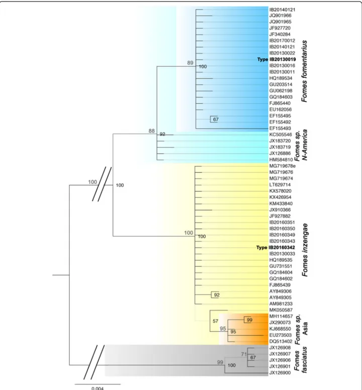
Phylogenetic analyses were performed with 60 rDNA ITS sequences obtained from our Fomes isolates and se-lected sequences currently available in public databases (GenBank). After a test for the best ML model, a Hasegawa-Kishino-Yano model was used for the ML ana-lysis. The ML tree with the highest log likelihood (− 1143.4536) is in accordance with the Bayesian tree (Fig.1). Bootstrap values were calculated with Maximum Parsi-mony (500 replicates), and the four most parsimonious trees (length = 83) were obtained with a Consistency Index of 0.951613, a Retention Index of 0.993890, and a Com-posite Index of 0.955663 for parsimony-informative sites.
The phylogenetic tree allows for the distinction of two well-supported major lineages within the F. fomentarius species complex in Europe, representing Fomes fomentar-iusand another species of Fomes. The four strains isolated from the alpine range fall within a clade of F. fomentarius sequences originating in Northern European countries (Russia, Poland, Latvia, Slovak Republic, Germany, Austria, Slovenia). Also, a strain from southern Italy grow-ing on Fagus falls in this clade (IB20140121). Typical plant substrates are Fagus sylvatica, Alnus spp., Acer negundo, and Picea abies. We consider this lineage as the Fomes fomentarius s. str. Lineage. It is sister to a clade from
North America growing on Betula spp., probably repre-senting another species of Fomes.
The sequences from the other European Fomes isolates cluster within a clade of Fomes sequences originating mostly from central to southern European countries (Italy, France, Portugal, Slovenia). In this case the plant sub-strates are Aesculus, Carpinus, Cerasium, Platanus, Popu-lus spp., Quercus spp., and Abies. This clade has a close relationship to a clade of Fomes from Asia that might rep-resent a fourth distinct species.
Internal clade sequence divergence was small, with 0– 3 base pair differences between the different strains of F. fomentarius s. str. (0.02%), and 0–1 f base pairs between the Mediterranean (F. inzengae) sequences (0.01%) (ITS1–5.8S-ITS2 region). Sequence divergence between the F. fomentarius s. str. and the F. inzengae clade was 9–18 base pairs (2.6%). Sequence divergence of the latter both to the outgroup F. fasciatus was 41–62 base pairs. Thus, pairwise distances confirm that F. fomentarius s. str. and F. inzengae can be considered as two distinct sis-ter taxa.
Phylogenetic analyses indicate a strong influence of the plant host substrate on speciation events in this genus of lignicolous, and opportunistically pathogenic basidiomycetes.
Pore diameter
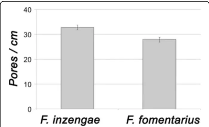
The basidiomes of F. fomentarius have 27–30 pores / cm (MW ± SD: 27.9 ± 0.9 pores / cm, n = 9), those of re-cently collected F. inzengae have 31–34 pores / cm (MW ± SD: 32.8 ± 0.9 pores / cm, n = 9). Thus, the F. inzengae strains produced significantly smaller pores than F. fomentarius (p = 0.000027, n = 9) (Fig. 2). The mean pore diameter of F. inzengae was 0.31 mm, and of F. fomentarius0.36 mm.
Basidiospore size
Basidiospores of F. inzengae are 9–12.5 × 3–4 μm (mean length = 10.8 ± SD = 0.9, mean width = 3.3 ± SD = 0.3, mean Q = 3.3 ± SD = 0.3, n = 37). This is smaller than the basidiospore size of 12–18 (− 20) × 4.0–7.0 μm as re-ported for F. fomentarius (Ryvarden & Gilbertson 1993, 1994), or as measured from our materials.
Mycelial characteristics in pure culture
Pure cultures of two strains, F. fomentarius IB20130016 and F. inzengae IB20160342, were comparatively investi-gated microscopically at all incubation temperatures. The best results were achieved with Congo red staining.
A typical trimitic hyphal system was constantly estab-lished at all temperatures by both strains: skeletal hy-phae, binding hyhy-phae, and generative hyphae with clamp connections, were always present, only varying in the composition of the three types of hyphae from strain to
strain and at different temperatures. At 32 °C and above, both strains formed inflated roundish terminal and
intercalary hyphal elements up to 10μm diam. Fomes
inzengaeformed these elements in greater quantities and more readily, already starting at 30 °C (Figs.3and4).
Differential characteristics of ground basidiomes
The powders resulting from ground basidiomes of F. fomentarius and F. inzengae could usually be differenti-ated by their consistency and pigmentation: the powder from F. fomentarius basidiomes was dark brown, and
Fig. 1 ITS-based Bayesian phylogeny of Fomes fomentarius s. lat rooted with F. fasciatus. Maximum Parsimony bootstrap values > 70% appear above the branches in grey. Bayesian probabilities > 65% appear in black, right of the respective node. Grey branches in the phylogeny are not supported. Fomes inzengae is strongly supported as a distinct species
arenaceous / granular, whereas that of F. inzengae basi-diomes was ochraceous brown and fluffy. However, there were also exceptions, such as a F. inzengae basi-diome that could not be unambiguously identified based on this character (Figs.3and4).
The basidiome powders also exhibited different behav-iours when mixed with water: the F. fomentarius powder floated, while that from F. inzengae swelled like a sponge.
Diameter of skeletal hyphae in pure culture and in basidiomes
The diameter of the skeletal hyphae was generally sig-nificantly different between F. fomentarius and F.
inzengae. In pure culture, the skeletal hyphae of F.
fomentarius ranged from 1.5–3.7 μm diam, and those
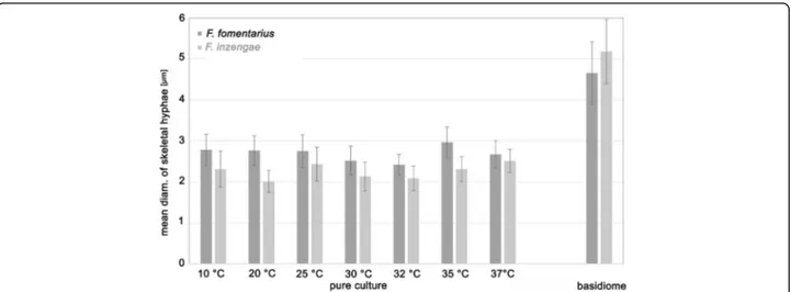
of F. inzengae from 1.3–3.5 μm. Through all tested temperatures, F. fomentarius had broader skeletal hyphae than F. inzengae. This difference was highly significant for the incubation temperatures 10, 20, 30, and 35 °C (p = 0.000000, n = 45 for each temperature) The diameter of the skeletal hyphae appears to be temperature-dependent in pure culture (Fig. 5).
The skeletal hyphae of the basidiomes were always signifi-cantly wider than ones produced in pure cultures. In the basidiomes, the diameter of F. fomentarius skeletal hyphae ranged from 3.0–6.4 μm, and those of F. inzengae from 3.2– 6.9μm. Thus, F. inzengae produced significantly wider skeletal hyphae in the basidiomes than F. fomentarius (p = 0.000027, nF.fom= 75, nF.inz= 90) (Fig.5). All Fomes strains
developed thicker skeletal hyphae in the harvested basidiomes than in pure cultures. Interestingly, the differ-ences between skeletal hyphae of the two species were al-ways significant but reversed: in harvested basidiomes F. inzengaehad wider skeletal hyphae than F. fomentarius, but in pure cultures F. inzengae had thinner ones than F. fomentarius.
Colony growth at different temperatures
All Fomes strains grew well at temperatures of 25–30 °C, and did not show any significant difference at these temper-atures. However, F. inzengae strains have a higher optimal temperature range of 30–32 °C. The performance of strains belonging to the two species at the other temperatures is clearly different: F. fomentarius strains grow significantly faster at 10 and 20 °C than the F. inzengae strains (10 °C: p= 0.018; 20 °C: p = 0.000010). At 25 °C, no significant dif-ference could be detected, but a slight tendency of the F. inzengae strains to growing larger colonies was observed. At higher temperatures (30–37 °C), the F. inzengae strains grew significantly faster (30 °C: p = 0.000000; 32 °C: p = 0.000000; 35 °C: p = 0.000002; 37 °C; p = 0.000000) com-pared to F. fomentarius (Table2, Fig.6).
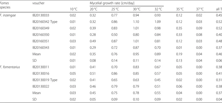
The mycelial growth rate per day was calculated for each isolate and the most relevant incubation tempera-tures (20, 25, 30, and 32 °C). This confirmed that F. fomentarius grows faster at 20 °C, and slower at 30 °C and 32 °C than F. inzengae strains. Strain properties ap-pear to be important, as some strains (e.g. F. inzengae IB20160342) grow extraordinarily fast, and others extra-ordinarily slow (F. fomentarius IB20130019) (Table2). Enzymatic activity
Laccase and phenol oxidase tests were always positive for all tested strains. Peroxidase tests gave ambiguous re-sults and were dependent on the age of the pure culture rather than on the particular strain.
Confrontation tests between heterokaryotic mycelia These were carried out at 25 °C as at that temperature there are no significant differences in growth rates be-tween the tested strains. When strains were tested against themselves, hyphal anastomoses were readily formed all over the confrontation zone (positive reac-tions). The strains tested (F. fomentarius IB20130019, IB20130022; F. inzengae IB20160349, IB20160351) did not show any kind of inhibition under the reflected-light microscope and grew easily into each other. However, when a strain was confronted with any other strain, the isolates formed distinct colony margins, and no anasto-moses were formed. Overall, the F. inzengae strains were more competitive than F. fomentarius strains at 25 °C, and F. fomentarius strains always exhibited reduced growth whenever they were matched with any other strain (Fig.7).
Volatile metabolites
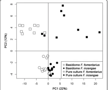
The PTR-TOF-MS dataset contained more than 300 mass peaks. Peaks with a concentration significantly higher than blanks were 232 for basidiome samples and 209 for pure culture samples. Data exploration by un-supervised PCA analysis of all samples (232 peaks) is
Fig. 2 Comparison of pore diameter (as pores / cm hymenophore surface) of Fomes inzengae and F. fomentarius. Pore diameter is significantly different (p = 0.000027, n = 9)
shown in Fig. 8. Different sample sets (basidiome and pure culture) are well separated by the second principal component. More interestingly, the first component indi-cates a certain separation of F. fomentarius from F. inzen-gaewhich is clearer for pure culture samples: despite the small amount of material used, freeze dried mycelial sam-ples provided a better resolution and separation. Based on a Kruskal-Wallis one-way analysis of variance, 91 mass peaks were significantly different between the pure culture samples of F. inzengae and F. fomentarius. Again, despite the larger amount of material available for the analysis, only 19 mass peaks were significantly different for the
basidiome samples. Figure9shows the concentration of a few selected compounds. Fomes inzengae is generally richer in VOCs than F. fomentarius, something true for many VOCs whose production is not dependent on the substrate such as some carbonyl compounds (Fig. 9, left and middle panels). However, as shown in data from nat-urally grown basidiomes, substrate or other environmental conditions result in differences in VOC production, as in the case of monoterpenes (Fig.9, right panels). Thus, the two Fomes species are producing species-specific volatile metabolites but the interaction with the substrate can mask this differences.
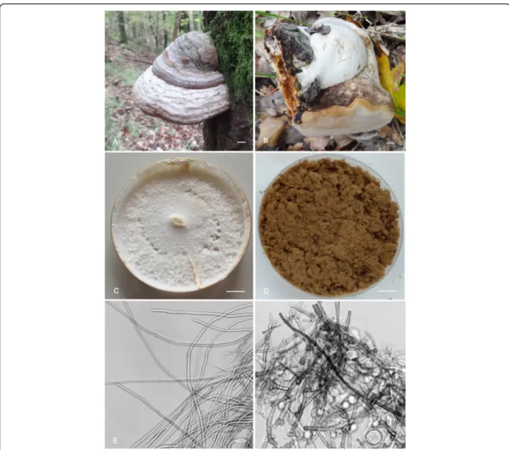
Fig. 3 Fomes inzengae. A. Basidiome of the lectotype (IB20160342) growing on Quercus cerris in the Natural Reserve of Cornocchia. B. Basidiome with new hymenophore formation (positive geotropical reaction) after falling of the host tree (IB20160343). C. Mycelia pure culture after 10 d on 3% MEA at 25 °C (IB20160342). D. Ground basidiome (IB20160342); note the ferruginous brown colour and fluffy consistence. E. Skeletal hyphae as formed after 5 d on 3% MEA at 37 °C (IB20160342). F. Inflated intercalar and terminal hyphal elements after 5 d at 37 °C, stained with Congo red (IB20160342). Bars A-D = 1 cm; E-F = 10μm
TAXONOMY
Fomes inzengae (Ces. & De Not.) Cooke, Grevillea 14 (69): 18 (1885).
Basionym: Polyporus inzengae Ces. & De Not., Erb. critt. Ital., ser. 1: no. 636 [typeset description on label with specimen] (1861).
Type: Italy: Sicilia: Palermo, on Populus dilatata, 1860– 1861, Inzenga [det. Cesati & De Notaris, Erb. critt. Ital., ser.1 no. 636 [intermixed with“Mycotheca Universalis”]
(SIENA – lectotypus hic designatus; IF556590); Prov.
Siena: Radicondoli, Riserva Naturale Cornocchia, on liv-ing Quercus cerris, 26 Oct. 2016, U. Peintner & C. Perini (IB20160342, epitypus hic designatus; IF556625). Diagnosis: Basidiomes macroscopically very similar to F. fomentarius from which it can be differentiated by the following characters: the pluriannual basidiomes have a hymenophore with 32–40 pores / cm; and the basidio-spores are (9.0–) 10–12 (− 12.5) x (2.8–) 3.0–3.5 (− 3.8), Q = (2.8–) 3.0–3.6 (− 3.7) μm.
Description: Basidiomes perennial, sessile, ungulate, tough, woody, to 20 cm wide. Upper surface quickly de-veloping a glabrous crust, grey (92LM) with a few dirty olivaceous spots (NP69), dull. Greyish coloured upper part the basidiome crust often conspicuously and irregu-larly marbled or brown-dotted. Marginal growth zone consisting of a distinctly zoned layer, zones 0.5–3 mm wide, in different shades of reddish brown (PR55), brown (NP67–69) or ochraceous brown (M70–71), minutely to-mentose; transitional zone between ochraceous brownish zonate margin and grey older crust sometimes conspicu-ous and darker brown. Pore surface concave, pale brown, pores circular, 31–34 (− 38) pores / cm, with thick to-mentose dissepiments. Tube layers indistinctly stratified, brown (PR59) and becoming stuffed; context tissue layer between the surface crust and the tubular layers, reddish brown (PR45), tough, azonate. Granular core developing at the upper part of the context, next to the substrate. Basidiospores cylindric, hyaline, smooth, not amyloid, (9.0) 10–12 (− 12.5) x (2.8–) 3.0–3.5 (− 3.8) μm, Q = (2.8-) 3.0–3.6 (− 3.7); n = 37; a large proportion ger-minate immediately. Basidia not observed. Cystidia not observed. Hyphal system trimitic, generative hyphae hya-line, thin-walled, with clamp connections, inconspicu-ous, 1.5–3.5 μm diam; Contextual skeletal hyphae thick-walled, non-septate, walls yellowish brown in KOH (3%), 3.2–6.9 μm diam, binding hyphae thick-walled, strongly branched, non-septate, 4.0–6.3 μm diam.
Cultures: Colonies reaching 4–6 cm diam after 5 d at 32 °C on 2% MEA; mycelium at first white, the cream to
orange pinkish buff, reverse cream to orange, with felty to cottony consistency and fluffy surface structure. Genera-tive hyphae with clamp connections, skeletal and binding hyphae readily formed, diam. of skeletal hyphae 1.3– 3.5μm, thick-walled, wall with yellow-ochraceous pig-ment. Inflated intercalary and terminal elements readily formed at temperatures of 32 °C and higher.
Habitat and distribution: On trunks of Quercus cerris, Q. pubescens, Castanea sativa, Carpinus betulus, Plata-nus acerifolia,and Populus spp., exceptionally also
Cera-sium avium and Abies alba. Based on sequences
deposited in public databases, it occurs in Italy, Slovakia, Slovenia, Switzerland, United Kingdom, France, China and Iran. It is likely to be present through the whole Mediterranean area on suitable hosts, but is often mis-identified as F. fomentarius (cfr, distribution of F. fomen-tariusshown in Bernicchia2005).
Nomenclature: Fomes inzengae has long been regarded as a synonym or form of F. fomentarius (Bondartsev 1953; Domański et al. 1967; Donk 1933, 1974; Lécuru et al. 2019; Pilát 1941; Saccardo 1881). The basionym Polyporus inzengae is based on material collected and documented by Giuseppe Inzenga, who sent his material to De Notaris for identification. Cesati and De Notaris published the name with a printed description as no. 636 (see Fig. 10) in Erbario Crittogamico Italiano (Soci-età crittogamologica italiana 1861; Sayre 1969), basing the description on the notes later reworked and twice published by Inzenga (1865, 1866) himself. Inzenga col-lected P. inzengae from Populus dilatata (now P. nigra) in Palermo (Italy, Sicily). The description from the pro-tologue and description and illustrations from Inzenga’s Funghi Sicilianiin black and white (Inzenga1865: 17, pl. 2 Fig. 1) and reproduced in colour (Inzenga 1866: pl. 7 Fig. 1), agree with our concept of the Mediterranean Fomes lineage. Donk (1933) believed this was a milky white form of F. fomentarius, and others in the 20th cen-tury followed.
The original basidiome collected by Inzenga was cut into slices and sent to various herbaria as parts of an exsiccatae set. One part of this original collection no. 636 was later inserted in another set, the Mycotheca
Universalis, conserved in Herbarium Universitatis
Senensis (SIENA). This collection is interpreted as a
syntype (cf. Wetzel and Williams 2018) and is here
selected as the lectotype for the name; all other parts deposited elsewhere are therefore now isolectotypes. Cooke (1885b) transferred the name to Fomes in a list that was a continuation of Fomes species started in a
previously published fascicle (Cooke 1885a) and is
considered to have done so validly (Turland et al. 2018: Art. 35.1 Ex. 5).
The lectotype of Fomes inzengae is damaged by insects, but important diagnostic characters can still be evalu-ated: the hymenophore has 33–40 pores / cm, and the diameter of the skeletal hyphae ranges from (3.4–) 4.5– 7.8 (− 10.0) μm (n = 30) with a mean value of 6.2 μm. A second collection of F. inzengae (Erb. critt. Ital. no. 977) collected in 1871 on Quercus (San Giuliano dal Sanno, Prov. Campobasso, Italy) has 32–38 pores / cm in the hymenium, and the skeletal hyphae range from 5.9 to 8.3 (− 9.4) μm. Unfortunately, we could not amplify DNA from these original collections of Fomes inzengae, and therefore we designate an epitype to fix the applica-tion of the name. Piccone (1876) recorded additional in-formation on the second collection by Pedicino noting
that it had also been included in Rabenhorst’s (1872) Fungi Europaei exsiccatino. 1508, which also consists of slices. Pedicino (1876) went on to record further obser-vations.
Comments: Fomes inzengae has considerably smaller ba-sidiospores than F. fomentarius. However, spores are dif-ficult to observe in many pluriannual polypores because they are formed either in small quantities or during spe-cial, restricted seasonal periods. Additional characters, which are always present, are therefore crucial to distin-guish these taxa: Fomes inzengae basidiomes can be sep-arated from those of F. fomentarius on hymenophore pore size, and the diameter of skeletal hyphae. Moreover,
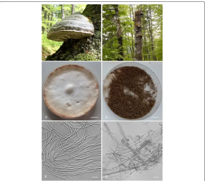
Fig. 4 Fomes fomentarius. A. Basidiome growing on Fagus sylvatica in Tyrol (Austria) (IB20170012). B. Several basidiomes growing on a dead stem of Fagus sylvatica. C. Mycelia pure culture (IB20130016) after 10 d on 3% MEA at 25 °C. D. Ground basidiome (IB20170012); note the dark brown colour and granular consistence. E. Generative hyphae with clamp connections (IB20130016) as formed after 5 d on 3% MEA at 30 °C. F. Inflated intercalary and terminal hyphal elements (IB20130016) after 5 d at 37 °C. Bars A, C-D = 1 cm; E-F = 10μm
substrate, growth rates, and volatile metabolites as well as pure culture characteristics help to distinguish these sister taxa. Barcoding rDNA ITS sequences are inform-ative for species distinction in Fomes.
Additional specimens examined: Italy: Prov. Siena: Radicondoli, Riserva Naturale Cornocchia, on living
tree of Quercus cerris, 29 Oct. 2013, M. N.
D’Aguanno (IB20130333); loc. cit., on Q. cerris, 26 Oct. 2016, C. Perini, R. Kuhnert-Finkernagel & U. Peintner (IB20160343); loc. cit., on living tree of Q. cerris, 1 Dec. 2017, C. Perini (IB20170300); Monti-ciano Riserva Naturale di Tocchi, on Castanea
sativa, 28 Oct. 2016, C. Perini, R.
Kuhnert-Finkernagel & U. Peintner (IB20160349); loc. cit., on dead deciduous tree, 28 Oct. 2016, C. Perini, R. Kuhnert-Finkernagel & U. Peintner (IB20160350); loc. cit., on Carpinus betulus, 28 Oct. 2016, C. Perini, R. Kuhnert-Finkernagel & U. Peintner (IB20160351); loc. cit., on Quercus cerris, 14 Jan. 2017, C. Perini (MSIENA8138); loc. cit., on living tree of Quercus pubescens, 14 Jan. 2017, C. Perini (MSIENA8062). Prov. Campobasso: San Giuliano dal Sanno, on
Quer-cus, Sep.1871, N. Pedicino (SIENA, Mycotheca Univ.,
Erb. critt. Ital. no. 977).
Fomes fomentarius (L.) Fr., Summa veg. Scand. 2: 321
(1849); nom. sanct. Syst. mycol. 1: 374 (1821)
Basionym: Boletus fomentarius L., Sp. Pl. 2: 1176 (1753). (Figs4,11)
Type: Bulliard, Herb. Fr. tab. 491 fig. II C–F (1791, sub
Boletus ungulatus Bull. (lectotypus hic designatus
IF556624) (Fig. 11). Austria: Tirol: Innsbruck, Magde-burger Hütte, alt. 1300 m, on living Fagus sylvatica, 20
Jul. 2013, K. Rosam & U. Peintner, (IB20130019,
epity-pus hic designatus, IF556623; GenBank KM360127
(ITS)).
Diagnosis: Fomes fomentarius basidiomes usually form on Fagus or Betula in boreal or temperate habitats. The pluriannual basidiomes have hymenophores with 27–30 pores / cm; the basidiospores are 12–18 × 4–7 μm. Description: Basidiomes perennial, sessile, ungulate, tough, woody, to 25 cm wide. Upper surface quickly de-veloping a glabrous greyish crust. Margin light brown, minutely tomentose; pore surface concave, pale brown, pores circular, 27–30 pores / cm, with thick tomentose dissepiments. Tube layers indistinctly stratified, reddish brown and becoming filled; context tissue a layer be-tween the surface crust and the tubular layers, yellowish brown, tough, azonate. Granular core developing at the upper part of the context next to the substrate. Basidio-spores cylindrical, hyaline, smooth, not amyloid, (12.5–) 13.5–18 (− 20.5) × 4.5–6.5 (− 7.5) μm, Q = (2.5–) 3.0–3.6 (− 3.5); n = 480. Usually produced in the spring in large quantities, difficult to observe during the rest of the year. Hyphal system trimitic, skeletal hyphae thick-walled, non-septate, with yellowish brown wall in 3% KOH, 3.0–
6.4μm diam, binding hyphae thick-walled, strongly
branched.
Pure cultures: Colonies reaching 2–4 cm diam after 5 d at 32 °C, mycelium first white, the cream to orange-pinkish buff, reverse cream to orange, with a velutinous-felty to cottony consistency. Generative hyphae with clamp connections, skeletal and binding hyphae readily formed, skeletal hyphae 1.5–3.7 μm diam, thick-walled,
Fig. 5 Diameter of skeletal hyphae in pure culture after 10 d incubation on 3% MEA at different temperatures and in naturally grown basidiomes. Differences between F. fomentarius and F. inzengae are always highly significant (p < 0.0001) with the exception of 37 °C (p < 0.05) (n = 45 for each temperature; n = 75 for F. fomentarius basidiomes; and n = 90 for F. inzengae)
wall with yellow-ochraceous pigment. Inflated inter calary and terminal elements formed at temperatures > 32 °C.
Habitat and distribution: In temperate habitats associated with Fagus sylvatica, and Betula spp., occasionally also with Picea abies, Acer negundo, Populus sp. or Alnus incana. Widely distributed in northern and central Eur-ope, including Latvia and Russia. In Russia also on Quer-cus. The records from Russia and Alaska (Betula neoalaskana) indicate a potential circumpolar distribution.
Occurring also in southern Europe on Fagus.
Comments: Fomes fomentarius s. str. is a temperate spe-cies with distinct morphological characters and host preference for Fagus and Betula, but in Russia it also grows on Populus and Quercus. The original diagnosis of Linné (1753) refers to a polypore growing on Betula. Fries (1821), in the sanctioning work, described the fungus as growing on Fagus. He also mentioned its use as tinder and as remedy against bleeding:“pro fomite aptissima. In hae-meragiis laudatus”. He also cited several illustrations, Table 2 Effects of temperature on mycelial growth (cm/day) of ten Fomes strains cultivated on 3% MEA. The mycelial growth rate per day [cm/day] was calculated for the first 7 days of incubation
Fomes species
voucher Mycelial growth rate [cm/day]
10 °C 20 °C 25 °C 30 °C 32 °C 35 °C 37 °C all T F. inzengae IB20130033 0.02 0.32 0.77 0.94 0.93 0.12 0.02 0.45 IB20160342 Type 0.01 0.32 0.86 1.10 1.09 0.12 0.03 0.52 IB20160349 0.02 0.39 0.83 1.01 0.98 0.35 0.09 0.52 IB20160350 0.01 0.28 0.50 0.80 0.84 0.33 0.08 0.40 IB20160351 0.03 0.49 0.87 1.01 0.81 0.12 0.03 0.48 IB20160343 0.01 0.29 0.72 0.87 0.70 0.01 0.00 0.37 Mean 0.02 0.35 0.76 0.95 0.89 0.19 0.04 0.46 SD 0.01 0.08 0.14 0.11 0.14 0.13 0.04 0.06 F. fomentarius IB20130011 0.01 0.41 0.70 0.83 0.67 0.05 0.00 0.38 IB20130016 0.05 0.51 0.86 0.85 0.57 0.05 0.00 0.41 IB20130019 Type 0.02 0.41 0.65 0.63 0.45 0.02 0.00 0.31 IB20130022 0.03 0.46 0.79 0.79 0.51 0.06 0.00 0.38 Mean 0.03 0.45 0.75 0.78 0.55 0.04 0.00 0.37 SD 0.02 0.05 0.09 0.10 0.09 0.02 0.00 0.04
Fig. 6 Mean colony diameter after 5 d on 3% MEA at different temperatures. F. inzengae grows significantly faster at temperatures of 30 °C and higher, but slower at 20 °C and below. With the exception of 25 °C, differences in growth rates between F. fomentarius and F. inzengae are always highly significant (p < 0.0001) (n = 45)
which can be used to select a lectotype as under Art. F.3.9 material cited in the protologue of a sanctioning work is treated as original material for the purposes of lectotypifi-cation. The illustration published by Bulliard (1791) was selected as lectotype here as it best represents the current concept of Fomes fomentarius. Moreover, it is easily avail-able online (https://doi.org/10.5962/bhl.title.5365). A epi-type is designated here in order to precisely fix the application of the name. We selected a collection from Austria on Fagus as epitype because all data are available
for this collection, including a pure culture.
Additional specimens examined: Austria: Tirol, Achen-kirch, Christlum, on Fagus, 26 Aug. 1991, U. Peintner (IB19910934); loc. cit., on Fagus, 21 May 2017, U.
Peint-ner (IB20170012); Gnadenwald, Gunggl, towards Maria
Larch, on Fagus, 1 May 1991, U. Peintner (IB19910047); Innsbruck, Hötting, alt. 817 m, on Fagus, 10 Jul. 2013, K. Rosam & U. Peintner(IB20130011, IB20130016); loc. cit., Stangensteig, alt. 820 m, on Picea, 25 Sep. 2013, K. Rosam & U. Peintner (IB20130022); Kärnten, Eberstein,
on Fagus sylvatica, 13 Jun. 1990, U. Peintner
(IB19901036). – Finland: Utsjoki, Kevo, Kevojokki, on dead Betula, 18 Aug. 1998, M. Moser (IB19980038). Sweden, Småland, Femsjö, Hägnan, Fagus, 21 Aug. 1976, M. Moser, IB19760143. – Italy: Corleto Monforte, Sal-erno, Parco Nazionale del Cilento e Vallo di Diano, 12 May 2008, Pecoraro (MSIENA8156); loc. cit., 12 May 2008, Pecoraro (MSIENA8157); loc. cit., 12 Nov. 2014, M. N. D’Aguanno (IB20140121). – Russia: Moskow
Ob-last: on Betula, 18 Oct. 2014, A. Shiryaev (SVER
926310); Sverdlovsk Oblast, Ekaterinburg City, on Betula, 4 Oct. 1978, N.T. Stepanova-Kartavenko (SVER 49614); loc. cit., Populus, 4 Aug. 1973, A. Sirko (SVER 10032); Orenburg Oblast, Orenburg State Nature Reserve, Popu-lus, 1 Oct. 2017, A.G. Shiryaev (SVER 926313); Volgo-grad Oblast, Volzhsky, Populus, 8 Oct. 2001, A.G. Shiryaev(SVER 420865); Novgorod Oblast, Ilmen, Popu-lus, 18 Aug. 1973, N.T. Stepanova-Kartavenko (SVER 229302); Smolensk Oblast, Dneper valley, Populus, 26 Sep. 2016, A.G. Shiryaev (SVER 867100); loc. cit., Vyazma, Quercus robur, 22. Aug. 1978, V. Ipolitov (SVER 155532); Samara Oblast, Zhiguli Nature Park, Q. robur, 10 Sep. 1983, F. Igorev (SVER 303495); Bashkiria: on
Fig. 7 Confrontations test of different isolates of Fomes fomentarius and F. inzengae after 6 d on MEA 3% at 25 °C. fomes inzengae is always growing faster and with a fluffier surface. F. inz49 = F. inzengae (IB20160349), F. inz51 = F. inzengae (IB20160351), F. fom19 = F. fomentarius (IB20130019), F. fom22 = F. fomentarius (IB20130022)
Fig. 8 VOC data exploration by unsupervised PCA analysis of all Fomes inzengae and F. fomentarius samples (232 peaks). Basidiome and pure culture samples are well separated by the second principal component (PC2 12.5%). Separation of F. inzengae from F.
fomentarius is more pronounced in pure culture samples than in basidiomes (PC1 22.9%)
Betula, 18 Aug. 1963, N.T. Stepanova-Kartavenko (SVER 19051); loc. cit., Nature Park Bashkiria, Q. robur, 19 Aug. 2012, A.G. Shiryaev (SVER 926313); Krasnodar Krai, on Betula, 5 Oct 1975, N.T. Stepanova-Kartavenko (SVER 22302); Perm Krai, Solikamsk, Populus, 23 Sep. 1999,
A.G. Shiryaev (SVER 72466); Kabardino-Balkar
Repub-lic, Q. robur, 27 Sep. 2006, A.G. Shiryaev (SVER 784532); Karelia Republic, Kivach Nature Reserve, Betula, 20 Sep. 2017, A.G. Shiryaev (SVER 926311); Tatarstan Repubic, Betula, 30 Sep. 1971, A. Sirko (SVER 38225).
DISCUSSION
Cryptic species revisited
The rDNA ITS region has been accepted as the barcod-ing gene for fungi (Schoch et al. 2012), and molecular phylogenetic methods are now widely applied for dis-tinction and definition of fungal taxa. This has led to the description of cryptic species representing distinct phylogenetic lineages (Krüger et al. 2004; Geml et al. 2006; Balasundaram et al. 2015; Obase et al. 2016; Sanchez-Garcia et al. 2016; Dowie et al. 2017; Mukhin et al. 2018). Meanwhile, multi-gene phylogenies have
proven to be especially reliable for species definition, confirming several of these cryptic taxa, as in Amanita
and Fomes (Pristas et al. 2013; Balasundaram et al.
2015). In this context it is especially important to screen for distinguishing characters, and to test them in a statis-tically significant number. This is tedious and time con-suming, and thus not often carried out. In this study we focussed on cryptic species in the genus Fomes, in search of characters which allow an easy, fast and reliable dis-tinction of these “cryptic” taxa without a need to se-quence. We based our evaluation on classical characters in addition to several that have previously rarely been used for species delimitation. Our results show that cryptic species can be recognized in Fomes by micro-morphological features, so providing valuable tools for a future more secure identifications of species in this im-portant group of wood-degrading fungi.
Basidiospores and hymenophoral pore size
When considering classical characters of basidiome morphology, basidiospore size and shape were clearly confirmed as valuable and important characters for the delimitation of species. However, basidiospore size can
Fig. 9 Three exemplar mass peaks with significantly different concentrations between Fomes inzengae and F. fomentarius: C4H8O.H+ (protonated butanal/butanone), C7H14O.H+ (protonated heptanal/heptanone) and C10H16.H+ (protonated monoterpenes) Pure culture samples had always better separation in VOCs concentration than basidiomes. The interaction with the substrate increases VOCs emission in F. fomentarius
be an overlapping character in closely related species, or in species with a wide basidiospore size ranges. Fomes
inzengae basidiospores are significantly smaller (9–
12.5 × 3–4 μm) than those of F. fomentarius. The latter have been reported to have a very wide range, e.g. 16– 24 × 5.5–6.5 (Jülich 1984), 18.5–19 × 5.5–6.0 μm (Brei-tenbach & Kränzlin 1986), 12–18 (20) × 4.0–7.0 μm (Ryvarden & Gilbertson 1993, 1994), or 12–15 (18) × 4.5–7.0 (Bernicchia 2005). Fomes fasciatus basidiospores are reported as 12–14 × 4.0–4.5 μm (Gilbertson & Ryvar-den 1986). Even for large spores, the distinction of F. inzengaeis always possible on spore width alone.
Polypore basidiomes often do not form basidiospores throughout the year, making it difficult to use them. As in many other polypores, Fomes basidiospores can be de-tected only during short periods, such as spring, or simi-lar periods without water or temperature stress. It is therefore important to find additional characters that can be used throughout the year. Hymenophore pore diameter emerges as such an important and reliable morphological character for the delimitation of taxa in Fomes. However, data need to be measured in a statisti-cally relevant numbers, and under a stereomicroscope.
Hymenophore pore diameter is not necessarily an inde-pendent character: we first hypothesized that hymeno-phore pore size could be positively correlated to basidiospore size. Fomes inzengae has smaller basidio-spores and also smaller hymenophoral pores then F. fomentarius. However, F. fasciatus has even smaller pores (4–5 / mm), although having intermediately sized spores. This type of correlation would be worthwhile to test in a wider range of polypore genera. Basidiospore size has been related to the size of the basidiomes and to the life-style of different polypore genera (Kauserud et al.2008,2011).
Skeletal hyphal diameter
The diameter of skeletal hyphae also turned out to be a valuable character for the delimitation of species in
Fomes when measured in a statistically significant
number. In naturally grown basidiomes, F. inzengae has significantly thicker skeletal hyphae than F. fomentarius. The diameter of skeletal hyphae is gener-ally significantly smaller when measured in pure cul-ture, reaching only about half that of skeletal hyphae in basidiomes. Moreover, our pure culture experiment
Fig. 10 Fomes inzengae: basidiome slice of Polyporus inzengae no. 636 (lectotype) with hand-written label and printed protologue (cut out from Erb. critt. Ital., ser. 1). The lectotype is currently secondarily intermixed with another series“Mycotheca Universalis” (SIENA). Bar = 1 cm
confirms that morphological characters are dependent on environmental characters such as temperature. Also, in pure culture, skeletal hyphal diameter is still significantly different between the two Fomes species, but it is reversed. In pure culture, F. fomentarius al-ways has significantly thicker skeletal hyphae than F. inzengae.
The morphology of fungal pure cultures from wood-inhabiting fungi was described for more than 1000 iso-lates (Stalpers1978), but a comparison to structures in the basidiome was not carried out. Cultivation was carried out on MEA 2% and isolates were incubated at room temperature and daylight. The culture diameter of F. fomentarius was reported to be 40– > 70 mm after 7 d. These data cannot easily be compared due to differences in incubation times; because of the fast growth of F. inzengae, we measured culture diameter after 5 d. The reported diameter of the skeletal hyphae
(1.5–3 (− 4) μm) is within the range of our data, but a distinction is not possible due to lack of statistically relevant data. The inflated intercalarly and terminal el-ements, as observed in our pure cultures, were also re-ported by Stalpers (1978); he called them “cuticular cells”.
A comparison of skeletal hyphal diameter reported for pure cultures (Stalpers1978) and basidiomes (Gilbertson & Ryvarden,1986) confirms that skeletal hyphae of poly-pores are usually thinner in pure cultures than in the basidiomes (e.g. Fomitopsis pinicola 1.5–2.0 vs. 3–6 μm,
Gloeophylum abietinum 2–4 vs. 3–6 μm, Lenzites
betu-lina 1–4 vs. 3–7 μm, Trametes gibbosa 1.5–3.5 vs. 4–
9μm). Skeletal hyphae have an important structural
function in basidiomes: thicker skeletal hyphae provide more stability and durability. Moreover, time could also be an important factor influencing the diameter of struc-tural hyphae.
Fig. 11 Fomes fomentarius (Bulliard tab. 491, fig. II C–F, 1791 – lectotype; as Boletus ungulatus). Parts of the original plate including another fungal species as well as the respective legend (orginally labeled Fig. I) were digitally removed. Reprint based on an original of Bulliard deposited in the New York Botanical Garden, The LuEsther T Mertz Library. Scanned version:https://doi.org/10.5962/bhl.title.5365
Growth characteristics in pure culture
Growth characteristics in pure culture, growth rates, and optimum growth temperatures are important characters for the delimitation of species in polypores (McCormick et al. 2013; Dresch et al. 2015). However, methods need to be standardized in order to obtain a meaningful com-parison of results. We propose using daily growth rates as a meaningful and easy measure for colony growth under standardized conditions. Fomes inzengae has an optimum growth temperature of 30 °C, with growth rates of 1.46 ± 0.20 cm / d. Fomes fomentarius has an optimum growth temperature of 25–30 °C, with signifi-cantly slower growth rates of 1.11 ± 0.80 cm / d at 30 °C. It is difficult to compare our growth rate data with that from other studies, but the optimum temperature is clearly higher for F. fasciatus, ranging between 32 and 39 °C (McCormick et al.2013).
Volatile organic compounds
Fungi emit a large spectrum of volatile organic com-pounds (VOCs). Recent studies have shown that fungal emission patterns can be species-specific, and chemotyp-ing is possible for some species and functional groups (Müller et al.2013; Redeker et al.2018). Species-specific VOCs have already been defined for a few polypore spe-cies (Marshall 1970; Cowan 1973; McAfee & Taylor 1999; Rapior et al.2000; Rosecke et al.2000; Ziegenbein et al. 2010; Konuma et al. 2015). More generally, this confirms that direct mass spectrometry allows for a reli-able species identification of wood decaying polypores, including a discrimination between F. fomentarius and Fomes inzengae(Pristas et al.2017).
Differences in the production of VOCs observed be-tween fungal basidiomes and pure culture are striking. At first, it is surprising that pure cultures produce a higher diversity and higher concentrations of VOCs than basi-diomes. Wood-decaying fungi produce specific VOCs dur-ing wood degradation, and emission patterns depend on both the cultivation stage and the substrate (wood chips or potato dextrose agar), suggesting that wood degrad-ation might activate synthetic pathways such as VOC
pro-duction (Konuma et al. 2015). Emission patterns of
basidiomes could differ because hyphae are not physiologically active any more: no wood degradation occurs in basidiomes, and in those the hyphae have mainly structural (skeletal hyphae) and reproductive functions. Thus, functional traits are different in basi-diomes, and they can be detected by VOC emission patterns. Moreover, VOCs have also been proposed as important substances for the interaction with other organisms (Chiron & Michelot 2005; Morath et al.
2012; Bennett & Inamdar 2015; Elvira
Sanchez-Fernandez et al. 2016), and interactions in the sub-strate are clearly different from those in basidiomes.
Substrate utilization
Our data confirm host substrate as important driver of speciation in wood degrading polypores (Kauserud et al. 2007; Skaven Seierstad et al. 2013). Long dis-tance spore dispersal appears to be common in wood-degrading fungi (Moncalvo & Buchanan 2008; James
2015), explaining the Northern Hemisphere
distribu-tion of the genus Fomes. However, basidiospores can only establish on a suitable substrate, as shown by our data: we collected and isolated typical Fomes fomentarius on Fagus growing in southern Italy. Espe-cially in white-rot lineages, host switching often leads to specialization to an angiosperm substrate, and thus to speciation (Krah et al. 2018). Substrate utilization reflects enzymatic capacities and the fungal metabolic properties. Host switches occur only rarely, and if no suitable host is available. Based on the available distri-butional data, it can be assumed that the ability to degrade different wood types is an important driver for speciation in Fomes.
Functional implications of the differences between F. inzengae and F. fomentarius
The differences detected between the two species of
Fomes reflect an optimal adaptation to environmental
conditions. Fomes inzengae appears to be well adapted to a warm and dry climate, and to the degradation of diffi-cult substrates containing a wide array of antifungal sub-stances, such as oak wood. The optimum growth temperature is higher, and ground basidiomes impres-sively show the ability of the tissues to absorbs water like a sponge. We speculate that the larger diameter of skel-etal hyphae and a less hydrophobic surface of hyphae might be responsible for this particular property. Fomes inzengaeis richer in VOCs, indicating a highly active and versatile natural product profile.
Potential diversity in the genus Fomes
The genus Fomes was originally circumscribed by Fries (1849, 1874) in a much wider sense than today, but the actual concept of the genus Fomes s. str. includes a com-paratively low species diversity (Justo et al. 2017) (Lowe 1955; Gilbertson & Ryvarden 1986, 1987; Ryvarden & Gilbertson1993, 1994; McCormick et al.2013).
Fomes graveolens (syn.: Globulifomes graveolens) is as potential sister taxon of F. inzengae based on analysis of a short ITS sequence (MG663229), but more data are needed for an exact placement and delimitation of this species.
Fomes fasciatus can easily be delimited based on the applanate-dimidate basidiomes and in growing on sub-tropical hardwoods in the southeastern USA. Delimita-tion can also be based on pore diameter, basidiospores size, and the optimum growth temperature of isolates:
Fomes fasciatus basidiomes have (3–) 4–5 pores / mm, the basidiospores are in the range 7.50–16.25 × 2.50– 6.25μm, mean 10.85 ± 0.10 × 4.15 ± 0.70 μm (n = 230), and the optimum growth temperature for isolates is higher than 30 °C.
However, our and other previous phylogenetic analyses indicate that Fomes diversity is higher than currently assumed (McCormick et al. 2013; Pristas et al. 2013). Phylogenetic analyses indicate at least one new Fomes spe-cies from Asia, and a potential new spespe-cies from North America (F. fomentarius II in McCormick et al. 2013). Hymenophores of F. fomentarius II from North America have 2–4 (− 5) pores / mm, and basidiospores in the range of 10.0–21.3 × 2.5–7.5 μm, mean 17.55 ± 0.05 × 5.27 ± 0.03μm (n = 805). Delimiting characters such as pores / cm and spore size overlap between the two lineages of F. fomentarius, and further comparative analyses (e.g. VOC profiles of basidiomes or culture, or the diameter of skel-etal hyphae) are needed to clarify whether F. fomentarius II is a distinct species or not. Finally, a BLAST analyses of ITS sequence (HM136871), the Fomes species reported from Mexico, reveals that collection does not belong to the genus.
Available epithets for Fomes lineages
Fomes fomentarius s. lat. Has a large number of syno-nyms, some of which could provide epithets for naming new Fomes lineages. For example, F. excavatus (syn. Polyporus fomentariusvar. excavatus) described on birch from Isle a la Crosse in Saskatchewan, Canada, and might possibly represent the North American clade of
Fomes or some other genus. The original description
(Berkeley, 1839) corresponds to F. fomentarius s. lat. However, the information provided, “Pores small, per-fectly round, fawn-coloured, cinnamon within.”, does not permit a distinction of Fomes taxa. Original material needs to be studied in order to test whether the distin-guishing characters for basidiomes defined in this study (e.g. pore size, skeletal hyphae diameter, spore size or
production of VOCs) enable an unambiguous
characterization of this North American Fomes taxon to be made.
Conclusions
Based on the proposed morphological and physiological characters, it should be easily possible to delimit new lineages of polypores as valid, and distinct species, in order to minimize the number of cryptic lineages in polypores. We also point out, that it is important to con-sider epithets, which were previously synonymised, as potentially available names for newly recognized phylo-genetic linages. Several morphological characters have been shown to be important and taxonomically valuable if evaluated in statistically relevant numbers, e.g.
hymenophore pore diameter or diameter of skeletal hy-phae. Physiological characters turned also out to be species-specific in this case, notably the daily mycelial growth rates, or temperature range of pure cultures. The production of volatile organic compounds also emerges as a promising tool for fast and reliable species delimita-tion in the future.
Abbreviations
BPP:Bayesian Posterior Probabilitiy; ESS: Estimated Sample Size; F: Fomes; MEA: Malt extract agar; MCMC: Markov Chain Monte Carlo; ML: Maximum Likelihood; PSRF: Potential Scale Reduction Factor; PCA: Principal Component Analysis; p: Probability value; PTR-TOF-MS: Proton Transfer Reaction Time of Flight Mass Spectrometer; rDNA ITS: Ribosomal DNA internal transcribed spacers; s. l.: Sensu lato; SPR: Subtree-Pruning-Regrafting; VOC: Volatile organic compound; w/v: Weight to volume ratio; w/w: Weight to weight ratio
Acknowledgements
We thank Johannes Falbesoner, Maria Nives D’Aguanno, Katharina Rosam and Elena Salerni for valuable help in the laboratory, and with collecting and isolating Fomes species. Moreover we like to express our special thanks to Carlo Saveri and Marco Landi from the Carabinieri Command for Forest, Enviornmental and Agri-food Protection. We thank Scott Redhead for his comments to the final version of the manuscript, especially for his valuable contributions on Fomes epithets. Moreover, we also thank Paul Kirk for sup-port in nomenclatural issues. Francesco Bellù is acknowledged for his help with original literature. We warmly acknowledge David Hawksworth for his careful and thorough editing of the paper.
Adherence to national and international regulations Not applicable
Authors’ contributions
CP and UP planned the study, designed the experiments, UP wrote the article. CP and AS collected material and carried out morphological analyses. RK-F and VW carried out the experiments, measurements and analyses, FB did the VOC analysis. RK-F, VW and FB were major contributors in writing the manuscript. All authors read and approved the final manuscript.
Funding
The project was financed by the Alpine Research Centre Obergurgl (Project P7180–017-014) of the University Innsbruck and by the Edmund Mach Foundation (FEM-CRI-AdP 2019).
Availability of data and materials
All data generated or analysed during this study are included in this published article [and its supplementary information files].
Ethics approval and consent to participate Not applicable
Consent for publication Not applicable
Competing interests
The authors declare that they have no competing interests.
Author details
1University Innsbruck, Institute of Microbiology, Technikerstr. 25, 6020
Innsbruck, Austria.2Food Quality and Nutrition Department, Edmund Mach
Foundation, Via Edmund Mach 1, 38010 San Michele all’ Adige, Italy.
3
Vegetation & Mycobiota Diversity Department, Institute of Plant and Animal Ecology (IPAE), Ural Branch of the Russian Academy of Sciences (UrB RAS), 8 March str., 202/3, 620144 Ekaterinburg, Russia.4Department of Life Sciences,