Università degli Studi di Messina
DIPARTIMENTO DI MEDICINA CLINICA E SPERIMETALE
Dottorato di Ricerca in
“Scienze Biomediche Cliniche e Sperimentali” XXXII ciclo
Coordinatore: Prof. Francesco Squadrito
_____________________________________________________________________________
Antioxidant treatment in neonatal population
Tesi di Dottorato di:
Dott.ssa Gabriella D’Angelo
S.S.D. MED/38
Relatore: Prof. Carmelo Salpietro
_____________________________________________________________________________ Anno Accademico 2018/2019
1 SUMMARY
The imbalance between pro-oxidant and antioxidant forces causes oxidative insults. Any augmented oxygen use increases the rate of production of reactive oxygen species that damages cells, especially those in development. Free radicals and the associated molecular damage are very likely critical components of several diseases of newborns. Melatonin, an endogenously produced indolamine in adult humans, but only minimally so in neonates, is a highly effective antioxidant and free radical scavenger, as demonstrated in vitro, in animal and in human studies. Melatonin detoxifies reactive species via electron donation. In the last several years, hundreds of publications have confirmed that melatonin is a broad-spectrum antioxidant: this molecule and its metabolites efficiently upregulates antioxidant enzymes (including glutathione peroxidases and
2
glutathione reductase), and down regulates pro-oxidant enzymes (nitric oxide synthases and lipoxygenases). Therefore, melatonin is an attractive agent in the treatment of “Oxidative stress related diseases” of the newborn.
In newborn humans, melatonin has been used at doses ranging from 0.1 to 100 mg/Kg; however, due to the lack of pharmacokinetic data in neonates, it is difficult to establish a therapeutic effective dose. Recent investigations on the
pharmacokinetic on newborns, reported that after
administration of melatonin, in a single bolus of 0.5 mg/Kg, by intragastric administration, resulted in higher serum melatonin level than adults. Such findings suggest that is possible to obtain and maintain therapeutic concentrations with this dose. However, no data were available on the therapeutic efficacy of these specific doses.
3
The aim of the present study is to investigate the effects of oral administration of melatonin on oxidative stress biomarkers in premature neonates. Therefore 0.5 mg/Kg/die of melatonin has been administrated orally to 35 preterm infants with gestational age <34 weeks, for the first week of life in comparison to placebo. Serum levels of oxidative stress biomarkers (F2 Isoprostanes, Advanced Oxidative Protein Products, Non-Protein Bound Iron) and serum melatonin were assessed at baseline T0 (0-2h of life), T1 (24h), T2 (72h).
We found no significant reduction in serum levels of oxidative stress biomarkers, except for F2 Isoprostanes at T1, which remains of unclear relevance. Analyzing serum melatonin levels, as expected, we found a significant increase from baseline T0 to T1 and T2 in the group receiving melatonin. However, endogenous melatonin levels
4
increased also in the placebo group, and this result may be considered a response to counteract the elevated oxidative stress associated with the preterm birth. We also found that, despite the fact that melatonin was administered based on the weight of the newborn, serum melatonin levels were dependent on the total amount of melatonin administered rather than related to the ratio melatonin/weight.
The data obtained indicate that studies with more preterm infants are needed to verify the efficacy of the antioxidant effect of melatonin and a higher, fixed dose, instead of a weight based dosage, is required to have a clearer therapeutic efficacy.
5 INTRODUCTION
Free radicals (FRs) damage has a well-known role in the pathogenesis of several diseases of newborns. In order to counteract FRs damage, many strategies to augment antioxidant status in ill-term and preterm infants have been proposed and several medications have been experimented
with mixed results. [1]
Many studies have tested the efficacy of melatonin to counteract oxidative damage in diseases of newborns. Melatonin is an endogenous indoleamine with pleiotropic functions on circadian rhythms, immune system, and FRs scavenge. In the last few years, melatonin as antioxidant has been integrated as an adjuvant in the treatment of oxidative stress (OS) related diseases, showing a decrease in OS
6
Melatonin is synthesized from the neurotransmitter serotonin and it has been recognised as a “ubiquitously distributed and functionally diverse molecule”. FRs are persistently produced in aerobic organisms; when generated in excess, reactive oxygen (ROS) and reactive nitrogen species (RNS) mutilate molecules and are important mediators of cell and tissue
damage.[1, 2]
Such findings suggested that melatonin supplementation can be considered in the management of the critical preterm newborns. Preterm babies are particularly susceptible to OS: their immature lungs have to face a hyperoxic environment
compared to the low intrauterine O2 levels; they often require
mechanical ventilation support which represents itself a hyperoxic insult which the preterm newborns is unable to counteract. Moreover, preterm infants are deprived of melatonin protection, which is provided by the mother through
7
the placenta, until they start producing circadian levels of melatonin at 52-60 weeks post-menstrual age.
In the last few years, melatonin supplementation at high doses in human newborns improved the clinical and biochemical outcomes of OS related diseases of the newborns such as Hypoxic Ischaemic Encephalopathy (HIE), Sepsis, Necrotizing Enterocolitis (NEC), Broncho Pulmonary Dysplasia (BDP),
and retinopathy of prematurity (ROP).[2]
Although pharmacokinetic profile of melatonin administration both via intragastric and intravenous route has been recently reported, the therapeutic threshold leading to a clinical and biochemical improvement remains unknown.
Therefore, in the present study we aim to investigate with a starting dose of 0.5 mg/Kg via oral administration, which resulted in higher levels than adults in previous
8
pharmacokinetic data, results in a therapeutic effect on the preterm newborns.
9 CHAPTER 1
MELATONIN
1.1 Properties and function
Melatonin is the main hormone produced by the pineal
gland. [1] Melatonin secretion, starting between the sixth and
eighth week of life with very low plasma levels, responds to a circadian rhythm, which is established once the baby acquires a proper sleep/wake cycle, enhanced by darkness and inhibited by light. The suprachiasmatic nucleus sends an electrical signal to the pineal gland via complex autonomic neural circuitry, which culminates in the release of norepinephrine from post-ganglionic neurons onto pinealocytes, activating melatonin production via adrenergic receptors. Melatonin is also produced in the retina, thymus, bone marrow, respiratory epithelium, skin, lens, intestine, reproductive organs and lymphocytes, from which it may
10
influence other physiological functions through paracrine
signalling. [1, 2] Due to its lipophilic nature, melatonin easily
crosses the blood-brain barrier and cell membranes, and, as a consequence of that, it is not stored in the pineal gland and its plasma levels faithfully reflects the pineal activity. It is synthesized starting from circulating tryptophan, which is hydroxylated by TPOH to 5-hydroxy-Trp (serotonin), then acetylated on its free amine by serotonin-N-acetyl-transferase, which is the limiting enzyme for the synthesis of melatonin, and finally O-methylated on the hydroxyl
group by hydroxyindole-Omethyltransferase. The
physiological peak occurs at night, with maximum plasma levels (varying from 80 to 120 pg/ml.) around 03:00/04:00a.m., whereas diurnal levels are nearly undetectable.
11
Melatonin receptors belong to two distinct classes of proteins: the G-protein coupled receptor superfamily (MT1 and MT2) and the quinone reductase enzyme family (MT3).
Melatonin main function is to regulate circadian rhythms such as core temperature and sleep wake cycling, but besides these well-known effects, melatonin is involved in blood pressure and autonomic cardiovascular regulation, immune system regulation, retinal functions, detoxification of FRs and antioxidant actions through its action on MT3 receptors protecting the brain from OS. Concerning the role of melatonin in immune regulation, melatonin stimulates the production of cytokines and more specifically interleukins (IL-2, IL-6, IL-12). Furthermore, melatonin reduces nitric oxide formation, which facilitates the decrease of the
12 1.2 Use of melatonin in newborn
The peculiar perinatal susceptibility to OS indicates that prophylactic use of antioxidants as melatonin could help to prevent or reduce OS related diseases in newborns. Several studies have tested the efficacy of melatonin to counteract oxidative damage in “oxygen radical diseases of newborn” such as distress syndrome, perinatal brain injury, and sepsis,
giving promising results. [2, 3]
Melatonin protective role in the newborns takes place from the very early beginning of gestation. In fact, melatonin receptors have been identified in the placenta playing a protective role against oxidative and nitrosative stress by increasing anti-oxidative enzymes and decreasing lipid
peroxidation. [4] The foetus benefits from maternal circadian
secretion of melatonin which is able to cross the placenta
13
melatonin from mid-gestation, whereas melatonin synthesis in the pineal gland begins in the postnatal period, about 52/60 weeks post-menstrual age. Therefore, preterm and early preterm infants deprivation of the indolamine is consistently longer than it is in term and near-term newborns. Moreover, melatonin production in preterm infants at 9 months is lower when compared with term
infants and it correlates with slower mental development. [6]
As a consequence of that, this susceptible population may
greatly benefit from melatonin supplementation.[7]
In the light of the antioxidative properties of melatonin and, on the other hand, the vulnerability of the newborns to OS, it is reasonable that a supplementation of melatonin could play a therapeutic role in OS related diseases of the newborns and in selected at-risk patients (preterm and early
14
In fact, melatonin supplementation has already been shown to prevent the increase of myeloperoxidase, MDA, nitrite/nitrate levels in hyperoxia damaged lung tissue after
mechanical ventilation in rats [9], to reduce pro inflammatory
cytokines [interleukin (IL)-6, IL-8 and tumour necrosis factor (TNF)-alpha] in tracheobronchial aspirate in
mechanically ventilated human newborns [10], to increase
plasma superoxide dismutase (SOD) and serum nitric oxide
in asphyxial newborns [11], to reduce the levels of lipid
peroxidation products, such as serum MDA
and4-hydroxylalkanal (4-HDA) in septic newborns [12], to increase
retinal ganglion cell survival in ischemic mouse retina by
the inhibition of HIF-1α. [13]
Furthermore, the pharmacokinetic profile of melatonin in newborns is different from adults pharmacokinetic, being the half-life of melatonin in preterm and term infants equal to approximately 15h rather than 20 to 50 minutes of the
15
adult population, probably due to the immature liver
metabolism and the poor renal excretion of the newborns [14]
its slow clearance though (0.045 l/h) makes replacement of
16 CHAPTER 2
OXIDATIVE STRESS
2.1 Oxidative stress in neonatal period
OS was first defined by Sies as the imbalance between
oxidants and antioxidants in favour of the oxidants, potentially leading to damage.[16]
FRs, mainly represented by ROS defined by "any chemical that contains one or more unpaired electrons and can stand
alone”, [17]
are normally produced during oxidative phosphorylation in the mitochondria. They are properly counter balanced by endogenous antioxidant agents, both enzymatic such as Superoxide dismutase, Glutathione
Peroxidase, Catalase, and non-enzymatic, mainly
represented by vitamin E; when antioxidant agents are insufficient, FRs rapidly react with DNA, lipid and proteins producing a cellular damage up to the death of the cell itself.
17
The peroxidation of lipid chains represents a danger for cell membranes function by modifying the membrane structure. ROS, mainly represented by superoxide anion (O2-), hydroperoxyl and hydroxyl (OH) radicals, are highly toxic for the mitochondria although it is the main place for their production; thus, ROS production and mitochondrial
dysfunction enhance each other. [18, 19]
OS is a physiological event in the fetal-to-neonatal transition, which is actually a great stress to the fetus. These physiological changes and processes greatly increase the production of FRs, which must be controlled by the antioxidant defense system, the maturation of which follows the course of the gestation. This could lead to several functional alterations with important repercussions
18
Why are newborns more vulnerable to OS, and, as direct consequence of that, to OS damage?
In the transition from intrauterine to extrauterine life, the foetus is transferred from an intrauterine 20–25 mmHg
oxygen tension (PO2) to 100 mmHg PO2.
[20]
On the other
hand, due to the low intrauterine PO2 levels, endogenous
antioxidant systems in newborns are not properly calibrated
to face this sudden change in O2 levels.
As a result of that, a perfectly normal delivery leads to a significant increase of OS. Therefore, pregnancy and perinatal complication such as preeclampsia, preterm birth, produce an additional charge of ROS, requiring a more consistent antioxidant action, which the newborn body it’s unable to provide.
Moreover, both OS and nitrosative stress are highly dangerous for the newborns brain, being the immature
19
cerebral white matter, mainly composed by promyelinating oligodendrocytes (O4 and O1), highly vulnerable to FRs which, indeed, induce their loss, leading to a decreased population of mature oligodendrocytes and to a
hypomyelination.[21]
Because of this high susceptibility to OS, the term Free Radicals Disease (FRD) was coined for a series of typical prematurity-related diseases in which oxidative damage plays a determining role.
High levels of OS biomarkers have been demonstrated in the biological fluids of these patients. Thus, elevated levels of Total Hydroperoxides (TH), Advanced Oxidative Protein Products (AOPP) and Non-protein Bound Iron (NPBI) in cord blood have been found to correlate with an increased risk for FRD. High TH plasma levels were found in preterm hypoxic newborns providing indirect evidence of an
20
increase in FRs generation during hypoxic conditions. The correlation between TH and hypoxanthine in plasma of preterm newborns strongly suggests that the deeper the hypoxia, the greater is the reactive oxygen metabolite production. The degree of hypoxia correlated with AOPP levels, indicating that plasma proteins are impacted in FRs
damage in preterm hypoxic newborns. [21]
Over the last decades, emerging data have suggested that OS is involved in the lung development and in the development of BPD in the preterm newborns. Human studies have shown a quantitative increase in oxidative damage to proteins and lipids in lung tissue and decrease in levels of antioxidants in biological fluids of ventilated
preterm infants. [22]
Several studies suggested a role of OS in the pathogenesis
21
NEC had significantly higher total oxidant status, and oxidative stress index levels compared with controls without NEC, with higher levels of TOS total oxidant status and oxidative stress index being associated with the severity of
NEC. [24]
NPBI has been found increased in cerebrospinal fluid of
preterm infants with post-haemorrhagic ventricular
dilatation, suggesting an association between
intraventricular haemorrhage (IVH) and subsequent white
matter damage. [25]
The presence of an association between biomarkers of OS measured in the first hours of life and brain damage (successfully evaluated through neuroimaging), emphasizes the possibility of early identification of newborns at greater risk of brain damage. Knowing also that after a hypoxic-ischemic insult cellular damage on energy substrates
22
continues to evolve over the first 12–48 h, it suggests that the introduction of new neuroprotective strategies and antioxidants in such an early stage of life could change the
long-term outcome of these infants. [26]
2.2 Oxidative stress related diseases
OS damage plays a crucial role in the onset and the
maintenance of many chronic diseases such as
atherosclerosis, ischaemia, diabetes, cancer,
neurodegenerative diseases. [27] As above mentioned, ROS
exceeding production is able to produce DNA damage with consequent genetic instability eventually leading to tumorigenesis. Moreover, ROS production is also involved in the induction of angiogenesis, epithelial-mesenchymal transition and “cross-talking” with surrounding cells that
23
As first suggested by Saugstad, [30] oxygen radical disease of
neonatology is nowadays a serious issue to deal with.
A brief overview on the main OS related diseases of newborns is provided below.
- Broncho Pulmonary Dysplasia
Defined by the need for supplemental oxygen and/or ventilator support for 28 days (mild) or beyond 36
weeks PMA (moderate and severe), [31] BPD is a
chronic lung disease of the premature which usually occurs in RDS affected preterm newborns.
Although oxygen therapy is essential in the treatment of respiratory disorders of newborn, hyperoxic exposure itself, is accepted as an important contributor to the development of chronic lung disease because induces excessive production of ROS/RNS in the respiratory system. The high
24
inspiratory concentrations of oxygen required to achieve adequate arterial oxygenation leads to a massive ROS production, which is responsible of the lung damage, whom specific targets are the vascular endothelial cells and the alveolar epithelial cells.
At the state of art, guaranteed volume ventilation and -in selected patients- the administration of exogenous
surfactant are the main strategies for BPD. [32]
- Sepsis
Clinical signs of sepsis in newborns are often non-specific such as tachy or bradycardia, respiratory distress (grunting, nasal flaring, cyanosis), mottled skin as a result of a poor perfusion, either irritability or lethargy; the clinical suspect is usually confirmed by isolating the responsible agent of the infection from a
25
The massive inflammatory response which is activated to face the noxa on one hand, is on the other hand responsible for the overwhelming production of ROS compared to the elevation of antioxidant enzymes, suggesting that antioxidant therapy might be useful in
the management of neonates with sepsis. [34] Melatonin
accumulates in mitochondria, and both it and its
metabolites have potent antioxidant and
anti-inflammatory activities and may be useful in sepsis [34].
- Hypoxic Ischaemic Encephalopathy
This condition is the result of a hypoxic–ischemic insult
-asphyxia- during the delivery, where two phases of
injury take place: the initial damage caused by the
interruption of brain perfusion, during which
inflammatory cascade and apoptosis of neurons begins, is followed by the reperfusion damage, mainly operated
26
by ROS production because of the shift to anaerobic
metabolism. [35] Therapeutic hypothermia is currently the
only recognized treatment for HIE. [36] The use of
synergic strategies, such as the association between hypothermia and melatonin supplementation, may lead to a larger neuroprotective effect on the brain, thus improving the neonatal outcome. In this regard, Haly et
al. [37] first studied the potential efficacy of 10 mg/kg of
melatonin when administered consecutively orally for five days, in combination with hypothermia, to infants with HIE. They noted, in the melatonin-hypotermia
group, less seizure activity detected by
electroencephalography, reduced white matter
abnormalities at the brain magnetic resonance imaging examination, and better neurological outcome at 6 month follow up, than in the hypothermia alone group. This study represents a promising starting point to
27
support the combinated use of melatonin and therapeutic hypothermia in infants with HIE.
- Necrotizing Enterocolitis
NEC is primarily a disease process of the gastrointestinal tract of premature neonates that results in inflammation and bacterial invasion of the bowel wall. It is a gastrointestinal surgical emergency in premature low BW neonates characterised by the necrosis of the intestinal mucosae. It responds to a multifactorial aetiology including immature gut barrier, enteral/parenteral feeding, inadequate perfusion of the gut. Though has not yet been demonstrated a direct cause-effect relationship between OS and NEC in humans, high levels of OS markers such as NPBI, AOPP and TH were measured in the cord blood of NEC
28
- Retinopathy of prematurity
ROP is a proliferative disease of the retinal vasculature of the premature, remaining one of the major cause of blindness in newborns. Its pathogenesis mainly responds to hyperoxia exposure and light exposure when the retina vessels development is not complete. In fact, 21% oxygen environment, compared to the hypoxic intrauterine environment, determines the suppression of VEGF, which is essential for normal retinal vascular development; if the abnormal neovascularization progresses through the retina into the vitreous, blood and fluid leakage will spread into the different parts of the eye. This leads to scar formation and traction on the retina which leads to complete retinal detachment and ultimately permanent
29 2.3 Biomarkers
“A biomarker is any substance, structure, or process that can be measured in the body or its products and influences or predict the incidence of outcome or disease” (WHO, 2001). To acquire clinical relevance, a biomarker should be (a) specific (b) have a prognostic value (c) correlate with disease activity (d) stable.
OS biomarkers are usually ROS-induced modifications on proteins (AAOP), lipids (Isoprostanes, MDA), and others and they reflects pharmacologic response to antioxidant interventions. Lipid peroxidation provides a number of possibilities for assays. It is a radical process whereby polyunsaturated fatty acid (PUFA) contained in the phospholipids of cellular membranes undergo a reaction with oxygen, yielding lipid hydroperoxides. The reaction occurs through a FR chain mechanism initiated by the
30
abstraction of a hydrogen atom from PUFA by a reactive free radical, followed by a complex sequence of propagative reactions. Hydroperoxides are the major initial molecular products of lipid peroxidation and can be measured in plasma by a variety of techniques. TH represents a measure of overall OS, because it is indicative of intermediate oxidative products of lipids and peptides. Lipid and protein damage from FRs exposure leads to lipid hydroperoxide generation from lipids and to carbonyl formation and protein hydroperoxide generation from proteins.
Lipid and protein hydroperoxide, in the presence of traces of free iron, produces several secondary reactive radical species, which can be measured collectively as organic hydroperoxide. Because of the rapid degradation in vitro, an accurate measurement of hydroperoxides is very difficult. The fact that the origin of lipid peroxidation products cannot be directly demonstrated represents a significant problem
31
with the lipoperoxidation tests. This limitation can be overcome by measuring a series of prostaglandin-like compounds, called Isoprostanes (IsoPs) and isofurans
(IsoFs). Isoprostanes and isofurans are produced
independently of the cyclooxygenase pathway, and their formation results respectively from oxidation of arachidonic acid (AA) and docosahexaenoic acid (DHA). The
F2-isoprostanes are prostaglandin-like products, which
originate from in vivo and in vitro peroxidation of arachidonic acid and phospholipids. They are not produced
by cyclooxygenase but just by free radicals’ reactions. [41]
F2-isoprostanes are protein G-like molecules generated by the peroxidation of arachidonic acid and phospholipids. They are more stable compared with other peroxidation products, such as aldehydes or peroxyl radicals; they are mainly measured in plasma and urine using methods such as the gas chromatography coupled to mass spectrometry (MS)
32
technique, liquid chromatography coupled to MS (LC-MS) and immunological assays. Normal adult humans are found to have stable plasma levels of F2- Isoprostanes. When compared with adults, the plasma F2-IsoP levels of newborns are significantly higher and an inverse relationship between IsoPs levels and gestational age was reported suggesting that lipid peroxidation is already active in the antenatal period and that it goes to fade during the last gestational weeks and throughout postnatal life. A recent study made it possible to determine the F2-IsoPs reference
levels in newborns. [42]
Iron, a highly reactive element, is one critical component for the generation of FRs being a strong biologic oxidant and a reducing agent. In particular, iron catalyses the formation of the highly reactive °OH from hydrogen peroxide by the Fenton reaction:
33
Fe2+ + H2O2 > Fe3+ + °OH + OH−
In moderate quantities and bound to protein, it is an essential element for growth and for all aerobic metabolic processes, but it is toxic when unbound. Physiologically, iron is safely sequestered in transport proteins such as transferrin and lactoferrin and stored in proteins like ferritin and hemosiderin. Iron ions cannot exist in plasma, so the term NPBI was introduced to indicate a low molecular mass iron form, free from the high-affinity binding to transferrin. In this case, iron is available to react with reduced
intermediates of oxygen and generate ROS [43]. These FRs
are capable of releasing even more iron by mobilizing it from ferritin. Therefore, the toxicity of iron is inversely proportional to the availability of ferritin necessary for sequestering and detoxifying ferrous ion, and directly proportional to the quantity of hydrogen peroxide available for producing hydroxyl radicals by the Fenton reaction.
34
Asphyxia and acidosis supply redox-cycling iron, predisposing the increase of the free iron content of erythrocytes. In newborns, the release of NBPI in erythrocytes correlates with plasma NBPI: the released iron has a tendency to diffuse from erythrocytes into the surrounding medium, suggesting the appearance of plasma NBPI. In hypoxic newborns, increased concentrations of plasma NBPI significantly correlate with the severity of the brain injury and alteration of neurodevelopmental outcome
until the second year of age.[44] Plasmatic free iron seems to
be a reliable index of brain damage, reaching 100% sensitivity and specificity at high concentration. Moreover, a supposed interrelation between NPBI and white matter injury in preterm hypoxic newborns has been advanced.
As plasma proteins are the first target of FRs, the detection of AOPP in biologic fluids can be an optimal strategy to detect and to estimate the degree of OS mediated protein
35
damage. Indeed, AOPP are terminal products of protein exposure to FRs without oxidant properties. AOPP levels
36 CHAPTER 3
OPEN QUESTIONS ON THE USE OF MELATONIN
Safety profile of melatonin has been largely demonstrated in preclinical and clinical data. No adverse effects have been reported. Melatonin has been proved to improve overall
survival in septic newborns when used at high doses [3], and
can reduce ventilator associated lung injury in preterm
infants [10], without any adverse event. Finally, while
melatonin supplementation aids the establishment of appropriate circadian rhythms, it does not suppress
endogenous secretion of the pineal hormone [46].
However, the translation from adults data to newborns leads
to different outcomes because of the different
pharmacokinetic of melatonin in infants; indeed, melatonin is extremely lipophilic and the body fat content of newborns
37
is very low, resulting in a different volume of distribution and higher plasma concentration.
Following this assumption, Merchant et al. reported the
pharmacokinetic profile of melatonin iv after infusion of 0.1 mg/Kg melatonin for two hours in preterm infants and they confirmed that, compared to adults and older children, the half-life and clearance rate of melatonin are longer and its
volume of distribution decreased. [15]
Carloni et al. reported the pharmacokinetic profile of pharmacological doses of melatonin in preterm neonates after intragastric administration using three different doses of melatonin: 0.1, 0.5 and 5 mg/Kg. After administration of a single 0.5 mg/Kg intragastric bolus, blood melatonin resulted in the high nM range (higher compared to adults physiological peak), and reached the μM range after
38 CHAPTER 4
THE STUDY
4.1 Materials and Methods
Study Population
The study was designed and conducted in accordance with the Declaration of Helsinki, and was approved by the Ethics Committee of the University Hospital of Messina (approval number E41/13). Prior to the study, written informed consent was obtained from the parents. Thirty-five premature newborns admitted to the Neonatal Intensive Care Units of the University Hospital of Messina and Catania, Italy, were enrolled in the study within 2 h after birth.
Inclusion and exclusion criteria of the enrolled newborns were as follows:
39
a. inclusion criteria: gestational age <34 weeks; normal liver function test (i.e., serum bilirubin, alkaline phosphatase, serum glutamic-oxaloacetic transaminase, serum glutamic pyruvic transaminase, etc.), normal kidney function test (i.e., serum creatinine levels, blood urea nitrogen);
b. exclusion criteria: obvious congenital malformation.
Newborns were fed with formula milk when mother’s milk was not available. The following data have been recorded: gestational age, birth weight, type of delivery, Apgar score, pH, mechanical ventilation, non-invasive ventilation,
oxygen requirement (FiO2), PCR as marker of sepsis.
Moreover, GMH, IVH and PVL were assessed by cerebral ultrasound scan. The occurrence of NEC, ROP and BPD were also recorded.
40
Demographic and clinical data are reported in Table 1.
Dose and Medication
Using a computer-generated randomization schedule, newborns were randomly assigned to two different groups. Group 1 received a daily intragastric bolus of 0.5 mg·kg−1 melatonin for 7 days, whereas group 2 received placebo.
Melatonin (Pisolino® Gocce, Pediatrica, Italy) was administered by a nurse through a nasogastric tube.
After administration, the tube was flushed with 0.5 mL of sterile water to ensure the full delivery of melatonin.
Melatonin Group Placebo Group p-value
Median Interquartile range Median Interquartile range Age (weeks) 31 28-34 32 29-34 0.112 Sex (M:F) 13:6 8:8 0.268 Weight (gr) 1550 1100-1850 1790 1150-2200 0.09 Apgar 1’ 7 4-9 8 6-9 0.155 Apgar 5’ 8 7-9 9 8-10 0.121
41
To determine the OS biomarkers (F2IsoPs, AOPP, NPBI) and melatonin levels, blood samples (1.5 mL) were collected through an indwelling arterial or venous catheter immediately before (time 0), 24h and 72 h after melatonin/placebo administration.
Samples were collected in plastic tubes without anticoagulant agents.
The serum was immediately separated by centrifugation, and stored at −20 ◦C until assayed.
4.2 Statistical Analysis
In order to describe the variables of interest, descriptive statistical measurements have been calculated (i.e. mean, sd, median, percentiles).
Moreover, absolute and relative frequencies have been reported to describe the behaviour of categorical variables.
42
The non-parametric approach has been used for the statistical analysis, both for the low sample size, either because the Kolmogorov Smirnov test confirmed the existence of significant differences with respect to normal distribution, which is why the most appropriate approach for data analysis is the non-parametric one.
The Mann Whitney test has been used to evaluate the differences between continuous variables (i.e. weight, days of life, Apgar, Isoprostanes…) respectively in the melatonin group and in the placebo group.
The categorical variables (i.e. birth type, sex, mechanical ventilation, non-invasive ventilation…) has been evaluated using the Chi- Square test.
The correlation between the melatonin level and some variables of interest as well as birth weight has been estimated using the Spearman correlation coefficient. The
43
Spearman p values have been calculated in order to evaluate the significance of each correlation coefficient.
The Wilcoxon test has been used to evaluate the differences between continuous variables before and after the treatment/placebo. A p value below 0.050 was considered statistically significant. The statistical software with which all statistical analyses were carried out is SPSS For Windows 22.
4.3 Results
Thirty-five preterm infants have been enrolled in the study: 19 were randomly assigned to the melatonin group and 16 to the placebo group. Median gestational age was 31 weeks (interquartile range 28-34w) for the melatonin group, versus 32 weeks (29-34) for the placebo group (p= 0.112). The two groups were comparable for sex distribution (p= 0.269),
44
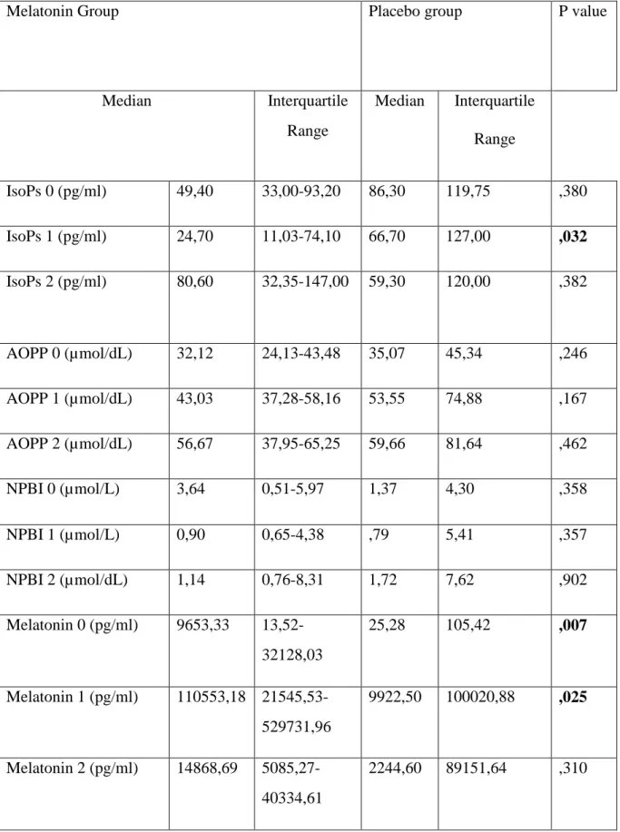
birth weight (p= 0.09), Apgar score at 1’ and 5’ (respectively p=0.155 and p=0.121) and type of delivery (p=0.82). No differences were found as well in needing of MV (p=0.077), NIV (p=0.142) and oxygen requirement (p=0.523), occurrence of sepsis (p=0.100), abnormalities at the CUS (p=0.893) between the two groups. No case of NEC occurred in either of the two groups, whereas a single case of BPD has been reported in the melatonin group and two cases of ROP occurred, one in each group. In the comparison of serum level of IsoPs, AOPP, NPBI and melatonin between the two groups, we found difference only in the IsoPs levels at T1, which were significantly lower in treated group were (p 0.032) than the placebo group (Table 2); as expected, serum melatonin levels as well were significantly higher in the treated group both at T1 and T2 (respectively p=0.007 and p=0.025) (Fig 1) (Table 2).
45
Table 2. A p value <0.05 has been considered significant.
Melatonin Group Placebo group P value
Median Interquartile Range Median Interquartile Range IsoPs 0 (pg/ml) 49,40 33,00-93,20 86,30 119,75 ,380 IsoPs 1 (pg/ml) 24,70 11,03-74,10 66,70 127,00 ,032 IsoPs 2 (pg/ml) 80,60 32,35-147,00 59,30 120,00 ,382 AOPP 0 (µmol/dL) 32,12 24,13-43,48 35,07 45,34 ,246 AOPP 1 (µmol/dL) 43,03 37,28-58,16 53,55 74,88 ,167 AOPP 2 (µmol/dL) 56,67 37,95-65,25 59,66 81,64 ,462 NPBI 0 (µmol/L) 3,64 0,51-5,97 1,37 4,30 ,358 NPBI 1 (µmol/L) 0,90 0,65-4,38 ,79 5,41 ,357 NPBI 2 (µmol/dL) 1,14 0,76-8,31 1,72 7,62 ,902 Melatonin 0 (pg/ml) 9653,33 13,52-32128,03 25,28 105,42 ,007 Melatonin 1 (pg/ml) 110553,18 21545,53-529731,96 9922,50 100020,88 ,025 Melatonin 2 (pg/ml) 14868,69 5085,27-40334,61 2244,60 89151,64 ,310
46
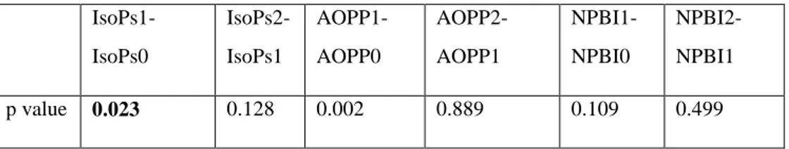
We evaluated the change in IsoPs, AOPP and serum Melatonin levels at time 0, 1, 2 with Wilcoxon test and we found that in the melatonin group, IsoPs at T1 levels were significantly decreased compared to IsoPs at T0 (p 0.023) (Table 3) (Fig. 1). Melatonin serum levels increased from T0 to T1 and from T1 to T2 both in placebo and treated group (Table 4). The difference between baseline and follow-up melatonin was significantly higher in the melatonin group: median melatonin levels increased from 96 pg/ml to 110553 pg/ml from T0 to T1 (p<0.001) followed by a decrease at T2 (14868 pg/ml; p=0.036). In the placebo group as well melatonin increased from a basal level of 25 pg/ml to 9922 pg/ml at T1 (p=0.033). Also, AOPP1 increased compared to AOPP0 (Table 3).
47 IsoPs1-IsoPs0 IsoPs2-IsoPs1 AOPP1-AOPP0 AOPP2-AOPP1 NPBI1-NPBI0 NPBI2-NPBI1 p value 0.023 0.128 0.002 0.889 0.109 0.499
Table 3. A p value of 0.05 has been considered significant
(Fig 1)
Melatonin1- melatonin0 Melatonin2-melatonin1
p value <0.001 0.036
Table 4.
48
Finally, through Rho Spearman test we investigate a possible relationship of interdependence between melatonin levels and the total dose of melatonin administered (based on weight) (p=0.475) and between melatonin levels and weight. We found a significant correlation between total amount of melatonin administered and serum melatonin levels at T1 and T2 (p=0.035 and p=0.045), whereas no correlation was found between weight and serum level of melatonin at none of the times examined (p=0.475, p=0.455, p=0.167 respectively for T0, T1 and T2).
4.4 Discussion
Antioxidant properties of melatonin has been demonstrated both in in vitro and in animal studies. In newborns humans, melatonin, administered both via oral and IV route at high doses (ranging from 10 to 100 mg/Kg), resulted in reduction
49
of serum OS biomarkers (MDA, nitrite/nitrate levels, SOD). In small clinical studies, melatonin improved clinical outcome in babies with septic shock (20 mg orally within 12 h of diagnosis), reduced ventilator-associated lung injury and inflammatory markers in preterm babies (10 doses of 10 mg over 72 h), reduced MDA and nitrate/nitrite in asphyxiated babies (80 mg orally over 16 h) and reduced cytokines and improved clinical outcome in surgical babies
(100 mg IV over 72 h). [3, 10-12] However, these doses have
been chosen on the base of results of in vitro and animal studies and the paucity of pharmacokinetic data in neonates led to a difficult establishment of a therapeutic effective dose.
Based on adult pharmacokinetic data, Merchant et al. [15]
first performed a pharmacokinetic study in preterm infants, starting with a iv dose of 0.1 mg kg-1 h-1 for 6 h. This resulted in plasma melatonin concentrations higher than
50
predicted probably due to several factors: melatonin is extremely lipophilic and the fat body content of the preterm newborns is very low; moreover, preterm infants present immature liver metabolism and poor renal excretion. Reliable pharmacokinetic data and melatonin concentrations closest to physiological adult concentrations were achieved with a 2 h infusion of 0.1 mg kg-1 h-1.
Recently, has been reported, for the first time, the pharmacokinetic profile of oral pharmacological dose of melatonin in preterm neonates. After administration of a single 0.5 mg/Kg bolus, blood melatonin resulted in the high nM range, pointing out the possibility to obtain and maintain protective concentrations of melatonin in blood using oral administration of the indoleamine repeated every 24h. These data has been considered helpful to prescribe proper dosage and frequency of administration of melatonin
51
in preterm infants. [47] However, no data were available on
the efficacy of these specific dose.
Therefore, in the present study we investigated the effect of treatment with 0.5 mg/Kg every 24h of oral melatonin on the serum levels of F2 Isoprostanes, AOPP and NPBI in comparison with placebo.
We found no significant reduction in serum levels of OS biomarkers except for IsoPs at time 1 which remains of unclear clinical relevance, even because it increases again at T2 almost reaching the placebo group levels. Analysing serum melatonin levels, as expected, we found a significant increase from baseline to T1 and T2 in the group receiving melatonin, but melatonin levels increased also in the placebo group at the same time, and this can be attributed to the attempt to counteract the oxidative stress associated with the preterm birth.
52
We investigated a possible relationship of interdependence between melatonin levels and the total dose of melatonin administered and between melatonin levels and weight and we found a significant correlation between total amount of melatonin administered and serum melatonin levels at T2 and T3 whereas no correlation was found between weight and serum level of melatonin at none of the times examined.
These results suggest that the serum melatonin level reached after administration is dependent on the total amount rather than on the dosage/Kg. Therefore, in our opinion, in the planning of future studies aiming to investigate the efficacy of melatonin as antioxidant, a higher standard fixed dose of melatonin should be preferred to a weight base dosage.
Although the sample size is too small to make a proper statistical analysis, no difference were found in the
53
incidence of OS related diseases (BPD, NEC, ROP, PVL) between the two groups.
A limitation of this study is the small number of patients enrolled. Larger studies with extended follow-up are needed to examine long-term clinical effects.
54 CHAPTER 5
CONCLUSIONS AND PERSPECTIVE
Despite melatonin is a promising molecule in providing reduction of OS biomarkers in newborns, further knowledges are needed to identify the proper dosage and frequency of administration.
Although the pharmacokinetic studies suggested that a regimen of 0.5mg/Kg/die lead to supraphysiologic levels of melatonin in preterm in newborns, our study demonstrated that this dose is not effective in reducing OS biomarkers in premature infants. Therefore, larger studied are needed to investigate the efficacy of higher doses of oral melatonin supplementation.
55 REFERENCES
[1]
B. Halliwell, Free radicals, antioxidants and human
disease: curiosity, cause, or consequence? Lancet
2009;344:721-724.
[2]
X. Wang. The antiapoptotic activity of melatonin in
neurodegenerative diseases. CNS Neurosci Ther
2009;15(4):345-357.
[3]
E. Gitto, M. Karbownik , R.J. Reiter et al. Effects of
melatonin treatment in septic newborns. Pediatr Res.
2001;50(6):756-760.
[4]
D. Lanoix, H. Beghdadi, J. Lafond, et al. Human
placental trophoblasts synthesize melatoninand express
its receptors, Journal of Pineal Research 2008;45(1):
50–60.
[5]
D. Lanoix, A.A Lacasse, R.J. Reiter, C. Vaillancourt.
56
model. Molecular and Cellular Endocrinology
2012;348(1): 1–11.
[6]
Y. Okatani, A. Wakatsuki, K. Watanabe et al. Melatonin
inhibits vasospastic action of oxidized low-density lipoprotein in human umbilical arteries, Journal of
Pineal Research 2000;2: 74–80.
[7]
B. Poeggeler. Melatonin replacement therapy in preterm
infants: the impact of pharmacokinetics. Expert Review
of Clinical Pharmacology 2013;6(4), 367–368.
[8]
H. Shoji, B. Koletzko, Oxidative stress and antioxidant
protection in the perinatal period. Current Opinion in
Clinical Nutrition and Metabolic Care 2007;10(3):324– 328.
[9]
L. Pan, J.H. Fu, X.D Xue et al. Melatonin protects
against oxidative stress damage in a neonatal rat model of bronchpulmonary dysplasia, World Journal of
57
[10]
E. Gitto, R.J. Reiter, G. Sabatino et al. Correlation
among cytokines, bronchopulmonary dysplasia and
modality of ventilation in preterm newborns:
improvement with melatonin treatment, Journal of
Pineal Research 2005;39(3):287-293.
[11]
F. Fulia, E. Gitto, S. Cuzzocrea, Increased levels of
malondialdehyde and nitrite/nitrate in the blood of asphyxiated newborns: reduction by melatonin, Journal
of Pineal Research 2001;31(4):343-349.
[12]
L. Marseglia, R. Reiter, G. Buonocore et al. Melatonin
and Neonatal Sepsis: A Promising Antioxidant Adjuvant Agent. American Journal of Perinatology 2017;34(14),
1382–1388.
[13]
S.W. Park, H.S. Lee, M.S. Sung et al. The Effect of
Melatonin on Retinal Ganglion Cell Survival in
Ischemic Retina. Chonnam Medical Journal
58
[14]
E.A Lane, H.B. Moss. Pharmacokinetics of melatonin in
man: first pass hepatic metabolism. Journal of Clinical
Endocrinology and Metabolism 1985;61:1214–1216.
[15]
N.M. Merchant, D.V. Azzopardi, A.F. Hawwa et al.
Pharmacokinetics of melatonin in preterm infants.
British Journal of Clinical Pharmacology
2013;76(5):725-733.
[16]
H. Sies, Oxidative Stress, Academic Press, London,
1985, pp. 1–507.
[17]
S.W. Ryter, A.M. Choi. Regulation of autophagy in
oxygen-dependent cellular stress, Curr. Pharm. Des.
2013;19(15):2747-2756.
[18]
JJ. Lemasters, T. Qian, L. He, et al. Role of mitochondrial inner membrane permeabilization in necrotic cell death, apoptosis, and autophagy.
Antioxidants and Redox Signaling 2002;4(5):769–781.
[19]
U. Förstermann, N. Xia, H. Li, Roles of vascular
59
atherosclerosis, Circulation Research 2017;120(4):713–
735.
[20]
R.L. Haynes, R.D. Folkerth, R.J. Keefe et al. Nitrosative
and Oxidative Injury to Premyelinating
Oligodendrocytes in Periventricular Leukomalacia.
Journal of Neuropathology & Experimental Neurology 2003;62(5):441–450.
[21]
S. Perrone, M. L. Tataranno, G. Stazzoni, Biomarkers of
oxidative stress in the fetus and in the newborn, Journal
of Matern-Fetal Neonatal Medicine 2012;25(12):2575-2578.
[22]
E. Gitto, R.J Reiter, S.P. Cordaro et al., Oxidative and
inflammatory parameters in respiratory distress syndrome of preterm newborns: beneficial effects of
melatonin. American Journal of Perinatology
2004;21(4):209-216.
[23]
S. Perrone, M. L. Tataranno, S. Negro, et al., May oxidative stress biomarkers in cord blood predict the
60
occurrence of necrotizing enterocolitis in preterm infants? The Journal of Maternal-Fetal & Neonatal
Medicine 2012;25(1):128–131.
[24]
C. Aydemir, D. Dilli, N. Uras, et al. Total oxidant status
and oxidative stress are increased in infants with necrotizing enterocolitis, Journal of Paediatric Surgery
2011;46 (11):2096–2100.
[25]
K. Savman, U.A. Nilsson, M. Blennow et al.
Non-protein-bound iron is elevated in cerebrospinal fluid from preterm infants with posthemorrhagic ventricular dilatation, Paediatric Research 2001;49 (2):208–212.
[26]
S. Tordjman, S. Chokron, R. Delorme. Melatonin:
Pharmacology, Functions and Therapeutic Benefits.
Current Neuropharmacology 2017;15(3):434-443.
[27]
I. Liguori,G. Russo, F. Curcio.Oxidative stress, aging,
and diseases, Clinical Interventions in Aging
61
[28]
V. Sosa, T. Moliné, R. Somoza, et al. Oxidative stress
and cancer: An overview. Ageing Research Reviews
2013;12(1):376–390.
[29]
A. Singh, R. Kukreti,L. Saso et al. Oxidative Stress: A
Key Modulator in Neurodegenerative Diseases.
Molecules 2019;24(8):1583.
[30]
Saugstad OD, Hypoxanthine as an indicator of hypoxia:
its role in health and disease through free radical production. Paediatric Research 1988;23:143–150.
[31]
A.H. Jobe, M. Ikegami, Prevention of
bronchopulmonary dysplasia. Curr. Opin. Pediatr.
2001;13:124–129.
[32]
J. Wang, W. Dong, Oxidative stress and
bronchopulmonary dysplasia. Gene 2018;678:177–183.
[33]
A.L.Shane, P.J Sánchez, B.J. Stoll. Neonatal sepsis. The
62
[34]
S. Batra, R. Kumar, K.R. Seema et al. Alterations in
antioxidant status during neonatal sepsis. Annals of
Tropical Paediatrics 2000;20:27–33.
[35]
E.P. Yıldız, B. Ekici, B. Tatlı, Neonatal hypoxic
ischemic encephalopathy: an update on disease pathogenesis and treatment. Expert Review of
Neurotherapeutics 2016;17(5): 449–459.
[36]
National Institute for Clinical Excellence Therapeutic
Hypothermia with Intracorporeal Temperature
Monitoring for Hypoxic Perinatal Brain Injury,
Interventional Procedures, Guidance NICE Guidelines 2010.
[37]
Aly H, Elmahdy H, El-Dib M, et al. Melatonin use for neuroprotection in perinatal asphyxia: a randomized controlled pilot study. J Perinatol 2015;35:186-191.
[38]
N. Baregamian, J. Song, C.E. Bailey, J.
Papaconstantinou, et al. Tumor necrosis factor-alpha
63
reactive oxygen species release, mitochondrial
autophagy, and c-Jun N-terminal kinase/p38
phosphorylation during necrotizing enterocolitis.
Oxidative Medicine and Cellular Longevity
2009;2:297–306.
[39]
J. Chen, Le. Smith. Retinopathy of prematurity,
Angiogenesis 2007;10(2):133-140.
[40]
S.K. Quimson. Retinopathy of Prematurity:
Pathogenesis and Current Treatment Options, Neonatal
Network 2015;34(5):284–287.
[41]
M. Comporti, C. Signorini, S. Leoncini et al. Plasma
F2-isoprostanes are elevated in newborns and inversely correlated to gestational age. Free Radical Biology and
Medicine 2004;37(5):724–732.
[42]
M. Longini, E. Belvisi, F. Proietti et al. Oxidative stress
biomarkers: establishment of reference values for isoprostanes, AOPP, and NPBI in cord blood.
64
[43]
M. Comporti, C. Signorini, G. Buonocore, et al. Iron release, oxidative stress and erythrocyte ageing, Free Radical Biology and Medicine 2002;32(7):568–576.
[44]
G. Buonocore, S. Perrone, M. Longini, et al. Non
protein bound iron as early predictive marker of neonatal brain damage. Brain 2003;126(5):1224–1230.
[45]
G. Buonocore, S. Perrone, M. Longini et al., Total
hydroperoxide and advanced oxidation protein products in preterm hypoxic babies, Paediatric Research
2000;47(2):221–224.
[46]
F. Waldhauser, M. Waldhauser, H.R. Lieberman et al.
Bioavailability of oral melatonin in humans,
Neuroendocrinology 1984;39:307–313.
[47]
S. Carloni, F. Proietti, M. Rocchi, et al. Melatonin
Pharmacokinetics Following Oral Administration in Preterm Neonates Molecules, Molecules 2017;12:1-22.
65 TABLE OF CONTENTS SUMMARY 1 INTRODUCTION 5 CHAPTER 1: MELATONIN
1.1 Properties and function 9 1.2 Use of melatonin in newborn 12
CHAPTER 2: OXIDATIVE STRESS 2.1 Oxidative stress in neonatal period 16 2.2 Oxidative stress related diseases 22
2.3 Biomarkers 29
CHAPTER 3: OPEN QUESTIONS ON THE USE OF MELATONIN 36
CHAPTER 4: THE STUDY
4.1 Material and methods 38
4.2 Statistical Analysis 41
4.3 Results 43
66
CHAPTER 5: CONCLUSIONS AND PERSPECTIVE 54