UNIVERSITA’ DEGLI STUDI DI NAPOLI FEDERICO II
DIPARTIMENTO DI FARMACIA
Dottorato di ricerca in Scienza del Farmaco
XXXI ciclo
Deorphanization of Adhesion GPCRs overexpressed in
Glioblastoma
Dott.ssa Sara Bottone
Tutors:
Prof. Mariano Stornaiuolo
Prof. Ettore Novellino
Coordinatore: Prof.ssa Maria Valeria D’Auria
2
Contents
ABSTRACT ... 5
Chapter 1 ... 7
INTRODUCTION ... 7
Folding is a physical process by which proteins reach their functional
three dimensional structure ... 7
Proteins fold moving in an energy landscape ... 8
Protein Folding is a multistep process ... 10
Chaperones assist protein folding ... 11
In the ER a sensitive surveillance mechanism ensures the degradation of
misfolded proteins ... 12
Accumulation of misfolded proteins can cause diseases ... 14
Ligands act as pharmacological chaperones ... 17
RESULTS ... 19
“Pharmacological Chaperone Readout” screening platform ... 19
FzM1 acts as a pharmacological chaperone of the mutant Fz4 receptor.. 21
FzM1 binding site on Fz4-wt receptor ... 23
FzM1 is an allosteric inhibitor of Fz4 ... 30
FzM1.8, a FzM1 derivate compound, acts as an allosteric agonist ... 32
FzM1.8 mechanism of action ... 37
Effects of FzM1 and FzM1.8 on tumor cells ... 42
DISCUSSION ... 47
MATERIALS AND METHODS ... 48
3
REFERENCES ... 57
Chapter 2 ... 64
INTRODUCTION ... 64
Orphan GPCRs ... 64
Orphan GPCRs and Glioblastoma ... 65
CELSR2: an orphan GPCR overexpressed in Glioblastoma ... 66
CELSR2 cellular functions ... 71
CELSR2 molecular partners ... 75
Spatiotemporal expression profiles of CELSR proteins in zebrafish ... 77
Inapplicability of traditional GPCR screening technologies to CELSR2
deorphanization ... 79
Deorphanization strategies ... 80
RESULTS ... 82
Development of a specific PC-platform to use for the identification of
CELSR2 modulators ... 82
CELSR2 mutants expression in mammalian cells ... 86
Generation of CELSR2 endogenous metabolite library ... 89
Rescue of ER-trapped CELSR2 mutants as readout to identify CELSR2
modulators ... 91
Three molecules from Ethyl acetate fraction act as CELSR2 modulators
... 93
Binding site prediction ... 96
DISCUSSION ... 101
MATERIALS AND METHODS ... 105
4
ACKNOWLEDGMENT ... 111
REFERENCES ... 112
5
ABSTRACT
G-protein-coupled receptors (GPCRs) represent, nowadays, one of the most productive source of drug targets. Some GPCRs members are, however, poorly studied and hence cannot be targeted for therapies yet. Among these, there are more than 90 GPCRs still orphan, i.e. with no described endogenous ligand and no clearly defined function. The orphan receptors CELSR2 and Frizzled4 (Fz4), have recently drawn the attention of the scientific community in virtue of their involvement in cancer progression. CELSR2 is an adhesion GPCR (class B GPCRs) overexpressed in glioblastoma. Until now, CELSR2 endogenous ligands have not yet been identified. Fz4 belongs to class F GPCRs and it is activated by the lipoproteins WNTs and Norrin. Misregulation of Fz4 activity is involved in tumor proliferation and cancer stem cell genesis in many types of malignancies, such as glioblastoma, colorectal and breast cancer. To date, the existence of low-molecular-weight organic molecules binding to and modulating Fz4 has not been reported. Thus, the identification of ligands/modulators of CELSR2 and Fz4 activity could pave the way for new therapeutic strategies to treat cancer. In this thesis, I attempted to identify CELSR2 and Fz4 ligands by using the “Pharmacological chaperone readout”, an innovative screening platform (PC-platform) that identifies ligands in virtue of their potency in affecting the tridimensional structure and the intracellular localization of a protein target.
The first chapter of the thesis describes the results obtained in the attempt of identifying new ligands of Fz4 receptor. To achieve this aim, a cell line expressing a Fz4 mutant, Fz4-L501fsX533, was generated. This frameshift mutation is responsible in vivo for the occurrence of the Familial Exudative Vitreoretinopathy (FEVR), a pathology of the retina. The resulting mutant, here referred to as Fz4-FEVR, aggregates intracellularly in the Endoplasmic Reticulum (ER) without reaching the Plasma Membrane (PM) of the cell where, in contrast, the wt receptor localizes at steady state. To identify Fz4 wt modulators, a library of organic molecules has been screened for pharmacological chaperones of Fz4-FEVR, i.e. for molecules able to rescue the folding and correct localization of Fz4-FEVR at PM. Using such read-out, the organic molecule FzM1 has been thus identified as Fz4-FEVR pharmacological chaperone. The pharmacological chaperone FzM1 acts as allosteric inhibitor of the Fz4-wt receptor, binding directly to the wt receptor and inhibiting the signalling pathway Fz4 is involved in. I also performed a structure-activity relationship (SAR) analysis using FzM1 as lead to identify the first allosteric agonist of Fz4, FzM1.8.
6
As discussed in the second chapter of this thesis, a specific PC-platform was developed for the identification of ligands of CELSR2. To achieve this aim, a misfolded version of CELSR2 was generated and then a library of metabolites was screened looking for compounds binding to the receptor and correcting its folding. Zebrafish eggs were used as biomass to obtain a library of metabolites which were screened for pharmacological chaperone efficiency. The outcome of the screening identified three compounds acting as modulators of CELSR2: cholesterol, PGE2 and β-carotene. They do not target the orthosteric binding site of CELSR2 but they are located in an hydrophobic region of the receptor at the interface between the TM-bundle and the lipid bilayer. The potential ability of these molecules to allosterically modulate CELSR2 function could have important implications both in physiological and pathological cell conditions.
7
Chapter 1
INTRODUCTION
Folding is a physical process by which proteins reach their functional
three dimensional structure
Proteins are pivotal units for cell function and vitality. To be properly active, a protein must “properly fold”, i.e. attain its correct three-dimensional structure. Final and correct “native-conformations” are achieved when unfolded nascent chains, emerging from the ribosomes, undergo a series of conformational changes and structural
transitions that characterize their specific folding pathways1.
The free energy endowed by a nascent chain is very high2. At the beginning of the
folding process, entropy forces the unfolded chain to assume and switch among all the
allowed structural conformations2. Later, during the folding process, the enthalpy
generated by the interactions established between different amino acids and domains of the protein counterbalances the entropic disorder and stabilize protein native states,
reducing total Free Energy to an absolute minimum3.
The native and the unfolded states have only a minimal difference of free energy (only
5-10 kcal/mole)4. However, only in the native conformation a protein can exert the
specific function to which it has been destined2. This is because only in this state the
amino acids contributing to the proteins’ active sites are correctly located in the space and relatively to each others.
During the folding process, proteins may assume the so called intermediate
conformations5. Compared with the native state, these conformations are less stable,
have already acquired secondary structures but miss the correct final tertiary structure.
One of the partially folded, intermediate states is named the “molten globule”4.
In vivo, most of the proteins achieve the native state in a time range from milliseconds
to seconds 6. Longer time is required, in contrast, for those proteins characterized by a
8
Proteins fold moving in an energy landscape
The energy landscape perspective is the most recent theory explaining the folding
process of a protein8,9. It describes the free energy endowed by the polypeptide chain
as a funnel-like landscape with many local minima corresponding to small energy traps (metastable states) and a single global minimum corresponding to the native
state10. In the energy funnel, the formation of conformations with lower free energy
occurs more likely than those with higher free energy and thus, the unfolded protein
irreversibly moves towards the minimal free energy of the native conformation11.
In the simplest case, the folding process involves only two states: the native state (N) and the unfolded state (U).
U ↔ N
In this case the folding process is represented by a smooth energy landscape and the folding and unfolding processes have the same running time (Fig. 1).
Figure 1. Energy landscape in which each point represents a protein conformation. The vertical axis represents the free energy (N stands for native state). (Adapted from Dill and Chan, 1997).
9
In most of cases, proteins take more time to roll down a rough funnel characterized by
many small hills and valleys12 (Fig. 2).
In these cases, the folding process alternates slow and fast phases. The slow phase depends on the presence of local minima in which partially folded intermediates can be trapped. The energy barrier that an intermediate has to overcome to achieve the native state is very high. For this reason, the intermediate state (I) is considered a
competitor of the native state12.
U ↔I ↔ N
Figure 2. Energy landscape showing energetic hills and traps which hamper the protein folding progress (adapted from Dill and Chan, 1997).
10
Protein Folding is a multistep process
How a protein reaches its three-dimensional conformation constitutes a central problem in biology. Anfinsen’s pioneristic experiments on the in vitro refolding of the
protein Ribonuclease I13, suggested that all the information required for a protein to
fold are contained in its primary sequence. Although these refolding experiments could be repeated with other small, single domain proteins, large multi-domain proteins
normally tend to aggregate when they are refolded in vitro14. This reflects what
happens in a cell, where most of the proteins requires cellular assistance to fold
properly and efficiently15.
Protein folding is a multistep process:
• The first step includes the formation of secondary structures (α-helix and β-sheets)
which fold rapidly because they are stabilized by intramolecular hydrogen bonds16,17.
• In a second phase of the folding process hydrophobic interactions, protein-solvent interactions and eventually disulfide bridges are established so that the hydrophilic and hydrophobic side chains can face to aqueous solvent and hydrophobic core of the
protein, respectively18.
• Finally, coarse movements of the different protein sub-domains lead to the acquisition of the final native conformation.
Despite enthalpy would push a folded protein toward an unfolded and chaotic organization of the amino-acidic chain, the native conformation is an irreversible state for a protein (at least for those kept at physiological condition of temperature, pH and
ionic strength)19. Intra-chain interactions, hydrogen bonds, hydrophobic interactions,
and disulfide bridges as well as interactions established between side chains and the solvent all help stabilizing the native conformation of the protein and contribute to
11
Chaperones assist protein folding
Although all information to reach the native, biologically active state of a protein is
present in the amino acid sequence21, cells invest in a complex network of molecular
chaperones which will assist the nascent chain and make the folding process more
efficient15. Chaperones are nanoscale molecular machines that recognize incompletely
or incorrectly folded proteins, arrest or unfold them and then either release them for
spontaneous refolding or target them for degradation22.
In the cytosol as well as in the Endoplasmic Reticulum (the two intracellular locations where protein folding occurs) two classes of chaperones can be found: general chaperones, which assist unfolded proteins by recognizing exposed hydrophobic patches, mobile loops, lack of compactness, or incomplete gycosylation, and private chaperones, which recognize a specific amino acid sequence or structure and bind devotedly to a specific protein (family), like for the chaperone RAP for the
LDL-receptor family23.
Besides chaperones, the ER possesses various enzymes that assist proteins during folding: oxidoreductases which are involved in the formation and rearrangements of
disulfide bonds24; Peptidyl-Prolyl-Isomerases (PPIases) which catalyze cis/trans
isomerization of proline residues25. PPIases are extremely important in protein folding
and their activity increase enormously the folding rate26. This is because the ribosome
attaches amino acids in the trans conformation, which requires an isomerization step
12
In the ER a sensitive surveillance mechanism ensures the degradation of
misfolded proteins
The Endoplasmic Reticulum hosts the folding of important proteins such as antibodies,
receptors and ion channels27. Incorrect folding of these proteins would be extremely
harmful not only for the cell, but for the entire organism. To avoid such events of misfolding, a stringent quality control system operates in the ER and discriminates between correctly folded proteins and misfolded proteins [either (partially) unfolded or
incompletely assembled protein complexes]28.
Only folded proteins are exported toward their final destinations. Unfolded proteins, on the contrary, are retained in the ER by chaperones, that recognizes improperly
misfolded proteins29. For example, protein carrying incorrect disulfide bonds are
recognized as misfolded and retained for further folding, aided by molecular
chaperones and folding catalysts such as protein disulfide isomerase 30. Prolonged
retention of misfolded proteins in the ER leads to ubiquitin-mediated degradation by
the ER-associated degradation pathway (ERAD)31. In the ERAD pathway, unfolded
proteins are recognized as aberrant molecules by quality control receptors. These
misfolded proteins are then sorted to an ER‐membrane‐associated
dislocation/ubiquitination complex containing adaptor proteins that help retro-translocation of the proteins into the cytosol, where they are polyubiquitinated and
13
Figure 3.The general ERAD pathway for degradation of misfolded ER proteins. ERAD is initiated on the recognition of non-native proteins as aberrant molecules by the unfolded protein response (UPR) receptors. After being extracted from its folding path, the substrate is retro-translocated in the cytosol where proteins are poly-ubiquitinated and then degraded from the proteasome (adapted from Douglas M. Cyr et al, 2009).
14
Accumulation of misfolded proteins can cause diseases
Despite the presence of a highly active chaperone machinery, in some cases, misfolded proteins cannot be rescued. The presence of misfolded proteins (and especially their
accumulation inside or outside the cells) can cause severe diseases32. These
pathologies are referred to as “Conformational Diseases”32. Most of the time,
misfolding is due to mutated amino acid residues that contributes negatively to the
stability of a protein32. More rarely, mutations have been shown to stabilize folding
intermediates which in turn slow down the folding process rate32.
The deleterious effects of misfolded proteins may depend on protein loss of function33
as observed in Cystic Fibrosis (CF) and Congenital Nephrogenic Diabetes Insipidus (cNDI). Cystic fibrosis is caused by mutations in both copies of the gene coding for
the cystic fibrosis transmembrane conductance regulator (CFTR) protein34. Although
several mutations in the CFTR sequence have been identified, the deletion of three nucleotides coding for the phenylalanine residue at position 508 (ΔF508 CFTR)
accounts for 70% of patients diagnosed with CF35. The ΔF508 allele of CFTR has been
confirmed as a trafficking mutation that blocks maturation of the protein in the ER and
targets it for premature proteolysis36. Similarly, congenital nephrogenic diabetes
insipidus is caused by mutations of the arginine vasopressin receptor (V2R) that cause
the trapping of the receptor in the ER37. Misfolded V2R is not expressed on the cell
surface and this results in a receptor loss of function37.
In other cases, deleterious effects of misfolded proteins may due to injurious ‘gain of function’ as seen in many neuro-degenerative diseases, in which protein misfolding
results in the formation of harmful amyloid fibrils33. Among these pathologies there is
the bovine spongiform encephalopathy (BSE), an infectious pathology whose
infectious agent is a protein named prion scrapie or Prpsc38. Compared to Prpc, the wt
uninfectious version of the prion protein, Prpsc misfolds and forms fibrils that are toxic
to the cells39. Moreover, Prpsc is able to drive the conformational change of Prpc
proteins that arein its close proximity40. This conformational change can be also the
result of mutations (single amino acid substitutions, deletions or insertions) of Prpc
primary sequence41. The beta-sheet content in Prpsc structure is higher than that of Prpc
that, on the other hand, is mainly enriched in alpha-helical conformation42.
Beta-sheet conformation seems to be a common feature of misfolded aggregated
15
well as of many other toxic fibrils43. Amyloid β peptide, α-synuclein and Hungtingtin
form amyloid fibrils in Alzheimer’s disease (AD), Parkinson’s disease (PD) and
Huntington’s disease (HD), respectively43. Beta-sheet conformation also exists in
many functional native proteins such as immunoglobulin44, but the transition from
alpha-helix to beta sheet is characteristic of amyloids deposits45. The abnormal
conformational transition from alpha-helix to beta-sheet exposes hydrophobic amino
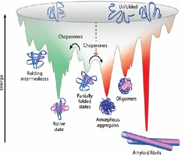
acid residues and promotes protein aggregation45 (Fig. 4).
There are different pharmacological approaches for conformational diseases. In some cases it is possible to protect the protein native state from misfolding and aggregation
events46. For BSE, a therapeutic approach is being consisting in the development of
antibodies which recognize and protect the native Prpc against the negative interactions
with Prps46.
In other cases it is possible to simplify the achievement of the correctly folded state. Recent studies have paved the way for the utilization of small molecules named
Pharmacological Chaperones (PC)47,48. PC are able to enter the cell and interact with a
16
Figure 4. Energy landscape of protein folding and misfolding. Protein folding and protein aggregation are competing reactions. During the folding process, the formation of energetically favorable intramolecular interactions (green) shifts the thermodynamic equilibrium towards the native conformations. However, proteins may adopt energetically favorable but non-native conformations that can slow down the folding process rate. These partially folded or misfolded states are prone to establish intermolecular interactions (red) which result in protein aggregation (amorphous aggregates, β-sheet-rich oligomers, and amyloid fibrils). Chaperones assist in overcoming the free energy barriers. Indeed, they inhibit intermolecular interactions and therefore promote folding to the native state conformation (adapted from Muntau et al. 2014).
17
Ligands act as pharmacological chaperones
Binding of orthostatic or allosteric drugs to their target proteins may promote the folding of the latter by shifting the thermodynamic equilibrium towards the native
conformation49. Native states together with folding intermediates, that are endowed
with properly organized pockets, are the only conformations able to form ligand binding interactions. Ligand binding lowers the free energy of the proteins, increasing
their stability49. Moreover, the low energy state of the ligand-protein complex
minimizes off-pathway interactions preventing misfolding and aggregation49.
Ligands have been shown to be successful in rescuing folding of membrane proteins and more specifically of GPCRs. Indeed, GPCRs explore a wide range of possible
native conformations, all endowed with very similar energetic minima50. Such
structure flexibility allows the binding of different class of ligands, all more or less able to stabilize their cognate receptor. For this reason, a large pool of candidates with a potential activity as PC have been identified. For example, β2AR-agonist promotes the folding of its cognate receptor by shifting the thermodynamic equilibrium towards
the native state51 (Fig. 5).
Figure 5. Agonist binding increases β2AR dynamics by decreasing the energy of intermediate and active states. G proteins further stabilize the active conformation and the formation of a signaling complex is required for the complete receptor transition to the active state (adapted from Nygaard, R et al 2012).
18
In vitro, there are several demonstrations of ligands acting as PC with potential for
therapeutic applications.
Misfolded versions of the V2-vasopressin receptor causes the congenital nephrogenic
diabetes insipidus (cNDI)37. Among these, the mutant R137H is retained in the ER and
not expressed on the cell surface52. Treatment of cells transiently expressing V2
mutants with the V2R antagonist SR49059, rescued the membrane localization and
signalling efficiency of the mutant receptor53. Moreover, the SR49059 chaperone
activity was tested in cNDI patients carrying R137H mutation54. Two-days after
administration, SR49059 had beneficial effects on urine volume and osmolality, that are clinical parameters associated with a functional V2R. However, further clinical trials of SR49059 were abandoned because of its potential interference with the
cytochrome P450 metabolic pathway54.
Another study showed that the antagonist Ipsen 17 acts as PC of its cognate receptor
melanocortin 4 receptor (MCR4)55. Mutations in MCR4 that cause intracellular
retention of the receptor are the most common monogenic cause for obesity56. Ipsen 17
rescued several intracellularly-retained MCR4 mutants, resulting in increased cAMP
production upon agonist stimulation55. Thus, the ability of Ipsen 17 to rescue a wide
variety of MCR4 mutants makes it particularly promising for therapeutic applications. However it has not yet been tested in animal models.
Janovick et al. showed that a non-peptide antagonist of the gonadotropin releasing
hormone receptor (GnRHR) could rescue folding and activity of five intracellularly
retained mutants (T32I, E90K, C200Y, C279Y, and L266R)57. GnRHR mutations can
affect the neural regulation of the reproductive system58. In a knock-in mouse model of
hypogonadism, treatment with GnRHR agonists restore reproductive parameters,
including spermatogenesis and androgen levels59.
Other examples of G-protein-coupled receptors (GPCRs) whose ligands were shown to
rescue the function of mutated receptors are: rhodopsin60, α1/β1/β2 adrenergic
receptors61, luteinizing hormone receptor (LH) and glucagon receptor62.
To date, known ligands of a protein were assayed for their pharmacological chaperone potential. Recently, we showed that the potential to rescue misfolded targets can be
19
RESULTS
“Pharmacological Chaperone Readout” screening platform
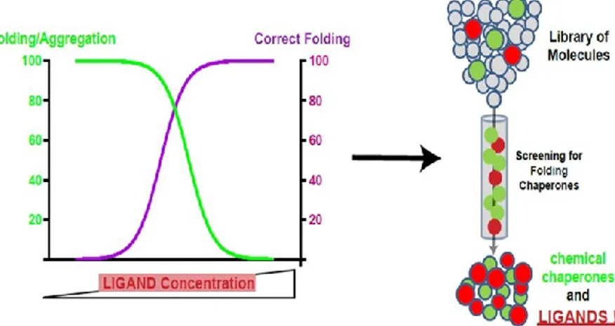
Recently we have set up a new screening platform named “Pharmacological
Chaperone Readout” (PC-Platform)63. This platform allows to identify ligands in
virtue of their ability to interact directly with the target and to affect its tridimensional structure (Fig. 6).
By using PC-platform we have identified the first allosteric modulator of the Frizzled 4 (Fz4) receptor, a member of the GPCR class F family.
To identify ligands of Fz4 we have generated a cell line expressing a Fz4 mutant, Fz4-L501fsX533, responsible in vivo for the occurrence of the Familial Exudative
Vitreoretinopathy (FEVR), a pathology of the retina64. The frame-shift mutation
L501fs533 dictates a change in the amino acidic sequence of the C-terminal tail of the receptor inhibiting its function. Moreover, the resulting mutant, here referred to as Fz4-FEVR, aggregates intracellularly in the ER without reaching the PM of the cell
where, in contrast, the wt receptor localizes at steady state65.
To identify Fz4 wt modulators, a library of organic molecules has been screened for pharmacological chaperones of Fz4-FEVR, i.e. for molecules able to rescue the folding and correct localization of Fz4-FEVR at PM. Using such read-out the organic
molecule FzM1 has been thus identified as Fz4-FEVR pharmacological chaperone63.
The pharmacological chaperone FzM1 acts as allosteric inhibitor of the Fz4-wt receptor, binding directly to the wt receptor and inhibiting the signalling pathway Fz4
is involved in63. Structure-Activity Relation analysis performed using FzM1 as lead
20
Figure 6. Schematic representation of “Pharmacological Chaperone Readout” platform.
21
FzM1 acts as a pharmacological chaperone of the mutant Fz4 receptor
The frameshift mutation that characterizes Fz4-FEVR C-terminal tail, affects receptor
stability and induces its aggregation and retention in the ER65. Notwithstanding,
Fz4-FEVR localization at the PM can be rescued by overexpressing the protein chaperone
α-B crystalline67. Fz4-FEVR can be thus included among the proteins that are
responsible for conformational diseases whose phenotype can be rescued by strategies aiming to improve folding. The marked difference in localization between the wt and mutant receptor makes Fz4-FEVR an ideal platform to screen for folding chaperones by offering an unambiguous readout.
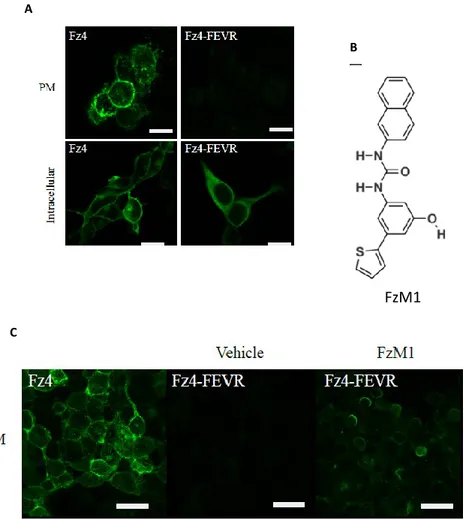
In order to identify pharmacological chaperones of Fz4-FEVR, we used as biological platform Hek293 cells stably expressing (HA)-tagged wt Fz4 (HA-WT) or Fz4-FEVR (Fz4-FEVR). As expected, immunofluorescence data showed that HA-Fz4-WT was localized in the Golgi complex and at the PM, whereas HA-Fz4-FEVR appeared to be trapped intracellularly, mainly in the ER (Fig. 7 A). HA-Fz4-FEVR Hek293 expressing cells were treated with a library of molecules for 48h (Supplementary Table S1). Chaperone activity was then assayed by measuring the recovery of HA-Fz4-FEVR localization at the PM.
We showed that FzM1 rescued HA-Fz4-FEVR PM localization with an efficiency of 15% and with a half-maximum effective concentration (EC50) in the micromolar range (Fig. 7 A, B, C). FzM1 is an ureido derivative harbouring a phenol, a thiophene and a naphthyl functional groups (Fig. 7 B)
22
Figure 7. FzM1 rescues Fz4-FEVR PM localization in Hek293 cells. (A) Cellular localization of Fz4 and Fz4-FEVR expressed in Hek293 cells. Confocal immunofluorescence of non permeabilized (PM) and permeabilized (intracellular) cells showing PM and ER localization of Fz4 and Fz4-FEVR (green), respectively. (B) Chemical structure of FzM1. (C) Rescue of Fz4-FEVR localization at the PM upon FzM1 treatment. Confocal immunofluorescence of non permeabilized cells showing PM localization of Fz4-FEVR upon treatment with FzM1. Fz4-WT expressing cells are shown for comparison.
A
C
B
A
23
FzM1 binding site on Fz4-wt receptor
We identified FzM1, a molecule acting as pharmacological chaperone of the mutant receptor Fz4-FEVR. We then confirmed that the molecule selected by the screening platform indeed addresses our target. In order to do this we identified the FzM1 binding site on Fz4-wt.
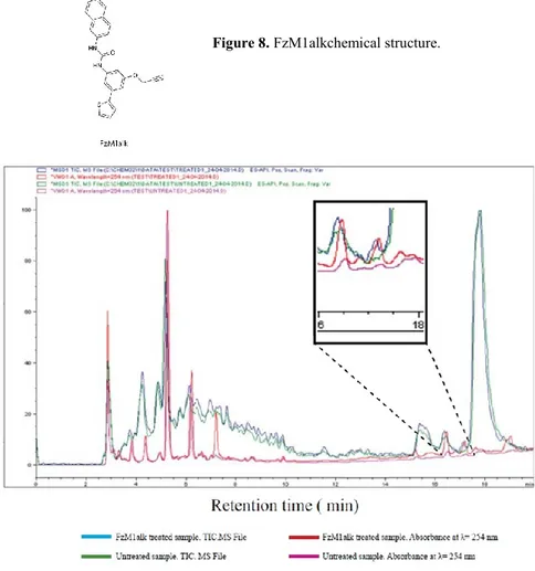
To achieve our aim, we modified the original FzM1 molecule by adding a reactive alkyne moiety. This new compound was called FzM1alk (Fig. 8). Alkyne groups may receive nucleophilic attacks from sulfhydrylic or hydroxylic groups of amino acids and generate covalent adducts. Thus FzM1alk would bind to serines, threonines and cysteines located at its binding sites.
HA-Fz4-WT Hek293 cells were cultured in the presence of FzM1alk. Fz4 was immunopurified, digested with trypsin and analyzed by LC/MS.
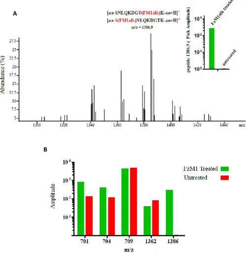
HPLC profiles of samples from FzM1alk-treated cells and untreated cells were compared. These almost totally overlapped, with the exception of a fraction eluting with a retention time of 17.2 min and present only in samples obtained from FzM1alk-treated cells (Fig. 9). Among the peptides eluting in this fraction, the one with an m/z of 1,386.9 Da corresponded, with a deviation (Δmass) of 1.6 Da from the theoretical mass, to amino acids 418–426 of Fz4 ICL3 (theoretical mass of 990 Da) carrying FzM1alk (molecular weight 398.5 Da) covalently linked either to S418 or to T425 (Fig. 10 A theoretical mass [M-H-FzM1alk]+ of 1,388.5 Da). A peptide with this m/z was present exclusively in the FzM1alk-treated sample. In contrast, the peptides that co-eluted with it (Fig. 10 B) were not unique as they were also present in the untreated sample in similar abundances, and thus they were not further considered.
Thus, the spectra indicated that FzM1alk interacted with Fz4 ICL3 by contacting at least S418 or T425.
24
Figure 9. LC/MS analysis of FzM1alk treated Fz4. HA-Fz4-wt Hek293 cells were treated with FzM1alk (10 μM) for 24 hours to be then lysed. After immunoisolation, Fz4 was digested with Trypsin and analyzed by LC/MS. Comparison between FzM1alk treated vs untreated Fz4 MS spectra enables to identify peptides presenting FzM1alk covalently attached (ΔM= + 398.5 Da). HPLC profiles of FzM1alk treated (blue curve) and untreated samples (green curve). Absorbance at λ=254 nm FzM1alk (red curve) and untreated (magenta curve) samples are reported. The boxes shows an enlargement of the region (retention time from 16.0 to 18.0 min) where a difference in the two HPLC profiles could be identified;
25
A
B
Figure 10. LC/MS analysis of FzM1alk treated Fz4. (A) MS spectra of the FzM1alk treated sample fraction eluting at retention time of 17.2 minutes. (B) Amplitudes of peptides with the indicated m/z in FzM1alk treated (green bars) and untreated (red bars) samples. Among the peptides eluting between 16.0 and 18.0 min of retention time, the pick corresponding to m/z of 1386-1387 was the only one to be exclusively present in FzM1alk treated sample.
26
Binding of FzM1alk or FzM1 to Fz4 ICL3 should cause a change in solvent accessibility in this region of the receptor. These changes can be highlighted by measuring the rate of hydrogen-to-deuterium exchange (HDX) at this site.
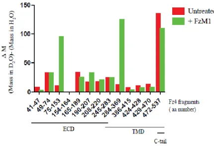
In the absence of FzM1, almost the entire receptor exchanged its hydrogen atoms with deuterium (with the exception of a helix of the ECD domain, amino acids 154–164;
Fig. 11). As expected, the C-terminal tail and the ECD of the receptor showed the
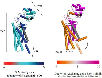
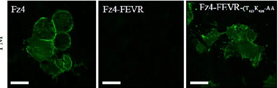
higher rate of exchange compared to the TMD (Fig. 12 A). Upon FzM1 binding, solvent accessibility of ICL3, TM5–7 and the C-terminal tail of the receptor was reduced, whereas the rest of the protein became more solvent accessible (Fig. 12 B). The burial of the ICL3 from solvent upon ligand binding appears compatible with ICL3 being contacted by FzM1 and with a change in the conformation of this loop. A burial of the C-terminal tail can be also hypothesized from the HDX data. The latter could be due to a tighter interaction of the tail with ICL3 or, more likely, as seen for many other GPCRs, with the tail interacting with the detergent micelles.
We envisaged that the diminished solvent accessibility of ICL3 upon FzM1 binding could have been the mechanism behind the folding chaperone effect of the molecule on Fz4-FEVR. To verify this, we replaced ICL3 residues with alanines to affect the fold of this ICL and its interactions with the solvent. We mutated some amino acids in the FZ4-FEVR ICL3 to then test the effect of such mutations on the PM localization of the receptor. Notably, the resulting mutant receptor regained PM localization only when T425 and K426 of FZ4-FEVR were mutated (Fig. 13). Thus, by interfering with the interactions established between the solvent and the amino acid involved in FzM1 and FzM1alk binding, we obtained the rescue of Fz4-FEVR folding and localization. By identifying the FzM1alk binding site at the ICL3 region, we proved that this pharmacological chaperone is indeed a ligand of Fz4-wt. FzM1 or FzM1alk binding induces a conformational change in Fz4-wt that reduces the solvent accessibility at the ICL3. Moreover, they act as pharmacological chaperones by stabilizing the Fz4-FEVR receptor and hampering its aggregation. Though the FEVR C-terminal tail induces Fz4-FEVR aggregation, ICL3 can counterbalance the effect of the tail, influencing the susceptibility of the full receptor to the aggregation. When FzM1 binds to ICL3 or T425A-K426A are mutated, the negative effects of the C-terminal tail on the entire receptor are compromised and the aggregation process is abolished.
27
Figure 11. Analysis of FzM1 binding by HD exchange. Immuno-purified Fz4-wt, treated or not with FzM1, was incubated on ice with D2O to allow H to D exchange.
The protein was then digested at low pH with Formic Acid and run on LC/MS (pH of the run less than 2.5 to minimize back exchange). Hydrogen to deuterium exchange rate (difference of peptide masses in D2O and in H2O) of Fz4 peptides in the presence
(green bars) or in the absence (red bars) of FzM1. Peptides are indicated by their aa numbering and by their position in Fz4 (Extracellular Domain, (ECD)), Transmembrane Domain (TMD) and cytosolic tail (C-tail);
28
Figure 12.Analysis of FzM1 binding by HD exchange. (A) Rate of deuterium exchange (change of mass upon incubation with D2O, Da) in untreated Fz4. (B) Difference in rate of
deuterium exchange upon FzM1 binding. The arrow indicates a possible movement of the C-tail toward the lipid bilayer. Segments in gray represent not detected peptides.
29
Figure 13. Cellular localization of Fz4, Fz4-FEVRand HA-Fz4T425AK426A-FEVR transfected in Hek293 cells. Immunofluorescence of
non permeabilized cells showing the PM localization of HA-Fz4-WT and HA-Fz4T425AK426A-FEVRbut not of HA-Fz4-FEVR. Scale bars, 10 μm.
30
FzM1 is an allosteric inhibitor of Fz4
By identifying the FzM1 binding site, we proved that this pharmacological chaperone is a ligand of Fz4-wt. Next, we confirmed that FzM1 modulates Fz4-wt activity. Fz4 signaling pathway is activated by Wnt ligands. Wnt binding to Fz4 recruits the
scaffold protein Dishevelled (Dvl) at the PM68. The complex Wnt-Fz4-Dvl inactivates
the Adenomatous Polyposis Coli (APC) destruction complex [formed by Glycogen synthase kinase 3 (GSK3β), Axin, and Adenomatous Polyposis Coli] and leads to the intracellular accumulation and nuclear translocation of catenin. In the nucleus, β-catenin regulates T-cell factor /lymphoid enhancer factor (TCF/LEF) dependent
transcription of Wnt/ β-catenin target genes like cyclin D1, c-myc, and lgr568.
We tested the ability of FzM1 to modulate the Wnt/β-catenin pathway with a
luciferase assay that measures the TCF/LEF dependent transcription activation. We
performed the assay on Hek293 cells transfected with HA-Fz4-wt. These cells do not express Fz4 endogenous ligands and, as expected, did not have a TCF/LEF basal activity. However TCF/LEF activity can be induced upon treatment with Norrin, an
high affinity ligand of Fz469.
In absence of the Fz4 agonist Norrin, FM1 did not induce the TCF/LEF dependent transcription (Fig. 14 B). Thus, FzM1 did not act as a Wnt/ β-catenin pathway activator.
On the contrary, after FzM1 treatment, Norrin-dependent TCF/LEF activation was abolished, suggesting that this molecule could be an inhibitor of the Wnt–β-catenin pathway (Fig. 14 A).
We further confirmed the FzM1 activity as Wnt pathway inhibitor by using LiCl, a GSK-3β inhibitor, that causes the β-catenin nuclear translocation (Fig. 14 B).
We also hypothesized the molecular mechanism behind Wnt pathway inhibition by FzM1. ICL3 is the region involved in the Dsh recruitment by Fz4. Contacting ICL3, FzM1 induced a conformational change of Fz4 which no longer was able to recruit Dvl, ultimately blocking β-catenin C-terminal phosphorylation played by CK1 and the consequent β-catenin nuclear translocation.
31
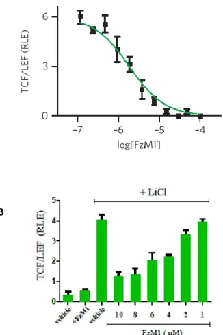
Figure 14. (A) Dose-response curve for FzM1 inhibition of Wnt signaling. Hek293 cells were transiently transfected with HA-Fz4-WT and treated with Norrin (40 ng ml−1) in the presence of the indicated concentrations of FzM1.
Measurements were done in triplicate and are shown as mean ± s.d. RLE, relative luciferase expression. (B) Hek293 cells were transiently transfected with HA-Fz4-WT treated with LiCl (30 mM) and or with the indicated concentrations of FzM1.
A
32
FzM1.8, a FzM1 derivate compound, acts as an allosteric agonist
We showed that FzM1 is an allosteric inhibitor of Fz4. With the aim to generate new Wnt/β-catenin pathway modulators we used FzM1 as lead compound to obtain a small library of FzM1 analogs.
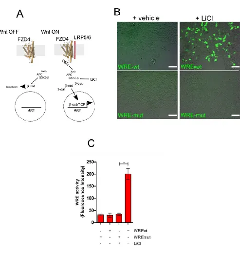
To evaluate the activity of the new compounds we used Hek293 cells transiently expressing both Fz4 and a Wnt reporter construct, the latter presenting the coding sequence of GFP under the control of an optimized Wnt Responsive Element (WRE-wt). Compared to mock transfected cells, transient expression of FZD4 did not induce activation of the Wnt reporter construct indicating a low level of basal activity of FZD4 in the absence of ligands. In contrast, activation of Wnt/β-catenin canonical pathway by the GSK3-β inhibitor LiCl (Fig. 15 A-C) induced GFP expression and increases the fluorescence of the cells.
We showed that the replacement of FzM1 thiophene with a carboxylic group transformed FzM1 in to a new allosteric agonist of Fz4. In the absence of any orthosteric ligand, the new compound 3-Hydroxy-5-(3-(naphthalen-2-yl)ureido) benzoic acid (FzM1.8, Fig. 16 and Fig. 17 A, B) was able to increase WRE activity in Fz4 expressing Hek293 cells.
Similarly to FzM1, FzM1.8 did not increase WRE activity in cells not expressing Fz4 (Fig. 17 C-D) indicating that the effect of the compound is Fz4 dependent. Moreover, FzM1.8 did not induce GFP expression in cells transiently expressing both Fz4 and either a mutated nonfunctional WRE reporter (WRE-mut, Fig. 17 E-F). These results confirm that FzM1.8 does not act as general transcriptional activator and that, instead, its activity relies on a functional WRE. Interestingly, in the range of concentrations tested, FzM1.8 modulated WRE activity with values fitting hormetic dose-response curves (described in Equation1). We measured both a stimulatory (EC50 of activation (logEC50act) ± s.e.m.=-6.4 ± 0.2, n=16) and an inhibitory activity (EC50 of inhibition (logEC50inh) ± s.e.m. = -5.5± 0.1, n=16) (Fig. 17 B).
Despite presenting a switch in its activity, FzM1.8 contacts the same binding sites of the original lead. As already shown for FzM1, Hek293 cells transfected with the Fz4 mutant FZD4T425A only partially responded to FzM1.8 treatment (Fig. 18). Moreover, HDX analysis reveals that FzM1.8 binding reduced solvent accessibility of ICL3. Interestingly, both the ligands affected the conformation of TM6, with FzM1.8
33
increasing the solvent accessibility of this section of the receptor more than FzM1.8 (Fig. 19).
34
Figure 15. Experimental platform used to measure Wnt/β-catenin pathway activity. (A) Schematic cartoon depicting Fz4 involvement in Wnt/β-catenin pathway; (B) Hek293 cells transfected with the constructs WRE-wt and WRE-mut (Merge of Bright Field and GFP channel, Bar = 50μm). Activation of the pathway by LiCl induces GFP expression in cells transfected with WRE-wt but not in those transfected with mutated nonfunctional WRE (WRE-mut); (C) The histogram shows the increase in WRE activity after treatment with LiCl. [Values are reported as mean +/- s.e.m. (n=5) *P<0.05].
35
Figure 17. FzM1.8 is an allosteric agonist of Fz4 and activates the Wnt/β-catenin pathway. (A-B) Dose-response curves for FzM1 and FzM1.8 modulation of Wnt/β-catenin pathway in Hek293 cells co-transfected with both Fz4-wt cDNA and the WRE-wt reporter construct. Values indicate changes in the WRE activity (expressed as the percentage of change over basal activity). (C-D) Dose-response curves for FzM1 and FzM1.8 modulation of WRE activity in Hek293 in the absence of Fz4; (E-F) Dose-response curves for FzM1 and FzM1.8 modulation of WRE activity in Hek293 cells co-transfected with Fz4-wt and WRE-mut.
Figure 16. Chemical structure of FzM1.8
36
Figure 19. FzM1.8 addresses Fz4 ICL3. Solvent accessibility of Fz4 upon binding to DMSO, FzM1 or FzM1.8 measured by HDX analysis. The color scale reflects the level of deuterium exchange measured as difference in peptide mass in the presence or in the absence of vehicle or of ligands. The arrows indicate the ICL3 and the TM6 of Fz4 and the level of deuteration upon vehicle, FzM1 or FzM1.8 binding. Numbers indicate TMD helices (5–6) and the C-terminal tail helix (8).
Figure 18. Dose-response curves for FzM1.8 modulation of WRE activity in Hek293 cells expressing Fz4-wt (red curve) or FZD4-T425A (black curve);
37
FzM1.8 mechanism of action
We showed that FzM1.8, upon binding to Fz4, initiates a signaling route resulting in an increased WRE activity. Thus, we started testing if FzM1.8, by either affecting Fz4/Dvl interaction or by inhibiting β-catenin proteasomal degradation, was acting via Wnt canonical pathway (Fig. 20 A).
As shown in Figure 20 B, in the presence of FzM1.8, Fz4 is still able of recruiting Dvl. Furthermore, differently from Wnt ligands or LiCl, FzM1.8 did not increase intracellular levels of β-catenin (Fig. 20 C). Finally, in the presence of CIK 7, a
pan-inhibitor of Casein Kinase I70, a protein involved in activation of the Fz4/Dvl
complex71, FzM1.8 stimulatory activity was mostly preserved (Fig. 20 D). Thus
FzM1.8/Fz4 complex stimulates WRE activity but does not affect proteins taking part in the Fz4/Dvl/APC axis.
To explain FzM1.8 activity, we thus hypothesized the involvement of a non-canonical pathway. The only pathway that has been so far described as positive modulator of the Wnt canonical pathway is the one involving Phosphatidylinositol-4,5-bisphosphate
3-kinases (PI3Ks)72. PI3K has been already shown to affect Wnt canonical pathway on
multiple levels. In differentiated myofibers, binding of the orthosteric ligand Wnt7a to
Fz7 directly activates PI3K pathway and induces myofiber hypertrophy73. Moreover,
via AKT, PI3K inhibits GSK3-β and leads to β-catenin stabilization74. Alternatively,
AKT has been shown to promote the activity of the histone acetyltransferases CBP and
P300, both trans-activators of the β–catenin/TCF transcription complex75. These
remodel the chromatin-bound to Wnt responsive genes, ultimately boosting the activity of the β–catenin/TCF transcription complex. Indeed, in the presence of the
PI3K inhibitor LY29400276, FzM1.8 lost its ability to induce WRE activity (Fig 21 A).
WB analysis of cell treated with FzM1.8 revealed an increase in AKT phosphorylation at Thr308 upon 30 min and 2 hour of incubation with the ligand, confirming the involvement of PI3K in the pathway activated by FzM1.8. Differently, we did not measure an increase in AKT phosphorylation at Ser473 (Fig. 22 A, B). Finally, Hek293 cells expressing both Fz4 and the AKT dominant negative mutant AKT K179M were insensitive to FzM1.8, further proving the involvement of the protein kinase in the WRE activation elicited by the ligand (Fig. 22 C).
Since FzM1.8 does not induce β-catenin stabilization (Fig. 20 C) we tested whether WRE activation by FzM1.8 was dependent on CBP/p300. In the presence of the
38
CBP/p300 specific inhibitor I-CBP 11277, FzM1.8 activity was drastically reduced
(Fig. 21 B) confirming the involvement of PI3K and CBP/p300 downstream the pathway activated by FzM1.8. The involvement of CBP/p300 indicates also that the Fz4/PI3K axis elicited by FzM1.8 is “ancillary” to the canonical Wnt pathway. Even if it bypasses the Wnt/Fz4/APC canonical route, the Fz4/PI3K axis acts ultimately on the nuclear pool of β-catenin (Fig. 21 C).
39
Figure 20. FzM1.8 does not affect canonical Wnt/β-catenin pathway. (A) Schematic cartoon depicting Fz4 involvement in Wnt/β-catenin pathway and the molecular target of the Casein Kinase I inhibitor CKI-7; (B) Non-confocal immunofluorescence showing recruitment of Dvl-GFP at the PM of HuH7.5 cells, in the presence or not of Fz4-wt and of the indicated amount of FzM1.8; (C) The histogram shows the intracellular levels of β-catenin measured in Fz4 expressing Hek293 cells upon treatment with LiCl (30 mM) or the indicated amount of FzM1.8; (D) Dose-response curves for FzM1.8 modulation of WRE activity in cells expressing Fz4-wt in the presence of CKI-7; [Values are reported as mean +/- s.e.m. (n=5) *P<0.05].
40
Figure 21. Fz4/FzM1.8 complex activates PI3K. (A-B) Dose-response curves for FzM1.8 modulation of WRE activity in cells expressing Fz4-wt in the presence of either PI3K (A) or CBP/p300 (B) inhibitors; (C) Schematic cartoon depicting Fz4 involvement in Wnt/PI3K pathway. [Values are reported as mean +/- s.e.m. (n=5).*P<0.05].
41
Figure 22. FzM1.8 signaling involves AKT. (A) Intracellular levels of AKT phosphorylated at Thr306 (P-Thr306), or at Ser473 (P-Ser473) in Hek293 cells transfected with Fz4 and treated for the indicated period of times with FzM1.8 (2 μM) or with the corresponding amount of vehicle (DMSO). In A the ratio between Phosphorylated and total AKT are indicated for each condition. [Values are reported as mean +/- s.e.m. (n=3)]. Panel B shows sections of gels representative of the replicates used for the quantification reported in A. The intensity of each band was normalized by decorating each filters with an anti α-tubulin antibody. Only the relevant portion of the gels are shown. (C) Dose-response curves for FzM1.8 modulation of WRE activity in Hek293 cells co-transfected with Fz4-wt and with the dominant negative AKT mutant AKT-K176M. [Representative of three independent experiments].
42
Effects of FzM1 and FzM1.8 on tumor cells
We also investigated if FzM1 and FzM1.8 by modulating the Fz4 receptor activity could affect cancer cells growth. Indeed, the intracellular signaling pathway activated by Fz4 culminates in the activation of genes that have been related to tumor cell
survival, differentiation and invasiveness78.
As first we looked at the effects of FzM1 treatment on growth, differentiation and migration of U87MG glioblastoma cells. Fz4 expression in these cells has already
been shown to relate to invasiveness and the differentiation state of the cells79. This
cell line, when cultured in dish, arranges to form neurospheres that are spherical cell clusters containing cells positive for Nestin, a neural stem cell marker (Fig. 23 A, B). Treatment with FzM1 hampers neurosphere formation and induces cells to acquire a more elongated or neuronal appearance (Fig. 23 and Fig. 24). Moreover, FzM1 reduces the number of cells positive for Nestin, suggesting that, upon FzM1 treatment, U87MG cells transitioned toward a more differentiated phenotype (Fig. 25 A, B). We confirmed the ability of FzM1 to act as inhibitor of tumor cell growth in intestinal Caco2 cell line. Similarly to U87MG cells, Caco2 cells are known to rely on an active Fz4-dependent pathway to maintain their undifferentiated proliferative state. FzM1 treatment promotes differentiation of Caco2 cells as shown by the marked change in their morphology (increase in cytoplasm volume) and by the expression of the differentiation marker E-cadherin (Fig. 26 A, B).
We also evaluated the effects of FzM1.8 treatment on Caco2 cell line. Differentially from FzM1, FzM1.8 does not change the differentiation state of these cells. However, the treatment causes an increase of the cells duplication rate (Fig. 27 B). Thus, we investigated if FzM1.8 influenced the Fz4-depedent gene regulation. We showed that FzM1.8 causes a Fz4-dependent upregulation of genes stimulating cell proliferation such as c-myc and cyclin-D (Fig. 27 A, B). On the contrary, FzM1.8 treatment does not induce changes in the colon stem cell marker lgr5, indicating the FzM1.8 actives Fz4 pathways that stimulates proliferation preserving stemness of the cells (Fig. 27 A,
B). It has been further showed that the ability of FzM1.8 to activate Fz4 plays an
important role in tissue repair lung cells66,80. Indeed, Fz4 is a positive modulator of cell
43
A
B
vehicle
FzM1
Figure 23. Effects of FzM1 treatment on U87MG glioblastoma cells. (A) U87MG cells were cultured in presence of vehicle (DMSO 0,1%) or FzM1 (10 µM). β-catenin is in green and nuclei are in blue (DAPI). Immunofluorescence shows vehicle-cells form peculiar neurospheres; upon compounds treatment they disassemble. Scale bar: 20µm. (B) U87MG cells immunofluorescence, β-catenin is in green and nuclei are in blue (DAPI). FzM1 treatment reduces dimension of neurospheres. Scale bar: 20µm.
44
FzM1
vehicle
Figure 24. Morphological effect of FzM1 on U87MG cells . FzM1 (10 µM) affects U87MG cell morphology. Upon treatment, cells acquire a more differentiated phenotype. Bright field images show nuclei. Scale bar: 100 µm
B
Figure 25. FzM1 influences on U87MG differentiation. (A) Immunofluorescence shows neural stem cell marker nestin in green and nuclei in blue (DAPI). FzM1 treatment reduces the number of nestin-positive U87MG cells. Scale bar: 100 µm (B) Upon FzM1 treatment U87MG cells show radical decrease of: % of Nestin-positive cells, neurosphere average diameter and number of neurospheres.
FzM1
vehicle
45
Figure 26. Effects of FzM1 on Caco2 differentiation. (A) FzM1 accelerates CaCo-2 cell differentiation. CaCo-2 cells were treated with vehicle (DMSO 0.1%, 3 d) or with FzM1 (10 μM, 3 d). Upon treatment, cells acquire a more differentiated phenotype (β-catenin is in green). Scale bars, 20 μm. (B) Quantification of differentiation of CaCo-2 (number of cells changed in morphology, E-cadherin–positive and β-catenin–positive cells) upon treatment with FzM1 (10 μM, 3 d); n = 3, data represent mean ± s.e.m.
FzM1
vehicle
A
46
Figure 27. FzM1.8 increases proliferation of undifferentiated colon cancer cells. (A) The effect of FzM1.8 or FzM.1 on the expression levels of genes regulated by Fz4 signalling pathway. Genes stimulating cell proliferation such as c-myc and cyclin-D are up-regulated. (B) Duplication rate of human colonic biopsies in culture medium supplemented with FzM1, FzM1.8 or with vehicle.
A
47
DISCUSSION
There are several evidences of the ability of ligands to act as pharmacological chaperones in rescuing the folding of their target proteins. We proved that, by transitive property, the potential to rescue misfolded targets can be used as a readout in screening campaigns toward the identification of new ligands. We screened a library of small molecules for their ability to rescue the ER-trapped folding defective mutant of Fz4, Fz4-FEVR. In this way we identified FzM1, the first allosteric modulator of
Fz4 that physically interacts with the ICL3 of Fz4.In our opinion, the change in the
conformation of ICL3 upon FzM1 binding is the mechanism behind the pharmacological chaperone activity it exerts on Fz4-FEVR. Fz4-FEVR bound to FzM1 is more resistant to receptor aggregation and is properly transported to PM. Interestingly, we showed that FzM1.8, that is an analog of FzM1, is not able to act as pharmacological chaperone of Fz4-FEVR. However, FzM1.8 acts as a positive allosteric modulator of Fz4-wt. Thus, the PC-platform seems to select ligands binding the inactive state conformations (such as antagonist and negative allosteric modulator) rather than the active state conformations of GPCRs (such as agonist and positive allosteric modulators). The active-state conformations of GPCRs are higher in energy
(less stable) than inactive-state conformations50. Moreover, it has been shown that
agonist binding even does not stabilize a unique native conformation51. In contrast, it is
associated with conformational heterogeneity that may be important to regulate several
alternative pathways51. In our opinion, these could be the reasons why agonist and
positive modulators fail in counterbalance the mutant protein tendency to aggregate and have been thus never identified with our PC platform.
PC-platform represents a useful alternative to traditional screenings. It might be especially useful in the case of protein targets difficult to purify in large quantities and proteins with unclear, unknown or even absent connections to signaling cascades, such as orphan GPCRs. Furthermore, we have already shown that the applicability of the platform can be extended also to the receptors for which mutations inducing misfolding and aggregations are not known. Indeed, we proved that appending the mutated tail of Fz4-FEVR to other TM proteins is sufficient to determine the unfolding
of the chimeric proteins and their ER retention65. Finally, GPCRs would represent
good candidates for PC-platform. Indeed, they are highly dynamic and can exist in a multitude of distinct conformations that allow the binding of different class of ligands.
48
MATERIALS AND METHODS
Reagents. Salts and organic solvents are from Sigma Aldrich (USA) and Applichem
(Germany), are handled according to manufacturer instructions.
Cell culture. Hek293 cells and U87MG were cultured in RPMI medium and DMEM
(Dulbecco’s Modified Essential Medium), respectively, supplemented with 10% FBS (Fetal Bovine Serum), Glutamax and Pen/Strep antibiotics. Transfections were performed with polyethylenimine (PEI) according to the manufacturer’s protocol. HA-Fz4-WT cell clones are obtained by transfecting Hek293 cells with the corresponding cDNA cloned in pcDNA 5.0, kind gift of B.T. Mac Donald (Harvard medical School).
Antibodies. The following antibodies were used: mouse monoclonal anti-HA peptide
(HA-7), (code: #H3663 Sigma-Aldrich) was used at 1/2,000 dilution for both western blotting (WB) and immunofluorescence (IF) while a 1/200 dilution was used for immunoprecipitation (IP); mouse monoclonal anti- α-tubulin (10D8, code: sc-53646, Santa Cruz biotechnology, Santa Cruz, CA, USA) (WB: 1/200); rabbit polyclonal anti- β-catenin (H-102, sc-7199, Santa Cruz biotechnology) (WB: 1/1,000); rabbit polyclonal anti- HA peptide, ( code: H6908Sigma-Aldrich) (WB: 1/2,000; IF : 1/500, IP: 1/500); rabbit anti- AKT (#9272, Cell Signaling Technology, USA) (WB: 1/1000); rabbit anti- Phospho-AKT (Thr308) (D25E6, #13038, Cell Signaling Technology, USA) (WB: 1/2000); rabbit anti- Phospho- AKT (Ser473) (D9E, #4060, Cell Signaling Technology, USA) (WB: 1/2000); goat anti-rabbit IgG (H&L), DyLight 594 conjugate, (code: GtxRb-003-D594NHSX, Immuno- Reagents, Inc. Raleigh, NC, USA) (IF:1/500); Donkey anti- Rabbit IgG (H&L), HRP conjugate, (code: DkxRb-003- DHRPX, ImmunoReagent, Inc.) (WB: 1/4,000), goat anti- Mouse IgG (H&L), DyLight 594 conjugate, ( code: GtxMu- 003-D594NHSX, ImmunoReagent, Inc.) (IF: 1/500 dilution for); goat anti-Mouse IgG (H&L), HRP conjugate, (code : GtxMu-003-DHRPX ImmunoReagent, Inc.) (WB: 1/4,000).
DNA-All DNA constructs were synthesized at Gene Script (USA). The cDNA coding
for N terminally HA-tagged FZD4wt (HA-FZD4-wt), and for the mutants HA-FZD4- T425A and HA-FZD4-T425D were all cloned in the expression vector pCDNA3.1 (Invitrogen). For the reporter construct WRE-GFP-wt: 8 repeats of the optimized TCF/LEF binding sequence [AGATCAAAGGGG-3’] (interspaced by the triplet 5’-GTA-3’) were positioned upstream to a minimal TATA box promoter [5’-
[5’-49
cttggcattccggtactgttggtaaaaagcttggcattccggtactgttggtaaagccacc-3’]. The sequence was cloned in the vector pCDNA 3.1 (+) GFP between the restriction sites for NruI and HindIII. This replacement substitutes the CMV promoter of the original vector with the TCF/LEF responsive sequences. The correctness of the sequences was verified by DNA sequencing. The control reporter construct WREGFP-mut was obtained by mutagenesis of WRE-GFP-wt and presents the 8 repeats of the TCF/LEF binding sequence mutated to [5’-AGGCCAAAGGGG-3’]. A reporter construct ( cmv-GFP) presenting GFP under the control of the cmv promoter was used as control. The plasmid necessary for the expression of the human AKT mutant AKT-K179M was kindly provided by Prof. G. Condorelli (Naples, Italy).
Immunofluorescence. Cells were grown up on glass coverslips and then were fixed in
3,7% formaldehyde/PBS pH 7.4 freshly made for 30 minutes at room temperature (RT). Formaldehyde was quenched by incubating the coverslips for 30 minutes in 0.1 M glycine/PBS. Cells were permealized in 0.1% Triton/PBS pH 7.4 for 10 minutes at RT. Glass coverslips were then incubated with primary and secondary antibodies diluted in PBS for 1 h and 30 min at RT, respectively. After fixing coverslips with glycerol/PBS 1:1 on slides, cells were analyzed with a Leica TCS-SMD-SP5 confocal microscope.
Compound synthesis. FzM1 FzM1alk and FzM1.8 were synthesized following a
procedure of urea derivatives formation by reacting amines and isocyanates. HNMR, CNMR, FTIR spectroscopy and HRMS were used to confirm of the compounds. The synthesis of the FzM1 analogues was realized in cooperation with Professor Romano Silvestri’s research group at University “La Sapienza” in Rome.
Immunoisolation from cultured cells and trypsin digestion. Cells were lysed in B
buffer (Hepes K-OH 50mM, 150 mM NaCl, 1% Triton X-100, supplemented with protease inhibitors). Lysates were centrifuged at 14,000 r.p.m. to remove cell debris and unbroken cells. Clarified lysates were incubated overnight with antibody at 4 °C, followed by treatment with Protein A–coupled Sepharose (45 min, 4 °C). Samples were extensively washed in B buffer and then resuspended in 40 μl of buffer containing trypsin and incubated overnight at RT. 20 μl of the tryptic digestion were processed for LC/MS.
HDX. Immunoisolated Fz4 (still bound to the Sepharose beads) was incubated in B
buffer at 4 °C in the presence of or without ligand (10 μM concentration). After 30 min, beads were resuspended in 40 μl of D20 for 1 min in the presence of or without
50
ligand. Formic acid was then added (5% final concentration), and the samples were
boiled four times at 95 °C for 15 min. DTT (50 mM) was added to reduce disulfide bonds. 20 μl of the tryptic digestion were processed for LC/MS.
HPLC/MS. All samples were analyzed by analytical HPLC/MS (Agilent 1200 series
HPLC system, Agilent 1260 UV-vis detector Infinity and Agilent Quadrupole 6110 LC/MS) equipped with a C18-bounded analytical reverse-phase HPLC column (Vydac 218TP104, 4.6 × 250 mm) using a gradient elution (10–90% acetonitrile in water (0.1% TFA) over 20 min; flow rate = 1.0 ml/min. For HDX experiments, the gradient buffer was kept at a pH lower than 2.5 (to reduce back exchange) by adding formic acid.
LC/MS spectra analysis. LC/MS spectra were analyzed with MetAlign51 with the
following settings: mass resolution/BIN (nominal mass mode, 0.65), peak slope factor (5× noise), peak threshold factor (5× noise), peak threshold abs. value (150), average peak width (three scans), autoscaling on total signal, tuning alignment (prealign processing iterative, mass peak selection set on min. factor (5× noise). Amplitudes of masses coming from treated and untreated samples were subtracted to identify mass exclusively present in each of the samples. Masses were assigned with Mascot (MatrixScience)52. Samples containing only trypsin or protein Sepharose or antibody were run as controls.
Statistical analysis. EC50 was calculated by fitting direct total binding data by
nonlinear regression analysis of dose-response curve using Prism software (GraphPad). All data were analyzed using the two-tailed Student’s t-test. P < 0,05 were considered statistically significant.
FZD4 receptor model. FZD4-wt three-dimensional model was built using the CCP4
suite. Crystal structure of the cysteine-rich domain of FZD8 (Protein Data Bank (PDB) code 4F0A) and the TMD of Smoothened receptor (PDB codes 4NAW and 4JKV) were used as templates for ECD and TMD and ILC of FZD4, respectively.
TCF/LEF activity measurement using the WRE-GFP constructs. Hek293 cells
were seeded (5 x 10³ per well) in 96-well black Optyplates (Perkin Elmer). After 24 hours, cells were co-transfected with both GFP-wt (or when indicated WRE-GFP-mut) and HA-FZD4-wt. Transfection mixtures were prepared as follow: for each well 0,25μg of PEI (pH 7.0) was supplemented with 0,08 μg of DNA(both diluted in 4 μl of DMEM). The mixture was incubated at room temperature for 30 min to be then
51
diluted in culture medium and added to the cells. 24 hours after transfection, the medium was replaced and cells incubated with FzM1, FzM1.8 at the indicated concentrations and for the indicated times. When reported, cells were supplemented with either CKI 7 dihydrochloride (5μM), NFAT inhibitor (10μM), Bisindolylmaleimide II (7,5μM ), Ly294002 (10μM), Suramin Hexasodium salt (10μM), Gallein (10μM), I-CBP 112 ( 7,5μM ) or Neomycin (500μM). At the end of the incubations, cells were fixed in 3.7% formaldehyde in PBS pH 7.4 for 30 min. Formaldehyde was quenched by incubating the cells for 30 min in 0.1 M Glycine in PBS 1X. The activity of the compounds was evaluated by measuring GFP expression.