UNIVERSITY
OF TRENTO
DEPARTMENT OF INFORMATION AND COMMUNICATION TECHNOLOGY
38050 Povo – Trento (Italy), Via Sommarive 14
http://www.dit.unitn.it
PREDICTING CELL ADHESION PROBABILITY
VIA THE BIOCHEMICAL STOCHASTIC PI-CALCULUS
Paola Lecca, Corrado Priami,
Carlo Laudanna and Gabriela Constantin
October 2003
BIO-122
PREDICTING CELL ADHESION PROBABILITY VIA THE BIOCHEMICAL STOCHASTIC π-CALCULUS
This paper presents a stochastic model of the lymphocytes recruitment in inflamed brain microvessels. The framework used is based on stochastic process algebras for mobile systems. The automatic tool used in the simulation is the biochemical stochastic π-calculus. The biochemical stochastic π-calculus is an efficient tool for describing the concurrency of the different interactions driving the phases of lymphocytes recruitment. It models a biochemical systems as a set of concurrent processes selected according to a suitable probability distribution in order to quan-titatively describe the rates and the times at which the reactions occur. We use here this tool to model and simulate the molecular mechanisms involved in encephalito-genic lymphocytes recruitment. In particular, we show that the model predicts the adhesion probability of the lymphocytes as a function of contact time of the cells with endothelium. The results of the model reproduce, within the experimental errors, the behavior of the data obtained either from laboratory measurements or from a classical deterministic treatment of the mechanics of cell adhesion.
1. Introduction
With recent progress in cell and molecular biology, the field of cell mechan-ics has grown rapidly over the last few years. The focus of these recent developments is on three interconnected areas: (a) the responses of the cell to mechanical forces, (b) the mechanics of cell adhesion and (c) the de-formation of biomolecules. In particular the biophysics of cell adhesion is the most intensively investigated area of cell mechanics. These studies have been driven by the strong interest in many biological processes of which cell adhesion is an important element, leading to the development of a number of mathematical models. Cell attachment to and detachment from a sur-face, such as, for example, endothelial surface that lines the blood vessel wall is a central aspect in the inflammatory processes. The lymphocytes are the cells whose mechanical properties are most deeply studied, because of their central role in the tissues response to inflammation.
Lymphocytes roll along the walls of vessels to survey the endothelial surface for chemotactic signals, which stimulate the lymphocyte to stop rolling and migrate through the endothelium and its supporting basement membrane. Lymphocyte adhesion to the endothelial wall is mediated by
binding between cell surface receptors and complementary ligands expressed by the endothelium. The dynamic of adhesion is regulated by the bond association and dissociation rates: different values of these rates give rise to different dynamical behaviors of the cell adhesion.
The most common approach to the simulation of rolling process of lym-phocyte is based on hydrodynamical models of the particle motion under
normal or stressed flow1,15,18. At a macroscopic scale, the process is
gen-erally modeled with the typical equations of mass continuity, momentum transport and interfacial dynamic. At a microscopic scale, the cell rolling is simulated as a sequence of elastic jumps on the endothelial surface, that result from sequential breaking and formation of molecular bonds between
ligands and receptors 15,6,8. This kind of model is able to simulate the
time-evolution of bond density. In general, these models are highly com-plex and with many parameters, yet they are still idealized and rely on a number of nontrivial assumptions, that often are not supported by sufficient quantitative experimental observations.
Moreover, an other difficulty for a mechanical approach is to treat the disparate scales between the cell (typically of the order of micrometers) and the bonds (of the order of nanometers). In fact, rolling involves either dynamical interaction between cell and surrounding fluid or microscopic elastic deformations of the bonds with the substrate cells. Moreover recent studies have revealed that the process leading to lymphocyte extravasation is a sequence of dynamical states (contact with endothelium, rolling and firm adhesion), mediated by partially overlapped interactions of different adhesion molecules and activation factors. The classical mechanical models are inefficent tools to describe in an easy and nimble way the concurrency of the molecular interactions; furthermore, also if they treat the physical system at the scale of intermolecular bonds with appreciable detail, they are not able to reproduce the sensitivity to the small pertubations in the reagent concentrations or in reaction rates typical of microscopic stochastic systems governed by complex and concurrent contributions of many differ-ent molecular reactions. The probabilistic nature of a biological system at the molecular scale requires new languages able to describe and predict the
fluctuations in the population levels. We rely on a stochastic extension22,23
of the π-calculus17, a calculus of mobile processes based on the notion of
naming. The basic idea of this biochemical stochastic π-calculus is to model a system as a set of concurrent processes selected according to a suitable probability distribution in order to quantitatively accommodate the rates and the times at which the reactions occur.
3
We use here this framework to model and simulate the molecular mech-anism involved in encephalitogenic lymphocyte recruitment in inflamed brain microvessels. In particular we show that the biochemical stochas-tic π-calculus model reproduces, within the estimated measurement errors, the same functional behaviour of the cells adhesion probability versus the
contact time, as it was found in laboratory experiments26
.
The paper is organized as follows. In the next section we report a very brief survey of the physiology of the lymphocytes interactions with endothelial surface. Section 3 briefly recalls the basics of the biochemical stochastic π-calculus. Then it shows our specification of the lymphocyte recruitment, and finally, it discusses the results of the stochastic simulation and compares them with the experimental observations. In the last section we show some conclusions.
2. Molecular mechanism of autoreactive lymphocyte recruitment in brain venules
A critical event in the pathogenesis of multiple sclerosis, an autoimmune disease of the central nervous system, is the migration of the lymphocytes from the brain vessels into the brain parenchima. The extravasation of lym-phocytes is mediated by highly specialized groups of cell adhesion molecules and activation factors. The process leading to lymphocytes migration,
il-lustrated in Fig. 1 , is divided into four main kinetic phases: 1) initial
contact with the endothelial membrane (tethering) and rolling along the vessel wall; 2) activation of a G-protein, induced by a chemokine exposed by the inflamed endothelium and subsequent activation of integrins 3) firm arrest and 4) crossing of the endothelium (diapedesis). For this study, we have used a model of early inflammation in which brain venules express
E-and P-selectin, ICAM-1 E-and VCAM-120. The leukocyte is represented by
encephalitogenic CD4+
T lymphocytes specific for PLP139-151, cells that are able to induce experimental autoimmune encephalomyelitis, the animal model of multiple sclerosis.
Tethering and rolling steps are mediated by binding between cell sur-face receptors and complementary ligands expressed on the sursur-face of the endothelium. The principal adhesion molecules involved in these phases are the selectins: the P-selectin glyco-protein ligand-1 (PSGL-1) on the autore-active lymphocytes and the E- and P-selectin on the endothelial cells. The action of integrins is partially overlapped to the action of selectins/mucins:
a less relevant role.
Chemokines have been shown to trigger rapid integrin-dependent
lym-phocyte adhesion in vivo through a receptor coupled with Gi proteins.
Integrin-dependent firm arrest in brain microcirculation is blocked by
per-tussis toxin (PTX), a molecule able to ADP ribosylate Giproteins and block
their function. Thus, as previously shown in studies on na¨ive lymphocytes homing to Peyer’s patches and lymph nodes, encephalitogenic lymphocytes also require an in situ activation by an adhesion-triggering agonist which exerts its effect via Gi-coupled surface receptor.
The firm adhesion/arrest is mediated by lymphocyte integrins and their ligands from the immunoglobulin superfamily expressed by the endothe-lium. The main adhesion molecules involved in cell arrest is integrin LFA-1 on lymphocyte and its counterligand ICAM-1 on the endothelium. The
action of α4 integrins is partially overlapped to the action of LFA-1: α4
integrins are involved in the arrest but they have a less relevant role20.
Hematic flow
3. Diapedesis 1. Tethering and rolling
2. Firm arrest
PSGL−1/E− & P−Selectin
Integrins / VCAM−1 1 β 4 α
of G proteinActivation Activationof integrins
leukocyte LFA−1/ICAM−1
Selectins/Mucins
Figure 1. The process leading to lymphocyte extravasation is a finely regulated se-quence of steps controlled by both adhesion molecules and activating factors. It involves: 1. initial contact (tethering) and rolling along the vessel wall mediated by selectins (PSGL-1/E- and P-selectin) and integrins(α4β1/VCAM-1) and (LFA-1/ICAM-1); 2.
chemoattractant-induced heterotrimeric G protein-dependent intracellular biochemical changes leading to integrins activation; 3. integrin-dependent firm arrest, due pricipally to LFA-1/ICAM-1 interaction: and 4. diapedesis.
5
3. The BioSpi model implementation and results
We first recall the syntax and the intuitive semantics of the stochastic
π-calculus23
. We then describe our specification of the lymphocyte recruit-ment process, and eventually we discuss the simulation results.
Biomolecular processes are carried out by networks of interacting pro-tein molecules, each composed of several distinct independent structural parts, called domains. The interaction between proteins causes biochemi-cal modification of domains (e.g. covalent changes). These modifications affect the potential of the modified protein to interact with other proteins. Since protein interactions directly affect cell function, these modifications are the main mechanism underlying many cellular functions, making the stochastic π-calculus particularly suited for their modeling as mobile com-municating systems. The syntax of the calculus follows
P ::= 0 | X | (π, r).P | (νx)P | [x = y]P | P |P | P + P |A(y1, . . . , yn)
where π may be either x(y) for input, or xy for output (where x is the subject
and y is the object) or τi for silent moves. The parameter r corresponds to
the basal rate of a biochemical reaction and it is an exponential distribution associated to the channel occurring in π. The order of precedence among the operators is the order (from left to right) listed above. Hereafter, the trailing 0 will be omitted.
The prefix π is the first atomic action that the process (π, r).P can per-form. The parameter r is the unique parameter of an exponential distribu-tion. The input prefix binds the name y in the prefixed process. Intuitively, some name y is received along the link named x. The output prefix does not bind the name y which is sent along x. The silent prefix τ denotes an action which is invisible to an external observer of the system. Summation denotes nondeterministic choice. The operator | describes parallel compo-sition of processes. The operator (νx) acts as a static binder for the name x in the process P that it prefixes. In other words, x is a unique name in P which is different from all the external names. A(y1, . . . , yn) is the
def-inition of constants (hereafter, ˜y denotes y1, . . . , yn). Each agent identifier
A has a unique defining equation of the form A(y1, . . . , yn) = P , where the
yiare distinct and fn(P ) ⊆ {y1, . . . , yn} (see below for the definition of free
names fn).
n(µ) = fn(µ) ∪ bn(µ) of a label µ.
µ Kind fn(µ) bn(µ)
τ Silent ∅ ∅
xy Output {x, y} ∅
x(y) Input {x} {y}
Functions fn, bn and n are extended to processes in the obvious way. Below we assume that the structural congruence ≡ on processes is defined as the least congruence satisfying the following clauses:
• P and Q α-equivalent (they only differ in the choice of bound names) implies P ≡ Q,
• (P/≡, +, 0) is a commutative monoid,
• (P/≡, |, 0) is a commutative monoid,
• (νx)(νy)P ≡ (νy)(νx)P, (νx)(R | S) ≡ (νx)R | S if x 6∈ fn(S), (νx)(R | S) ≡ R | (νx)S if x 6∈ fn(R), and (νx)P ≡ P if x 6∈ fn(P ). • A(˜y) ≡ P {˜y/˜x}, if A(˜x) ::= P is the unique defining equation of
constant A
The biological interpretation is as follows. Processes model molecules and domains. Global channel names and co-names represent complemen-tary domains and newly declared private channels define complexes and cellular compartments. Communication and channel transmission model chemical interaction and subsequent modifications. The actual rate of a reaction between two proteins is determined according to a constant basal
rateempirically-determined and the concentrations or quantities of the
re-actants . Two different reactant molecules, P and Q, are involved, and the reaction rate is given by Brate × |P | × |Q|, where Brate is the reac-tion’s basal rate, and |P | and |Q| are the concentrations of P and Q in the
chemical solution computed via the two auxiliary functions, Inx, Outxthat
inductively count the number of receive and send operations on a channel x enabled in a process.
The semantics of the calculus thereby defines the dynamic behaviour of the modeled system driven by a race condition, yielding a probabilistic model of computation. All the activities enabled in a state compete and the fastest one succeeds. The continuity of exponential distributions ensures that the probability that two activities end simultaneously is zero.
7 (. . . + (xhzi, r).Q)|((x(y), r).P + . . .) x,rb·1·1 −−−−−→ Q|P {z/y} P x,rb·r0·r1 −−−−−−−→ P0 P |Q x,rb·r 0 0·r01 −−−−−−−→ P0|Q , r 0 0= r0+ Inx(Q) r0 1= r1+ Outx(Q) P x,rb·r0·r1 −−−−−−−→ P0 (ν x)P−−−−−−−→ (ν x)Px,rb·r0·r1 0 Q≡P,P−−−−−−−→x,rb·r0·r1 P0,P0≡Q0 Q−−−−−−−→x,rb·r0·r1 Q0
A reaction is implemented by the three parameters rb, r0 and r1, where rb
represents the basal rate, and r0and r1denote the quantities of interacting
molecules, and are computed compositionally by Inxand Outx.
3.1. Specification
The system of interacting adhesion molecules that regulate the lympho-cytes recruitment on endothelial surface illustrated in Fig. 1 has been im-plemented in the biochemical stochastic π-calculus. The system is com-posed by eight concurrent processes, corresponding to the eight species of adhesion molecules, that regulate the cell rolling and arrest: PSGL1, PSELECTIN, CHEMOKIN, CHEMOREC, ALPHA4, VCAM1, LFA1 and ICAM1. The code implements the four phases of the lymphocyte recruit-ment: the interaction between PSGL1 and PSELECTIN, the ALPHA4 and LFA1 activation by chemokines and the firm arrest mainly caused by the interaction between the active form of LFA1, LFA1 ACTIVE, and ICAM1 and in part also due to the interaction of the active form of ALPHA4, ALPHA4 ACTIVE, with VCAM1. Its specification is
We simulated the role and the contribution of the different interactions as bi-molecular binding processes occuring at different rates. The selectins interaction PSGL1/PSELECTIN plays a crucial role in guaranting an ef-ficient rolling, therefore the channels rates for the communication in the binding process between PSGL1 and PSLECTIN have been calculated from the deterministic rates of the Bell model, that reproduce the tethering and rolling motion. Analogously, for the ALPHA4 ACTIVE/VCAM1 interac-tion, that contributes to rolling and, in part, also to cell arrest, the channels rate have been calculated from the Bell model rates that recreate the rolling motion. The interaction LFA1 ACTIVE/ICAM1 is the main responsible of
SY ST EM ::= P SGL1|P SELECT IN |CHEM OKIN |CHEM OREC|ALP HA4 |V CAM 1|LF A1|ICAM 1
P SGL1 ::= (ν backbone)BIN DIN G P SIT E1
BIN DIN G P SIT E ::= (bindhbackbonei, RA).P SGL1 BOU N D(backbone) P SGL1 BOU N D(bb) ::= (bb, RD0).P SGL1
P SELECT IN ::=
(bind(cross backbone), RA).P SELECT IN BOU N D(cross backbone) P SELECT IN BOU N D(cbb) ::= (cbb, RD0).P SELECT IN
CHEM OKIN ::= (ν chemobb)BIN DIN G CSIT E
BIN DIN G CSIT E ::= (lighchemobbi, RA C).CHEM OCHIN BOU N D(chemobb) CHEM OCHIN BOU N D(chemobb) ::= ACT 1|ACT 2|ACT 3(cbb)
ACT 1 ::= (alpha acthsign1i, A).ACT 1 ACT 2 ::= (lf a acthsign2i, A).ACT 2 ACT 3(chb) ::= (chb, RD C).CHEM OKIN CHEM OREC ::=
(lig(cross chemobb), RA C).CHEM OREC BOU N D(cross chemobb) CHEM OREC BOU N D(ccr) ::= (ccr, A).CHEM OREC
ALP HA4 ::= (alpha act(act a), A).ALP HA4 ACT IV E LF A1 ::= (lf a act(act l), A).LF A1 ACT IV E
ALP HA4 ACT IV E ::= (ν backbone2)BIN DIN G ASIT E
BIN DIN G ASIT E ::= (bind2hbackbone2i, RA).ALP HA4 BOU N D(backbone2) ALP HA4 BOU N D(bb2) ::= (bb2, RD1).ALP HA4
V CAM 1 ::= (bind2(cross backbone2), RA).V CAM 1 BOU N D(cross backbone2) V CAM 1 BOU N D(cbb2) ::= (cbb2, RD1).V CAM 1
LF A1 ACT IV E ::= (ν backbone3)BIN DIN G SIT E3
BIN DIN G SIT E3 ::= (bind3hbackbone3i, RA).LF A1 BOU N D(backbone3) LF A1 BOU N D(bb3) ::= (bb3, RD2).LF A1 BOU N D
ICAM 1 ::= (bind3(cross backbone3), RA).ICAM 1 BOU N D(cross backbone3) ICAM 1 BOU N D(cbb3) ::= (cbb3, RD2).ICAM 1 BOU N D
RA = 6.500 RA C = RD0= 0.051 RD1= 5.100
RD2= 1.000 RD C = 3.800 A = inf inite
Radius of vessel = 25 micromenters Length of vessel = 100 micromenters V olume of vessel = 1.96 × 10⁵
cubic micrometers Radius of lymphocyte = 5µm
firm arrest of the cell on the endothelium and thus the rates of communi-cation between LFA1 ACTIVE and ICAM1 ACTIVE have been calculated from those reproducing the firm adhesion in Bell model simulations.
The activation of ALPHA4 and LFA1 integrins by the chemokines is im-plemented in two steps: firstly a chemokine CHEMOKIN binds to its recep-tors CHEMOREC and changes to a “bound” state CHEMOKIN BOUND. Then the complex CHEMOKIN BOUND sends two names sign1 and sign2 on the channels act alpha and act lfa, on which the processes ALPHA4 and LFA1 are ready to receive them as inputs. After ALPHA4 and LFA1 have received the signals from CHEMOKIN BOUND, they change to the active
9
form ALPHA4 ACTIVE and LFA1 ACTIVE.
In our model, the whole process of lymphocyte recruitment occur in
a space of V = 1.96 × 105µm3, corresponding to a volume of a vessel of
25µm of radius and 100µm of length, and in a simulated time of 15 sec. In
the considered volume V , the number of mulecules is of the order of 106.
In our simulations the values of the volume and of the molecules number have been proportionally re-scaled by this factor, in order to make the code computationally faster.
The stochastic reaction rates for bimolecular binding/unbinding reac-tion are inversely proportial to the volume of space in that the reacreac-tions
occur9
, in particular for the stochastic association rate we have that RA =
kon/V and for the stochastic dissociation rate we have RD = 2kof f/V ,
where the ki’s are the deterministic rates with values shown in the
follow-ing table.
Process kon (sec−1) kof f (sec−1)
Tethering 84 1
Rolling 84 100
Chemokines activation 0.5 75
Firm adesion 84 20
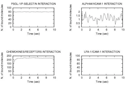
The output of simulation is the time-evolution of number of bonds
(shown in Fig. 2) assuming the following densities expressed in
µm−2: PSGL-119 and P-SELECTIN 5600, ALPHA45 and VCAM-1 85,
CHEMOREC and CHEMOKINES 15000, LFA-110and ICAM-1 5500. The
characterization of the steps and the adhesion molecules implicated in lym-phocyte recruitment in brain venules was performed by using intravital microscopy, a potent technique allowing the visualization and analysis of the adhesive interactions directly through the skull in live animal.
The BioSpi simulations reproduce the hyperbolic behavior (−1/x + const) predicted by the classical mechanical hydrodynamical models pre-sented in the literature6,7,8,11,15,18.
Only lymphocytes that express high levels of PSGL-1, high levels of
LFA-1 and or α4 integrins and the corresponding receptors for the
acti-vating factors presented by the endothelium will be able to efficiently be
recruited in inflamed venules. Starting from this experimental evidence20,
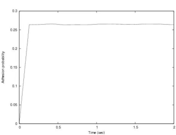
Figure 2. BioSpi simulation of 4-phases model of lymphocyte recruitment. P r(adhesion) = 1 Nl X i wiNi (1)
where Nl = Sendothelium/Scontact is the total number of lymphocytes on
the laminar flux in contact with endothelium given by the ratio between
endothelial surface (∼ 15700µm2) and cell contact area (∼ 200µm2), N
i
indicate the number of bound molecules for the i-th molecular interaction, and the w’s are the weights of the linear model, that quantify the statis-tical influence of the different molecular interactions in the cell adhesion mechanism. In our model the weights can take values in the range be-tween 0 and 1. Because of the lack of experimental quantifications for the statistical influence of the different molecular interactions, we assume that
wi = 1/8 = 0.125 for all the considered interactions of the eigth molecular
species.
The model given in (1) is plotted in Fig. 3. It is in agreement, within the errors ranges, with the theoretical and experimental results in, one of
11
Figure 3. BioSpi model of cell adhesion probability versus contact time (eq. (1).
Adhesion probability
Time (sec)
Figure 4. PSGL-1/P-SELECTIN binding curve.
which illustrated in Fig.4a 16. A more complete presentation of the recent
experimental observations and of the physical treatment of the adhesion
mechanics, in agreement with our BioSpi model, is given in16,21,26.
a
At the present the most studied molecular interaction of the cell adhesion machanics if the PSGL-1/P-SELECTIN interaction.
4. Conclusion
The usage of new languages such as stochastic π calculus to describe and simulate the migration of autorective lymphocytes in the target organ will help us better understand the complex dynamics of lymphocyte recruit-ment during autoimmune inflammation in live animal. Furthermore, our approach may represent an important step toward future predictive studies on lymphocyte behavior in inflamed brain venules. The stochastic calcu-lus may, thus, open new perspectives for the simulation of key phenomena in the pathogenesis of autoimmune diseases, implicating not only better knowledge, but also better future control of the autoimmune attack. Fi-nally, the obtained results show the efficiency of biochemical stochastic π-calculus to simulate experimental data, offering the possibility to model and predict data of biological observations on a computer (in silico experi-ments). This new opportunity provided by the computer science may allow the biologists and medical researchers to save time by reducing the number of needed experiments in the case in which the computer simulation can exclude inadequate hypothesis.
References
1. Bell G. I., Science 200, 618-627, 1978
2. The BioSpi project web site: http://www.wisdom.weizmann.ac.il/∼aviv 3. Chang K. C. and Hammer D. A., Langmuir 12, 2271-2282, 1996.
4. Chang K., Tees D. F. J. and Hammer D. A., The state diagram for cell ad-hesion under flow: leukocyte adad-hesion and rolling, Proc. natl. Acad. Sci. USA 10.1073/pnas200240897, 2000.
5. Chigaev A, Blenc AM, Braaten JV, Kumaraswamy N, Kepley CL, Andrews RP, Oliver JM, Edwards BS, Prossnitz ER, Larson RS, Sklar LA. Real time analysis of the affinity regulation of alpha 4-integrin. The physiologically acti-vated receptor is intermediate in affinity between resting and Mn(2+) or anti-body activation. J Biol Chem. 2001 Dec 28;276(52):48670-8.
6. Dembo M., Torney D. C., Saxaman K. and Hammer D, The reaction-limited kinetics of membrane-to-surface adhesion and detachment. Proc. R. Soc. Lon. B. Vol. 234, pp. 55-83, 1998.
7. Dong C., Cao J., Struble E. J. and Lipowsky H., Mechanics of leukocyte deformation and adhesion to endothelium in shear flow, Annual of biomedical engineering, Vol. 27, pp 298-312, 1999.
8. Fritz J., Katopodis A. G., Kolbinger F. and Anselmetti D., Force-mediated ki-netics of single P-selectin/ligand complexes by atomic force microscopy, Proc. Natl. Acad. Sci USA, Vol. 95, pp.12283-12288, 1998.
9. Gillespie D. T., Exact stochastic simulation of coupled chemical reactions, Journal of Physical Chemistry, 81(25): 2340 - 2361, 1977.
13
in peripheral leukocyte adhesion molecule expression and density. Psychosom Med. 2000 Sep-Oct;62(5):664-670.
11. Goldman A. J., Cox R. G. and Brenner H., Slow viscous motion of a sphere parallel to a plane wall: couette flow, Chem. Eng. Sci, 22: 653 - 660, 1967. 12. Hammer D. A. and Apte S. M. Biophys. J. 63, 35-57, 1992.
13. Kuo S. C., Hammer D. A., and Lauffenburger D. A., Biophys. J. 73, 517-531, 1996.
14. C. Laudanna, J. Yun Kim, G. Constantin and E. Butcher, rapid leukocyte integrin activation by chemokines, Immunological Reviews, Vol. 186: 37-46, 2002
15. Lei X. and Dong C., Cell deformation and adhesion kinetics in leukocyte rolling, BED-Vol. 50, Bioengineering Conference, ASME 1999 (available at http://asme.pinetec.com/bio1999/data/pdfs/a0081514.pdf)
16. B. Marshall, R. P. McEver, C. Zhu, Kinetics rates and their force depen-dence of the P-SELECTIN/PSGL-1 interaction measured by atomic force mi-croscopy, BED-Vol. 50, Bioengineering Conference ASME, 2001
17. Milner R., Communicating and Mobile Systems: the π-calculus. Cambridge University Press, 1999
18. N’dri N., Shyy W., Udaykumar and H. S. Tran-Son-tay R., Computa-tional modeling of cell adhesion and movement using continuum-kinetics ap-proach, BED-Vol. 50, Bioengineering Conference, ASME 2001 (available at http://asme.pinetec.com/bio2001/data/pdfs/a0012976.pdf)
19. Norman KE, Katopodis AG, Thoma G, Kolbinger F, Hicks AE, Cotter MJ, Pockley AG, Hellewell PG. P-selectin glycoprotein ligand-1 supports rolling on E- and P-selectin in vivo. Blood. 2000 Nov 15;96(10):3585-3591.
20. Piccio L., Rossi B., Scarpini E., Laudanna C., Giagulli C., Issekutz A. C., Vestweber D., Butcher E. C. and Costantin G., Molecular mechanism involved in lymphocyte recruitment in inflamed brain microvessel: critical roles for P-selectin Glycoprotein Ligand-1 and Heterotrimeric Gi-linked receptors, The
Journal of Immunology, 2002
21. Pierres A., Benoliel A. M., Bongrand P., Measuring the lifetime of bonds made between surface-linked molecules, J. Biol. Chem. 270:26586-92, 1995. 22. Priami, C., Stochastic π-calculus, The Computer Journal, 38, 6,578–589, 1995 23. Priami, C., Regev A., Shapiro E. and Silverman W.., Application of a stochas-tic passing-name calculus to representation and simulation of molecular pro-cesses, Information Processing Letters, 80, 25 -31, 2001
24. Schmidtke D. W. and Diamond S. L., Direct observation of membrane tethers formed during neutrophil attachment to platelets or P-selectin under physio-logical flow, The Journal of Cell Biology, Vol. 149 Number 3, 2000.
25. Udaykumar H. S., Kan H. C.,Shyy W. and Tran-Son-Tay R., Multiphase dynamics in arbitrary geometries on fixed cartesian grids, J. Comp. Phys., Vol. 137 pp. 366 - 405, 1997.
26. Zhu C., Bao G. and Wang N., Cell mechanics: mechanical response, cell ad-hesion and molecular deformation, Annual Review of Biomedical Engineering 02:189-226.