1
Molecular epidemiology of Blastocystis and association with
intestinal parasites among patients in Negrar hospital, Italy
By
Hossein Soleymanpoor
A Thesis Submitted for the degree of Doctor of Philosophy
Medical Faculty
Polytechnic University of Marche
2
Dedicated with love to my wife
3
Acknowledgements
First of all, I am heartily thankful to my coordinator, Professor Andrea Giacometti, whose encouragement, guidance and support from the initial to the final level enabled me to develop an understanding of the subject.
It is a pleasure to thank Dr. Zeno Bisoffi and Dr. Francesca Perandin who made this thesis possible and with whom I shared ideas and their assistance in lab matters and their patience is highly appreciated.
I also extend my sincere thanks to Dr. Fabio Formenti for his help. I am thankful to Dr Rune Stensvold for his guidance.
Lastly, I extend my deepest gratitude to my parents, my brothers for their unconditional love and support.
4
Abbreviations
DNA Deoxyribonucleic acid IBS Irritable bowel syndrome ST Subtype
GI Gastrointestinal
SSU-rDNA Small-subunit ribosomal DNA PCR Polymerase chain reaction Rt-PCR Realtime PCR
5
Abstract in English
Blastocystis is a common intestinal protozoan, but its clinical significance and
its role in the human gut microbiome is still not completely understood.
Clinical manifestations among symptomatic subjects are mainly
gastrointestinal (GI) or cutaneous symptoms, but also several asymptomatic cases have been described. The aim of the present study was to characterize the presence of Blastocystis in fecal specimens from patients attending to our center for tropical diseases, in order to explore the prevalence and the diversity of Blastocystis infections in our study population. We characterized the presence of the 4 most common subtypes in the selected cohort, composed by subjects with different geographical origins (mainly Italians and Africans). Analysis of co-infections with other parasites has been also performed and description of symptoms found in different subtypes or co-infections combinations has been reported.
Blastocystis resulted to be the most prevalent parasite in our population and
we found that 48.1 % of Blastocystis positive subjects presented GI symptoms. ST3 was the most prevalent subtype in Italians, while in Africans ST1 and ST3 were found with the same frequency. Interestingly, in all the analyzed geographical areas, the most prevalent group was composed by subjects infected by more than one Blastocystis ST. No association between a particular subtype or STs-combination with symptoms has been detected. We observed the presence of co-infecting parasites in the 48.5 % of our cases. An association between Nationality and GI symptoms has been highlighted (P=0.031).
Our study confirms that Blastocystis infection symptomatology cannot be completely explained neither by the different subtypes presence nor by other parasite co-infections, thus supporting the hypothesis that the host condition is the key aspect that can influence the pathogenicity of Blastocystis spp colonization. Future studies on the association between Blastocystis infection
a d patie ts gut i o iota a d/o i u ologi al o ditio s, ould elu idate
6
Riassunto in Italiano
La Blastocisti è un protozoo intestinale comune, ma il suo significato clinico e il suo ruolo nel microbioma intestinale umano non sono ancora completamente
compresi . Le manifestazioni cliniche tra i soggetti sintomatici sono
principalmente sintomi gastrointestinali (GI) o cutanei, ma sono stati descritti
anche diversi casi asintomatici . Lo scopo del presente studio era di
caratterizzare la presenza di Blastocisti in campioni fecali da pazienti che frequentano il nostro centro per malattie tropicali, al fine di esplorare la prevalenza e la diversità delle infezioni da Blastocisti nella nostra popolazione di studio. Abbiamo caratterizzato la presenza dei 4 sottotipi più comuni nella coorte selezionata, composta da soggetti con diverse origini geografiche (principalmente italiani e africani). E 'stata anche eseguita un'analisi delle coinfezioni con altri parassiti e sono stati riportati i sintomi trovati in diversi
sottotipi o combinazioni di coinfezioni.
La Blastocisti è risultata essere il parassita più diffuso nella nostra popolazione e abbiamo scoperto che il 48,1% dei soggetti positivi a Blastocisti presentava sintomi GI. ST3 era il sottotipo più diffuso negli italiani, mentre in Africani ST1 e ST3 erano stati trovati con la stessa frequenza. È interessante notare che, in tutte le aree geografiche analizzate, il gruppo più prevalente era composto da soggetti infettati da più di un Blastocisti ST. Non è stata rilevata alcuna associazione tra un sottotipo particolare o una combinazione ST con sintomi. Abbiamo osservato la presenza di parassiti co-infettanti nel 48,5% dei nostri casi. È stata evidenziata un'associazione tra nazionalità e sintomi gastrointestinali (P = 0,031).
Il nostro studio conferma che la sintomatologia dell'infezione da Blastocisti non può essere completamente spiegata né dalla diversa presenza di sottotipi né da altre parassiti parassitarie, supportando quindi l'ipotesi che la condizione ospite sia l'aspetto chiave che può influenzare la patogenicità della colonizzazione di Blastocisti spp. Studi futuri sull'associazione tra l'infezione da blastocisti e il microbiota intestinale dei pazienti e / o condizioni immunologiche potrebbero chiarire la causa della penetranza patogena variabile osservata di questo parassita.
7
Contents:
Acknowledgements ... 3
Abbreviations ... 4
Abstract in English ...
5
Abstract in Italian ... 6
CHAPTER 1 INTRODUCTION ... 9
1.1 Introduction ... 10
1.2 Taxonomy ... 10
1.3 Morphology ... 11
1.4 Life cycle and Transmission ... 13
1.5 Pathogenesis ... 13
1.5.1 Intestinal symptoms ... 14
1.5.2 Extraintestinal Symptoms ... 15
1.5.3 Blastocystis and Irritable bowel syndrome ... 15
1.6 Treatment of Blastocystis ... 16
1.7 Epidemiology and Prevalence ... 17
1.8 Diagnosis ... 19
1.9 Aims and Objectives ... 21
CHAPTER 2 MATERIALS AND METHODS
... 22
2. Materials and Methods ... 23
2.1 Samples selection ... 23
8
2.3 Real-Time PCR ... 24
2.4 Blastocystis subtype analysis ... 24
2.5 Statistical analysis ... 25
CHAPTER 3 RESULTS ... 26
3.1
Patie ts’ features ... 27
3.2 Characterization of Blastocystis subtypes ... 27
CHAPTER 4 DISCUSSION AND CONCLUSION ... 30
4.1 Discussion ... 31
4.2 Conclusions ... 33
REFERENCES ... 34
LIST OF TABLES ... 47
9
CHAPTER 1:
10
1.1 Introduction
Blastocystis is an unusual protistan enteric parasite classified under a highly
diverse group of organisms called stramenopiles, and is the only known member of this group associated with human pathology (1).
Blastocystis is commonly identified in stool specimens and it is one of the most
common parasites that reside in the human intestinal tract. The disease it causes is called blastocystosis but most publications refer it to as Blastocystis infections. Clinical symptoms attributed to Blastocystis infections include recurrent watery diarrhea, mucous diarrhea, vomiting, abdominal cramps and flatulence. Blastocystis can infect both children and adults and its geographical distribution appears to be global with prevalence ranging from 30 to 50% in developing countries (2).
At first, the name B. enterocola was proposed by Alexeieff (3) and later it was isolated from human feces and the name B. hominis was coined (4). Initially, it was described as harmless intestinal yeast. Its association with human disease was suggested by a number of reports and eventually work by Zierdt increased the awareness of Blastocystis infections in humans. In spite of its description about a century ago, the exact pathogenesis mechanisms of Blastocystis infections are uncertain (5). A number of clinical and epidemiological studies implicate the parasite as a potential pathogen, while others exonerate it as an etiology of intestinal disease (6,7). Significant progress has been achieved on descriptions of the morphology and genetic diversity of Blastocystis but most aspects of its life cycle, molecular biology, and pathogenicity remain unresolved (2,6).
1.2 Taxonomy
The taxonomic classification of Blastocystis is a controversial subject and there are many disagreements among researchers. Blastocystis was earlier described to be a yeast or a fungus (3,8), a cyst of another protozoa (9), or a degenerating cell (10). Blastocystis was described as a protist on the basis of morphological and physiological features (11). These protistan features included presence of one or more nuclei, smooth and rough endoplasmic reticulum, Golgi complex, mitochondria-like organelles, inabilty to grow on fungal medium, ineffectiveness of antifungal drugs, and susceptibility to some
11
antiprotozoal drugs. Later, Blastocystis was classified as a sporozoan (5) and finally reclassified as a sarcodine. Molecular sequencing studies of Blastocystis partial small-subunit rRNA (ssrRNA) showed that Blastocystis is not monophyletic with the yeasts, fungi, sarcodines, or sporozoans (12) and it was concluded that Blastocystis is not related to yeasts. In another study, the complete Blastocystis ssrRNA gene was sequenced and phylogenetic analysis suggested that Blastocystis should be classified within the Stramenopiles (also known as Heterokonta) (13).
Molecular phylogenetic analysis showed that Blastocystis is closely related to the Stramenopile Proteromonas lacerate (14). Another study involving molecular analysis of Blastocystis ssrRNA, cytosolic-type 70-kDa heat shock
protein, translation elongation factor 2, and the non- atalyti B su u it of
vacuolar ATPase confirmed that Blastocystis is a Stramenopile (14). Stramenopiles characteristically possess flagella with mastigonemes. Interestingly, since Blastocystis does not have flagella and is non-motile, it was therefore placed in a newly formed Class Blastocystea in the Subphylum Opalinata, Infrakingdom Heterokonta, Subkingdom Chromobiota, and Kingdom
Chromista (15). In addition, elongation factor- α EF- α) sequencing for
phylogenetic analysis also showed that Blastocystis is not a fungus and suggested that it diverged before Trypanosoma, Euglena, Dictyostelium and other eukaryotes. Most studies in the past named Blastocystis species according to host origin and this may have resulted in confusion regarding specificity, cell biology and pathogenicity of the parasite. A consensus report on the terminology for Blastocystis genotypes was published (16). Based on this report humans can be host to Blastocystis from a variety of animals including mammals (subtype 1), primates (subtype 2), rodents (subtype 4), cattle and pigs (subtype 5), and birds (subtype 6 and 7) (17,18).
1.3 Morphology
The morphological forms exhibited by species are many. Cysts develop into vegetative forms. The well characterized forms are the vacuolar, granular and the amoeboid forms. Other vegetative forms such as avacuolar or multi-vacuolar also have been identified. Other rareforms observed include medusa head and chest nut burr cells especially in aging cultures and on exposure to oxygen. All these above forms can be viewed by phase contrast microcopy and bright field microscopy of wet mounts, stained smears and electron microcopy (2,5,19).
12
The vacuolar form has a large central vacuole filling the entire cell space and limiting the cytoplasm and its intra cellular components to a thin peripheral
i . The e is size a iatio f o μ to μ , though ost human isolates
measure 5- μ . Su se ue tly, the e t al a uoles e e a tually fou d to
be the membrane bound bodies containing carbohydrate and lipid dispersed as flocculent or granular material. These central bodies are probably storage organelles and take part in apoptosis (1,20). The cytoplasmic rim contains one or two nuclei and the mitochondria are observed as rosettes around the nuclei. These structures may bulge inwardly into the central body and appear as filaments. Rarely, in fresh clinical isolates a surface coat or capsule has been observed, which presumably protects the organism from osmotic shock and trap bacteria for nutrition (1,5).
The granular form resembles the vacuolar form except for the presence of granules both in the central body and cytoplasm. The granules may be metabolic, reproductive or lipid containing. The reproductive granules have a possible role in schizogony. The granular forms exhibit lesser degree of
pleomorphism and range in size from 15-80 μ (19).The amoeboid form is less
frequently encountered. As its name implies, they are irregular in shape, possess one or two pseudopodia but are non-motile. The cytoplasm contains a single large vacuole and this form converts to cyst. This form is more frequently observed in symptomatic patients suggesting its pathogenic potential (21). Because they resemble neutrophils and macrophages, they can be easily missed in conventional stool examination. To identify them, Zierdt suggested simultaneous gram staining of an unfixed smear where these forms lyse on exposure to air, while the leucocytes remain intact (1,5). The cyst form
is spherical or ovoid and is smaller in size (3- μ . So e ysts e ou te ed i
animals are larger (22). Each cyst has a thick multilayered wall with or without a surface coat. The condensed cytoplasm has several mitochondria and storage vacuoles.
The number of intracystic nuclei may vary from one to four. The cyst can remain viable for up to one month at 25 °C and on exposure to air. These cysts serve as transmissible infective forms(1). Vegetative forms transform into other vegetative forms of various morphology and hence these can be easily overlooked in fecal samples (20). Though the vacuolar and granular forms appear irregularly stained under the microscope, Vdovenco was able to demonstrate uniformly stained live organisms in fresh cultures. So the vacuolar and granular forms may represent degenerative changes in the organism. The avacuolar forms and the multivaculoar forms have recently been recognized to be the most predominant forms in vivo and also the forms which are frequently missed during microscopic examination (23).
13
1.4 Life cycle and Transmission
Many life cycles have been proposed for Blastocystis (2,3,6) owing to a lack of controlled experimental studies and the pleomorphic nature of the organism. The first life cycle was proposed by Alexeieff and it described the involvement of binary fission and autogamy (3). Some of the reports suggest modes of division like plasmotomy and schizogony (24). Most of these observations were based on microscopic analysis. Although Blastocystis had been isolated from laboratory animals, the lack of a suitable animal model was considered to be a major reason for the disagreement on its life cycle (6). Recent studies have shown successful experimental infection of Blastocystis in chickens (25) and rats (25,26,27). Rats appear to be good animal models for Blastocystis infection but reproducibility of animal infection needs to be ascertained.
A life cycle proposed by Tan states that infection is initiated when cysts of
Blastocystis are orally ingested by humans or animals. Ingested cysts develop
into vacuolar forms in the large intestine and later reproduce by binary fission. Some of the vacuolar forms encyst and are passed through the feces and the cycle is repeated. The role of the amoeboid and granular form in the life cycle of Blastocystis is not understood and remains to be elucidated (6). More recently, Tan revised the life cycle and included findings from molecular typing suggesting that Blastocystis isolated from humans actually comprise human and zoonotic genotypes of varying host specificities (28). A modified life cycle of Blastocystis must take into consideration the large reservoir of this parasite in a range of animal populations with humans as potential hosts(28).
1.5 Pathogenesis
There is still much debate about the pathogenicity of Blastocystis in humans. Though many authors have given credit to it as a pathogen (29,30), there are still many that doubt the role of Blastocystis in human disease (7,31). The most common symptoms associated with Blastocystis infection include diarrhoea, abdominal pain and vomiting. There are many reports of single patients that show there was no other cause of sickness identified in patients, with
Blastocystis being the only infection detected. There have been several case
reports suggesting that Blastocystis is related to urticaria (28). The amoeboid forms of Blastocystis ST3 were found in a case of acute urticaria and the authors suggested that cutaneous symptoms may be caused by disruptions to
14
the immune homeostasis as the host produces an inflammatory response against the amoeboid forms (32).
Another case showed the presence of Blastocystis ST2 in a severe case of gastrointestinal symptoms and chronic urticaria in the absence of any other infectious agent. Symptoms persisted after initial antibiotic therapy but were finally eradicated after combined metronidazole and paromomycin treatment (33). A retrospective study reported 8/8 (11%) Blastocystis infected patients to have skin manifestations as well as gastrointestinal symptoms (34). Unfortunately this study relied solely on microscopy, so no information on ST related to cutaneous lesions can be gathered; however all of these studies do show the potential for Blastocystis to cause cutaneous symptoms. It was recently suggested that gastrointestinal symptoms related to Blastocystis might be ST related but results remain inconclusive (35,36,37). It was suggested that ST1 may be related to pathogenicity with a higher subtype-symptom relationship being noted (38).
There have been conflicting reports on the pathogenicity of ST2 with some studies showing high symptom- infection rates (33,34) whereas others have seen no link (39,40). A study in Colombia showed that 100% of patients with diarrhoea had ST2 where asymptomatic people all had ST1 (41). There have been two previous studies that have suggested ST4 to be a pathogenic strain due to the high incidence of this ST in patients with severe diarrhoea (42,43). It was also suggested that ST8 could be a pathogenic strain. ST8 is a rare subtype found in humans and in two studies has been related to severe symptoms (35,44). Even though ST3 is the most common ST found in humans, there is a low association between ST and symptoms shown by patients (35). An animal study in rats showed that ST1 was statistically related to pathogenicity and that there may be pathogenic and non- pathogenic strains within ST3 and ST4 (27).
1.5.1 Intestinal symptoms
Blastocystosis frequently presents with diarrhea and abdominal pain (28), along with other nonspecific gastrointestinal symptoms including nausea, vomiting, constipation, dysentery, flatulence, bloating, anorexia and weight loss (45). Symptoms range from mild chronic diarrhea to acute enteritis (46). There is some evidence associating parasite density with severity of clinical symptoms caused by Blastocystis (47,48). Greater than five parasites per high-power field (× 400) for wet mounts or oil immersion (× 1000) in permanent stained smears are associated with higher frequency of acute intestinal symptoms (47,48). Recent studies however focus on association of subtypes with human pathology, without any information on infection density (45,49).
15
Since that data is limited to rule out the association of parasite density with intestinal symptoms, future studies might benefit by investigating Blastocystis induced intestinal pathology, focusing on parasite density along with subtype association.
1.5.2 Extraintestinal Symptoms
A correlation between Blastocystis and cutaneous lesions, particularly urticaria, has been reported (32,50). Multiple case studies suggest a causal link between acute or chronic urticaria and Blastocystis. There are reports on association between delayed-pressure urticaria, angioedema, and palmoplantar pruritus with Blastocystis as well (28,33). Resolution of cutaneous symptoms after chemotherapeutic treatment observed in these studies, re-enforce the role of
Blastocystis with skin disorders (51).
1.5.3 Blastocystis and Irritable bowel syndrome
Irritable bowel syndrome (IBS) is "a functional disorder of the gastrointestinal tract characterized by regular occurrence of abdominal pain or discomfort along with alteration in frequency or consistency of the stool in the absence of organic etiology" (52,53,54). It is a very common disorder with a worldwide prevalence of 10-20% (55). Young adult patients are more frequently diagnosed with IBS than people over the age of 50 years (56) and most studies find a female predominance (57,58) although only a few people see their family doctor. The disease results in a reduced quality of life and is a multi-billion pound health-care problem (59). In the UK, only 50% of IBS cases are thought to be diagnosed (60). The symptoms of IBS differ from one person to another and may include irregular bowel movements, abdominal pain or discomfort, flatulence, and diarrhoea or constipation (61). Stress worsens IBS rather than being causative in any way (52).
The cause and pathophysiology of IBS are complicated and not well explained, and the main significant abnormalities include visceral hypersensitivity, irregular. Gut motility and autonomous nervous system dysfunction (61). Barbara et al. (62) demonstrated that there is reliable evidence showing that IBS may be the adverse result of an acute episode of infectious gastroenteritis, the so-called postinfectious (PI) IBS, and the infectious agents involved in the development of IBS include pathogenic bacteria, parasites, and viruses. Studies have illustrated that genetic factors, chronic stress and enteric infections can predispose persons to developing IBS (63). Most drug therapies to date are unable to make a major impact on the quality of life for sufferers (63).
16
Hence, an important problem is to understand what lies behind the development of symptoms in IBS (64).
An understanding of the role of Blastocystis in IBS is restricted by the ambiguity surrounding its pathogenicity (65). Nevertheless, symptoms that have been attributed to infection with Blastocystis are non-specific, IBS-like and include diarrhoea, abdominal pain, cramps or discomfort, and nausea (66,67). Furthermore, chronic excretion of Blastocystis with persistent symptoms has been reported (67).
Hussain et al. showed that IgG antibody levels to Blastocystis in patients with IBS were significantly higher compared with asymptomatic controls, demonstrating immune activation, and suggesting some association between
Blastocystis and IBS (68). In a study by Yakoob et al. Blastocystis was more
frequently demonstrated in the faecal samples of IBS patients (46 %) than the control group (7%) (69).
Giacometti et al. evaluated a possible link between Blastocystis infection and IBS and their findings support a link between the two(70) . In contrast, Tungtrongch et al. found no relationship between presence of Blastocystis in faeces and IBS diagnosis (31).
Some authors have suggested that an intestinal tract that is abnormal for any reason may provide conditions suitable for proliferation of Blastocystis (71,72). It is possible that Blastocystis infection is an indicator of intestinal dysfunction or resident intestinal flora disorders rather than a cause of IBS. Whenever
Blastocystis is detected in stool samples of patients with IBS it does not
necessary mean that the symptoms are due to this organism and other infective and non-infectious causes should be investigated (65). In summary, accumulating reports suggest an association between Blastocystis infection and IBS. However, it is unclear whether Blastocystis is a primary etiological agent in IBS, and it has been suggested that an abnormal intestinal situation like IBS may give an environment in which parasite numbers can increase (71).
1.6 Treatment of Blastocystis
Since pathogenesis of the parasite is controversial, antibiotics are seldom prescribed for Blastocystis infections. The mild nature of the disease and self-limitation of symptoms also make clinicians skeptical about prescription of chemotherapy against the parasite (73). Several drug trials and clinical studies suggest the efficacy of metronidazole against Blastocystis (73), but frequent reports of treatment failure make antiparasitic therapy against it even more
17
controversial (46). Chronic infections in which all other etiologies have been excluded, metronidazole is the treatment of choice (46). Several factors might influence treatment outcomes, including infection density, acquisition of mutations, resistance of certain developmental stages to metronidazole and, most importantly, subtype or strain-to-strain variation in drug susceptibility (46). In fact, in-vitro studies have suggested a varying response of different
Blastocystis isolates to metronidazole (74).
The most commonly prescribed alternatives to metronidazole for Blastocystis infections are co-trimoxazole and paramomycin (73). Unfortunately, resistance to these drugs has also been reported in the parasite (73). There is a pressing need to identify alternative treatment option of Blastocystis infections. Major roadblocks in development of new treatments against the parasite are the absence of efficient drug resistance and susceptibility screening tools. A lack of knowledge concerning molecular mechanisms of Blastocystis antibiotic resistance as well as pathogenesis further complicates the situation. Since
Blastocystis infections are predominantly reported in developing countries,
interest and resources required to develop anti-Blastocystis treatment options are also limited.
1.7 Epidemiology and Prevalence
Blastocystis is reported to be one of the most common protozoans found in
fecal samples of both symptomatic patients and asymptomatic individuals (75,76,77). There is a significant increase in prevalence reports which has helped us to better understand the distribution of genotypes, mode of transmission and pathogenicity aspects. Blastocystis has a worldwide distribution and findings of many surveys reported it to be most frequently isolated protozoan parasite (76,78-80).
Prevalence of Blastocystis infection is higher in developing countries than in developed countries (81) and occurrence as high as 60% were reported from some developing countries (78). Occurrence of Blastocystis varies from country to country. A low prevalence of 0.5% has been reported among asymptomatic healthy individuals in Japan (82). A moderate prevalence of 14-21% and 23% was reported in Thailand (83) and United States (84) respectively.
A high prevalence of 40.7% and 60% was reported in Philippines (80) and Indonesia (78) respectively. High incidences (36.9-44%) of Blastocystis were also observed in Thai military personnel (76,85). Prevalence of Blastocystis may vary widely within various geographical regions of the same country. In
18
Thailand, a prevalence of 0.8% and 45.2% was reported from Nan province (86) and Pathum Thani province (87) respectively.
Variations in the same geographical region may represent true differences between communities or living conditions. Nevertheless, these reported variations might be due to lack of a standardized diagnostic methodology and difficulty in identifying parasitic forms other than the common vacuolar form. Recent studies have used PCR-based approaches to further elucidate genotype information which has shed light on the distribution of Blastocystis genotypes in humans and animals. Studies have found that Blastocystis subtype 3 was the most common subtype among isolates from countries including Turkey (37), Greece (88), Singapore, Japan, Pakistan, Bangladesh, and Germany (39). In summary, studies suggest that there is no association between specific genotype and geographic origin; and due to its predominance in urbanized countries, subtype 3 is probably the subtype of human origin. It has been observed that humans with compromised health and poor hygiene are more susceptible to Blastocystis infections.
Blastocystis infections are also of special clinical interest to developed
countries as millions of travelers going to developing countries are at risk of acquiring infection (90). Blastocystis infections are more common during hot weather and during the pre-monsoonal months (2). Based on current knowledge, it is generally accepted that Blastocystis is transmitted by the fecal-oral route. This assumption is strengthened by animal infection studies (26,91) and reports showing high prevalence of Blastocystis in population living in poor hygiene (92). Therefore, control measures should consist of good hygiene practices and community sanitary facilities. Because Blastocystis is generally regarded as a zoonotic parasite, animals and their fecal material represent a risk for human infection. Contamination of food, water, and environment by animal fecal material should be prevented. High prevalence of Blastocystis has been shown in pets particularly dogs and cats and it was suggested that these domestic animals could be an important source of infection to humans (93). Routine antiparasitic treatment practice for pet animals may be useful to eliminate the parasite. Animal handlers must take additional precautions for their personal hygiene and may go for stool examination especially if experiencing any gastrointestinal symptoms. In unhygienic and high
Blastocystis prevalence areas, sterilization of water is recommended.
Currently, the best sterilization method is to boil water as chemical methods of water sterilization have not been extensively studied for Blastocystis. Travelers to high prevalence areas should ensure that they consume clean water and cooked food. Blastocystis has been found in sewage (94) and there is growing
19
evidence for waterborne transmissions (84) which makes it necessary to develop preventive measures to ensure water sanitation.
1.8 Diagnosis
Because of its uncertain pathogenesis, reasonable clinical significance is seldom given to Blastocystis infections. Generally, diagnosis and other important aspects of Blastocystis infections are not included in the curriculum of medical studies and thus diagnosis of Blastocystis remains a challenging task for a diagnostic laboratory. Although an experienced laboratory technician can perform diagnosis in direct fecal smears, most diagnostic laboratories do not have expertise on identification of this parasite and there is a need for training to enable identification of all forms of Blastocystis in fecal samples. Identification of Blastocystis in direct fecal smears is relatively difficult as the parasite can be confused with yeast, cyclospora, or fat globules. In the past, laboratory diagnosis of Blastocystis was based on the identification of vacuolar and granular forms in direct fecal specimens (95).
Direct i os opy of fe al spe i e s is pe fo ed y et ou ts ith Lugol s
iodine or permanent fixed smears with Giemsa, acid-fast, t i h o e a d Field s
staining. Rather than the characteristic vacuolar form, the cyst form may predominate fecal samples. Cyst forms might be difficult to identify by direct
microscopy because of their small size (3-5 μ ut these a e effe ti ely
concentrated by density-gradient methods (74).
Diagnostic labs should therefore include the fecal cyst form as an indicator of
Blastocystis infection. Many researchers suggest that when all other known
bacterial, viral or parasitic causes of symptoms are absent and Blastocystis is present in large numbers it should be treated as a pathogen. More than five organisms per high power field (×400 magnification) should be considered as a heavy infection. For confirmative diagnosis in stool samples, in vitro culture in Jo es ediu is a ethod of hoi e 96). It was reported that in vitro culture of fecal samples was six times and twice more sensitive than direct fecal smears and trichrome staining methods respectively (97). However, it was also reported in this study that the in vitro culture method failed to detect some parasites suggesting that not all Blastocystis isolates can be readily cultured in
laboratory. Blastocystis can be cultured in various mediums i ludi g Jo e s
ediu , Boe k a d D ohla s i spissated ediu o diphasi aga slant ediu ith Jo e s as a ediu of hoi e fo patient samples. Diphasic agar
20
slant medium was reported to be good for the culture of Blastocystis from pigs, cattle and chickens (98,99). In axenized cultures, cell densities of up to 2.5 ×10⁷ can be achieved (100) and doubling time may vary from 6 to 23 h, depending on type of medium and isolate (101). Colony growth of Blastocystis has been shown on solid medium and cultures were viable for up to 2 weeks (102). Molecular approaches, particularly PCR-based diagnosis have been described for Blastocystis (103). PCR amplification using subtype specific primers is suggested to be useful for identifying and genotyping Blastocystis from patient samples. Knowledge of the genotype can be extremely valuable if certain Blastocystis genotypes are found to be more virulent than others. A study has demonstrated that PCR-based detection of Blastocystis from fecal specimens is more sensitive than in vitro propagation (104). A sensitive and specific real-time light cycler PCR assay was developed to detect a 152 bp sequence in an uncharacterized region of the Blastocystis genome and 11 strains of Blastocystis from subtypes 1, 3, and 4 were with this method (105). Using this method, Blastocystis was detected in stool samples that were found
Blastocystis negative during microscopy and conventional PCR. In addition, this
method showed no cross-reactivity with other common gastrointestinal pathogens.
Other methods like enzyme-linked immunosorbent assay (ELISA) and immunofluorescence detection have not been comprehensively investigated for Blastocystis. Although development of monoclonal antibodies against
Blastocystis has been reported (106), antigenic diversity of Blastocystis seems
to be a limiting factor in the use of immunological methods.
Blastocystis infections have been reported to induce IgG and IgA responses in
patients and detected by indirect fluorescent antibody test (IFA) and ELISA (68,107-110). ELISA titers ranged from 1:50-1:1,600 (108) and it was observed that high titers were associated with symptomatic infections of Blastocystis (68,107,108,110). In a recent study using ELISA, secretory IgA, serum IgA and serum IgG levels were detected in Blastocystis infected patients with and without clinical symptoms (110). It was found that serum from only symptomatic patients had significantly higher antibody levels. On the other hand, Kaneda et al. (109) reported asymptomatic patients with serum antibodies to Blastocystis and high levels were observed in chronic cases. Overall, it may be desirable to develop specific monoclonal antibodies against different genotypes and evaluate different serological assays for the diagnosis of Blastocystis infections.
Diagnosis of blastocystosis has been reported with the help of invasive diagnostic techniques like endoscopy but it has not been evaluated.
21
detected in the microscopic examinations of the lumen fluids aspirated during endoscopy (111). As Blastocystis can be detected in feces and no characteristic intestinal lesions are associated with infection, invasive diagnostic techniques are not recommended for routine examinations. In brief, a number of methods have been described for the diagnosis of Blastocystis. Direct microscopy of stained fecal smears is useful and it should be supplemented with numbers of parasites observed per high power field to help clinicians ascertain parasitic load. For confirmatory diagnosis, microscopic examination should be supplemented by in vitro culture and/or PCR-based methods.
1.9 Aims and Objectives
In the present study, we analyzed all fecal specimens for which, in previously molecular diagnosis, we observed the presence of Blastocystis DNA. The stool specimens were collected from patients suspected of harboring intestinal
parasites and attending to our center for tropical disease in two years (2014 –
2015). The aim of the study was to characterize Blastocystis genotype distribution in our patients, evaluating the influence of different geographical origins, the dynamics of mixed STs and the association with other parasite co-infections, in order to explore the prevalence and the diversity of Blastocystis infections in our cohort population. A description of symptoms found in patients carrying different subtypes or co-infection combinations has been reported.
22
CHAPTER 2:
23
2. Materials and Methods
2.1 Samples selection
This retrospective study was performed at the Center for Tropical Diseases of Sacro Cuore-Don Calabria Hospital in Negrar (Verona), a referral center for tropical and parasitic infections in Italy. In this center receives patients coming from all Italian regions, people who need medical assistance and immigrants lived in temporary accommodation centers.
The sample identification code was retrieved from the electronic archive of the molecular parasitology laboratory searching among all specimens collected from January 2014 to December 2015. In this period, a total of 1778 fecal samples were screened by three separate multiplex Real time polymerase chain reaction (Rt-PCR) for detecting Entamoeba histolytica - Entamoeba
dispar - Cryptosporidium spp, Giardia intestinalis - Dientamoeba fragilis - Blastocystis spp, Strongyloides stercoralis - Schistosoma spp - Hymenolepis nana. Among these, 756 samples were subjected to the molecular test for the
presence of Blastocystis spp.
260 Samples positive for Blastocystis spp. were further analyzed for the 4 most frequent human subtypes (ST1, ST2, ST3 and ST4) molecular characterization, as described in the following paragraphs. The samples investigated for
Blastocystis subtypes were divided into three groups based on the presence of
gastrointestinal symptoms, itching or absence of symptoms. The geographical origin of the patients was also considered (Italians, non-Italian Europeans, Africans, Asians and South Americans).
2.2 Fecal samples collection and DNA extraction
According to the routine procedure of our laboratory, fecal samples collected for molecular test were stored in 95% ethanol prior to be processed. DNA extraction was performed as previously described (112). 200 mg of each stool
sample was stored at –20 °C overnight in a solution of PBS 1X with 2% of
polyvinylpolypyrrolidone (PvPP) (Sigma-Aldrich, Milan, Italy). In each sample, Phocine Herpes Virus type-1 (PhHV-1, kindly provided by Dr. Pas S., Erasmus MC, Department of Virology , Rotterdam) was added within the S .T.A.R. buffer (Roche, Milan, Italy), serving as an internal control for the isolation and
amplification steps. All the samples were then frozen and boiled for 10 min at ̊ C. The DNA was extracted by MagnaPure LC.2 instrument (Roche
24
Diagnostic, Monza, Italy), using the DNA isolation kit I (Roche). The DNA was eluted in a final volume of 100 µl. DNA samples were appropriately labelled and stored at -20°C for subsequent molecular tests.
2.3 Real-Time PCR
Each DNA was amplified by Real-Time PCR (CFX96-Biorad) as described by Stensvold et al. (113). The realtime is a multiplex PCR able to detect at the same time the possible presence of 3 protozoa (D. fragilis-G.
intestinalis-Blastocystis spp). Moreover, this multiplex PCR detect the PhHV DNA; it is an
exogenous DNA added to the samples before to start the extraction. It is necessary to verify the good performance of extraction and the presence of inhibitory for the polymerase enzyme. Also, in each PCR run are presents 2 positive controls (high and low DNA quantity) and a negative control.
2.4 Blastocystis subtype analysis
Nested-PCR was performed according to the protocol described by Scanlan et al (114). with minor modifications. First step PCR was performed to provide a
Blastocystis specific 18S rDNA template for each of the subsequent ST-specific
PCRs (ST1, ST2, ST3, and ST4). RD5, BhRDr, ST1-F, ST2-F, ST3-F, ST4-F primers sequences were retrieved from (114) and PCR was performed using iTaq DNA polymerase (Bio-Rad, Milan, Italy) in 50 µL of reaction volume, according to the a ufa tu e s i st u tio s. The follo i g y li g o ditio s e e used fo the first step PCR: initial denaturation 95°C for 3 min, 30 cycles at 94°C for 1 min, 59°C for 1 min, 72°C for 1 min, final elongation 72°C for 5 min. 5 µL of DNA sample was used ( Table 1). The ST-specific PCRs were performed as follows: initial denaturation 95°C for 3 min, 35 cycles at 94°C for 30 sec, Tannealing primers for 30 sec, 72°C for 1 min, final elongation 72°C for 5 min. The following Tannealing were used: 56° C for ST1 and ST2, 48° C for ST3 and ST4. 1 µL the initial PCR product was used per each reaction (Table 2). A no-template control was always included in each PCR run. PCR products were analyzed by 2.5 % agarose gel electrophoresis, to detect the specific DNA bands. 2 examples of gel images are reported in figure1 and figure 2.
25
2.5 Statistical analysis
Descriptive statistics were used to analyze the characteristics of the entire cohort and separately for each continent of origin of patients. The statistical analysis was performed on data collected in our electronic database. Data entry of each specimens cover: ID, age, sex, nationality, symptoms, Blastocystis
ST, other parasites (Entamoeba histolytica, Entamoeba dispar, Giardia intestinalis, Dientamoeba fragilis, Strongyloides stercoralis, Schistosoma spp, Hymenolepis nana). We then investigated on associatio s et ee all patie ts
characteristics through univariate logistic regression models and parametric and non-parametric statistical test such as Chi-Squared test. The data were statistically assessed using SPSS 16.0. Chi square test and percentages were used for data analysis, the P values below or equal to 0.05 were regarded as significant.
26
CHAPTER 3:
27
3. Results
3.1 Patie ts’ features
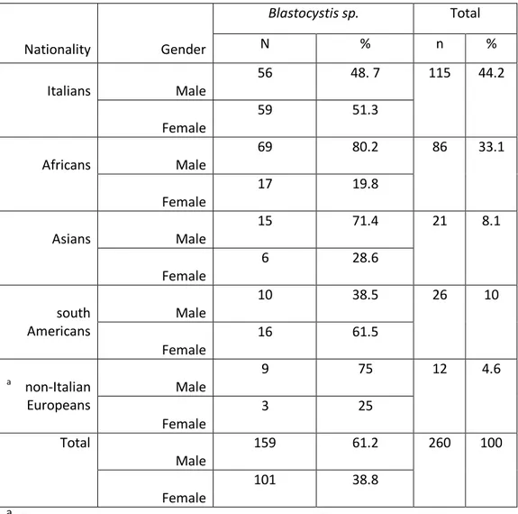
In a group of 1778 samples subjected to molecular tests for the investigation of intestinal parasite infection, 756 were tested for Blastocystis. Among these, we identified 260 (34.4 %) positive samples for Blastocystis spp. From a total of 260 persons infected with Blastocystis sp. 159 (61.2%) were male and 101 (38.8%) were female. Our study population was composed by people of different geographical origins, with Italians 115 (44.2%) and Africans 86 (33.1%) being the predominant ones; other patiens were from South Americans 26 (10%), Asians 21 (8.1%) and non-Italian Europeans 12 (4.6%) (Table 3).
According to table 4, 29.2% of subjects were more than 50 years old. Prevalence in other age groups were 10.8% at below 10 years old, 12.7% at 11-20, 19.2% at 21-30, 14.2% at 31-40 and 13.9% at 41-50 years old.
3.2 Characterization of Blastocystis subtypes
We further analyzed the Blastocystis positive subjects by a specific Nested-PCR assay (114), in order to evaluate the presence of the 4 most prevalent
Blastocystis STs and we were able to characterize a total of 260 samples.
Obtained results are reported in Table 5. In particular, we found that ST3 and ST1 were the most frequent subtypes (single subtype carriers), with a prevalence of (24.6%) and (22.7%) respectively, also ST2 and ST4 were detected in the (8.1%) and (7.3%) of samples. The most prevalent group was composed by subjects infected by more than one Blastocystis ST (37.3%). Among mixed STs co-infections, the most frequent was clearly the ST1-ST3 (16.9%) (Table 5). We found multiple double infection, ST3-ST4 (3.8%), ST1-ST2 (2.3%), ST2-ST3 (1.9%), ST1-ST4 (0.4%) and ST2-ST4 (0.8%). Although with a lower frequency, triple STs infections were also detected, like ST1-ST3-ST4 (4.6%) and ST1-ST2-ST3 (4.2%) and ST2-ST3-ST4 (1.6%), and finally (0.8%) of quadruple infection by ST1-ST2-ST3-ST4 (Table 5).
Table 6 shows the prevalence of subtypes of Blastocystis according to gender that 26.4% of male were infected with ST1 also 28.7% of female were infected
28
with ST3. No sig ifi a t o elatio as fou d et ee patie tsʼ ge de a d
Blastocystis subtypes infection (P= 0.381).
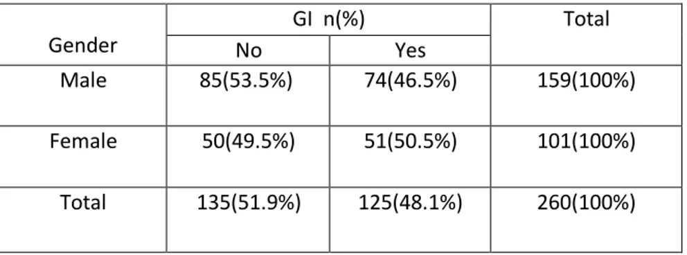
Analyzing the clinical phenotype of our study, we found that 48.1% % of
Blastocystis positive subjects presented GI symptoms (abdominal pain,
diarrhea, irritable bowel syndrome) (Table 7), 18.8 % reported itching (P= 0.074) (Table 8). No association between GI symptoms and gender has been observed (p = 0.310) (Table 7).
A slightly correlation (P=0.038) was observed between the gender and infected with Blastocystis and other parasites (Table 9).
Table 10 shows distribution of Blastocystis subtypes among age groups.
25.4% of individuals that were infected with ST1, had more than 50 years old. Also in the individuals infected with ST2, 33.3% were observed in the 21-30 years old group (Table 10). There was correlation between age groups and
Blastocystis subtypes (P= 0.044).
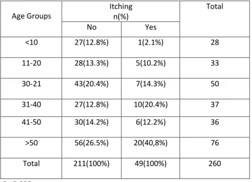
Among age groups 32.8% of more than 50 years old presented GI symptoms. 9.6% of Subjects less than 10 years old, also 10.4% of 11-20, 20.8% of 21-30, 14.4% of 31-40 years old have GI symptoms. (P= 0.660) (Table 11). 40.8% of more than 50 years old reported Itching (P=0.080) (Table12).
According to the Table 13, 78.6% of subjects less than 10 years old that, in addition to Blastocystis were infected with other parasites. Between the age groups and infected with other parasites were correlation (P= 0.008).
Table 14 shows Frequency of individuals infected with Blastocystis subtypes According to Nationality (P= 0.125).
According to the correlation between Nationality and GI Symptoms, 58.3% of the Italian subjects presented GI symptoms. There was significant between GI sypmtoms and Nationality (P=0.031) (Table 15).
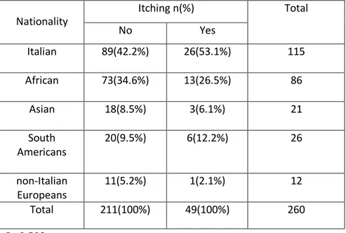
Frequency of individuals infected with Blastocystis sp. according to Nationality and Itching shows 53.1% of subjects were Italian (P=0.508) (Table 16).
Frequency of individuals infected with Blastocystis sp. only and Blastocystis with other parasites According to Nationality shows there was significant
29
between Nationality and infected with other parasites (P= 0.009). 76.2% of Asian subjects infected with Blastocystis sp. and other parasites (Table 17). Among single subtype carriers, 59.3% of individuals infected with ST1 presented GI symptoms. 33.3% of ST2, 51.6% of ST3 and 47.4% of ST4 subjects presented GI symptoms (P= 0.353) (Table 18).
According to the table 19, 50.7% of subjects infetcted with Blastocystis only have GI symptoms also 45.2% of subjects infected with Blastocystis and other parasites were presented GI symptoms (P= 0.222) (Table 19).
In patients carrying Blastocystis, we analyzed also the presence of other co-infecting parasites: Entamoeba histolytica, Entamoeba dispar, Giardia
intestinalis, Dientamoeba fragilis, Strongyloides stercoralis, Cryptosporidium spp, Schistosoma spp, Hymenolepis nana. We analyzed the possible association
between the presence of GI symptoms and co-infection with different parasites, but no statistically significant difference has been detected (P = 0.754) between samples infected only by Blastocystis (54.4 % presenting symptoms, 68/134) and samples with multiple parasite coinfections (45.2 % presenting symptoms, 57/126). Table 20 reports the results on the association between symptoms and Blastocystis, both with and without other parasitic infections (P= 0.754).
Table 21 shows distribution of subtypes of Blastocystis according to infected with Blastocystis only or infected with other parasites (P= 0.401). 27.6% of subjects infected with Blastocystis only reported ST3 and 26.2% of subjects infected Blastocystis and other parasites reported ST1.
30
CHAPTER 4
DISCUSSION AND
CONCLUSIONS
31
4. DISCUSSION AND CONCLUSION
4.1 Discussion
As a result of our retrospective study, Blastocystis emerged as the most frequent parasite in samples tested for intestinal parasite infections at our Centre for Tropical Diseases, confirming previous reports indicating that the prevalence of Blastocystis infection is higher than that of other intestinal parasites (115). We could characterize the Blastocystis subtype for 260 positive samples. Our analysis highlighted that ST3 was the most prevalent subtype in Italians of our regional area, and this result reflected previous report by another Italian group (116). ST1 was also present with a slightly lower frequency.
The findings showing the dominance of ST3 are similar to most previous studies in Europe and Asia, such as Jamtemtor’s study in Thailand that reported ST3 as the most dominant subtype (57.1%), followed by ST1 (21.4%), ST7 (17.9%) and ST6 (3.6%) (117) Similarly, Wong et al. study in Singapore found ST3 to be the most dominant subtype (78%), followed by ST1 (22%) (89). Boondit et al. study also reported ST3 as the most dominant subtype (76%), followed by ST1 (20%) (118). Meloni et al. in Italy found the following ST distribution: ST3 (47.1%), ST2 (20.6%), ST4 (17.7%), ST1 (8.8%), and ST7 and ST8 (2.9%) (119). A study by Dogruman et al. reported similar results, with ST3 being the most dominant in the symptomatic (59.3%) and asymptomatic groups (48.5%), followed by ST2 with 15.3% and 33.3% and ST1 with 20.3% and 15.2%. Similar subtype prevalence rates were also reported by Ozyurt et al. in Turkey and other countries such as China, Germany, Japan, and Denmark (40). A different result was reported in a study by Awatif et al. in Libya where ST1 was the most dominant subtype in outpatients (51.1%), followed by ST2 (24.4%) and ST3 (17.8%) (120). An alternate result was also reported in a study by Dominguez et al. in Spain where ST4 was the most dominant subtype at 94.1%, followed by ST1 (2%) and ST2 (3.9%) (42). Souppart et al. stated that
Blastocystis infection may not link to certain subtypes but to risk factors in
infection transmission, including environmental factors (transmission route and source of contamination), parasite factors (pathogenic potential and zoonosis), and host factors (genotype, immunity, and age) (121).
In our study, we had the opportunity to compare subjects coming from different geographical areas. In Africans, the second predominant group, ST1
32
and ST3 were the most prevalent subtypes, present with equal frequencies. Forsell and colleagues reviewed the subtype prevalence in Africa and, depending on the considered country, they reported ST1 and ST3 as actually the most prevalent subtypes (122). For sake of clarity, we have to consider that STs distribution in our population could be also influenced by the time of permanence in Italy of the foreign subjects, but this variable was not retrievable in our retrospective study. This aspect could also explain the presence of ST4 in few African individuals, since ST4 has been reported as being absent in African populations by several studies (reviewed in (122,123)).
Anyway, globally, our cohort confirmed that ST3 and ST1 were the most prevalent subtypes. An intriguing result of our analysis was the frequent presence of mixed subtype infections, across all the geographical areas. This observation confirms how a subtype-specific PCR assay could highlight a frequent presence of mixed subtype co-infections (114). This aspect has received little attention in the past, since it was often underestimated by methodological limits and considered just an incidental finding. Recently, the presence of mixed subtype infections has been highlighted as an important characteristic to be studied, in order to explore the diversity and distribution of this parasite in the human gut (114). Applying the state-of-the-art subtype-specific method (114) we were able to detect mixed infection in the 37.3 % of our cases, with ST1-ST3 being the most common mixed subtype combination, as already observed in other studies (reviewed in (124)). This data could indicate that the mutual presence of different subtypes could be a successful cooperation strategy for host colonization by Blastocystis spp. The same concept could also be applied for the presence of multiple parasite co-infections: the association between Blastocystis and D. fragilis could also indicate a cooperative interaction between the two protozoa. Association between Blastocystis spp and other parasites has already been observed in literature (e.g. with G. intestinalis (122). and in particular with D. fragilis (125)) but no conclusive association with symptoms has been highlighted.
Although we did not detect any statistically significant correlation between any STs and symptoms (probably due to the limited number of samples in our study, or to the intrinsic genetic variability and/or immunological factors), neither in mixed subtypes presence or other parasite co-infections, we observed a slightly higher proportion (48.1 %) of symptomatic versus asymptomatic subjects in Blastocystis positive subjects, with a prevalence of ST1 and ST3 in patients with GI symptoms, while ST2 seems to have a lower impact; moreover, the presence of both Blastocystis and D. fragilis, in our
33
study, seems to increase the percentage of patients with GI symptoms, respect to Blastocystis alone or in co-infection with other tested parasites. Anyway, since no statistically significant clue has been obtained, additional data are needed to confirm these indications.
This study has several limitations, mainly related to the retrospective study design. In particular, symptom reporting may not be sufficiently accurate and this might be a further reason for the lack of statistically significant correlation between the molecular findings and the clinical characteristics.
4.2 Conclusions
Our study confirms that Blastocystis infected subjects present a highly variable symptomatology, that is not completely explicable by the different subtypes presence or by other parasite co-infections; although the variability of the samples could require a higher number of observations to reach a statistically significant indication, our data support the hypothesis that the host condition is the key aspect that can influence the pathogenicity of Blastocystis spp
olo izatio . Futu e studies o patie ts i u ologi al o ditio s a d gut
microbiota, associated to Blastocystis infection, could fill the gap of information to explain the cause of the observed variable pathogenic penetrance of this parasite.
34
REFERENCES
1. Tan KS. New insights on classification, identification, and clinical relevance of
Blastocystis spp. Clin Microbiol Rev 2008, 21:639–65.
2. Stenzel DJ, Boreham PFL. Blastocystis hominis revisited. Clin Microbiol Rev 1996, 9:563-584.
3. Alexeieff A. Sur la nature des formations dites kystes de Trichomonas
intestinalis. C R Soc Biol 1911, 71:296-298.
4. Brumpt E. Blastocystis hominis n. sp. et formes voisines. Bulletin de la
Societe de Pathologie Exotique et de Ses Filiates (Paris) 1912, 5:725–730.
5. Zierdt CH . Blastocystis hominis-past and future. Clin Microbiol Rev 1991, 4: 61-79.
6. Tan KSW. Blastocystis in humans and animals: new insights using modern methodologies. Vet Parasitol 2004, 126:121-144.
7. Leder K, Hellard ME, Sinclair MI, Fairley CK, Wolfe R. No correlation between clinical symptoms and Blastocystis hominis in immunocompetent individuals. J Gastroenterol Hepatol 2005, 20:1390-1394.
8. O Co o F W. Intestinal protozoa found during acute intestinal conditions
amongst members of the Egyptian expeditionary force, 1916–1917.
Parasitology 1919, 11:239–253.
9. Bensen, W. Trichomonas intestinalis und vaginalis des Menschen. Arch
Protistenkd 1909, 18:115–127.
10. Swellengrebel NH. Observations on Blastocystis hominis. Parasitology 1917,
9:451– 459.
11. Zierdt CH, Rude WS, Bull BS. Protozoan characteristics of Blastocystis
hominis. Am J Clin Pathol 1967, 48:495-501.
12. Johnson AM, Thanou A, Boreham PF, Baverstock PR. Blastocystis hominis: phylogenetic affinities determined by rRNA sequence comparison. Exp Parasitol 1989, 68:283-288.
35
13. Silberman JD, Sogin ML, Leipe DD, Clark CG. Human parasite finds taxonomic home Nature. 1996, 380:398.
14. Arisue N, Hashimoto T, Yoshikawa H, Nakamura Y, Nakamura G, Nakamura F, Yano TA, Hasegawa M. Phylogenetic position of Blastocystis hominis and of stramenopiles inferred from multiple molecular sequence data. J Eukaryot Microbiol 2002, 49:42-53.
15. Cavalier-Smith T. A revised six-kingdom system of life. Biol Rev Camb 861 Philos Soc 1998, 73:203-266.
16. Stensvold CR, Suresh GK, Tan KS, Thompson RC, Traub RJ, Viscogliosi E, Yoshikawa H, Clark CG. Terminology for Blastocystis subtypes--a consensus. Trends Parasitol 2007, 23:93-96.
17. Noël C, Dufernez F, Gerbod D, Edgcomb VP, Viscogliosi PD, Ho LC, Singh M, Wintjens R, Sogin ML, Capron M, Pierce R, Zenner L, Viscogliosi E. Molecular phylogenies of Blastocystis isolates from different hosts: Implications for genetic diversity, identification of species, and zoonosis. J Clin Microbiol 2005, 43:348-355.
18. Yan Y, Su S, Ye J, Lai X, Lai R, Liao H, Chen G, Zhang R, Hou Z, Luo X.
Blastocystis sp. subtype 5: a possibly zoonotic genotype. Parasitol Res 2007,
101:1527- 1532.
19. Stenzel DJ, Boreham RE. Blastocystis. In: Gillespie S& Pearson RD editors. Principles and practice of Clinical parasitology. October 2001 edition .John Wiley& sons 2001: 355-367.
20. Parija SC, Jeremiah SS. Blastocystis: Taxonomy, biology and virulence. Trop Parasitol 2013,3:17-25.
21. Vassalos CM, Spanakos G, Vassalou E, Papadopoulou C, Vakalis N. Differences in clinical significance and morphologic features of Blastocystis sp subtype 3. Am J Clin Pathol 2010,133:251-8.
22. Stenzel DJ, Lee MG, Boreham PF. Morphological differences in Blastocystis cysts-an indication of different species? Parasitol Res 1997,83:452-7.
36
23. Vdovenko AA. Blastocystis hominis: Origin and significance of vacuolar and granular forms. Parasitol Res 2000,86:8-10.
24. Singh M, Suresh K, Ho LC, Ng GC, Yap EH. Elucidation of the life cycle of the intestinal protozoan Blastocystis hominis. Parasitol Res 1995,81:446-450.
25. Iguchi A, Ebisu A, Nagata S, Saitou Y, Yoshikawa H, Iwatani S, Kimata I. Infectivity of different genotypes of human Blastocystis hominis isolates in chickens and rats. Parasitol Int 2007,56:107-112.
26. Yoshikawa H, Yoshida K, Nakajima A, Yamanari K, Iwatani S, Kimata I. Fecal-oral transmission of the cyst form of Blastocystis hominis in rats. Parasitol Res 2004,94:391-396.
27. Hussein EM, Hussein AM, Eida MM, Atwa MM. Pathophysiological variability of different genotypes of human Blastocystis hominis Egyptian isolates in experimentally infected rats. Parasitol Res 2008,102:853-860.
28. Tan KS. Blastocystis spp. N.A. Khan (ed.), Emerging Protozoan Pathogens. Taylor and Francis 2008
29. Andiran N, Acikgoz ZC, Turkay S, Andiran F. Blastocystis hominis–an
emerging and imitating cause of acute abdomen in children. J Pediatr Surg
2006, 41(8):1489–1491.
30. Levy Y, George J, Shoenfeld Y. Severe Blastocystis hominis in an elderly
man. Journal of Infect 1996, 33(1):57–59.
31. Tungtrongchitr A, Manatsathit S, Kositchaiwat C, Ongrotchanakun J, Munkong N, Chinabutr P, Leelakusolvong S, Chaicumpa W. Blastocystis hominis infection in irritable bowel syndrome patients. Southeast Asian J Trop Med
Public Health 2004, 35(3):705–710.
32. Katsarou-Katsari A, Vassalos CM, Tzanetou K, Spanakos G, Papadopoulou C, Vakalis N. Acute urticaria associated with amoeboid forms of Blastocystis sp.
subtype 3. Acta Derm Venereol 2008, 88(1):80–81.
33. Vogelberg C, Stensvold CR, Monecke S, Ditzen A, Stopsack K, Heinrich-Grafe U, Pohlmann C. Blastocystis sp. subtype 2 detection during recurrence of
37
34. Balint A, Doczi I, Bereczki L, Gyulai R, Szucs M, Farkas K, Urban E, Nagy F, Szepes Z, Wittmann T, Molnar T. Do not forget the stool examination!-cutaneous and gastrointestinal manifestations of Blastocystis sp. infection.
Parasitol Res 2014, 113(4):1585–1590.
35. Roberts T, Stark D, Harkness J, Ellis J. Subtype distribution of Blastocystis isolates identified in a Sydney population and pathogenic potential of
Blastocystis. Eur J Clin Microbiol Infect Dis 2013, 32(3):335–343.
36. Bohm-Gloning B, Knobloch J, Walderich B. Five subgroups of Blastocystis
hominis from symptomatic and asymptomatic patients revealed by restriction
site analysis of PCR-amplified 16S-like rDNA. Trop Med Int Health 1997,
2(8):771–778.
37. Ozyurt M, Kurt O, Molbak K, Nielsen HV, Haznedaroglu T, Stensvold CR. Molecular epidemiology of Blastocystis infections in Turkey. Parasitol Int 2008,
57(3):300–306.
38. Yan Y, Su S, Lai R, Liao H, Ye J, Li X, Luo X, Chen G. Genetic variability of
Blastocystis hominis isolates in China. Parasitol Res 2006, 99(5):597–601.
39. Yoshikawa H, Wu Z, Kimata I, Iseki M, Ali IK, Hossain MB, Zaman V, Haque R, Takahashi Y. Polymerase chain reaction-based genotype classification among human Blastocystis hominis populations isolated from different countries.
Parasitol Res 2004, 92(1):22–29.
40. Dogruman-Al F, Dagci H, Yoshikawa H, Kurt O, Demirel M. A possible link between subtype 2 and asymptomatic infections of Blastocystis hominis.
Parasitol Res 2008, 103(3):685–689.
41. Ramirez JD, Sanchez LV, Bautista DC, Corredor AF, Florez AC, Stensvold CR.
Blastocystis subtypes detected in humans and animals from Colombia. Infect
Genet Evol 2013, 22:223–228.
42. Dominguez-Marquez MV, Guna R, Munoz C, Gomez-Munoz MT, Borras R. High prevalence of subtype 4 among isolates of Blastocystis hominis from symptomatic patients of a health district of Valencia (Spain). Parasitol Res
38
43. Stensvold CR, Christiansen DB, Olsen KE, Nielsen HV. Blastocystis sp. subtype 4 is common in Danish Blastocystis-positive patients presenting with
acute diarrhea. Am J Trop Med Hyg 2011, 84(6):883–885.
44. Stensvold CR, Arendrup MC, Nielsen HV, Bada A, Thorsen S. Symptomatic infection with Blastocystis sp. subtype 8 successfully treated with
trimethoprim-sulfamethoxazole. Ann Trop Med Parasitol 2008, 102(3):271–
274.
45. Stensvold CR, Lewis HC, Hammerum AM, Porsbo LJ, Nielsen SS, Olsen KE, Arendrup MC, Nielsen HV, and Mølbak K. Blastocystis: unravelling potential risk factors and clinical significance of a common but neglected parasite. Epidemiol Infect 2009,137: 1655-1663.
46. Tan KS, Mirza H, Teo JD, Wu B, and Macary PA. Current Views on the Clinical Relevance of Blastocystis spp. Curr Infect Dis Rep 2010,12: 28-35.
47. Kaya S, Cetin E, A idoğa B, Arikan S, and Demirci M. Pathogenicity of
Blastocystis hominis, a clinical reevaluation. Turkiye Parazitol Derg 2007,31:
184-187.
48. Moghaddam DD, Ghadirian E, and Azami M. Blastocystis hominis and the evaluation of efficacy of metronidazole and trimethoprim/sulfamethoxazole. Parasitol Res 2005,96: 273-275.
49. Eroglu F, Genc A, Elgun G, and Koltas IS. Identification of Blastocystis hominis isolates from asymptomatic and symptomatic patients by PCR. Parasitol Res 2009,105: 1589-1592.
50. Hameed DM, Hassanin OM, and Zuel-Fakkar NM. Association of
Blastocystis hominis genetic subtypes with urticaria. Parasitol Res
2011,108:553-560.
51. Pasqui AL, Savini E, Saletti M, Guzzo C, Puccetti L, and Auteri A. Chronic urticaria and Blastocystis hominis infection: a case report. Eur Rev Med Pharmacol Sci 2004,8:117-120.
52. Agrawal A, Houghton L A, Lea R, Morris J, Reiily B & Whorwel P. Bloating and distention in irritable bowel syndrome: the role of visceral sensation. Gastroenterology 2008, 134:1882-9.