Table of Contents
LIST OF ABBREVIATIONS ... III ABSTRACT ... IX CHAPTER 1: INTRODUCTION ... 1 1.1 DNA METHYLATION ... 1 1.1.1 DNA methylation: an historical overview ... 1 1.1.2 DNA methylation in plants: sequence context and methylation pathways ... 2 1.1.3 Molecular functions of DNA methylation in plants ... 6 1.1.4 DNA methylation and its involvement in plant growth and development ... 8 1.1.5 Involvement of DNA methylation in plant response to abiotic stress ... 11 1.1.6 Cross‐talk between epigenetic mechanisms and hormone network ... 13 1.2 PHASES OF PLANTS RESPONSES TO STRESS ... 14 1.3 CADMIUM TOXICITY IN HIGHER PLANTS ... 16 1.3.1 Cadmium distribution in the environment ... 16 1.3.2 Effects of cadmium exposure on plant growth and development ... 17 1.3.3 Uptake and transport of Cd in higher plants ... 17 1.3.4 Morphological, physiological and biochemical plant responses to Cd toxicity ... 19 1.3.5 Role of phytohormones in plant response to Cd stress ... 21 1.4 STUDY MODEL: ARABIDOPSIS THALIANA ... 24 1.4.1 Classification, geographical distribution and principal characteristics of Arabidopsis thaliana. 24 1.4.2 DNA methylation landscape of the A. thaliana genome ... 26 1.4.3 DNA methyltransferases‐defective mutants of Arabidopsis thaliana. ... 27 AIM OF THE WORK ... 30 CHAPTER 2: MATERIAL AND METHODS ... 32 2.1 PLANT LINES ... 32 2.2 GROWTH CONDITIONS ... 32 2.3 GERMINATION TEST ... 33 2.4 GROWTH PARAMETERS ANALYSIS ... 33 2.5 CD QUANTIFICATION ... 342.6 TOTAL RNA EXTRACTION ... 34
2.7 RNA‐SEQ ... 35
2.8 PREPROCESSING AND ANALYSIS OF RNA‐SEQ DATA ... 36
2.9 HEATMAP CONSTRUCTION ... 37
2.10 GENE ENRICHMENT ANALYSIS ... 37
2.11 ANALYSIS OF HORMONE‐RELATED PATHWAYS ... 38
2.12 SINGLE STRAND CDNA SYNTHESIS ... 38
2.13 QUANTITATIVE REAL‐TIME PCR (QRT‐PCR) ... 39
2.14 LIBRARIES RESULTS VALIDATION ... 42
2.15 HORMONE LEVEL QUANTIFICATION ... 43
2.15.1 Extraction and purification ... 43
2.15.2 HPLC analysis ... 43
2.15.3 GC‐MS analysis ... 43
2.16 CONFOCAL VISUALIZATION OF GFP EXPRESSION AND SIGNAL QUANTIFICATION ... 44
2.17 ANALYSIS OF ROOT MERISTEM SIZE AND PATTERN ... 45
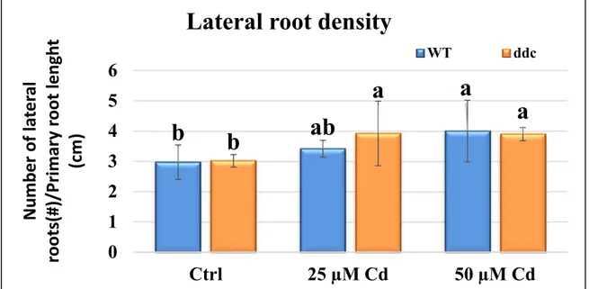
3.1 IMPACT OF CD EXPOSURE ON PLANT GROWTH AND DEVELOPMENT OF DDC MUTANT AND WT A. THALIANA SEEDLINGS.
... 46
3.2 QUANTIFICATION OF CD ABSORPTION IN DDC MUTANT AND WT SEEDLINGS OF A. THALIANA... 51
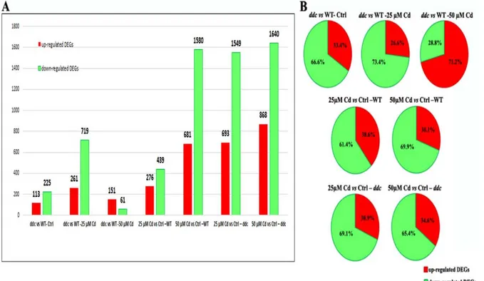
3.3 RNA‐SEQ ANALYSIS ... 52
3.3.1 Analysis and Identification of Differentially Expressed Genes (DEGs) ... 52 3.3.2 Gene Enrichment Analysis ... 55 3.3.3 Differential expression of genes involved in hormones metabolism in ddc mutant and WT under Cd treatment. ... 63 3.3.3.1 Auxin ... 63 3.3.3.2 Cytokinins ... 73 3.3.3.3 Gibberellins ... 84 3.3.3.4 Jasmonic acid ... 91 3.3.3.5 Abscisic acid ... 95 3.3.3.6 Ethylene ... 107 3.3.4 Differential expression of genes involved in hormones signalling in ddc mutant and WT under Cd treatment. ... 112 3.3.4.1 Auxin ... 112 3.3.4.2 Cytokinins ... 114 3.3.4.3 Gibberellins ... 116 3.3.4.4 Jasmonic acid ... 118 3.3.4.5 Abscisic acid ... 120 3.3.4.6 Ethylene ... 123 3.3.4.7 Brassinosteroids ... 125 3.4 LIBRARIES RESULTS VALIDATION: QUANTIFICATION OF THE EXPRESSION LEVELS OF GENES RELATED TO HORMONE BIOSYNTHESIS AND SIGNALLING IN DDC MUTANT AND WT IN CTRL CONDITION AND UNDER CD TREATMENT BY QRT‐PCR. ... 128
3.5 PHYTOHORMONES QUANTIFICATION IN DDC MUTANT AND WT UNDER CD TREATMENT. ... 131
3.5.1 IAA quantification ... 131 3.5.2 GAs quantification ... 132 3.5.3 JA quantification ... 135 3.5.4 ABA quantification ... 136 3.5.5 SA quantification ... 136 3.6 HOW ARE THE DETECTED ALTERATION IN HORMONE PATHWAYS LINKED TO THE PHENOTYPE AND DIFFERENTIAL RESPONSE TO CD OF DDC MUTANT? ... 137 3.6.1 An insight into the involvement of auxin distribution pathway ... 137 3.6.2 Effects of Cd toxicity on Root Apical Meristem pattern in ddc mutant and WT A. thaliana seedlings. ... 144 3.6.3 Cd Impact on SCARECROW expression pattern in ddc mutant and WT A. thaliana seedlings. . 149 CHAPTER 4: DISCUSSION ... 153 CHAPTER 5: CONCLUSIONS ... 163 REFERENCES ... 165 ACKNOWLEDGEMENTS ... 212
LIST OF ABBREVIATIONS
12,13-EOT 12,13S-epoxy-octadecatrienoic acid 12-OPDA 12-oxo-phytodienoic acid
13-HPOT 13S-hydroperoxy-octadecatrienoic acid
AAO Abscisic aldehyde oxidases
ABA Abscisic acid
ABA-GE ABA-glucosyl ester
ACO 1-aminocyclopropane-1-carboxylate oxidase
ACS 1-aminocyclopropane-1-carboxylate SYNTHASE
ACX Acyl-CoA oxidase
AGO ARGONAUTE
AHK Histidine Kinane
AMI1 AMIDASE 1
AOC Allene oxide cyclase
AOS Allene oxide synthase
ARF AUXIN RESPONSE FACTOR
ARR1 ARABIDOPSIS RESPONSE REGULATOR 1
AtBG1 ß-glucosidase
AuxREs Auxin-responsive promoter elements
BAH Bromo Adjacent Homology
BAK1 BRI1-ASSOCIATED RECEPTOR KINASE 1
BAM Binary Alignment Map
BCH ß-carotene hydroxylases
BER Base Excision Repair
BES1 BRI1-EMS-SUPPRESSOR BIN2 BR-INSENSITIVE 2 BR Brassinosteroids BR Brassinosteroids BRI1 BRASSINOSTEROID-INSENSITIVE 1 BSK BRs signalling kinase
BZR1 BRASSINAZOLE-RESISTANT 1
CAM Crassulacean acid metabolism
CAT1 CATALASE1
CDG Constitutive differential growth
cDNA Complementary DNA
cDNA Complementary DNA
CK cytokinins
CKXs Cytokinin oxidase/dehydrogenase enzymes
CMT Chromomethylase
COI1 CORONATINE INSENSITIVE 1
Col-0 Columbia-0
CPS ent-CDP synthase
CYP Cytochrome P450 monooxygenases
cZ cis-zeatin
DCL3 DICER- LIKE PROTEIN 3
ddc drm1 drm2 cmt3
DDM1 DECREASED DNA METHYLATION 1
DEG Differentially Expressed Gene
DHZR Dihydrozeatin riboside
DMAPP Dimethylallyl diphosphate
DME DEMETER
DME TRANSCRIPTIONAL ACTIVATOR DEMETER
DML2 DEMETER-LIKE 2
DML3 DEMETER-LIKE 3
DNA Deoxyribonucleic acid
Dnmt2 DNA methyltransferase homologue 2 DRM Domains rearranged methyltransferase
dsRNA Double-stranded RNA
EPF2 EPIDERMAL CATTERNING FACTOR 2
ER Endoplasmic Reticulum
ET Ethylene
FIE FERTILIZATION-INDEPENDENT ENDOSPERM
FIS2 FERTILIZATION-INDEPENDENT SEED 2
FPKM Fragments Per Kilobase Million
FWA FLOWERING WAGENINGEN
GA Gibberellins
GA20ox GA 20-oxidase
GA3ox GA 3-oxidase
GAMT GAs methyltransferases
GFP Green Fluorescent Protein
GGPP Geranylgeranyl diphosphate
GO Gene Ontology
HMA Heavy Metal-transporting P-type ATPase
HPt Histidine phospho¬transferase
IAA indole-3-acetic acid
IAM Indole-3-acetamide
IAOX indole-3-acetaldoxime
IGP Indole-3-glycerol phosphate
IND Indole
INS Indole synthase
IPA Indole-3-pyruvic acid
IPP C5-isopentenyl diphosphate
IPT Adenylate isopentenyltransferase
IRT1 IRON-REGULATED TRANSPORTER 1
JA Jasmonic acid
JAZ Jasmonate-ZIM domain
KAO ent-kaurenoic acid oxidase
KAT 3-ketoacyl-CoA thiolase
KO ent-kaurene oxidase
KS ent-kaurene synthase
KYP KRYPTONITE
LOG LONELY GUY
MEA MEDEA
MEG Maternally Expressed Gene
MES 2-N-morpholine ethane sulphonic acid
MET Methyltransferase
Mez1 maize Ez-like gene 1
miRNA MicroRNA
mRNA Messenger RNA
MS Murashige and Skoog
MT metallotionein
NCED 9-cis-epoxycarotenoid dioxygenase
NIT NITRILASE
NO Nitric oxide
NPR1 NONEXPRESSER OF PATHOGENESIS-RELATED PROTEIN 1
NRAMP Natural Resistance-Associated macrophage
OPR3 OPDA reductase 3
PA Phaseic acid
PC Phytochelatins
PEG Paternally Expressed Gene
PIN PINFORMED
PM Plasma Membrane
POL IV RNA Polymerase IV
POLV RNA Polymerase V
PP2Cs Phosphoprotein phosphatase 2Cs
PSY Phytoene synthase
QC Quiescent Center
qRT-PCR Quantitative Reverse Transcriptase Polymerase Chain Reaction
RAM Root Apical Meristem
RdDM RNA-directed DNA methylation
RDR2 RNA- DEPENDENT RNA POLYMERASE 2
RIN transcription
factor RIPENING-INHIBITOR transcription factor
RNAseq RNA sequencing
ROS Reactive Oxygen Species
ROS1 REPRESSOR OF SILENCING 1
SA Salicylic acid
S-AdoMet S-adenosyl-L-methionine
SAM Sequence Alignment Map
SAM S-adenosylmethionine
SAM S-adenosylmethionine
SAM Shoot Apical Meristem
SCF Skp-Cullin-F-box
SCN stem cell niche
SCR SCARECROW
sd Standard Deviation
siRNA Short interfering RNA
SnRK2s SNF1-related protein kinases 2 SRA domain SET and RING associated domain
ssRNA Single-stranded RNA
StAR Steroidogenic acute reg¬ulatory
TAA1 Tryptophan aminotransferase 1
TAM Tryptamine
TDC Tryptophan decarboxylase
T-DNA Transfer DNA
TE Transponsable Elements
TE Transposable Element
TGA TGACG Sequence-specific Binding Proteins
TIR1/AFB TRANSPORT INHIBITOR RESPONSE/AUXIN SIGNALING
F-BOX
TPL TOPLESS
TPR TPL-RELATED
TPS Terpene synthase
tRNA-IPT tRNA isopentenyltransferase tZ trans-zeatin
UGTs O-glucosyltransferases
WT Wild type
YUC YUCCA
ZEP Zeaxanthin epoxidase
ZIP ZRT-IRT-like Proteins
ABSTRACT
Due to their sessile life style, plants are continuously exposed to a variety of abiotic and biotic stresses which could potentially hinder their growth, development, productivity and survival. In this scenario, it appears evident the relevance of epigenetic mechanisms in assuring growth plasticity to the plant and withstanding stresses through a rapid and extensive modification of gene expression in a manner that overcomes the restrictions of a highly stable DNA sequence.
Epigenome landscape is largely related to DNA methylation process, which is one of the most significant players in the control of plant responses to environmental changes and stressors. On the other hand, all these responses are also under the control of an intricate signalling network which strongly involves the phytohormones, whose action is in turn influenced by epigenetic mechanisms. Despite this information, the complex mechanisms by which DNA methylation modulates plant stress responses are yet largely unresolved, mainly with respect to heavy metal stress, for which a metal- and species-specific response was evidenced.
In order to gain further insight into these aspects, in the present work we performed a comparative transcriptomic analysis on the drm1 drm2 cmt3 (ddc) mutant of A. thaliana, defective in both maintenance and de novo DNA methylation, and WT plants exposed to a long lasting (21 days) Cd treatment at 25 and 50 µM concentrations. Attention was focused on Cd as one of the most toxic pollutants, widespread in both terrestrial and marine environment. The mutant was chosen as a suitable tool for investigating mechanisms and molecular processes that act in and are regulated by DNA methylation. Analyses of growth parameters and targeted cytophysiological features were also carried out.
Concerning the results, transcriptomic analysis highlighted photosynthesis, stress responses and hormone biosynthesis as the genetic pathways more impacted by Cd treatment in both ddc mutant and WT. All these pathways are highly relevant for plant development. A more detailed analysis carried out on the pathways related to the phytohormones suggested that, under a prolonged heavy metal exposure, plant activity was directed to enhance and/or maintain the level and signalling of hormones which are relevant in sustaining the growth (auxins, cytokinins and gibberellins) more than those of hormones specifically related to stress response (jasmonic acid, abscisic acid and salicylic acid). This could represent the plant strategy to avoid the negative effects of long-lasting
activity of stress-related hormones. Interestingly, such strategy could be more efficient in
ddc mutant than in the WT. Indeed, likely due to a higher genome plasticity conferred to the mutant by its DNA hypomethylated status, in the ddc mutant the described transcriptomic differences have already been observed in the treatment with 25 μM Cd, while in the WT only in the treatment with 50 μM Cd. The outcome of this different modulation of gene expression was a better growth performance in ddc vs WT, as evidenced by growth parameters analysis. A tight relationship between the hormone-related transcriptomic differences and the different cyto- morphophysiological features of ddc mutant vs WT under Cd treatment was also revealed.
ABSTRACT
Le piante, a causa della loro natura sessile, sono continuamente esposte a una varietà di stress abiotici e biotici, i quali potrebbero potenzialmente ostacolare la loro crescita, sviluppo, produttività e sopravvivenza. In questo scenario, appare evidente l'importanza dei meccanismi epigenetici nell’ assicurare alla pianta una certa plasticità di crescita e la capacità di tollerare situazioni stressanti, attraverso una modulazione rapida ed estesa dell'espressione genica, in modo da superare le restrizioni imposte dalla fissità dell’informazione codificata nel DNA.
Il “landscape epigenetico” di un organismo eucariotico è in gran parte correlato al processo di metilazione del DNA, che è uno dei meccanismi maggiormente coinvolti nel modulare la risposta delle piante ai cambiamenti ambientali e ai fattori di stress. D'altra parte, tutte queste risposte sono anche controllate da un intricato signalling, in cui un ruolo fondamentale è svolto dalle diverse classi di fitormoni, la cui azione è a sua volta influenzata da meccanismi epigenetici. Nonostante la vasta letteratura al riguardo, i complessi meccanismi con cui la metilazione del DNA modula le risposte della pianta allo stress rimangono ancora in larga misura incompresi, principalmente per quanto riguarda lo stress da metalli pesanti, per il quale è stata evidenziata l’esistenza di risposte metallo- e specie-specifiche.
Al fine di ottenere ulteriori informazioni riguardo questi aspetti, nel presente lavoro abbiamo effettuato un'analisi trascrittomica comparativa tra il mutante drm1 drm2 cmt3 (ddc) di A. thaliana, difettivo sia nella metilazione di mantenimento che de novo del DNA e piante WT, esposti ad un trattamento prolungato (21 giorni) con Cd, alle concentrazioni di 25 e 50 µM. L'attenzione è stata focalizzata sul Cd in quanto è uno degli inquinanti più tossici, diffuso sia in ambiente terrestre che marino. Il mutante è stato scelto in quanto strumento adatto per studiare i meccanismi e i processi molecolari che agiscono e sono regolati dalla metilazione del DNA. All’analisi trascrittomica è stata inoltre associata l’analisi di alcuni parametri di crescita e di alcune mirate caratteristiche citofisiologiche.
Per quanto attiene i risultati, l'analisi trascrittomica ha evidenziato il processo di fotosintesi, la risposta allo stress e la biosintesi degli ormoni come i pathways genetici più influenzati dal trattamento col Cd, sia nel mutante che nel WT. Tutti questi pathways sono estremamente rilevanti nello sviluppo delle piante. Inoltre, il quadro emerso da un’analisi più dettagliata dei pathways relativi ai fitormoni ha portato ad ipotizzare che, nel caso di
una prolungata esposizione a metalli pesanti, l'attività delle piante venga indirizzata a migliorare e/o mantenere il livello e il signalling degli ormoni atti a sostenere la crescita (auxine, citochinine e gibberelline) più che di quelli specificamente correlati alla risposta allo stress (acido jasmonico, acido abscissico e acido salicilico). Tutto ciò potrebbe rappresentare una strategia messa in atto dalla pianta per evitare gli effetti negativi di una prolungata attività degli ormoni legati allo stress. Interessantemente, tale strategia sembrerebbe essere realizzata in modo più efficiente dal mutante. Infatti, probabilmente a causa di una maggiore plasticità del genoma conferita dallo stato ipometilato del suo DNA, nel mutante le differenze a livello del trascrittoma sono state osservate già nel trattamento con 25 µM Cd, mentre nel WT solo nel trattamento con 50 µM Cd. Questa diversa modulazione dell'espressione genica ha trovato riscontro in una migliore performance di crescita del ddc rispetto al WT, come evidenziato dall'analisi dei parametri di crescita. È stata inoltre osservata una stretta relazione tra le differenze trascrizionali riguardanti i pathways ormonali e le diverse caratteristiche citomorfofisiologiche del ddc vs WT quando trattati col Cd.
CHAPTER 1: INTRODUCTION
1.1 DNA methylation
DNA methylation refers to the covalent addition of a methyl group to the cytosine bases of DNA, to form 5-methylcitosine (He et al.,2011). It is one of the principal epigenetic mechanisms leading to heritable and non-heritable genome modifications, which occur beyond changes in nucleotide sequence. Like other epigenetic marks, such as the histone code, DNA methylation contributes to chromatin remodelling processes and it is involved in the regulation of gene expression (He et al.,2011).
DNA methylation can be found in both prokaryotes and eukaryotes. In bacteria, its major function is to act as a defence mechanism against invading phages: methylation differentiate host genome from that of the phage, which becomes preferential target of cleavage action of host restriction enzymes. It also plays an important role in DNA repair and replication (Chinnusamy and Zhu, 2009). In eukaryotes, DNA methylation is a fundamental mechanism for the maintenance of genome stability and the regulation of gene expression in response to both external and internal stimuli; as such, it plays a relevant role in plant diversity and development (Becker et al.,2011; Lauria and Rossi, 2011; Schmitz et al.,2011; Zhang et al.,2018).
1.1.1 DNA methylation: an historical overview
Covalent modifications of the DNA were firstly described by Hotchkiss in 1948. However, the first suggestion that DNA methylation could have some relevant biological roles, through the modulation of gene expression was made only in 1969 by Griffith and Mahler, while studying the basis of the long-term memory in the brain. In 1975, similar models for gene activity regulation and for the hereditability of DNA active or inactive status, based on the enzymatic methylation of cytosine in its sequences, were independently proposed by Riggs (Riggs, 1975) and Holliday and Pugh (1975). In particular, these authors supposed the existence of enzymes (methylases) that directly or indirectly act on specific sequences (context) modifying DNA methylation pattern and, consequently, changing gene expression. They also supposed the action of specific methylases that recognize
hemi-methylated DNA after the replication assuring the maintenance and the hereditability of DNA methylation pattern.
Unfortunately, no experimental evidence supported the brilliant models proposed by Riggs (1975) and Holliday and Pugh (1975) until the advent and the progressive upgrading of molecular techniques that allowed to screen DNA methylation in the sequences of interest. For example, the restriction of DNA with isoschizomers (i.e. enzymes that cut DNA at specific sequences context and in relation to methylation presence/absence) followed by Southern blot made possible to know whether DNA target sequences were or not methylated in the selected restriction sites. Thanks to this new technique, Doerfler (1981; 1983) finally proved the correlation between DNA methylation and gene expression modulation, discovering that the promoter regions of many inactive genes were methylated; vice versa, the corresponding active genes resulted unmethylated. Moreover, by using the 5-azacytidine, an analogue of the cytidine that inactivates the DNA methyltransferases, it was shown that 5-azacytidine reactivated silent genes, including the inactive X chromosomes (Holliday, 1991).
Over the years, additional studies and the recent genome-wide sequencing technologies allowed to gain further insights into DNA methylation pattern and role, which includes a lot of other processes, like genomic imprinting (Surani et al.,1984), inactivation of transposable elements (TEs) (Pray, 2008), and the control of telomere length (Chan and Blackburn, 2004).
1.1.2 DNA methylation in plants: sequence context and methylation pathways
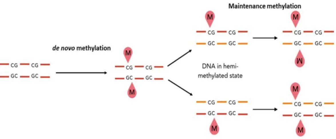
DNA methylation can be distinguished in de novo methylation and maintenance methylation. De novo methylation consists in the methylation of DNA sequences not previously methylated (Fig. 1.1). It is involved in the rearrangement of methylation pattern during the embryogenesis and in the cell differentiation processes during development (Razin and Cedar, 1993). Maintenance methylation preserves the methylation status of symmetric (palindromic) sites after DNA duplication through recognition of hemimethylated sites and methylation of the newly synthesized filament (Finnegan and Dennis, 1993; Saze et al.,2003) (Fig. 1.1).
Figure 1.1: Schematic representation of de novo and maintenance methylation of DNA. The methyl group is indicated by the letter “M”.
In animals, cytosine methylation is mainly restricted to the symmetric CpG dinucleotide, except for the embryonic stem cells (Ramsahoye et al.,2000), the adult mouse cortex and human brain (Lister et al.,2013), where CpH methylation was found. By contrast, plant DNA can be methylated in any sequence context (i.e. CG, CHG and CHH, where H is A, T or C), but most commonly in “symmetric cytosines” CpG and CpHpG (Meyer et al.,1994).
DNA methylation is catalysed by specific enzymes, the DNA methyltransferases, which recognise specific DNA sequences through their variable N-terminal domain and transfer a methyl group from S-adenosyl-L-methionine (S-AdoMet) to carbon 5 of cytosine residues through the activity of the C-terminal catalytic domain (Pòsfai et al.,1989; Kumar et al.,1994; Pavlopoulou and Kossida, 2007).
So far, plant DNA methyltransferases have been classified in 4 different families on the basis of their linear domain arrangement: METHYLTRANSFERASE (MET) family, CHROMOMETHYLASES (CMTs) family, DOMAINS REARRANGED METHYLTRANSFERASES (DRMs) family, and DNA METHYLTRANSFERASE HOMOLOGUE 2 (Dnmt2) family, whose role in DNA methylation has still to be further elucidated (Pavlopoulou and Kossida, 2007).
MET is the family principally involved in the maintenance of DNA methylation pattern at the symmetric CpG sites after replication events (Finnegan and Dennis, 1993; Genger et al.,1999). As far, in A. thaliana, the gene family encoding these DNA methyltransferases comprises 5 members and among them only 4 are partially
characterized (METI, METIIa, METIIb, and METIII). The conserved structure of these genes suggests that they were originated from one ancestral gene through a series of duplication events (Finnegan and Dennis, 1993; Genger et al.,1999). METI encodes a functional methyltransferase (Genger et al.,1999) expressed in both vegetative and floral tissues, with its highest expression in meristematic cells (Ronemus et al.,1996), while no specific function has been still attributed to the proteins encoded by the other members of this gene family (Genger et al.,1999).
CMTs is a family of DNA methyltransferases unique of plants, discovered by Henikoff and Comai in 1998 and characterized by the presence of a chromodomain amino acid motif between the conserved motifs II and IV. In A. thaliana, the gene family encoding CMTs enzymes includes 3 members, CMT1, CMT2 and CMT3, originated by gene duplication events. In A. thaliana, CMT1 is generally mutated or disrupted by the insertion of transposable elements (TEs) within the coding region of the gene. In both cases, the result will be the premature end of the translational process of the CMT1 protein (Henikoff and Comai, 1998). CMT2 activity seems to have an important role in keeping high levels of DNA methylation at TEs in the heterochromatic fraction (Zemach et al. 2013). This enzyme is principally involved, together with DRM2, in the establishment and maintenance of the asymmetrical methylation on CpHpH sites, depending on the genomic region. Namely, DRM2 catalyses CpHpH methylation at TEs, usually located in heterochromatin, or at other repeated sequences in euchromatic chromosome arms. Whereas, CMT2 catalyses CpHpH methylation at histone H1-containing heterochromatin (Huettel et al.,2006; Zemach et al.,2013; Liu et al.,2014), but at certain extent it can also participate in CpHpGp methylation maintenance, which is mainly catalysed by CMT3 (Lindroth et al.,2001; Stroud et al.,2014). In particular, CMT3 recognises H3K9me2 epigenetic mark and binds to the DNA nucleosome through the Bromo Adjacent Homology (BAH) domain and the chromo domains. Note that a similar recognisance mechanism is activated by the SET and RING associated (SRA) domain of KRYPTONITE (KYP, also known as SUVH4) enzyme, that catalyses the methylation of the H3K9. In such a way, CpHpG and H3K9me2 modifications reinforce each other through a positive regulatory feedback (Stroud et al.,2013; Du et al.,2014).
In A. thaliana, DRMs family includes DRM1and DRM2. Both these proteins mediate de novo methylation in all DNA sequence context (CpG, CpHpG, CpHpH) by an
RNA-directed DNA methylation process (RdDM) (Matzke and Mosher, 2014). In the canonical RdDM pathway, RNA Polymerase IV (POL IV) and RNA- DEPENDENT RNA POLYMERASE 2 (RDR2) work to produce double stranded DNA (dsDNA) which is cleaved by DICER- LIKE PROTEIN 3 (DCL3) into siRNA. These siRNAs are loaded onto ARGONAUTE (AGO) proteins and paired to complementary RNAs scaffold produced by RNA Polymerase V (POLV) to recruit DOMAINS REARRANGED METHYLASE 1 (DRM1) and DOMAINS REARRANGED METHYLASE 2 (DRM2), which methylate the DNA in a sequence-independent manner (Fig. 1.2) (Law and Jacobsen, 2010; Zhang and Zhu, 2011; Pikaard et al.,2012; Matzke and Mosher, 2014).
Figure 1.2: Schematic representation of the RNA-directed DNA methylation (RdDM) pathway (Matzke and Mosher, 2014).
Finally, methylome dynamics is further assured by DNA demethylation. Indeed, active DNA demethylation is extremely important for both genome-wide epigenetic reprogramming and for the activation of target gene loci during plant development (Hsieh et al.,2009). In particular, it consists in the enzymatic removal of methylated cytosine by the combined action of a family of DNA glycosylases including DEMETER (DME), REPRESSOR OF SILENCING 1 (ROS1), LIKE 2 (DML2), and
DEMETER-LIKE 3 (DML3). Subsequently, a base excision repair (BER)-dependent mechanism completes the process (Penterman et al.,2007; Zhu, 2009; reviewed by Li et al.,2018). 1.1.3 Molecular functions of DNA methylation in plants
For long time, most of the knowledge on DNA methylation in plants was obtained from studies carried out on A. thaliana (Zhang et al.,2006; Cokus et al.,2008).In the last decade, genome-wide sequencing technologies allowed us to obtain information on DNA methylation pattern in several other species which include important crops such as Zea mays (Eichten et al.,2013), Solanum lycopersicum (Fray and Zhong, 2015) and Oryza sativa (reviewed by Deng et al.,2016). In plants, DNA methylation primarily take places on transposon repeat sequences. Transposons, also called “jumping genes”, are mobile elements of the genome. Based on their structure and transposition mechanism, there are two major classes of transposons: the class I transposons (or retrotransposons), that translocate from a site to another through a reverse transcription process with an RNA intermediate, resulting in an increase of the final copy number, and the class II transposons, that translocate the transposon from the integrated site to a new site of the genome, keeping the final copy number unaltered (Engels et al.,1990). Even though transposable elements activity contributes to genome evolution and diversity, it can also cause, depending by the site of its insertion, mutations of functional genes and/or chromosome instability (Saze et al.,2012). The heavy methylation state of transposons detected in several plants, preventing their expression and transposition, is addressed to avoid the risk of a bursts of transposition activity. In such a way, genome integrity and transcriptional homeostasis is assured (Pray, 2008; Zemach, 2010; Saze et al.,2012; Kim and Zilberman, 2014).
The presence of methylated transposons close to or within a gene can affect gene expression leading in most cases to gene silencing (Rodrigues and Zilberman, 2015). Moreover, also the promoter region and the coding regions of actively expressed genes can be methylated (Zilberman, 2008; Bewick and Schmitz, 2017). Until now, although several functions have been proposed, the exact role of gene body methylation in plants is not yet well understood (Zilberman, 2008; Takuno and Gaut, 2013; Bewick and Schmitz, 2017). More defined is, instead, the role of cytosine methylation at the gene promoter region. In this case, methylated status prevents the binding of transcription factors to their target sequences and represents a direct mechanism by which gene expression can be regulated
by DNA methylation in both mammals and plant. Therefore, increasing methylation at promoters’ level results in gene silencing, whereas reduced methylation results in gene activation. (Iguchi-Ariga and Schaffner, 1989; Bell and Felsenfield, 2000; Campanero et al.,2000; Hark et al.,2000).
The inhibition of transcription by DNA methylation can occur in different ways and also involves an interplay with other epigenetic mechanisms. Indeed, transcription can be modulated: i) by inhibiting or promoting the binding of transcriptional activators or repressor, respectively ii) by promoting repressive histone modifications, like H3K9me2 methylation, and inhibiting the permissive ones, like histone acetylation. (Boyes and Bird, 1991; Henderson and Jacobsen, 2007; Zhang et al.,2011; Matzke and Mosher, 2014).
DNA methylation is also involved in genomic imprinting (Surani et al.,1984) and in the control of telomere length (Chan and Blackburn, 2008). Genomic imprinting consists in the differential activation or inactivation of alleles of a gene depending from the paternal or maternal origin of the chromosome. Namely, one of the alleles is silenced, and only the one from the other parent will be expressed. (Bajrami and Spiroski, 2016). Even though the mechanisms underlying this process aren’t yet completely defined, it was observed an involvement of DNA methylation. In particular, it has been demonstrated that the repressed allele is methylated, while the active allele is unmethylated. In human, the most studied cases of methylation-related genomic imprinting deal with Prader-Willi syndrome and Angelman syndrome, associated to the imprinting on the long arm of chromosome 15 (Bajrami and Spiroski, 2016). In plants, several imprinted genes, which also show parental differences in DNA methylation, have been identified, including FERTILIZATION-INDEPENDENT SEED 2 (FIS2) (Jullien et al.,2006), FLOWERING WAGENINGEN (FWA) (Kinoshita et al.,2004), FERTILIZATION-INDEPENDENT ENDOSPERM 1 (ZmFie1) (Hermon, 2007), FERTILIZATION-INDEPENDENT ENDOSPERM 2 (ZmFie2) (Gutiérrez-Marcos et al.,2006) and maize E(z)-like gene 1 (Mez1) (Haun, 2007). However, there are some exceptions to these evidences of DNA methylation role in genomic imprinting, as in the case of the MEDEA (MEA) gene of A. thaliana. MEA acts as suppressor of endosperm development, and loss of function mutations could cause precocious endosperm formation before fertilization, prolonged endosperm nuclear proliferation after fertilization and embryo abortion (Grossniklaus et al.,1998, Kiyosue et al.,1999). The MEDEA locus is the first example of an imprinted region that doesn’t
present differential DNA methylation between the silenced allele and the active allele, suggesting a DNA methylation-independent mechanism(s) and the existence of other factors that determines imprinting at the MEA locus (Wöhrmann et al.,2012).
Finally, DNA methylation is also implicated in telomere length regulation. Telomeres consists of structures formed by proteins and repetitive DNA situated at the end of the chromosomes. Both telomeric and sub-telomeric regions in the chromosome are bound to telomere binding proteins and present heterochromatinic structure (Blasco, 2007). Telomeric heterochromatin is usually devoid of functional genes, and it plays an important role in chromosome end protection and telomere length regulation (Ottaviani et
al.,2008). When this protective structure fails, the results are chromosome degradation or fusion with neighbouring chromosomes (Chan, 2004).
One interesting evidence of the DNA methylation involvement in maintenance of telomeric heterochromatin regards the role of some telomeric repeat–containing transcripts in A. thaliana. These transcripts, in fact, are processed in small interfering RNAs that promote the methylation of asymmetric cytosines in telomeric (CCCTAAA)n repeats (Vrbsky et al.,2010). Although these siRNAs-directed mechanisms contribute to telomere length control, this process is determined and reinforced by several independent mechanisms, of which many are epigenetic ones (Vrbsky et al.,2010).
1.1.4 DNA methylation and its involvement in plant growth and development
In line with the above described roles of DNA methylation, this epigenetic mechanism plays a relevant role in the control of plant growth and development throughout its whole life cycle, from the gametophyte development, throughout the fecundation process, during the vegetative development until flowering, fruit formation and ripening (reviewed by Zhang et al.,2018).
In particular, a very complex and dynamic pattern of methylome, tightly related to other epigenetic modifications, has been assessed in A. thaliana during gametogenesis and fecundation (reviewed by Zhang et al.,2018). Indeed, the male gametophyte presents a de-repression of TEs activity, due to the global demethylation mediated by TRANSCRIPTIONAL ACTIVATOR DEMETER (DME) activity, and the down-regulation of DECREASED DNA METHYLATION 1 (DDM1) (Slotkin et al.,2009;
maintaining DNA methylation in symmetric cytosine sequences (Jeddeloh et al.,1999; Zemach et al.,2013). As a consequence, high level of siRNAs is produced from the demethylated and de-silenced transposons which accumulate in sperms (Fig. 1.3 A). After fecundation, siRNAs will be further processed through the RdDM canonical pathway, thus reinforcing the CpHpH methylation at transposons (Gehring et al.,2009; Ibarra et al.,2012; Ingouff et al.,2017; reviewed by Zhang et al.,2018) (Fig. 1.3 A).Alsothe central cell of female gametophyte undergoes DME-mediated global demethylation (Fig. 1.3 A); as a result, the endosperm formed following its fertilization by the sperm cell will be globally demethylated, although a reinforced CpHpH methylation at transposons will be also present due to the siRNAs activity (Gehring et al.,2009; Ibarra et al.,2012) (Fig. 1.3 A). Moreover, the endosperm is subject to gene imprinting (Pignatta et al.,2014; reviewed by Zhang et al.,2018). In the maternally expressed genes (MEGs), the maternal allele is hypomethylated, and the paternal one is methylated and repressed (Fig. 1.3 B), while in the paternally expressed genes (PEGs) the maternal allele is marked by the H3K27me3 repressive histone modification, while the paternal one presents the active modification H3K36me3 (Dong et al.,2017) (Fig.1.3 B).
Figure 1.3: (A) In the male vegetative cell of A. thaliana siRNAs are produced and transported into the two sperm cells. One of the cells fertilizes the egg cell, where the siRNAs are further processed through the RdDM canonical pathway, reinforcing the CpHpH methylation. The other sperm cell fertilizes the female
central cell, globally demethylated. As a consequence, the resultant endosperm will be globally demethylated but will also present a reinforced CpHpH methylation at transposons due to the siRNAs presence. (B) the endosperm presents maternally expressed genes (MEGs), characterized by DNA hypomethylation and trimethylation of histone H3 lysine 4 (H3K4me3), with the paternal allele silenced by DNA hypermethylation or H3K27me3, and paternally expressed genes (PEGs), characterized by H3K36me3, whereas the maternal allele can be silenced by H3K27me3 (Zhang et al.,2018).
Concerning more specifically plant vegetative growth and pattern formation, DNA methylation has a crucial role. A de novo DNA methylation through the RdDM pathway is essential for the proliferative activity of meristems, on which plant growth relies (Kawakatsu et al.,2016). According to this assumption, meristem defects have been detected in Zea mays RdDM mutants, which display strong developmental abnormalities (Alleman et al.,2006; Erhard et al.,2009; Moritoh et al.,2012; Wei et al.,2014). Interestingly, in peach (Prunus persica (L.) Batsch) changes in the level of DNA methylation have been found to mark the transition of apical vegetative shoot meristem towards floral bud (Bitonti et al.,2002).
Methylation has been found to play a significant role also in cell differentiation. For example, a differential CpG and CpHpG methylation pattern among the division zone, transition zone, elongation zone and mature zone of developing leaves has been evidenced in Zea mays plants, related to the different regulation of maintenance DNA methyltransferases (Kawakatsu, et al.,2016). Furthermore, a different DNA methylation pattern in Zea mays leaves was found also in genes involved in chromatin remodelling, cell cycle progression and growth regulation. All these evidences indicate that DNA methylation has an important role in leaf growth in Zea mays (Candaele et al.,2014). In addition, in A. thaliana DNA methylation has been found to control stomata formation. In fact, an hypermethylation of EPIDERMAL CATTERNING FACTOR 2 (EPF2) gene, whose product is a peptide ligand that represses stomata formation, leads to an over-production of stomatal lineage cells (Yamamuro et al.,2014).
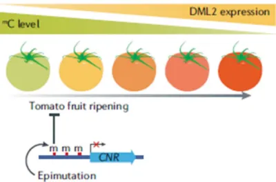
Recently, it has been evidenced a relevant role for methylation-related epigenetic control on fruit development. In particular, it has been demonstrated that an increase of DME-LIKE2 (DML2) DNA demethylase expression, is required for the activation of ripening-induced genes during Solanum lycopersicum fruit development. Actually, the demethylated status of the ripening-induced genes let the binding of
RIPENING-INHIBITOR (RIN) transcription factor to the promoter of these genes and consequently their transcription, that starts the ripening process (Zhong et al.,2013; Lang et al.,2017) (Fig. 1.4).
Figure 1.4: Solanum lycopersicum fruit ripening is accompanied by a diminution of genomic methylation and an increase of DML2 expression (Zhang et al.,2018).
DNA methylation has been found to play a role also in plant growth plasticity in response to environmental condition and adaptive strategies, as evidenced in relation to the heterophylly phenomenon exhibited by the aquatic plant Trapa natans L. Indeed, clear differences in DNA methylation level were detected in the floating and submerged leaves produced by this plant, which are strikingly different in morphology. Namely, while the floating leaves were normally expanded, the submerged ones presented a root-like shape, likely to adapt to water presence (Bitonti et al.,1996).
1.1.5 Involvement of DNA methylation in plant response to abiotic stress
The role of DNA methylation in plant response to stress has been also widely investigated. Indeed, modifications in DNA methylation pattern, allowing rapid and reversible changes in the chromatin structure, can enable the activation of defence pathways through the combination of genetic and epigenetic mechanisms (Peng et al.,2009). Moreover, this epigenetic mechanism establishes a DNA methylation-dependent stress memory in plants in presence of a persistent stress (Jiang et al.,2014; Sanchez et al.,2014; Wibowo et al.,2016), principally charged to GC-rich sequences methylation, that
ensures the faithfully transfer of the “memory” to the offspring (Mathieu et al.,2007) (Fig. 1.5).
Figure 1.5: Following stress exposure, plant activate stress responsive pathways accompanied by modifications in DNA methylation status, that could be genome-wide or at specific loci level. In presence of a persistent stress, this “stress memory” genomic configuration is inherited by the next generations (Zhang
et al.,2018).
Both biotic and abiotic stresses were found to induce modifications in DNA methylation pattern either genome-wide or at specific loci, usually associated to transcriptional regulation of genes involved in plant stress responses (Yong et al.,2015; Xu et al.,2015; Zhang et al.,2016) that, in turn, control important genetic functions like transcription, replication, DNA repair, gene transposition and cell differentiation (Madlung and Comai 2004; Angers et al.,2010; Sahu et al.,2013).
For example in Zea mays, under cold stress, the presence of methyltransferases and, consequently, the level of genomic methylation decreases. This modification seems to be organ- and site- selective, with a decrease of DNA methylation in roots in Ac/Ds transposons, suggesting a possible role for transposable elements in stress response (Shan et al.,2013). Whereas, in Solanum lycopersicum plants, cold down-regulates the expression of DML2, which led to a hypermethylation and silencing of genes responsible for the biosynthesis of volatile compounds, thus causing the loss of flavour of Solanum
lycopersicum fruits under cold storage (Zhang et al.,2016). In Mesembryanthemum crystallinum exposed to salt stress, water deficit caused by the osmotic pressure determines the switching to the Crassulacean acid metabolism (CAM) (Bohnert et al.,1988). This switchover to the CAM pathway is accompanied by the hypermethylation of satellite DNA, that probably enables the process thanks to the formation of chromatin structures that let the simultaneous regulation of all the genes involved (Dyachenko et al.,2006).
Under heavy metal stress, a reduction of DNA methylation has been detected in Trifolium repens L. and Cannabis sativa L. after exposure to nickel, cadmium and chromium (Aina et al.,2004), while in Brassica napus heavy metal exposure promotes genomic methylation (Li et al.,2016). An increase of DNA methylation, related to an overexpression of PoCMT gene was also observed in plants of Posidonia oceanica L. Delile exposed to Cd (Greco et al.,2012). These data suggest that methylome dynamic under stressful condition depends on both the plant species and the kind of heavy metal. Therefore, further studies are required to fully elucidate the network of processes that act in and are regulated by DNA methylation, mainly under stress conditions.
1.1.6 Cross-talk between epigenetic mechanisms and hormone network
In the last years, an emerging cross-talk between epigenetic modifications and the phytohormones action has been highlighted by several studies. As known, epigenetic modifications include not only DNA methylation but also histone modification, chromatin remodelling, non-coding RNAs, which interplay each other rather than act alone in the control of gene expression (reviewed by Yamamuro et al.,2016).
On the other hand, as above discussed, there is large evidence that epigenetic modifications, including DNA methylation, can override genetic programs in response to environmental cues, thus conferring growth plasticity to the plants and contributing to their survival strategies (Dowen et al.,2012). However, the biochemical signals that alter the epigenome and the transduction of such signals are still largely unknown, while the detected relationship with hormone action begins to shed light to this research field.
At this respect, available data largely deal with the involvement of histone modifications (reviewed by Yamamuro et al.,2016). However, some interesting evidences are emerging also in relation to DNA methylation (reviewed by Zhu, 2010; Yamamuro et al.,2016). For example, it has been shown that the induction of auxin responsive genes
mediated by the AUXIN RESPONSE FACTORS (ARFs) is modulated by microRNAs, histone modifications and chromatin remodelling factors (Jones-Rhoades and Bartel 2004; Mallory et al.,2005). In addition and very interestingly, in met1 null allele embryos of A. thaliana, the distribution of auxin and its efflux carrier PIN-FORMED 1 (PIN1) resulted abnormal (Friml 2003, Weijers et al.,2005), although the PIN1 gene doesn’t result to be methylated in both wild type (WT) and met1 null mutant. This result is consistent with and indirect involvement of MET1 activity in the modulation of PIN1 expression (Xiao et al.,2006).
1.2
Phases of plants responses to stressIn a biological context, stress is defined as a condition related to abiotic and/or biotic factors, that exerts a disadvantageous influence on the plant and determines a significant deviation of the optimal condition of life (Taiz and Zeiger, 2003; Larcher, 2004; Cramer et al.,2011).
The abiotic stress factors that affect the geographical distribution of the plants, limit their productivity and threaten food security are numerous: extreme levels of light (high and low), radiation (UV-B and UV-A), temperature (high and low, that lead to chilling and freezing), water (drought, flooding, submergence), salinity (excessive Na+), deficiency or
excess of essential nutrients, gaseous pollutants (ozone, sulphur dioxide), chemical factors like extreme pH and heavy metals in the soil (Fedoroff et al.,2010; Pereira, 2016).
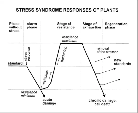
Following exposure to stress, plant response can be summarized in four distinct phases: the alarm phase, the resistance phase, the exhaustion phase and the regeneration phase (Lichtenthaler 1998; Larcher, 2004).
The alarm phase, or response phase, is characterized by a decline of one of several physiological functions, like photosynthetic performance and/or uptake and translocation of nutrient, due to the exposition to the stressful factor (Fig. 1.6). This is a crucial step for the plant, that needs to perceive promptly the deviation from their normal physiological standard and respond in a rapid and efficient manner (Lichtenthaler, 1998; Duque et al.,2013; Ben Rejeb et al.,2014). Stress sensing in plants still remains a largely unresolved topic. Namely, only few putative sensors have been so far identified, since dysfunction in one gene does not cause significant phenotypes in stress responses due to the functional redundancy of genes encoding sensor proteins (Zhu, 2016). Another reason concerns the
mechanisms of the signalling; in fact, the most common model of sensing external stimuli is based on the binding of a chemical ligand to a specific receptor. However, while it could be a suitable model for chemical stresses, it isn’t the same for physical stresses such as, for example, temperature stress (Verslues et al.,2006).
Following stress perception, the activation of one or more signalling transduction cascades, varying depending from the different kind of stresses, determines the beginning of the resistance phase (Lichtenthaler, 1996; Lichtenthaler, 1998). Classically, many actions can be triggered: the activation of specific ion channels and kinase cascades (Verslues et al.,2005), the production of reactive oxygen species (ROS) (Dat et al.,2004) and the activity of phytohormones like abscisic acid (ABA), salicylic acid (SA), jasmonic acid (JA), and ethylene (ET) signalling (reviewed by Verma et al.,2016). All these mechanisms lead to a reprogramming of gene expression resulting in long-term metabolic and morphological adaptations addressed to increase plant tolerance and alleviate biological damages caused by stress. This allows to establish a new physiological standard (optimum stage), that corresponds to the plant resistance maximum (Fig. 1.6) (Lichtenthaler, 1998). If the stress dose overloads the plant capacity to put in place mechanisms for coping with it, especially during long-term stress, physiological processes slow down more and more, and inevitably vitality becomes progressively lost, causing severe damage and cell death (exhaustion phase or end stage) (Fig. 1.6) (Lichtenthaler, 1998). However, if the stressor agents are removed in time, before the senescence process becomes widespread, plants can regenerate themselves and, also in this case, adjust to a new physiological standard (regeneration phase) (Fig.1.6) (Lichtenthaler, 1996; Lichtenthaler, 1998). This new physiological standard depends from the time and the stage of exhaustion, that determines the range of minimum and maximum resistance that the plants acquire following exposure to stress. (Lichtenthaler, 1996; Lichtenthaler, 1998).
Figure 1.6: Phase sequences and responses induced in plants by stress exposure (Lichtenthaler, 1998). 1.3 Cadmium toxicity in higher plants
1.3.1 Cadmium distribution in the environment
Metals with density higher than 5 g cm-3 are classified as heavy metals, and they
represent fiftythree of the ninety natural occurring elements (Weast, 1984). Among them, iron (Fe), molybdenum (Mo), and manganese (Mn) are fundamental micronutrients for the organisms. Zinc (Zn), nickel (Ni), copper (Cu), vanadium (Va), and chromium (Cr) are toxic elements at high concentrations, but as trace elements they are useful as components of the active sites of some enzymes, while silver (Ag), arsenic (As), mercury (Hg), lead (Pb), antimony (Sb) and cadmium (Cd) have no known metabolic and/or nutritional function and are toxic for both plants and animals (Nies, 1999).
Heavy metals are dangerous environmental pollutants, whose presence is particularly relevant in areas subjected to anthropogenic pressure. Cadmium (Cd, density = 8.6 g cm -3), in particular, is a widespread heavy metal considered as one of the most
al.,2004). In natural environment Cd is present mostly as a “guest metal” in Pb/Zn mineralization (Baker et al.,1990). As environment pollutant, Cd is mainly released by power stations heating systems, metal-working industries, waste incinerators, urban traffic, cement factories and as by-product of phosphate fertilizers (Sanità di Toppi e Gabbrielli, 1999). Soils containing a Cd concentration that ranges between 0.04 to 0.32 µM are classified as non-polluted, while soils with a Cd concentration varying from 0.32 to 1 µM are considered moderately polluted. When the concentration reaches 35 µM, the soil is classified as highly polluted, and only Cd hyperaccumulating species can survive in such environment (Sanità di Toppi and Gabbrielli, 1999).
1.3.2 Effects of cadmium exposure on plant growth and development
The toxic effects of Cd are well known since its absorption induces complexes changes at genetic, biochemical and physiological level in both plants and animals (Bingham et al.,1976; Das et al.,1997: Sanità di Toppi and Gabbrielli, 1999; Benavides et al.,2005; Greco et al.,2012). In particular, in plants, it has been showed that Cd alters the uptake of minerals, reducing their availability from the soil by decreasing the soil microbes population (Moreno et al.,2002), reduces the absorption of nitrate and its transport from root to shoot, inhibiting the nitrate reductase in the shoot (Hernandez et al.,1996), and inhibits Fe (III) reductase, that causes a Fe (II) deficiency (Alcàntara et al.,1994). Cd was also found responsible, although not directly, of Reactive Oxigen Species (ROS) production (Heyno et al.,2008). In addition, Cd can inhibit or enhance the activity of several enzymatic or non-enzymatic antioxidants (Salin, 1988) and increase lipid peroxidation, causing severe oxidative stress that enhance its toxic effects (Benavides et al.,2005). The most obvious consequence of Cd toxic effects is a reduction of plant growth due to the direct and indirect inhibition of photosynthesis, respiration and nitrogen metabolism, as well as to a reduction in water and nutrient availability (dos Santos et al.,2012).
1.3.3 Uptake and transport of Cd in higher plants
Cd absorption depends principally from the soil characteristics, the metal concentration in the soil and the plant species and or varieties/ecotypes. Humic acid, solid
solution and pH can modify Cd availability from the soil and, consequently, affect its absorption (Cabrera et al.,1988; Mench and Martin, 1991).
Cd absorption from the soil is mostly charged by root tip, and it involves three principal pathways:
i. Cd2+ can be absorbed through a rapid exchange with H+ produced by the H 2CO3
dissociation in H+ and CO
3- during plant respiration in the root epidermal cells and
enter into the root epidermis layer through the apoplast pathway. This process is rapid and doesn’t require energy (Yamaguchi et al.,2011);
ii. Cd2+ can be transported by ion channels for Fe2+, Zn2+ and Ca2+ and subsequently
enter the root epidermis layer through the symplastic pathway (reviewed by Song et al.,2017);
iii. Cd2+ can be chelated by molecular compounds produced by plants to enhance the
availability of ions in the soil and enter the root epidermis layer through yellow stripe 1 like (YSL) proteins (Curie et al.,2009).
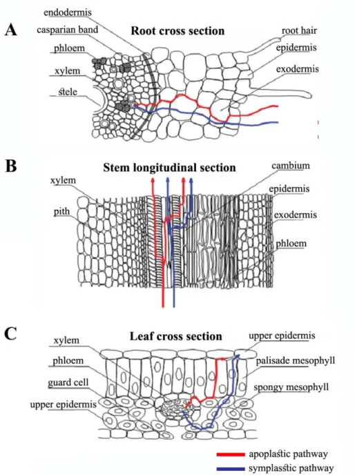
After its uptake from the root, Cd can be transported through tissues and organs via apoplastic pathway, that involves transfer through extracellular fluids and gas spaces between and within cell walls, and symplastic pathway in which water and solutes are intracellularly transferred, and it involves the ion transmembrane transport (Fig. 1.6 A) (reviewed by Song et al.,2017). At the level of the endodermis, metal is complexed by several ligands, such as organic acids and phytochelatins (PCs) and transported to xylem elements in the stele, through it can reach aerial organ via xylematic flow (Fig. 1.6 A, B) (Akhter et al.,2014). In leaves, metal transport and distribution occur through both apoplastic and symplastic pathways and Cd can be finally sequestered into extracellular and subcellular compartment (reviewed by Song et al.,2017), or loaded into phloematic elements that dislocate it from the shoot to the roots as a part of detoxification processes (Fig. 1.6 C) (van Belleghem et al.,2006).
As a result, only small amounts of Cd are transported in shoots (Sanità di Toppi and Gabbrielli, 1999), and the content of Cd in plants decreases in the order roots > stem > leaves > fruits > seeds (Blum, 1997).
Figure 1.6 Uptake and transport of Cd through apoplastic and symplastic pathways in higher plant (A) root, (B) stem and (C) leaf (Song et al.,2017).
1.3.4 Morphological, physiological and biochemical plant responses to Cd toxicity In order to withstand the heavy metals, plants adopt either of two strategies: avoidance and tolerance. Avoidance is the strategy that plant adopts at the beginning, after heavy metal exposure: it enhances the production of organic acids and chelate and sequesters Cd to prevent its access to the root cells, thus protecting the plant from the external stress influence. Tolerance mechanisms, instead, let the plant survive the effects
of an internal stress, enabling the normal functioning of physiological processes even in presence of high concentrations of toxic substances (Song et al.,2017).
To prevent metal uptake inside the cells, plant roots secrete exudates that have the function to chelate metals in the soil matrix (Marschner, 1995). Moreover, plants try to reduce Cd uptake by binding the metal to cellular walls, sequestering it into the apoplast or inhibiting its transport (Manara, 2012). Root cells present on their walls pectic sites, histidine groups and extracellular carbohydrates, like callose and mucilage, that bind the heavy metals and prevent their uptake into the root cells. In this way the cell wall modulates plant metal absorption (Manara, 2012).
Metal uptake and homeostasis is further modulated by the presence at the level of plasma membrane of a range of transporters like those belonging to the ZRT-IRT-like Proteins (ZIP) family transporters and the Natural Resistance-Associated macrophage (NRAMP) family proteins. Both these proteins are involved in the transport of divalent cations, such Cd2+ and Zn2+, across membranes (Manara, 2012). ZIP transporters are
necessary, but not sufficient, for the enhanced accumulation of metal ions in hyperaccumulator plants (Guerinot, 2000), while Nramp5 has been identified as a major Cd uptake transporter in Oryza sativa (Sasaki et al.,2012).
If this first line of defence fails, metal ions penetrate the cells and are conveyed to the shoots by transporters such as the Heavy Metal-transporting P-type ATPases (HMAs), that have the double functions of efflux pump, to remove metal ions from the cells, and internal transporter of Cd and Zn from the tissues to the xylem (Manara, 2012). Heavy metals can also be chelated inside plant cells by phytochelatins (PCs), metallotioneins (MTs), organic acids, amino acids and phosphate derivatives and, eventually, sequestered into the vacuole. HMA and NRAMP family transporters are also involved in this process (Manara, 2012).
Finally, when all of these mechanisms are exhausted, plants activate oxidative stress defence mechanisms based on mitogen-activated protein kinases (MAPK) signalling cascade and the synthesis of stress-related proteins and signalling molecules, like heat shock proteins, hormones and ROS, that induce the production of enzymatic and non-enzymatic antioxidants and the activation of antioxidant mechanisms (Dat et al.,2000).
1.3.5 Role of phytohormones in plant response to Cd stress
Recently, particular attention has been paid to the role of phytohormones in alleviating the effects of the Cd-induced toxicity, through the modulation of their level and signalling (Asgher et al.,2015; Bücker-Neto et al.,2017). In particular, it has been shown that these plant growth regulators have also a protective role against the Cd negative effects on plants by: i) regulating the antioxidative defence system and osmolytes production; ii) restricting Cd uptake in plants; iii) activating stress tolerance genes. In such a way, they enhance plant adaptation and survival (Arasimowicz-Jelonek et al.,2011; Masood et al.,2011; Piotrowska-Niczyporuk et al.,2012; Iqbal et al.,2013; Khan and Khan 2014; Asgher et al.,2015).
Among the various phytohormones, auxin plays a pivotal role in growth and development, being involved in cell division, elongation and differentiation (Litwack 2005; Mockaitis and Estelle, 2008; Jain and Khurana 2009; Ljung, 2013). This makes particularly interesting to study the relationship between this hormone homeostasis and heavy metal toxicity. Data in literature showed that both auxin metabolism and polar distribution are modulated by heavy metal stimuli (Wang et al.,2015). Under Cd exposure, a decrease in indole-3-acetic acid (IAA) content, which is the predominant representative of auxin in plants (Hu et al.,2013) and a down-regulation of numerous auxin-responsive genes (Weber et al.,2006; Van de Mortel et al.,2008) were detected in A. thaliana plants. It has been shown that such Cd effect was related to a nitric oxide (NO)-mediated reduction of PIN-FORMED 1/3/7 (PIN1/3/7) auxin efflux carrier in the meristem and the repression of IAA signalling (Yuan and Huang, 2016). Therefore, Cd exposition negatively affects IAA metabolism, transport and signalling. By contrast, an increase of IAA concentration was observed by both Sofo et al. (2013) and Vitti et al. (2013) in A. thaliana roots as a result of Cd-mediated up-regulation of YUCCA2 gene and NITRILASE family genes (NITs), which are involved in the indole-3-acetaldoxime (IAOX) auxin biosynthetic pathway. This increase in IAA level was correlated to an enhanced lateral root formation, due to the Cd-induced suppression of primary root elongation (Besson-Bard et al.,2009; Fattorini et al.,2017).
In addition, an exogenous addition of auxin, or the stimulation of endogenous levels, was found to prevent plant growth inhibition under metal exposure and to increase heavy metal tolerance in plants (Srivastava et al.,2014). Even though the mechanisms
driving these responses are still poorly understood, it was suggested that auxin could enhance heavy metal tolerance by decreasing the Cd-induced disorder in membrane organization (Hac-Wydro et al.,2016) and/or increase metal retention in roots by fixing it to hemicellulose (Zhu et al.,2013). From these evidences, it’s clear that auxin regulation in response to heavy metal stress is very complex and needs to be further investigated.
Regarding cytokinins, (CKs), under Cd exposure, a decrease of their level due to the enhancement of CKs oxidation/degradation was documented in Triticum durum (Veselov et al.,2003), while Cd-mediated decrease of CKs fractions (zeatin and zeatin riboside) was reported in Glycine max (Hashem, 2013). By contrast, just like for IAA, an increase of CKs amount was also detected in Cd-treated plants. Namely, Vitti et al. (2013) reported a significant increase of trans-zeatin riboside (t-ZR) and dihydrozeatin riboside (DHZR) in Cd-treated shoots of A. thaliana seedlings, while Sofo et al. (2013) observed an increase of t-ZR and DHZR in both root and shoot of A. thaliana seedling. Therefore, as for auxin, the emerging picture is somehow controversial. On the other hand, there is large evidence that CKs activation alleviates stress by the restoration of photosynthetic pigments and chloroplast membranes, strongly damaged by Cd, determining, in turn, an enhancement of the photosynthetic capacity. Moreover, they induce plant metabolism, leading to an increase of primary metabolite levels in Cd-treated plants (Piotrowska-Niczyporuk et al.,2012). An enhancement of antioxidant capacity in plants under Cd-stress after application of exogenous CKs was also reported in Solanum melongena (Singh and Prasad, 2014).
Concerning gibberellins (GAs), also their role in protecting plants against Cd stress has been extensively reported (Mansour and Kamel, 2005; Iqbal et al.,2011; Zhu et al.,2012; Masood and Khan, 2013; Hadi et al.,2014). In fact, it was shown that in A. thaliana GAs action alleviated Cd toxicity by reducing both NO accumulation and the expression of IRON-REGULATED TRANSPORTER 1 (IRT1) gene, which encodes an iron transporter, partially responsible for Cd-uptake into root cells (Zhu et al.,2012). Furthermore, in Brassica juncea plants, GAs activity decreased Cd oxidative stress, determining as a result an increase of net assimilation rate and relative growth rate (Masood and Khan, 2013).
Other evidences deal with jasmonic acid (JA) ability to protect plants against abiotic stresses (Wilen et al.,1994; Velitchkova and Fedina 1998; Maksymiec et al.,2007;
Qiu et al.,2014). In particular, it has been shown that JA causes the neutralization of Cd toxic effects by inducing the accumulation of osmolytes and enhancing antioxidants enzyme activity and carotenoids biosynthesis (Poonam et al.,2013; Chen et al.,2014;). Moreover, in A. thaliana plants this hormone was also found to up-regulate the transcription of GSH-metabolic genes and the phytochelatins accumulation, thus leading to a higher plant tolerance to Cd (Maksymiec et al.,2007).
Almost predictable is the involvement of abscisic acid (ABA), which is a central regulator of abiotic stress response in plants (Bartels and Sunkar, 2005; Tuteja, 2007; Danquah et al.,2014). High concentrations of Cd could impair water balance (Rauser and Dumbroff, 1981; Schat et al.,1997; Mukhopadhyay and Mondal, 2015). Therefore, under Cd stress, ABA signalling pathway induces stomatal closure which causes a suppression of transpiration flow, resulting in a restriction of transpiration and of the root-to-shoot translocation of metals (Bücker-Neto et al.,2017). Moreover, it was hypothesized that ABA may have a role in the activation of MAPKs signalling, thus providing Cd tolerance. Although it is already known that ABA is able to induce transient MAP kinases activity (Knetsch et al.,1996; Burnett et al.,2000), the mechanisms downstream the role of ABA in MAP kinases activation in response to heavy metal toxicity need to be further elucidated (Bücker-Neto et al.,2017).
Concerning ethylene, in A. thaliana an increase of hormone biosynthesis was detected under Cd treatment, related to an up-regulation of the expression of ACS2 and ACS6 genes, the main isoforms involved in Cd-induced ethylene production in A. thaliana (Schellingen et al.,2014). Evidences of the involvement of ethylene in Cd tolerance were reported also in Brassica juncea, where the ethylene-mediated protection of photosynthesis was related to its involvement in the regulation of glutathione (GSH) synthesis and the modulation of antioxidant system components (Masood et al.,2011). Ethylene-mediated Cd-tolerance was reported also in Lycopersicon. esculentum seedlings (Iakimova et al.,2008; Liu et al.,2008), Allium cepa (Maksymiec, 2011) and Glycine max (Chmielowska-Bąk et al.,2013).
An important role in Cd tolerance is also documented for salicylic acid (SA), another key hormone in plant response to stress. In particular, Shi et al. (2009) reported on the capacity of SA to reduce Cd uptake, enhance antioxidant activities and improve photosynthetic capacity in Cannabis sativa. Furthermore, under Cd stress, it was reported
that, in Pisum sativum plants, SA activity preserved membrane stability by modulating redox balance through up-regulation of antioxidant responses, thus safeguarding photochemical activity of chloroplast membranes and photosynthetic carboxylation reactions (Popova et al.,2009).
Finally, also brassinosteroids (BRs) activity was found to reduce the adverse effects of Cd stress in plants. Namely, BRs could reduce Cd toxicity on photochemical processes by reducing the damage on photochemical reaction centres and the activity of oxygen evolving centre, as well as by maintaining efficient photosynthetic electron transport (Janeczko et al.,2005), also through up-regulation and down-regulation of many Cd-stress responsive genes (Villiers et al.,2012).
Globally, literature data clearly evidence the role of the different hormone classes in plant response to stress. In some cases, like for auxin and cytokinins, results appear sometime controversial, but it must be underlined that they were derived in the context of a different experimental background related to different plant species, plant growth stage and specific treatment. Moreover, under stress condition, several cross-talks come in action between the different players of plant signalling network, making more complex the picture of plant response.
1.4 Study model: Arabidopsis thaliana
In 1907 Friedrich Laibach, during his PhD, described the correct chromosome number of A. thaliana (Laibach, 1907) and, some years later, proposed this plant as a model organism, founding the experimental Arabidopsis research (Laibach, 1943) that, however, began effectively only in 1980s, thanks to the opening of the Third International Arabidopsis Conferences at Michigan State University, that created the basis for the formation of an electronic Arabidopsis newsgroup (Meinke et al.,1998).
1.4.1 Classification, geographical distribution and principal characteristics of Arabidopsis thaliana.
Arabidopsis thaliana belongs to the Brassicaceae family (ord. Capparales) (Mitchell-Olds, 2001), and the genus Arabidopsis contains about 10 species native of Eurasia, North Africa and North America (Fig. 1.7). A. lyrata and A. halleri, the closest