I.S.Mu.L.T - Rotator Cuff Tears Guidelines
9 Shoulder and Elbow Unit Biomechanics Laboratory “M. Simoncelli” D. Cervesi Hospital, Cattolica, Italy 10Department of Orthopaedics and Traumatology,
San Jacopo Hospital, Italy
11Department of Orthopaedics and Traumatology, San Camillo Hospital, Rome, Italy
12Unit of Arthroscopic and Sports Medicine, Hesperia Hospital, Modena, Italy
13Sport Science, University e-Campus, Novedrate, Italy; Tunisian Research Laboratory “Sports Perfor-mance Optimization”, National Center of Medicine and Science in Sport, Tunis, Tunisia
14UO Pediatric Orthopaedics, Humanitas Research Hospital, Milano, Italy
15Hystology ed Embriology, University of Salerno, Italy 16Shoulder and Elbow Unit, IRCCS Humanitas
Insti-tute, Rozzano, Milano, Italy
17Department of Orthopaedic and Traumatology, Isti-tuto Chirurgico Ortopedico Traumatologico (ICOT), Latina, Italy
18Head of Department of Orthopaedics and Traumatol-ogy, Azienda Ospedaliera San Giovanni di Dio e Ruggi d’Aragona, University of Salerno, Italy; Queen Mary University of London, Barts and the London School of Medicine and Dentistry, Centre for Sports and Exercise Medicine, Mile End Hospital, London, UK
Corresponding author: Francesco Oliva
Department of Orthopaedics and Traumatology University of Rome “Tor Vergata”
Viale Oxford, 81 00133 Rome, Italy
E-mail: [email protected]
Abstract
Despite the high level achieved in the field of shoul-der surgery, a global consensus on rotator cuff tears management is lacking. This work is divided into two main sessions: in the first, we set questions about hot topics involved in the rotator cuff tears, from the etiopathogenesis to the surgical treatment. In the second, we answered these questions by mentioning Evidence Based Medicine. The aim of the present work is to provide easily accessible guidelines: they could be considered as recommendations for a good clinical practice developed through a process of sys-tematic review of the literature and expert opinion, in
Guidelines
Francesco Oliva1 Eleonora Piccirilli1 Michela Bossa8 Alessio Giai Via1 Alessandra Colombo14 Claudio Chillemi17 Giuseppe Gasparre7 Leonardo Pellicciari8 Edoardo Franceschetti6 Clelia Rugiero1 Alessandro Scialdoni1 Filippo Vittadini7 Paola Brancaccio2 Domenico Creta3 Angelo Del Buono4 Raffaele Garofalo5 Francesco Franceschi6 Antonio Frizziero7 Asmaa Mahmoud8 Giovanni Merolla9 Simone Nicoletti10 Marco Spoliti11 Leonardo Osti12 Johnny Padulo13 Nicola Portinaro14 Gianfranco Tajana15 Alex Castagna16 Calogero Foti8 Stefano Masiero7 Giuseppe Porcellini9 Umberto Tarantino1 Nicola Maffulli18
1 Department of Orthopaedics and Traumatology, University of Rome “Tor Vergata”, Italy
2 Service of Sports Medicine, II University of Naples, Italy
3 Physical Therapy and Rehabilitation Service, Private Hospital “Madre Fortunata Toniolo”, Bologna, Italy 4 Orthopaedics and Traumatology, Ospedale
Sant’An-na, Sanfermo della Battaglia, Como, Italy
5 Shoulder Service, Miulli Hospital, Acquaviva delle Fonti, Bari, Italy
6 Department of Orthopaedic and Trauma Surgery, Campus Bio-Medico University of Rome, Italy 7 Department of Physical and Rehabilitation
Medi-cine, University of Padua, Italy
8 Department of Physical and Rehabilitation medi-cine, School of Medimedi-cine, University of Rome “Tor Vergata”, Italy
order to improve the quality of care and rationalize the use of resources.
KEY WORDS: rotator cuff tears, Guidelines.
Introduction
The pathologies of the rotator cuff are common and they can be considered as a natural decline of the
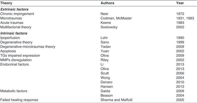
muscle-tendon unit in aging with statistically significant increase in frequency after 50 years. The painful shoulder is re-lated in 30-70% of cases to disorders of the rotator cuff. The incidence of rotator cuff tears varies between 5 and 40%, although it is very difficult to establish the real inci-dence of these lesions, which are often asymptomatic. Currently, the pathology of the rotator cuff is considered to be multifactorial, because extrinsic and intrinsic fac-tors play important roles, although it remains unclear the specific weight of each of these factors (Tab. 1). F. Oliva et al.
Table 1. Etiological factors analyzed.
Theory Authors Year
Extrinsic factors
Chronic impingement Neer 1972
Microtraumas Codman, McMaster 1931, 1993
Acute traumas Keene 1983
Multifactorial theory Soslowsky 2002
Intrinsic factors
Ipoperfusion Lohr 1990
Degenerative theory Sano 1999
Degenerative-microtraumas theory Yadav 2009
Apoptosis Yuan 2002
TGs impaired expression Oliva 2009
MMPs disregulation Riley 2002 Endocrinal factors Li 2013 Oliva 2013 Scutt 2006 Wong 2004 Denaro 2010 Hansen 2013
Metabolic factors Gaida 2009
Beason 2004
Failed healing response Sharma and Maffulli 2005
Table 2. Histopathological classification of the degeneration of the rotator cuff according to Riley. Organization of the fibers Nuclei of tenocytes Hyalinization Grade 1
Normal The bundles of fibers are well The nuclei are elongated with No hyalinization. tendon oriented with a wavy pattern. a pattern of unrecognizable
The single fibers are easily chromatin. The cores are distinguished within the bundle. arranged with their axis parallel
to the bundles of collagen fibers. Grade 2
Little The collagen fibers are relatively The nuclei are shorter but more No hyalinization. degeneration well aligned but the ripple oval. It can be observed a darker
is irregular. chromatin. The cores are often arranged in short chains that have an aspect in Indian file.
Grade 3
Moderate Loss of orientation of the collagen The cell nuclei are round or oval Moderate hyalinization, areas of degeneration fibers. and often increased in number. staining eosinophilic
There is a loss of orientation of homogeneous preparation the cores in relation to the bundles with hematoxylin/eosin. of collagen fibers. Chromatin has
a dark color. Grade 4
I.S.Mu.L.T - Rotator Cuff Tears Guidelines
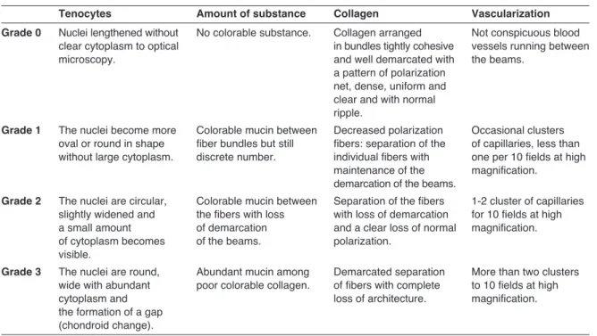
Table 3. Histopathological classification of tendinopathies of the rotator cuff according to Bonar.
Tenocytes Amount of substance Collagen Vascularization
Grade 0 Nuclei lengthened without No colorable substance. Collagen arranged Not conspicuous blood clear cytoplasm to optical in bundles tightly cohesive vessels running between
microscopy. and well demarcated with the beams.
a pattern of polarization net, dense, uniform and clear and with normal ripple.
Grade 1 The nuclei become more Colorable mucin between Decreased polarization Occasional clusters oval or round in shape fiber bundles but still fibers: separation of the of capillaries, less than without large cytoplasm. discrete number. individual fibers with one per 10 fields at high
maintenance of the magnification. demarcation of the beams.
Grade 2 The nuclei are circular, Colorable mucin between Separation of the fibers 1-2 cluster of capillaries slightly widened and the fibers with loss with loss of demarcation for 10 fields at high a small amount of demarcation and a clear loss of normal magnification. of cytoplasm becomes of the beams. polarization.
visible.
Grade 3 The nuclei are round, Abundant mucin among Demarcated separation More than two clusters wide with abundant poor colorable collagen. of fibers with complete to 10 fields at high
cytoplasm and loss of architecture. magnification.
the formation of a gap (chondroid change).
Table 4. Geometric classification according to Burkhart.
Type Description Pre-operative MRI Treatment Prognosis
1 Crescent shape Short and wide break Repair end-to-bone Good or excellent
2 Longitudinal (L or U) Long and narrow break Convergence of margins Good or excellent 3 Massive contracted Long and large, >2 × 2 cm Partial repair Good
4 Arthropathy of the rotator cuff Arthropathy of the cuff Arthroplasty Good
Table 5. References Guidelines present in literature.
Paper Authors Journal Year of publication
Clinical practice guidelines Beaudreuil J, Dhénain M, Orthop Traumatol Surg Res. 2010;96(2):175-179. for the surgical management Coudane H, Mlika-Cabanne N
of rotator cuff tears in adults.
Optimizing the management Pedowitz RA, Yamaguchi K, American Academy 2011;19(6):368-79 of rotator cuff problems. Ahmad CS, Burks RT, Flatow EL, of Orthopaedic Surgeons.
Green A, Iannotti JP, Miller BS, J Am Acad Orthop Surg. Tashjian RZ, Watters WC 3rd,
Weber K, Turkelson CM, Wies JL, Anderson S, St Andre J, Boyer K, Raymond L, Sluka P, McGowan R
Rehabilitation after van der Meijden OA, Westgard P, Int J Sports Phys Ther. 2012; 7(2): 197–218. arthroscopic rotator cuff Chandler Z, Gaskill TR,
repair: current concepts Kokmeyer D, Millett PJ review and evidence-based
guidelines.
Clinical Practice Guidelines Hopman K, Krahe L, by The University of New Australia. 2013 for the Management Lukersmith S, McColl A, Vine K South Wales Rural Clinical
of Rotator Cuff Syndrome School, Port Macquarie
in the Workplace.
AAOS appropriate use criteria: Pappou IP, Schmidt CC, J Am Acad Orthop Surg. 2013;21(12):772-775. optimizing the management Jarrett CD, Steen BM, Frankle MA
of full-thickness rotator cuff tears.
Methodology
The Authors were divided into four groups:
- Coordinator: he conceived and organized the work and the group and selected the most impor-tant QUESTIONS (Q) on this topic.
- Control group: controlled the development of the work and discussed the recommendations. - Group of the experts: they individually received a
question and developed the ANSWERS (A) ac-cording to the rules of EBM, when it was possible. - Group of preparation and evaluation of literature: drew up the text and assisted the group of experts in evaluation the literature.
Methods and criteria study selection
For research were consulted the following databases: - PubMed
- Embase - Google Scholar - Cochrane Library
Randomized controlled trials (RCTs); Systematic re-views; to follow if missing the first two, the other levels of evidence. The literature is updated at December, 2014. F. Oliva et al.
The natural history of rotator cuff tears is to progress over time. Lesions may develop a tendon retraction and a fatty degeneration that make it more uncertain to re-pair. The final stage is the Rotator Cuff Arthropathy. Few studies were performed to quantify the histopatho-logical alterations of tendon ruptures and different histopathological (Tabs. 2, 3) and geometric (Tab. 4) clas-sifications were drawn in order to feature tendinopathies. Despite the high level reached in the field of shoulder surgery in our country, Italian orthopaedic surgeons have never produced a Consensus Protocol on this topic. To the best of our knowledge in literature we found several attempts to simplify the management of the tears of the rotator cuff through the compilation of guidelines or documents (Tab. 5).
Approach to Guidelines
These recommendations developed through a process of systematic review of the literature and expert opin-ion, that can be used to improve the quality of care and rationalize the use of resources. Clinical decisions on individual patients require the application of the recom-mendations, based on the best scientific evidence and clinical experience of the physician.
According to the literature, there is not a general con-sensus regarding the diagnostic criteria and the valid-ity of the physical examination in patients with
shoul-Question n. 1: Clinical tests
Though many tests have been described for the clini-Level of evidence
% %
%
%
Level of evidence Criteria for analysis and inclusion
I Meta-analyzes and systematic reviews of randomized controlled trials (RCTs) of high quality, or RCTs with minimum or low risk of bias.
Systematic reviews of high quality relative to cohort studies or case-control.
II Cohort studies or randomized case-control high quality, with minimal risk of confounding or bias and with high or discrete probability of causation.
III Case-control studies and retrospective comparison of well-conducted with reasonable probability of causation.
IV Non-analytic studies as case series or individual cases.
%
!
Level of recommendation
% %
%
%
Level of recommendation Criteria for analysis
A Supported by at least two studies of level Ib or from a review level Ia (“it was shown”). B Supported by at least two independent studies of level II or extrapolations from studies
of level I (“it is possible”).
C Not supported by adequate studies of level I or II (“indications”). D Indications of experts (“there is no evidence”).
!
!
the clinical tests are not accurate in distinguishing rotator cuff disorders compared to other diseases. Information about the mechanism of injury and type and onset of pain should be collected; therefore, conventional X-ray and MRI studies provide addition-al details.
Level of recommendation: A Key points:
The most reliable and sensitive tests for the proper evaluation of the rotator cuff lesions are:
• The Jobe test, for supraspinatus tendon. • The Patte test for infraspinatus tendon.
• The lift off and belly press test, according to the range of movement, for the subscapularis tendon. Key words: diagnostic accurancy, clinical test, physi-cal examinations, crossed with rotator cuff tears.
Question n. 2: Instrumental diagnosis
Scientific literature focuses on three points:
a) After the clinical suspicion of RCT what is the gold standard imaging technique to confirm the diagnosis?
b) Is there an imaging recommended technique to improve the diagnostic accuracy of RCT?
c) What are the relevant informations that could be provided by each imaging modality and that could encourage therapeutic decision?
Conventional radiography (RX): its use for soft tis-sue injuries of the shoulder is not validated. X-rays can be useful to exclude other possible causes of shoulder pain.
Ultrasound: the diagnostic accuracy of the ultra-sound is good and comparable to that of conventional MRI to identify and quantify the complete injuries (full thickness) of the rotator cuff, although there are con-trasting results about its validity in partial RCT. Magnetic Resonance Imaging (MRI): the diagnostic accuracy of MRI for the detection of full-thickness RCT is excellent, but it is more limited for partial tears. Arthro-MR: despite Arthro-MR is a less invasive techniques, it has a limited usefulness if compared to MRI and ECO. However, it may still be considered for its high sensitivity and specificity. The use of di-agnostic imaging is useful after 6 weeks of symp-toms suggestive of RCT and Ultrasound (combined or not with conventional radiography to determine osteoarthritis, bone abnormalities and the pres-ence/absence of calcification) has been recommend-ed as the most valid method of imaging to exclude a rupture of the rotator cuff after an unresponsive con-servative treatment.
Key points:
• A complete physical examination help to correctly select the most appropriate imaging technique for accurate diagnosis.
• MRI or ECO can confirm a possible full-thickness tears, but however, if a patient has an implantable device that does not allow the execution of MRI, conventional radiography should be considered as a viable alternative.
• Actually, there is no consensus on which ap-proach is more precise, convenient, appropriate or not invasive for the diagnosis of a complete or partial RCT.
• The best imaging test is based on several factors such as: sensitivity and specificity, operator’s ex-perience in the performance and interpretation of the study, timing, cost and contraindications of the test for the patient.
Key words: controlled randomised trials and system-atic reviews, rotator cuff tear in combination with imaging OR X ray OR ultrasound OR magnetic reso-nance OR radiological diagnoses.
Question n. 3: Rehabilitation approach in the
rotator cuff tears
It is difficult to obtain double blind studies analyzing the most appropriated rehabilitation approach in the rotator cuff tears.
In literature, different conservative treatments were studied. Comparing addiction or not of proprioceptive stimuli to stretching and muscular strengthen exercis-es, followed by cryotherapy, there were not statisti-cally significantly differences. According to literature, there are no differences between occupational thera-py and home-based exercises in conservative treat-ment of rotator cuff tears.
There are also studies which are in favor of surgery in small and medium-sized tears of rotator cuff, while other ones support conservative treatments.
However, it is not possible to establish which is the better treatment because high quality clinical random-ized studies are necessary.
Level of recommendation: B Key points:
• There are some advantages from the utilization of Therapeutic Exercise (TE), singularly or in an In-dividual Rehabilitation Project (PRI), in patients with rotator cuff tears.
• Despite its efficacy, it is unknown when PRI should be started, what programs should include or time necessary for a surgery indication. • High quality Clinical Randomized Studies, utilizing
standard outcome scores, are necessary to esta-blish the best PRI, including TE.
Key words: rotator cuff, supraspinatus, infraspinatus, subscapolaris, teres minor, in combination with other words such as tear, lesion, pathology, injury, exercise, exercises, physiotherapy, rehabilitation, intervention.
Question n. 4: Drug therapy
In patients affected by symptomatic rotator cuff tears, the aim of treatment is the pain’s reduction and the improvement of movements and life’s quality. In liter-ature, drug therapy is still debated.
NSAIDs (Non Steroid Anti-Inflammatory Drugs) are the most studied drugs used in this pathology. The Non Specific NSAIDs were compared with Placebo, I.S.Mu.L.T - Rotator Cuff Tears Guidelines
Corticosteroid injections and COX-2 inhibitors. NSAIDs therapy reduces pain in the first 3-4 weeks, however it is necessary planning a different treatment for a complete pain’s suppression and to improve functions.
Level of recommendation: C Key points:
• There is not a definitive drug therapy for rotator cuff tears.
• NSAIDs therapy reduces pain in the short term, while does not improve functions.
• Corticosteroid injections and NSAIDs have similar effects in the short term.
Key words: shoulder, gleno-humeral joint, rotator cuff, acromion, supraspinatus muscle, crossed with lesion, rupture, tendinophaty, impingement, deficien-cy, disorder.
Question n. 5: Shoulder injections
Shoulder injection is an argument of different studies yet. According to literature, injective treatment with glucocorticoids, local anaesthetic, hyaluronic acid (HA) or Platelet-Rich Plasma (PRP) is efficacious to reduce pain and to improve functions in patients with rotator cuff tendinopathy. However, it is not possible recommending a particular type of injections through those studied.
According to literature, glucocorticoid or HA injective treatment is indicated in patients with complete or partial rotator cuff tear because it reduces pain and improve functions. Also in this case, it is not possi-ble to indicate which type of injective drug is more efficacious.
Level of recommendation: B Key points:
• Glucocorticoids, local anaesthetics, HA or PRP are used to improve pain and performance in pa-tients with rotator cuff tendinopathy.
• No sufficient evidences in comparison between different injective therapies.
• Glucocorticoids and local anaesthetics have cyto-toxic effects on tenocytes.
• HA decreases pain in patients with partial tear of the rotator cuff tendons.
• At the moment, there are no evidences that PRP injections must be used in patients with partial or complete tear of the rotator cuff tendons.
Key words: rotator cuff tendinopathy injection, rota-tor cuff tendinopathy glucocorticoids injection, rotarota-tor cuff tendinopathy hyaluronic acid injection, rotator cuff tendinopathy Platelet-Rich Plasma injection, rota-tor cuff tear injection.
Question n. 6: Surgery indications and
reparability criteria
It is not easy to decide the most appropriated surgery
The outcome depends on different factors such as age, gender, symptom’s duration, “surgery timing”, function-ality, tear’s anatomy and the presence of “worker com-pensation”. Thus, it is necessary evaluating these vari-ables to make an adequate surgery choice.
Level of recommendation: D Key points:
• There is no a “cut-off” age for surgery indication, which must be evaluated basing on patient’s ac-tivities and on difference between chronological and physiological age.
• Despite women being epidemiologically more af-fected, this is not a limitation to surgery indication. • In traumatic tears, it is suggested surgery
repara-tion by 4 months.
• Anatomical and anatomopathological severity of the tear is a determining factor for the clinical outcome. • A preserved preoperative functionality is a
posi-tive prognostic factor.
Key words: rotator cuff repair, rotator cuff tears, sub-ject headings, surgical indication, operative indication and indication surgery, prognostic factors.
Question n. 7: Miniopen vs arthroscopy
Scientific studies do not demonstrate statistically signifi-cant differences in the outcomes between the all-arthroscopic and mini-open rotator cuff repair. The out-comes have been evaluated with different methods: VAS, Costant-Murley, DASH, UCLA, ASES, RMN, etc. The main differences between the two techniques are total cost and operating room time which are both in-creased in arthroscopy technique.
In literature, there is not a consensus on these re-sults. The small number of patients is the only limit of the precedent randomized studies; the number of pa-tients increases only in retrospective studies.
Level of recommendation: B Key points:
• Surgical technique depends on surgeon’s and pa-tient’s preference because the outcome is not in-fluenced by surgical decision.
• There are no statistically significant differences between the two techniques considering relapse, complications and functional outcomes.
• Arthroscopy is more expensive and requires more operative time than miniopen technique.
Key words: rotator cuff miniopen arthroscopy, rotator cuff tear miniopen arthroscopy, rotator cuff repair miniopen arthroscopy, rotator cuff tear repair miniopen arthroscopy.
Question n. 8: Arthroscopic treatment of
par-tial tears of the rotator cuff: where and when
Current scientific evidence does not allow to deter-mine which is the best treatment for symptomatic par-tial lesions of the rotator cuff (LPSCR). The arthro-scopic debridement with or without acromioplasty, F. Oliva et al.
treatments. Debridement is generally indicated as a treatment of injuries involving less than 50% of the tendon thickness or tendon lesions of grade I/II ac-cording to Ellman.
It is hard to compare the results of different treat-ments for the LPSCR because of the heterogeneity of the type of lesions and the tools used in evaluating the results. It is impossible to propose treatment guidelines due to the low level of evidence proposed by the studies in the literature.
Key points:
LPSCR grade I-II of Ellman (involvement of less than 50% of the thickness tendon):
• Lack of studies of level I, II and III.
• The arthroscopic debridement with or without acromioplasty results in clinical improvement in patients with grade III of LPSCR Elmann.
• There are no studies that compare repair to de-bridement in these patients.
Level of recommendation: D
LPSCR grade III of Ellman (involvement of more than 50% of the thickness tendon):
• In one study (level of evidence IV), Weber demonstrates the superiority of repair compared to debridement in patients with grade III LPSCR Ellman.
• There are three studies evaluating the results of the trans-tendon repair and three studies evaluat-ing the results after completion of the repair. All studies report a clinical improvement after the re-pair of the lesion.
• There are no studies of level I, II, III that compare repair to debridement in these patients.
Level of recommendation: D
Repair technique in patients with LPSCR grade III of Ellman:
• The presence of two prospective randomized studies level II allows us to conclude that there is no statistically significant difference between the repair techniques.
Level of recommendation: C
Key words: partial rotator cuff, rotator cuff, rotator cuff tears, rotator cuff lacerations, arthroscopic cuff repair, partial thickness rotator cuff.
Question n. 9: Management of the condition
of the long head of the biceps in association
with lesions of the rotator cuff
When long head of the biceps (LHB) tears are associ-ated with rotator cuff tears, surgical exploration and possible treatment is recommended if symptoms per-sist for more than 3 months after conservative treat-ment. The two main treatments involve the tenodesis of the LHB and tenotomy of the LHB.
Many studies suggest tenotomy of the LHB as a fast treatment, well tolerated by the patient, with the possi-bility to reduce the time of rehabilitation after surgery. Other studies suggest that tenodesis of LHB leads to a better ability to return to activity sports and to a good restoration of the anatomy of the LHB despite
the longer-term rehabilitation and the greater difficulty in the surgical technique.
According to the results reported by the literature it is not possible to give an absolute recommendation on which is the best type of treatment for the pathology of the LHB. Tenotomy of the biceps is indicated in older patients with a sedentary lifestyle and low func-tional demand, and in obese patients who can accept cosmetic problems. Tenodesis of the LHB is instead recommended in young patients under the age of 40 years who practice physical activity.
Key points:
• The tenotomy and tenodesis of the LHB have shown clinical and functional overlap.
• The tenotomy hesitates more often in aesthetic al-terations compared to tenodesis.
• Among the different types of tenodesis present in the literature it is not possible to decide which is the best technique because there is no literature about this.
Level of recommendation: C
Key words: biceps tenotomy, biceps tenotomy ver-sus tenodesis or tenodesis, biceps tendon, long-head biceps lesions.
Question n. 10: Surgical suture
In the past, open repair surgical techniques were con-sidered the gold standard. Concerning arthroscopic repair, there was an evolution of the repair techniques in “single row”, “double row” and “transosseous equiv-alent” towards the idea of reproducing the area of reinsertion of the tendons of the rotator cuff.
Key aspects for effective repairs of the rotator cuff in-clude:
• good initial stiffness and strength of the surgical repair;
• good stability during the movement of intra and external rotation occurring in the immediate post-operative period;
• optimization of the contact tendon-bone surface. The most common technique is the “single row” (SR). The documented failure rate with these repair tech-niques appeared high, up to 90% in the case of large and massive injuries, at the tendon-suture interface. The “double-row” (DR) repair is more resistant than a “single-row”, but it is important to consider the greater strain on the repaired tendon. Trans-bone repair tech-niques (without anchors) have been introduced in arthroscopy in order to restore a tendon insertion at least 20% stronger than any other surgical technique, even if the concentration of the stress is moved from the tendon-suture junction to the bone.
In conclusion, at this time, there is no evidence that could support the use of a repair technique over another. Level of recommendation: B
Key points:
• The double-row techniques increase costs in terms of materials and time of the operating room (EBM). • Current evidence of the literature lead to consider
a repair type single-row in the lesions less than 3 I.S.Mu.L.T - Rotator Cuff Tears Guidelines
cm and in the presence of a good quality of ten-don tissue, while the double-row repair would be considered in cases of injury larger than 3 cm and with poor quality of tendon tissue.
• In large lesions, chronic and retracted, even a double-row repair has a high risk of failure. • The transbone techniques seem biomechanically
promising, but not yet supported by sufficient ran-domized clinical trials.
Key words: rotator cuff, cuff tears, cuff repair, asso-ciate a suture anchor, double-row, single-row, arthro-scopically repair.
Question n. 11: Massive and irreparable
ro-tator cuff tears
There are different types of treatment utilized for massive and irreparable rotator cuff tears. However it is not possible to identify an ideal treatment.
Conservative treatment, arthroscopy debridement and reverse prosthesis are useful above all in the el-derly, while tendon transposition is usually used in the young people.
Other two types of treatment are long head of the bi-ceps tenotomy and partial reparation of the tear: they are both useful to decrease pain. The use of scaffolds is still studied.
Latissimus dorsi transposition is used for the posteri-or-superior tears while pectoralis major transposition for the anterior-superior tears.
Level of recommendation: D Key points:
• There is not an ideal treatment for massive and ir-reparable rotator cuff tears.
• There are no controlled randomized studies com-paring conservative and surgical treatment or the different types of surgical treatment.
• It is necessary an accurate clinical and radiologi-cal evaluation to choose an adequate treatment. • After treatment, the results are good both in the
ear-ly and in the middle time but they could decrease. Key words: irreparable rotator cuff tear and massive ro-tator cuff tear crossed with randomized controller study and systematic review.
Question n. 12: Regenerative strategies in
surgical repair
Three options are arising interest in rotator cuff repair strategies: PRP, scaffolds and mesenchimal cells. PRP (Platelet-Rich Plasma)
The best evidence on PRP use in clinical practice are about the risk of a new tear that can be globally re-duced or arised according to the section area and de-pending on the patient’s age. There are no differences of the clinical outcome after a short period of follow-up. Some studies showed that PRP reduces pain after the surgical procedure and improves the functional
recov-Key points:
• Poor evidence on pain reduction and risk of a new tear.
• At this time, EBM does not support PRP in rotator cuff tears.
• It is necessary to classify and standardize differ-ent PRP preparations available.
Question n. 13: Use of scaffold, patch and
augmentation
Autograft
Literature showed good result in using periosteal au-tologous flap with a low percentage of re-tears and with poor complications such as etherotopic calcifica-tions. Positive results have been shown also in clini-cal and in instrumental/ultrasound outcome.
Xenograft
Some studies showed positive results using graft for not completely repairable lesions, with good results without complications at three years follow up. Other studies revealed inflammatory complications in 40% of cases and worst clinical outcome.
Allograft
Allograft are a novel technique but clinical result are still controversial. Some Authors found positive re-sults in repairing rotator cuff with inconsistent clinical outcome; on the other hand, other studies demon-strated an improvement in functional outcome without complications.
Synthetic scaffold
Patch and synthetic scaffold are used for augmenta-tion in non anatomically repairable rotator cuff tears. They have positive results on a clinical and instru-mental point of view.
Level of recommendation: D Key points:
• Scaffold should be used only in massive rotator cuff tears, with inconsistent tissue that does not permit a complete repair of the tendon.
• At this time, EBM does not support scaffold for lack-ing of RCT level I and for small number of patients. • Patch and xenograft have significant
immunologi-cal complications.
Key words: tissue, graft, augmentation, mesenchi-mal, stem cells, supraspinatus, rotator cuff, repair. Mesenchimal stem cells
Clinical use approved by RTC in literature concerns fracture healing.
Level of recommendation: D Key points:
• At this time, the use of mesenchimal stem cells is not supported by evidence.
Key words: tissue, graft, augmentation, mesenchi-mal, stem cells, supraspinatus, rotator cuff, repair.
Question n. 14: Latissimus dorsi transfer
F. Oliva et al.tion, particularly in young patients with massive pos-terior-superior rotator cuff tears, when the surgical repair is no longer considered a possible solution. Today is also used in combination with reverse shoulder prosthesis implantation in older patients with a severe loss of external rotation. The length of the tendon is very important because an insufficient mobilization of the tendon can determine a limitation of the rotations, a decentralization of the humeral head and an increase in pressure of the head against the glenoid. In literature, all Authors have reported good functional results after LDT surgery, in particu-lar in external rotation recovery. The integrity of the tendon of the subscapularis muscle is important to have a good clinical outcome. This technique showed a poor functional outcome in patients with severe glenohumeral osteoarthritis. There is no agreement on what is the best place to insert the tendon. In summary, after this procedure, we can expect an im-provement of about 35° vertically, 10° of external ro-tation, and a recovery of abduction strength in up to about 70% compared to the contralateral shoulder healthy, but that we can not wait for a return to nor-mality. Although the results so far are encouraging, it is not possible to establish clear recommendations on the use of the LDT.
Level of recommendation: D Key points:
• Absence of studies of evidence level I.
• The results of this transfer in posterior superior and massive irreparable rotator cuff tears are en-couraging with respect to the recovery of the ROM, external rotation, the strength and function of the shoulder.
• Negative prognostic factors appear to be the glenohumeral osteoarthritis, the glenohumeral joint space narrowing, the rupture of the tendon of the subscapularis muscle and fatty degeneration of advanced teres minor muscle.
• The follow-up is still short to assess the long-term results.
Key words: shoulder, rotator cuff tear, massive rota-tor cuff tear, tendon transfer, latissimus dorsi transfer, randomized controlled trial, young patients.
Question n. 15: Reverse prosthesis in
ir-reparable rotator cuff tears
The reparation is more difficult when there is a pro-gression of the rotator cuff tear. In literature, there is no consensus on the most appropriated treatment. Reverse Shoulder Arthroplasty (RSA) was introduced for patients with rotator cuff arthropathy, but now it is in-dicated also in other pathologies such as pseudoparal-ysis, proximal humeral fractures, instability or oncologic surgery. Good results have been reported in patients with irreparable rotator cuff tears and in patients over and under 60 years old.
However, in literature there are many limits such as the absence of randomized prospective studies of level I and the short follow-up. Thus, there are no
de-finitive conclusions for the long term follow-up after RSA. Furthermore, many studies include different in-dications for the reverse prosthesis and utilize differ-ent evaluation scales which do not allow to compare the results.
Level of recommendation: D Key points:
• No studies with level of evidence I and II.
• Reverse shoulder prosthesis could be advise in symptomatic patients with massive and irrepara-ble tears of the rotator cuff, if associated with one or more of these conditions:
- pseudoparalysis
- humeral head shifted up with subacromial space < 6 mm
- gleno-humeral arthrosis.
Key words: rotator cuff tear, massive rotator cuff tear, reverse shoulder arthroplasty, reverse shoulder replacement, hemiarthroplasty, randomized con-trolled trial, reverse in young, young patients.
Question n. 16: Rehabilitation protocol after
rotator cuff repair
Despite the growth of the clinical interest and of the studies, there is a partial scientific evidence on the therapeutic strategies to improve post-operative out-come after reparation of the rotator cuff tears. The type of surgical and rehabilitative treatment must be personalized considering factors such as size and type of the tear, age of the patients, presence of co-morbidities, compliance to treatment. A rational reha-bilitative approach is based on a gradual mobilization of the shoulder. The aim is the articular preservation and the prevention of the excessive tension on the re-paired tendons.
The different types of exercises and of treatment must be introduced at the right time during the reha-bilitation protocol. It is also recommended a gradual restart of sportive and recreational activities, only af-ter an adequate functional re-education and without pain. Articulation and strength are complete and simi-lar to the contralateral arm only 6 months after surgi-cal repair of the rotator cuff, with consequent restart of the sportive activities.
Key points:
• The rehabilitation program must be personalized in each patient. It depends on intrinsic and extrin-sic factors which could influence tendons healing and functional recovery. For example, early or late mobilization could both lead to negative ef-fects on biomechanical properties of the healed tissues.
• The basic knowledge and the mechanobiological studies have led to scientific evidences. This knowledge help us taking the correct clinical deci-sion to control post-operative pain, deciding shoulder immobilization time, time and type of neuromuscular rehabilitation.
Key words: rotator cuff tear, rotator cuff surgical re-pair, rotator cuff post surgical rehabilitative treatment I.S.Mu.L.T - Rotator Cuff Tears Guidelines
crossed with randomized controlled trial and system-atic review.
Question n. 17: Return to sport after repair of
the rotator cuff tears
Shoulder tear management is based on conserva-tive or surgical treatment and on rehabilitation until functional recovery of the state before the trauma. In athletes, there are 2 phases: the first leads to recov-ery of the physical activities in the daily life (normal population), while the second leads to return to the sportive performance.
However, in literature the most of the studies have a level III of evidence or they are expert opinions, so there are not univocal data and Evidence Based Recommendations are necessary for the athletes. The I.S.Mu.L.T. guidelines for the muscular trauma re-covery have introduced the concept of motor re-educa-tion in phases IV and V as the final part of the rehabili-tation period, which gradually leads the athlete to train-ing again. The aims durtrain-ing these two phases are: • Recovery of the proprioception and the
coordina-tion in the specific sports.
• Metabolic specific readjustment (aerobic-anaero-bic-mist).
• Recovery of the most important strength’s charac-teristic for the performance (maximum, explosive, elastic, resistant).
Level of recommendation: C Key points:
• In the sportive population, there are no studies which describe methodologic approaches for the different physiological variables to recover the sportive performance after rotator cuff tear. • Physical trainer must choose personalized protocols
for the athletes considering detraining effects during
the sport interruption period and the temporal pro-gression in the sportive performance’s recovery. Key words: management of rotator cuff lesion in ath-letes, rehabilitation of rotator cuff lesion, return to sport after rotator cuff repair, rotator cuff lesion in ath-letes, recovery after rotator cuff lesion.
Question n. 18: Rotator cuff tears in the
child-hood
The incidence of the rotator cuff tears in the childhood is about 1%, but it could be underestimated. Typically, the young patient has a persistent pain, not associated with a particular trauma. The overuse activities of the upper extremities is the main risk factor, e.g. in tennis, basket and volleyball players and in the pitchers. Path-ogenic mechanism is usually gleno-humeral internal im-pingement and MRI (Magnetic Resonance Imaging) is the diagnostic examination of first level.
According to the literature, it is indicated the conserv-ative treatment. The surgical treatment is used when symptoms don’t disappear. Arthroscopy treatment is still debated.
Level of recommendation: D Key points:
• Rotator cuff tears are rare but probably underesti-mated in the childhood.
• The tears are often partial. • MRI is examination of first level.
• Conservative treatment is the first choice. • Arthroscopy is more advisable after conservative
treatment failure.
Key words: rotator cuff tears, sport injuries in combi-nation with pediatric, adolescent, adolescent athletes.
Answer n. 1: Clinical tests
F. Oliva et al.Supraspinatus tendon tests
Tests Author Result Sensitivity % Specificity % PPV
%
NPV %
Accuracy
Jobe test Noel et al. 1989 (1) Strength deficit 95 65 86 85 85 Itoi et al. 1999 (2) 77 68 44 90 70 Itoi et al. 2006 (3) 87 43 79 67 79 Leroux et al. 1995 (4) 79 67 56 85 73 Kim et al. 2006 (5) 76 71 92 51 69
!
(to be continued)I.S.Mu.L.T - Rotator Cuff Tears Guidelines
Cont.
!
Tests Author Result Sensitivity % Specificity % PPV
% NPV % Accuracy Kelly at al. 2010 (6) 89 60 - - 53
Jobe Test Itoi et al. 1999 (2) Pain 63 55 31 82 57 Itoi et al. 2006 (3) 78 40 - - 71 Leroux et al. 1995 (4) 86 50 96 22 Kim et al. 2006 (5) 94 46 46 94 62 Kelly at al. 2010 (6) 80 60 81 58 73 Full can test Itoi et al. 1999 (2) Strength deficit 77 74 49 91 75 Itoi et al. 2006 (3) 83 53 - - 78 Kim et al. 2006 (5) 77 68 54 86 71 Kelly et al. 2010 (6) 45 75 - - 49 Full can test Itoi et al. 1999 (1) Pain 66 64 37 85 64 Itoi et al. 2006 (3) 80 50 - - 74 Kim et al. 2006 (5) 71 68 52 91 69 Kelly et al. 2010 (6) 34 25 - - 33
!
F. Oliva et al.
Infraspinatus tendon tests
+
+
+
+
Tests Author Result Sensitivity % Specificity% PPV
%
NPV %
Accuracy
Patte test Itoi et al.
2006 (3) Strength deficit 84 53 - - - Kelly et al. 2010 (6) 52 67 - - 53 Leroux et al. 1995 (4) 83 61 21 97
Patte test Itoi et al. (3) Pain 54 54 - - -
Kelly et al. 2010 (6) 34 100 - - 42 Leroux at al. 1995 (4) 92 30 29 93 - External rotation lag sign Hertel et al. 1996 (7) 70 100 100 56 78 Castoldi et al. 2009 (8) 12 98 73 73 73 Walch et al. 1998 (9) 98 98 - - - Bak et al. 2010 (10) 45 91 87 57 65 Miller et al. 2008 (11) 46 94 77 78 -
Drop sign Hertel et al.
1996 (7) 21 100 99 32 43 Bak et al. 2010 (10) 45 70 65 50 56 Miller et al. 2008 (11) 73 77 61 85 -
Atrophy Litaker et al.
2000 (12)
55 73 81 43 -
I.S.Mu.L.T - Rotator Cuff Tears Guidelines
Subscapularis tendon tests
+
+
+
+
Tests Author Result Sensitivity % Specificity% PPV
%
NPV %
Accuracy
Lift off test Itoi et al. 2006 (3) Strenght deficit 79 59 - - - Leroux et al. 1995 (4) 0 61 0 88 - Barth et al. 2006 (13) 18 100 100 77 78 Itoi et al. 2006 (3) Pain 46 69 - - - Internal rotation lag sign Hertel et al., 1996 (7) Strenght deficit 97 96 97 96 96 Scheibel et al. 2005 (14) 75 - - - - Rigsby et al. 2010 (15) 98 94 - - - Bak et al. 2010 (10) 31 87 75 50 56 Miller et al. 2008 (11) 100 84 28 100 -
Belly press test Barth et al. 2006 (13) 40 98 89 80 81 Scheibel et al. 2005 (14) 38 - - - - Rigsby et al. 2010 (15) 88 97 - - -
Napoleon test Barth et al. 2006 (13) 25 98 83 76 77 Scheibel et al. 2005 (14) 69 - - - - Rigsby et al. 2010 (15) 98 97 - - -
+
!
Answer n. 2: Instrumental diagnosis
F. Oliva et al.'
'
'
'
'
'
Authors (year) N. of cases or studies included
Type of study Imaging techniques Sensitivity Specificity Lenza et al.(16) (2013) 20 studies (1147 shoulders) Systematic review - Magnetic resonance (MRI) - Arthro-MR - Ultrasound (US)
- for any rupture of the cuff: MRI 98% (6 studies, 347 shoulders); US 91% (13 studies, 854 shoulders); - full thickness tears: MRI 94% (7 studies, 368 shoulders); ArthroMR 94% (3 studies, 183 shoulders); US 92% (10 studies, 729 shoulders)
- for any rupture of the cuff: MRI 79% (6 studies, 347 shoulders); US 85% (13 studies, 854 shoulders); - full thickness tears: MRI 93% (7 studies, 368 shoulders); ArthroMR 92% (3 studies, 183 shoulders); US 93% (10 studies, 729 shoulders) Smith et al.(17) (2012) 44 studies (2751 shoulders in 2710 patients) Systematic review and misanalysis MRI in full thickness or partial rotator cuff tears - partial tears: 0.80; - full thickness: 0.91 - partial tears: 0.95; - full thickness: 0.97 Smith et al.(18) (2011) 62 studies (6066 shoulders in 6007 patients) Systematic review and misanalysis Ultrasound in full thickness or partial rotator cuff tears - partial tears: 0.84; - full thickness: 0.96 - partial tears: 0.89; - full thickness: 0.93 Ottenheijm et al. (19) (2010) 44 studies: - 22 full thickness tear; - 15 partial tear; - 3 subacromial bursitis; - 2 tendinopathies; - 2 calcifications Systematic review and misanalysis Ultrasound in subacromial disorders - partial tears: 0.72; - full thickness: 0.95; - subacromial bursitis: 0.79-0.81; - tendinopathies: 0.67-0.93; - calcifications: 1.00 - partial tears: 0.93; - full thickness: 0.96; - subacromial bursitis: 0.94-0.98; - tendinopathies: 0.88- 1.00; - calcifications: 0.85-0.98 Kelly, Fessell (20) (2009) 67 studies Systematic review - US - MRI - ArthroMR - partial tears: US 0.67; MRI 0.44 - full thickness: US 0.87; MRI 0.89; Arthro-MR 0.95 - partial tears: US 0.94; MRI 0.90 - full thickness: US 0.96; MRI 0.93; Arthro-MR 0.93 Dinnes et al.(21) (2003)
10 cohort studies Systematic review and misanalysis - US - MRI - ArthroMR - all tears: US 0.33-1.00 MRI 0.83 ArthroMR 0.95 - all tears: US 0.43 to 1.00 MRI 0.86 ArthroMR 0.93 (to be continued)
I.S.Mu.L.T - Rotator Cuff Tears Guidelines
Cont.
Authors (year) N. of cases or studies included
Type of study Imaging techniques Sensitivity Specificity Ardic et al.(22) (2006) 59 shoulders in 58 patients Transversal study - US - MRI (compared to clinical tests) - all supraspinatus tears: US 98.1%; clinical tests for impingement 78.3% - full thickness: US 54.2% in underestimation vs MRI 71.2% (10 cases) - partial tears: US 37.3% in overestimation vs MRI 27.1% (6 cases) - all supraspinatus tears: US 60%; clinical tests for impingement 50% Blanchard et al. (23) (1999) 104 patients Transversal study - MRI - ArthroMR full thickness: RM 81%; ArtroMR 50% full thickness: RM 78%; ArtroMR 96% Singson et al.(24) (1996)
177 MRI images Randomized retrospective study - RM fast spin-echo T2 with fat suppression - RM fast spin-echo T2 without fat suppression - full thickness: with fat suppression 100%;
without fat suppression 100% - partial tears: with fat suppression 92%; without fat suppression 67% Normal tendons: both the techniques 86%
!
Answer n. 3: Rehabilitation approach in the rotator cuff tears
F. Oliva et al.Studies comparing conservative treatments in rotator cuff tears
(
(
(
(
(
(
(
(
(
Author (year) No Type of
study Follow-up period Outcome Martins, Marziale (25) (2012) 18 participants: - 9 in the control group - 9 in the experimental group Randomized Clinical Study
Follow-up absent - Shoulder ROM measurement with goniometer;
- Western Ontario Rotator Cuff Index (WORC);
- Occupational Stress Indicator (OSI); - Visual Numeric Scale (VNS) Krischak et al.(26) (2013) 43 participants Randomized Clinical Study 2 months after therapy
- Primary outcome: VAS (Visuo Analogic 10-point Scale); - Constant-Murley score; - Shoulder ROM; - Clinical impingement; - Grade of strength in
abduction/adduction and rotation Moosmayer et al. (27) (2010) 103 participants Randomized Clinical Study 6-12 months after surgery
- Primary outcome: Constant score; - American Shoulder and Elbow Surgeons (ASES) score;
- Short form (SF-36) Health survey; - Subscores on grade of movement, pain, shoulder strength and grade of satisfaction of the patient
Kukkonen et al. (28) (2014) 180 participants Randomized Clinical Study 3-6-12 months after surgery
- Primary outcome: Constant score; - Subjective evaluation comparing pre and post-surgery;
- Subjective grade of satisfaction Seida et al. (29) (2010) 3 Reviews Cochrane and 14 Randomized Clinical Studies Systematic Review — — Huisstede et al. (30)(2011) 137 Studies Systematic Review — —
!
Answer n. 4: Drug therapy
I.S.Mu.L.T - Rotator Cuff Tears Guidelines
' ' '
'
'
'
Authors Analyzed drugs n. studies
(f.u. days)
n. pt Results Evidence
Boudreault et al. (31)
12 RTCs
FANS vs Placebo 4 (14) 120 In favour of FANS for pain
II
NS FANS vs Cox2 3 (14) 608 Similar both in pain and in tolerance
III
FANS vs Corticost. Injections
3 (33) 200 No differences III
van der Sande et al. (32) 3 SR + 5 RCTs FANS vs laser (Naproxen 550 mg x2/die 14 gg vs Laser 902 nm) 1 RCT (14) Moderate evidence in favour of laser II Ibuprofen (600 mg 4/die) vs Ibuprofen slowly released (1200 mg 2/die) 1 RCT (168) 147 In favour of traditional ibuprofen II FANS vs Corticost. Injections 3 RCTs (28-42) 120 No differences II
Van der Windt et al. (33)
18 RCTs
FANS vs Corticost. Injections
4 RCTs (28) N.D. In favour of injections both for pain and for functions
III
FANS vs Placebo 4 RCTs (14) N.D. In favour of FANS III
!
Answer n. 5: Shoulder injections
F. Oliva et al.Randomized studies indicating efficacy of injective treatments in rotator cuff tendinopathy
*
*
*
* *
*
* *
*
*
*
*
Author (year) No Type of
study
Follow-up
Result Outcome Level of
evidence Adebajo et al. (1990) (34) Corticosteroid (n=x) Diclofenac per os (n=x)
Randomized // Major efficacy with injective treatment // II Alvarez et al. (2005) (35) Corticosteroid (n=30) Xilocaine (n=28) Randomized 2,6,12,24 weeks No statistically significantly differences between the 2 groups in each follow-up WORC, DASH, Shoulder and Elbow Surgeons, active ROM I Eyigor et al. (2010) (36) Corticosteroid + Mepivacaine (n=20) TENS (n=20) Randomized 1,4,12 weeks Statistically significantly differences in favour of group I after 1 week
VAS for pain, ROM, Shoulder Disability Questionnaire (SDQ), Short Form-36 (SF-36), Beck Depression Scale (BDS) II Rha et al. (2013) (37) PRP (n=20) Dry Needling (n=19) Perspective Randomized 24 weeks PRP treatment more efficacious than dry needling treatment Shoulder Pain and Disability Index, passive ROM, global health state questionnaire II Holt et al. (2013) (38) Corticosteroid + Lidocaine (n=19) Lidocaine (n=21) Randomized 2,4,12 weeks No statistically significantly differences between the 2 treatments in the middle-long term Oxford Shoulder Score (OSS) II Kesikburun et al. (2013) (39) PRP (n=20) Placebo (n=20) Randomized 3,6,12,24 weeks 1 year No statistically significantly differences between the 2 groups Western Ontario Rotator Cuff Index (WORC), Shoulder Pain and Disability Index (SPADI), 100-mm VAS for pain, ROM
I Rabini et al. (2012) (40) 92 patients Corticosteroid (group 1) Local Microwave Diathermy (group 2)
Randomized 24 weeks No statistically significantly differences between the 2 treatments DASH, Constant-Murley score, VAS for pain
II
I.S.Mu.L.T - Rotator Cuff Tears Guidelines
Answer n. 6: Surgery indications and reparability criteria
Randomized studies indicating injective treatments efficacy in partial or complete tear of the rotator cuff tendons
*
*
*
*
*
* *
* *
*
* *
*
*
*
*
!
Author (year) No Type of study Follow-up Result Outcome Level of
evidence Shibata et al. (2001) (41) 78 patients HA (group 1) Dexametasone (group 2)
Randomized 24 weeks No statistically significantly differences between the 2 treatments UCLA score and articular ROM II Chou et al. (2010) (42) HA (n=25) Placebo (n=20) Randomized Double blind 5 weeks and 33.1 months No statistically significantly improvement in the group treated with HA Constant-Murley and VAS for pain II
!
Correlations between demographical and clinical variables to make an adequate surgery decision for rotator cuff tears reparation
+
+
+
+
+
+ +
+ +
+
+
+
+
+
+
+
+
!
Author No Follow-up Variable studied Result Level of
evidence Rhee et al. (43) 238 14.6-13.2
months
Age (60-79 years) Patients < or > 70 years old have an equivalent increased in clinical scores
III
Dwyer et al. (44) 344 24 months Age (< 55 years) Patients < or > 55 years old have equivalent results
II
Cofield et al. (45) 105 13.4 years Gender Less pain and major abduction mobility in male
IV
Petersen et al. (46)
36 31 months Surgery “timing” Traumatic tears! reparation is recommended by 4 months
IV
Bartolozzi et al. (47)
136 20 months Symptoms period Symptoms period > 1 year: bad response to surgery
IV
Gladstone et al. (48)
38 12 months Fat infiltration in the cuff with RMN
Tear size influences reparation. Fat degeneration is not improved by tendinous suture
II
!
Answer n. 7: Miniopen vs arthroscopy
F. Oliva et al.
' ' '
' '
'
!
Author (year) No Type of study Follow-up Outcome Level of
evidence Chul-hyun et al. (49)
(2012)
ASC (n=30) MO (n=30)
Randomized 6 months Similar ROM, rehabilitation period, shoulder stiffness, complications; VAS < in ASC first week
I
Peer van der Zwaal et al. (50) (2013)
ASC (n=47) MO (n=48)
Randomized 13 months Similar DASH, Costant-Murley, active flex/rotat ext, pain, ROM
II
Kasten et al. (51) (2003)
ASC (n=17) MO (n=17)
Randomized 6 months Similar ROM, RMN, Costant-Murley, pain < in ASC first week
III
Kim et al. (52) (2003)
ASC (n=42) MO (n=34)
Retrospective 2-6 years Similar UCLA, ASES, VAS, strength, ROM Warner et al. (53)
(2005)
ASC (n=9) MO (n=12)
Retrospective 5 years Similar active flex/rotat ext, pain, strength
III
Severud et al. (54) (2003)
ASC (n=35) MO (n=29)
Retrospective 4 years Similar UCLA, ASES; MO some cases adhesive capsulitis
Youm et al. (55) (2005)
ASC (n=42) MO (n=42)
Retrospective 3 years Similar UCLA, ASES
Shan et al. (56) (2014)
ASC (n=422) MO (n=348)
Meta-analysis 4 years Similar UCLA, ASES, Costant, VAS
IV
!
I.S.Mu.L.T - Rotator Cuff Tears Guidelines
Answer n. 8: Arthroscopic treatment of partial tears of the rotator cuff: where and when
'
'
'
'
'
'
'
'
'
'
'
'
'
'
'
'
A u th o r S tu d y N ° le s io n s G ra d e T re ta m e n t F o ll o w -U p (m o n th s ) U C L A s c o re P re -o p / P o s t-op A n a lo g p a in s c a le P re -o p / P o s t-op A S E S S c o re P re -o p / P o s t-op C o s ta n t s c o re P re -o p / P o s t-op S a ti s fa c to r N e e r S c o re P o s t-op L ’I n s a la ta s c o re P o s t-op L e v e l o f e v id e n c e S n y d e r e t a l. (5 7 ) Ca s e s e ri e s 31 NI De b ri d e m e n t 23 32 nr nr nr 93% nr IV G a rt s m a n n e t a l. ( 5 8 ) Ca s e s e ri e s 111 E llm a n I,II,III De b ri d e m e n t 32 nr 6 .7 /1 .2 nr nr nr nr IV Co rd a s c o e t a l. ( 5 9 ) Ca s e s e ri e s 52 E llm a n I I De b ri d e m e n t 53 nr nr nr nr nr 90 IV P a rk e t a l. (6 0 ) Ca s e s e ri e s 37 E llm a n I,II De b ri d e m e n t 42 nr 6 .2 /1 .1 38/ 88 nr nr nr IV K a rt u s e t a l. (6 1 ) Ca s e s e ri e s 26 E llm a n I I De b ri d e m e n t 101 nr nr nr 7 2 (p o s t-o p ) nr nr IV O z b a y d a r e t a l. ( 6 2 ) Ca s e s e ri e s 19 < 50% De b ri d e m e n t nr 1 6 .8 /2 9 p < 0 .0 5 nr nr nr nr nr IV Liem et a l. (6 3 ) Ca s e s e ri e s 46 E llm a n I,II De b ri d e m e n t 50 nr nr 3 7 .4 /8 6 .6 p < 0 .0 0 1 8 7 .7 nr nr IV Id e e t a l. ( 6 4 ) Ca s e s e ri e s 17 E llm a n III T ra n s te n d in o us re p a ir 39 1 7 .3 /3 2 .9 p < 0 .0 1 n r nr nr nr nr IV T a ub e r e t a l. (6 5 ) Ca s e s e ri e s 16 E llm a n II,III T ra n s te n d in o us re p a ir nr 1 5 .8 /3 2 .8 p < 0 .0 1 7 .9 /1 .2 p < 0 .0 1 nr nr nr nr IV Ca s tr ic in i e t a l. ( 6 6 ) Ca s e s e ri e s 31 E llm a n II,III T ra n s te n d in o us re p a ir 33 nr nr nr 4 4 .4 /9 1 .6 nr nr IV De ut s c h e t a l. (6 7 ) Ca s e se ri e s 41 E llm a n III C om p letion of th e r e p a ir 38 nr 6 .5 /0 .8 p < 0 .0 0 1 42/ 93 p < 0 .0 0 1 nr nr nr IV (to be continued)F. Oliva et al. o r S tu d y N ° le s io n s G ra d e T re ta m e n t F o ll o w -U p (m o n th s ) U C L A s c o re P re -o p / P o s t-op A n a lo g p a in s c a le P re -o p / P o s t-op A S E S S c o re P re -o p / P o s t-op C o s ta n t s c o re P re -o p / P o s t-op S a ti s fa c to r N e e r S c o re P o s t-op L ’I n s a la ta s c o re P o s t-op L e v e l o f e v id e n c e t e t a l. ) Ca s e s e ri e s 36 > 5 0 % C om p letion of th e r e p a ir 42 1 7 .2 /3 1 .5 p < 0 .0 5 nr n r nr nr nr IV a th e t a l. ) Ca s e s e ri e s 42 > 5 0 % C om p letion of th e r e p a ir 39 nr 6 .5 /2 .7 p < 0 .0 0 1 4 7 .0 /8 2 .7 p < 0 .0 0 1 nr nr nr IV n c e s c h i e t 7 0 ) R CT 64 > 5 0 % 3 0 p z T ra n s te n d in o us re p a ir 3 0 p z c o m p le ti o n o f th e r e p a ir 38 nr nr 4 5 .6 /9 1 p < 0 0 0 1 47/ 90 p < 0 .0 0 0 1 48/ 92 p < 0 .0 0 0 1 4 6/ 91 p < 0 .0 0 0 1 nr nr II e t a l. ) R CT 48 > 5 0 % 2 4 p z T ra n s te n d in o us re p a ir 2 4 p z c o m p le ti o n o f th e r e p a ir 31 nr nr 5 4 .9 6 4 .6 5 7 .9 7 0 .4 nr nr II
!
Answer n. 9: Management of the condition of the long head of the biceps in association with
lesions of the rotator cuff
Answer n. 10: Surgical suture
I.S.Mu.L.T - Rotator Cuff Tears Guidelines
' ' '
' '
'
' '
'
'
' '
'
' '
'
'
' '
'
'
'
'
Author Cases Follow-up Examined
variable Result Level of evidence De Carli et al. (72) 35 pz tenotomy (group A) 30 pz tenodesis (group B) 24 months SST Costant score Strenght Popeye sign
Scales: satisfactory results in both groups, with no significant difference (ns). Popeye sign was found in 5 patients (17%) in group B and no patient in group A. The ultrasound examination showed the LHB within the bicipital groove in 80% of group A and group B. Power Doppler ultrasound showed signs of vascularization of the LHB in 20% of patients in group A and in 40% of the groups B and signs
of vascularization rotator cuff repaired in 28% of group A and 40% of group B.
II Slenker NR et al. (73) (systematic review) 433 pz tenodesis 699 pz tenotomy - Clinical and functional evaluation Aesthetic evaluation
Similar results in terms:
outcome good/excellent (74% tenodesis
vs 77% tenotomy); residual pain
(24% tenodesis vs 19% tenotomy). Higher percentage of cosmetic deformity in patients treated with tenotomy than the tenodesis (43 vs 8%)
IV
!
' '
'
'
'
!
Authors N. of cases Follow-up Chosen parameter Results Level of
evidence Gartman et al. (74) 83 (40 SR 43 DR) 10 months (6-12)
Healing evaluatd with Ultrasounds 75% SR 93% DR (suture bridge) I Carbonel et al. (75) 160 (80 SR 80 DR)
24 months UCLA ASES RM
Better clinical outcome (UCLA ASES) in DR No differences in RM I Lapner et al. (76) 80 (40 SR 40 DR)
24 months Constant, ASES US RM No clinical differences DR Better healing I Ko et al. (77) 71 (37 SR 34 DR) Not less then 24 months ASES CONSTANT UCLA RM No clinical differences No differences in healing I Burks et al. (78) 40 (20 SR 20 DR)
1 yars ASES UCLA CONSTANT No clinical differences No differences in healing I Ayadin et al. (79) 68 (34 SR 34 DR) Not less then 2 years
CONSTANT SCORE No differences II
Charousset et al. (80)
66
(35 SR 31 DR)
6 months Constant score Arthro-TC No clinical differences Better healing in DR II Grasso et al. (81) 80 (40 SR 40 DR)
2 years DASH Constant No clinical differences II
Franceschi et al. (82) 60 (30SR 30 DR) 2 years UCLA ROM Arthro-RM No differences I