Journal of Human Genetics (2018) 63:683–686 https://doi.org/10.1038/s10038-018-0427-x
BRIEF COMMUNICATION
The somatic
FAH C.1061C>A change counteracts the frequent FAH
c.1062
+5G>A mutation and permits U1snRNA-based splicing
correction
Daniela Scalet1●Claudia Sacchetto1●Francesco Bernardi1●Mirko Pinotti1●Stan F. J. van de Graaf2●Dario Balestra1 Received: 27 September 2017 / Revised: 24 December 2017 / Accepted: 30 January 2018 / Published online: 1 March 2018
© The Author(s) under exclusive licence to The Japan Society of Human Genetics 2018
ABSTRACT
In tyrosinaemia type 1(HT1), a mosaic pattern of fumarylacetoacetase (FAH) immunopositive or immunonegative nodules in liver tissue has been reported in many patients. This aspect is generally explained by a spontaneous reversion of the mutation into a normal genotype. In one HT1 patient carrying the frequent FAH c.1062+5G>A mutation, a second somatic change (c.1061C>A) has been reported in the same allele, and found in immunopositive nodules. Here, we demonstrated that the c.1062+5G>A prevents usage of the exon 12 5′ splice site (ss), even when forced by an engineered U1snRNA specifically designed on the FAH 5′ss to strengthen its recognition. Noticeably the new somatic c.1061C>A change, in linkage with the c.1062+5G>A mutation, partially rescues the defective 5′ss and is associated to trace level (~5%) of correct transcripts. Interestingly, this combined genetic condition strongly favored the rescue by the engineered U1snRNA, with correct transcripts reaching up to 60%. Altogether, thesefindings elucidate the molecular basis of HT1 caused by the frequent FAH c.1062+5G>A mutation, and demonstrate the compensatory effect of the c.1061C>A change in promoting exon definition, thus unraveling a rare mechanism leading to FAH immune-reactive mosaicism.
Introduction
Hereditary tyrosinemia type I (HT1)(OMIM 276700) is an autosomal recessive disorder caused by genetic defects in the fumarylacetoacetate hydrolase (FAH), the last enzyme in the catabolic pathway of tyrosine [1]. The accumulation of toxic metabolites in liver is believed to cause liver failure, cirrhosis, hepatocellular carcinoma, and death.
Several patients (28% and up to 100% in the Saguenay-Lac-Saint-Jean region of Quebec) [2] carry the FAH c.1062 +5G>A mutation at the exon 12 5′splice site (ss). Investi-gations in patients [3] and with minigenes [4] demonstrated that this change induces exon 12 and exons 12-13 skipping as well as partial intron 12 inclusion with usage of an
intronic cryptic 5′ss, thus explaining the FAH immune-negativity of liver sections from homozygotes. Differently, the presence of the new c.1061C>A (c.P354Q) somatic change with the c1062+5G>A mutation was found to be associated with hepatic FAH immune-positivity [5]. Here we demonstrated the compensatory effect of the c.1061C>A change in promoting FAH immune-positivity, which also renders the defective FAH 5′ss highly responsive to cor-rection by a compensatory U1snRNA.
Material and methods
To create the pFAHwt minigenes, the 804-bp genomic fragment spanning FAH intron 11 (from position c.960-357) trough intron 12 (until position c.1062+327) was amplified from genomic DNA of a normal subject using high-fidelity PfuI DNA-Polymerase (Transgenomic, Glasgow, UK) with primers 5′-CATATGGACTGGAGGGTGTTCCCA-3′ (for-ward) and 5′-CATATGCCACCTCATCCTGGGAGGGT-3′ (reverse), and cloned in the pTB expression vector by exploiting the NdeI restriction site within primers (under-lined) [6]. The mutant pFAH constructs were generated by site-directed mutagenesis (QuickChange II XL Site-directed * Dario Balestra
1 Department of Life Sciences and Biotechnology, University of
Ferrara, Ferrara, Italy
2 Tytgat Institute for Liver and Intestinal Research, Amsterdam
Gastroenterology and Metabolism, Academic Medical Center, Amsterdam, The Netherlands
123456789
Mutagenesis Kit; Agilent Technologies, Santa Clara, CA,
USA) using primers 5
′-CATCAGCGGGCCGGTGAA-TATCTGGCTGCACTGAG-3′ and
5′-CTCAGTGCAGC-CAGATATTCACCGGCCCGCTGATG-3′, to introduce the c.1062+5G>A change, and primers 5
′-CCAT-CAGCGGGCDGGTGAGTATCTG-3′ and 5
′-CAGA-TACTCACCHGCCCGCTGATGG-3′, to introduce the c.1061C>A/T/G changes. The pU1FAHexpression vector was created as previously described [7].
HepG2 cells were transfected with Lipofectamine 2000 (ThermoFisher SCIENTIFIC, Carlsbad, CA, USA) in 12-well plate with pFAH minigenes (1μg) alone or with a molar excess (1.5×) of pU1FAH. Total RNA extraction was performed 24 h post-transfection using TRIreagent (ThermoFisher SCIENTIFIC) and reverse transcribed using the M-MLV (ThermoFisher SCIENTIFIC). The
primer couples α-2,3 (5′-CAACTTCAAGCTCCTAA
GCCA CTGC-3′) and Bra
(5′-TAGGATCCGGTCAC-CAGGAAGTTGGTTAAATCA-3′) in the neighboring exons of the hybrid minigene, or FAH12F in exon 12
(5′-ACATGTACTGGACG ATGCTGCA-3′) and Bra,
indicated by arrows in Fig. 1, were used for the RT-PCR that was run for 40 cycles at the following conditions: 95 °C for 30 s, 56 °C for 30 s, 72 °C for 40 s. The densitometric analysis of bands was performed using ImageJ software.
For the denaturing capillary electrophoresis [8] on the automated ABI-3100 instrument, the RT-PCR has been performed with primerα-2,3 and Bra, the latter labeled with FAM dye (BraFAM).
Results and discussion
To dissect the mechanisms triggered by the HT1-causing FAH c.1062+5G>A mutation alone or in combination with the somatic FAH c.1061C>A change, we performed expression studies with FAH minigenes (Fig. 1a). As the splicing process is cell specific, the experiments were con-ducted into human hepatoma cell lines (HepG2), being liver the major physiologic site of FAH synthesis.
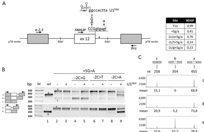
Fig. 1 Splicing patterns of FAH minigenes in HepG2.a Schematic representation of the FAH minigene, cloned within the NdeI sites (indicated) in the pTB vector, with the sequence of the exon 12 5′ss and of the investigated changes as well as of the 5′ tail of the com-pensatory U1FAH. The asterisk indicates the crypric 5′ss. The table
reports the predicted score of the wild type or mutated 5′ss (http://www.fruitfly.org/seq_tools/splice.html). b FAH splicing pat-terns in HepG2 cells transiently transfected with pFAH minigenes (1 μg) alone or with a molar excess (1.5×) of pU1FAH. RT-PCR was
conducted with primers α-2,3 and Bra (upper panel) or primers FAH12F and Bra (lower panel), and amplicons were separated on 2% agarose gel. The scheme of amplicons is reported on the left together with primers used (arrows).c FAH splicing patterns as in the upper panel of section b (lanes 2, 8, and 9) evaluated byfluorescently labeled RT-PCR with primers α-2,3 and BraFAM, followed by denaturing
capillary electrophoresis. The scheme of amplicons and the length (nt) are reported on top. Numbers below peaks indicate the relative amount (%, mean from three independent experiments) of each transcript
Consistently with previous data [3–5], the c.1062 +5G>A mutation, as revealed by both densitometric ana-lysis of bands or fluorescent RT-PCR followed by dena-turing capillary electrophoresis, induced exon 12 skipping (~31% of total transcripts) and partial intron retention (~69%), with no traces of correct transcripts (Fig.1b, upper panel and Fig. 1c), a splicing profile that validated our experimental approach. Interestingly, the introduction of the c.1061C>A substitution in the c.1062+5G>A background partially rescued the defective 5′ss recognition and led to residual levels of correct transcripts (5.2 ± 0.9%) (Fig.1c), which was further demonstrated by a RT-PCR focused on exon 12 (Fig.1b, lower panel).
Computational analysis of 5′ss scores (Fig. 1a, inset table), an estimate of the complementarity between the 5′ss sequence and the 5′ tail of the key spliceosomal U1 small nuclear RNA (U1snRNA) [9], predicts that the c.1061C>A change, but not the other c.1061C>T or c.1061C>G sub-stitutions, strengthen the affected 5′ss. This would favor the utilization of the mutated 5′ss and the production of correct transcripts. Consistently, the impact of the other changes (c.1061C>T or c.1061C>G) on the splicing profile of the c.1062+ 5G>A mutation was negligible (Fig.1b).
Altogether these observations demonstrated that in the c.1062+5G>A background the c.1061C>A somatic change accounts for trace levels of correct transcripts. In turn, this would explain the residual FAH protein expression and immune-positivity in HT1 liver nodules with the above mentioned genetic profile, thus providing an additional mechanism leading to FAH immune-reactive mosaicism. It is worth noting that the FAH immune-reactive mosaicism has been so far attributed to the reversion of the mutated allele into the wild type [10], as also reported in other diseases [11]. Both mechanisms would be favored by the high mutation rate of HT1 hepatocytes, [11] followed by positive selection of FAH-expressing hepatocites, which would lead to the formation of FAH-immunopositive nodules.
Splicing mutations, relatively frequent in metabolic dis-orders (http://www.hgmd.cf.ac.uk) as well as in others human diseases [12], represent potential targets for RNA-based therapies. Several studies have indicated that splicing mutations can be counteracted by engineered U1snRNAs with increased complementarity with the 5′ss of the defective exon [13–15]. We therefore created a U1snRNA variant specifically designed on the FAH exon 12 5′ss (U1FAH). However, in co-expression experiment, the com-pensatory U1FAH was ineffective on the c.1062+5G>A mutation, which demonstrated the severe impairment of the 5′ss recognition. Notably, and consistently with a slightly improved 5′ss, the combination of the HT1-causative mutation with the c.1061C>A change resulted in remark-able responsiveness to the U1FAH that promoted exon 12
inclusion, as witnessed by the robust synthesis of correct transcripts (62.5 ± 1.2%) (Fig.1b, c).
Conclusion
Altogether these findings elucidate the molecular basis of HT1 caused by the frequent FAH c.1062+5G>A mutation, and demonstrate the compensatory effect of the c.1061C>A change in promoting exon definition, thus unraveling a rare mechanism leading to FAH immune-reactive mosaicism.
Acknowledgements The study was supported by grants from the Telethon Foundation (GGP14190 to MP and DB), the AMC Foun-dation (to SFJvdG) and the University of Ferrara.
Compliance with ethical standards
Conflict of interest The authors declare that they have no conflict of interest.
References
1. Lindblad B, Lindstedt S, Steen G. On the enzymic defects in hereditary tyrosinemia. Proc Natl Acad Sci USA. 1977;74:4641–5.
2. Grompe M, St-Louis M, Demers SI, al-Dhalimy M, Leclerc B, Tanguay RM. A single mutation of the fumarylacetoacetate hydrolase gene in French Canadians with hereditary tyrosinemia type I. N Engl J Med. 1994;331:353–7.
3. Rootwelt H, Kristensen T, Berger R, Høie K, Kvittingen EA. Tyrosinemia type 1—complex splicing defects and a missense mutation in the fumarylacetoacetase gene. Hum Genet. 1994;94:235–9.
4. Carro R, Sánchez-Alcudia R, Pérez B, Navarrete R, Pérez-Cerdá C, Ugarte M, Desviat LR. Functional analysis and in vitro correction of splicing FAH mutations causing tyrosinemia type I. Clin Genet. 2014;86:167–71.
5. Bliksrud YT, Brodtkorb E, Andresen PA, van den Berg IE, Kvittingen EA. Tyrosinaemia type I—de novo mutation in liver tissue suppressing an inborn splicing defect. J Mol Med. 2005;83:406–10.
6. Tajnik M, Rogalska ME, Bussani E, Barbon E, Balestra D, Pinotti M, Pagani F. Molecular basis and therapeutic strategies to rescue factor IX variants that affect splicing and protein function. PLoS Genet. 2016;12:e1006082.
7. Balestra D, Barbon E, Scalet D, Cavallari N, Perrone D, Zani-bellato S, Bernardi F, Pinotti M. Regulation of a strong F9 cryptic 5′ss by intrinsic elements and by combination of tailored U1snRNAs with antisense oligonucleotides. Hum Mol Genet. 2015;24:4809–16.
8. Cavallari N, Balestra D, Branchini A, Maestri I, Chuamsunrit A, Sasanakul W, Mariani G, Pagani F, Bernardi F, Pinotti M. Acti-vation of a cryptic splice site in a potentially lethal coagulation defect accounts for a functional protein variant. Biochim Biophys Acta. 2012;1822:1109–13.
9. Roca X, Krainer AR, Eperon IC. Pick one, but be quick: 5′ splice sites and the problems of too many choices. Genes Dev. 2013;27:129–44.
10. Kvittingen EA, Rootwelt H, Berger R, Brandtzaeg P. Self-induced correction of the genetic defect in tyrosinemia type I. J Clin Invest. 1994;94:1657–61.
11. Hirschhorn R. In vivo reversion to normal of inherited mutations in humans. J Med Genet. 2003;40:721–8.
12. Sterne-Weiler T, Howard J, Mort M, Cooper DN, Sanford JR. Loss of exon identity is a common mechanism of human inherited disease. Genome Res. 2011;21:1563–71.
13. Fernandez Alanis E, Pinotti M, Dal Mas A, Balestra D, Cavallari N, Rogalska ME, Bernardi F, Pagani F. An exon-specific U1 small
nuclear RNA (snRNA) strategy to correct splicing defects. Hum Mol Genet. 2012;21:2389–98.
14. Scalet D, Balestra D, Rohban S, Bovolenta M, Perrone D, Ber-nardi F, Campaner S, Pinotti M. Exploring splicing-switching molecules for seckel syndrome therapy. Biochim Biophys Acta. 2017;1863:15–20.
15. Balestra D, Scalet D, Pagani F, Rogalska ME, Mari R, Bernardi F, Pinotti M. An exon-specific U1snRNA induces a robust factor IX activity in mice expressing multiple human FIX splicing mutants. Mol Ther Nucleic Acids. 2016;5:e370.