In vitro effects on calcium oxalate
crystallization kinetics and crystal morphology
of an aqueous extract from Ceterach
officinarum: Analysis of a potential antilithiatic
mechanism
Roberta De BellisID1☯*, Maria Piera Piacentini1☯, Maria Assunta Meli1, Michele MattioliID2, Michele MenottaID1, Michele MariID1, Laura Valentini1, Letizia Palomba1,
Donatella DesideriID1, Laura Chiarantini1
1 Department of Biomolecular Sciences, University of Urbino Carlo Bo, Urbino (PU) Italy, 2 Department of
Pure and Applied Sciences University of Urbino Carlo Bo, Urbino (PU) Italy
☯These authors contributed equally to this work. *[email protected]
Abstract
Ceterach officinarum Willd is a plant widespread throughout Europe and used in southern Italy as a diuretic. Beliefs in the benefits of C. officinarum aqueous extract in the treatment of calcium oxalate kidney stones are widely held. Little is known, however, about the actual mechanism of its antilithiatic action. Our results in this in vitro study corroborate C. offici-narum aqueous extract as a good source of antioxidants with a high antioxidant effects. Our results also demonstrate a major impact of C. officinarum aqueous extract on in vitro induced calcium oxalate crystallization kinetics and crystal morphology, showing its critical role in kidney stone formation and/or elimination. We show that progressively increasing doses of C. officinarum aqueous extract cause a sequence of effects. A powerful inhibitory action on calcium oxalate monohydrate (COM) growth and aggregation is first observed. C. officinarum aqueous extract also appears highly effective in stimulating nucleation increas-ing the number and reducincreas-ing the size of COM crystals, which become progressively thinner, rounded and concave in a dose-dependent manner. These shape-modified COM crystals are known to be less adherent to renal tubular cells and more easily excreted through the urinary tract preventing kidney stone formation. Further, C. officinarum aqueous extract pro-motes the formation of calcium oxalate dihydrate (COD) rather than the monohydrate so that, at the highest concentrations used, only COD crystals are observed, in significant greater numbers with a clear reduction in their size, in a dose-dependent manner. Further-more, AFM analyses allowed us to reveal the presence of C. officinarum component(s) on the surfaces of COD and modified COM crystals. The crystal surface adsorbed component (s) are shown to be similarly active as the total aqueous extract, suggesting a trigger factor which may direct crystal modification towards COD forms. In urolithiasis pathogenesis COD crystals are less dangerous than the COM forms due to their lower affinity for renal tubular cells. Our results are important in understanding the mechanisms which guide the
a1111111111 a1111111111 a1111111111 a1111111111 a1111111111 OPEN ACCESS
Citation: De Bellis R, Piacentini MP, Meli MA,
Mattioli M, Menotta M, Mari M, et al. (2019) In vitro effects on calcium oxalate crystallization kinetics and crystal morphology of an aqueous extract from Ceterach officinarum: Analysis of a potential antilithiatic mechanism. PLoS ONE 14(6): e0218734.https://doi.org/10.1371/journal. pone.0218734
Editor: Yogendra Kumar Mishra, Institute of
Materials Science, GERMANY
Received: January 30, 2019 Accepted: June 8, 2019 Published: June 25, 2019
Copyright:© 2019 De Bellis et al. This is an open access article distributed under the terms of the Creative Commons Attribution License, which permits unrestricted use, distribution, and reproduction in any medium, provided the original author and source are credited.
Data Availability Statement: All relevant data are
within the manuscript and its Supporting Information files.
Funding: The authors received no specific funding
for this work.
Competing interests: The authors have declared
modification induced by C. officinarum on the crystallization process. Based on these data, together with no adverse toxic effect being observed on the in vitro model of human intesti-nal enterocytes, C. officinarum aqueous extract could represent an attractive natural therapy for the treatment of urolithiasis.
Introduction
Urolithiasis is a common and frequent human pathology [1] characterized by a high recur-rence rate, complex pathophysiological bases and multifactorial etiology [2]. Urine is usually rich in minerals producing a high tendency towards stone formation which, in healthy individ-uals, is naturally inhibited through reduced crystal aggregation [3]. The most common urolith-iasis produces calcium oxalate (CaOx) crystals. Stone formation proceeds through various and complex physicochemical steps beginning with crystal nucleation and growth, followed by aggregation, crystal adhesion on renal tubular cells, and internalization into renal epithelial cells [4]. All of these processes occur in a complex environment containing both promoters and inhibitors [5] and when crystals nucleate, grow and are retained within the kidney, they lead to injuries in renal epithelial cells creating stone nidi [6].
High concentrations of CaOx crystals, or of oxalate itself, lead to toxic effects on renal cells which induce cell surface alterations, thus unmasking attachment sites for adhesion and/or internalization of crystals by renal epithelial cells [7–8]. The interaction between oxalate and/ or crystals with renal tubular epithelial cells is an important factor in stone formation [9–11]. Furthermore, renal exposure to oxalate leads to reactive oxygen species products with subse-quent lipid peroxidation and modification of cell structure, physiology, gene expression and cell death [2,12–13].
Calcium oxalate (CaOx) crystals are usually present in different forms: calcium oxalate monohydrate (COM), dihydrate (COD) and the rarer trihydrate (COT) [14]. The COM crystal form is reported to be more dangerous in urolithiasis pathogenesis because of their greater affinity for renal tubular cells [15–16] while COD forms are frequently found also in urine from healthy subjects [17]. There are various current therapies against urolithiasis and patients are often treated with some of them contemporarily (surgical procedures, medications, and diets). None of these are totally effective with frequent relapses in patients [18–21]. Despite progress made in such treatments, the problem has still to be solved [22] and without an effec-tive therapy, the use of phytotherapy and medicinal plants are to be considered as a valuable support [23–24] in providing some relief.
Historically, medicinal plants have been used as therapeutic remedies due to their antilithia-tic activities [2]. Unfortunately, the mechanism of action of the majority of these plants is little known and it is thus necessary to identify the active ingredients, and their antilithiatic mecha-nisms. Several studies have found that medicinal plants exert their antiurolithiatic effect at dif-ferent stages of urolithiasis, with difdif-ferent typologies of their pharmacological actions. These plants also show analgesic and anti-inflammatory properties as well as antioxidant, antispas-modic, astringent, crystallization inhibition, diuretic and litholytic effects. It has also been demonstrated that medical plants can change the ion concentrations in urine increasing mag-nesium and citrate excretion, or decreasing calcium and oxalate concentrations [2,24–26]. Most of these herbs and plants are taken as extracts in complementary and alternative medi-cine, also serving as an interesting source of potential drug candidates for pharmaceutical research [27].
Ceterach officinarum Willd. (syn. Asplenium ceterach L or Ceterach officinarum DC), a fern
species commonly known as "rusty back", is a spontaneous perennial herb belonging to the Aspleniaceae family with a widespread distribution in Western and Central Europe, including the Mediterranean region. It is characterized by a short rhizome which gives rise to green fronds with pinnated lamina having orange-brown trichomes only on their back and hence its name. It grows in fissures of carbonate rocks and between stone and brick walls.C. officinarum
aerial parts are extensively used in traditional medicine and its components include mineral salts, mucilages, tannins, flavonoids, caffeic acid and chlorogenic acid [28–30]. Tisanes of its aerial parts are used as antihypertensive and hepatic anti-inflammatory agents [31] while the decoction is used as an expectorant[32].C. officinarum tea is traditionally used in Southern
Italy for its diuretic properties and as a therapy against kidney stones so that, in this region, it is also called "stone-breaker" [33]. A decoction of aerial parts was also reported to eliminate renal calculus [34] but the actual mechanism of this antilithiatic action remains unclear.
The present study aims to investigate the antilithiatic ability of aC. officinarum aqueous
extract at various stages of stone formation, its possible mechanisms and effects on anin vitro
model of human intestinal enterocytes to evaluate potential therapeutic use ofC. officinarum
to treat and/or to prevent CaOx nephrolithiasis.
Materials and methods
Chemicals and reagents
All chemicals used were of analytical grade. Sodium oxalate (Na2Ox, Na2C2O4) was obtained
from ScienceLab; anhydrous calcium chloride (CaCl2), and the other conventional reagents
used were purchased from Sigma Chemical Company, St. Louis, MO, USA. Folin-Ciocalteu’s reagents were obtained from Biorad, CA, USA.
Plant material
Dried leaves and aerial parts ofCeterach officinarum Willd. was purchased from
Omeosalus-Vet (Italy).
Ceterach officinarum aqueous extract (AE) preparation
The dried plant was reduced to a coarse powder using a dry grinder. The dried fine powdered
C. officinarum (1.5 g) was soaked in distilled water (25 ml) and left at 72˚C for 30 min, stirring
frequently.
The extract was paper-filtered followed by centrifugation at 12,000 rpm for 15 min at 4˚C. The supernatant was then filtered using a single-use filter unit 0.22μm (Minisart, Biotech) and the filtrate, referred to asC. officinarum AE was freshly used or stored at -20˚C; aliquots of 1
ml were lyophilized overnight in a rotary evaporator and the dried powder was used for deter-mining the dry weight (dw), 1 ml of the extract corresponding to 20,6± 2,7 mg dw. In the experiments various aliquots ofC. officinarum AE were used.
Quantification of total phenols and flavonoids
The total polyphenol content ofC. officinarum AE was spectrophotometrically determined by
using the Folin–Ciocalteu method described by Singleton et al (1999) [35] with slight modifi-cations [36]. The extract was properly diluted in 80% ethanol; increasing aliquots of the sample were added to distilled water reaching a 100μl final volume and mixed with 50 μl of Folin– Ciocalteu reagent; a reagent blank containing only water was used. After 3 min at room tem-perature, 300μl of sodium carbonate 20% (w/v) were added and the volume brought to 1 ml
with distilled water. The range of AE concentrations used was 22–110μg dw/ml. The tubes were vortex mixed for 15 s and allowed to stand for 30 min at room temperature for colour development, followed by centrifugation at 10,000 rpm for 10 min. The supernatant absor-bance was measured at 725 nm (Uvikon spectrophotometer). The number of total phenols was expressed as caffeic acid equivalent (CAE)/g dw of extract using a standard calibration curve obtained with different concentrations of caffeic acid between 1 and 15μg/ml (R2= 0.9973). The method used to determine the concentration of total flavonoids inC. officinarum AE was
a modified aluminium chloride cholorimetric method [37] protocol with some modifications and adjusted for measurements in 96-well micro titer plates. In brief, 25μl of properly diluted AE was added to 5μl of 10% AlCl3, 5μL of 1M CH3COOK and 215μl of water. The
absor-bance of the mixture was measured at 405 nm. All measurements were conducted in triplicate. Total flavonoids were expressed as milligrams of quercetin equivalents (QE) per gram of AE dry matter using a standard calibration curve obtained with concentrations of quercetin rang-ing from 0.1 to 0.5 mg/ml (R2 = 0.9964).
Oxygen radical absorbance capacity (ORAC) assay
The antioxidant capacity ofC. officinarum AE was determined by the ORAC method [38] using a Fluostar Optima Plate reader fluorimeter (BMG Labtech, Offenburgh, Germany) equipped with a temperature-controlled incubation chamber (37˚C) and an automatic injec-tion pump. The ORAC assay measures a fluorescent signal from fluorescein as the fluorescent probe which is quenched in the presence of Reactive Oxygen Species (ROS). Addition of an antioxidant absorbs the generated ROS, allowing the fluorescent signal to persist. The ORAC assay is unique in that its ROS generator, AAPH (2,2’-azobis(2-methylpropionamidine) dihy-drochloride), produces a peroxyl free radical upon thermal decomposition. This free radical is commonly found in the human body, thus making this reaction biologically relevant. The fol-lowing mix was used: 200μl of 0.096 μM fluorescein sodium salt in 0.075M Na-phosphate buffer (pH 7.0), 20μl of sample or Trolox or 0.075 M Na-phosphate buffer (pH 7.0) as blank. The reaction was initiated with 40μl of 0.33 M of AAPH. Fluorescence was read at 485 nm ex. and 520 nm em. until complete extinction [38]. A calibration curve was made each time with the Trolox standard in 0.075 M Na-phosphate buffer (pH 7.0). ORAC values were expressed asμmol Trolox Equivalents (TE)/g dw.
DPPH radical scavenging assay
The antioxidant activity was evaluated using the stable free radical DPPH•(2, 2-Diphenyl-1-Picrylhydrazyl) and the DPPH•free radical scavenging assay conducted as reported in Salt-arelli [39].
In brief, 850μl of a freshly prepared 100 μM DPPH•ethanol solution was added to 150μl of sample properly diluted in 80% ethanol with concentrations of 5,95–96.5μg dw /ml. After 10 min at room temperature, the absorbance decrease at 517 nm was measured. Ethanol was used as a blank and the scavenging capacity calculated as DSA (DPPH Scavenging Activity) % = [(A517 nmof blank—A517 nmof sample)/A517 nmof blank] x 100. The EC50was calculated as
concentration (μg/ml) of extract dried weight required to provide 50% free radical scavenging activity.
Protein determination
The protein concentration was quantified by Bio-Rad Protein Assay based on Bradford dye reagent (Coomassie brilliant blue G-250) using BSA as a standard [40].
HPLC analysis
The samples were analysed in a Waters instrument equipped with Alliance HT 2795 high-per-formance liquid chromatography (HPLC), 2996 photo diode array (PDA) and micromass LC/ MS ZQ 2000 detector as follows. A C18 column, LiChroCART (250× 4 mm), with a particle size of 5μm, was used. The mobile phase consisted of acetonitrile (solvent A) and 0.1% aque-ous formic acid (solvent B). The gradient was changed as follows: 0–4 min from 5% A to 15% A, 4–20 min to 18% A, 20–35 min to 93% A, 35–40 min to 100% A. The total run time was 40 min. The injected sample volume was 50μl and the flow rate was 0.8 ml/min. UV spectra were recorded from 210 to 420 nm. Electrospray ionization (ESI) was operated in positive and nega-tive ion mode over a range of 100–700 amu. Capillary voltage was set at 3 kV, source tempera-ture at 100˚C and desolvation temperatempera-ture at 300˚C. The cone and desolvation nitrogen gas flows were 50 and 500 l/h, respectively. Data were processed using MassLynx 4.1 (Waters, Mil-ford, USA). The products were identified by comparison with analytical standards.
Cell culture and viability
Caco-2 cells were differentiated as described by Tunisi et al. [41]. Briefly, cells were seeded in 48-well plates at 5×10⁴cells/well and cultured at 37˚C in Eagle’s minimal essential medium (Life Technologies) supplemented with 10% bovine fetal serum (Life Technologies), penicillin (50 units/ml) and streptomycin (50μg/ml) (Life Technologies), gassed with an atmosphere of 95% air-5% CO2. After 20–21 days in culture, the differentiated cells were, finally, treated with various concentrations ofC. officinarum AE (250, 500 and 1000 μg dw/ml) for 24 h. Cellular
viability was assessed by measuring the reduction of 3-(4,5-dimethylthiazol-2-yl)-2,5-diphe-nyltetrazolium bromide (MTT), as described by Palomba et al. [42]. After 24 h of treatment, MTT (25μg/ml) was added directly to the culture medium 2 h prior to the end of the incuba-tion period. Cells were washed twice with phosphate-buffered saline (Sigma-Aldrich) and cel-lular MTT reductase activity was determined by measuring the absorbance of dimethyl sulfoxide extracts at 595 nm. Results are expressed as the percentage of MTT-reducing activity of treated versus untreated cells.
CaOx crystallization analysis
Stock solutions. Stock solutions were freshly prepared as follows: Sol A 10.0 mM calcium
chloride (CaCl2) in 200 mM sodium chloride (NaCl) and 10 mM sodium acetate, pH 5.7; Sol B
1.0 mM sodium oxalate (Na2C2O4) in 200 mM sodium chloride (NaCl) and 10 mM sodium
acetate, pH 5.7. Both were filtered through 0.22μm cellulose acetate filter [43]. The pH of 5.7 chosen for the stock solutions is the pH value frequently observed in the first morning urines of subjects with urolithiasis problems [44].
Spectrophotometric measure. CaOx crystallization was performed in the presence and
absence ofC. officinarum AE. The crystallization of CaOx was studied using a time-course
measurement of optical density as described by Mittal et al. [43]. Solutions A and B were kept at 37˚C. In a 3ml glass cuvette were subsequently added: 950μl of Sol. A, a gradually increasing aliquot of AE in a 100μl final volume with distilled water, and 950 μl of Sol B in order to achieve final assay concentrations of 5.0 mM calcium and 0.5 mM oxalate. The range of con-centrations used was 125–1000μg AE dw/ml reaction mix. The final solutions were continu-ously stirred and maintained at 37˚C. The turbidity of the crystal suspension was measured at an absorbance of 620 nm (Uvikon spectrophotometer). The rates of crystal formation were obtained by recording absorbances every 30 seconds over 50 minutes. The effect of AE on CaOx crystallization was evaluated against distilled water as a blank. All crystallization experi-ments were performed at least in triplicate.
Light microscopy observation. CaOx crystallization was observed by light microscopy in
the presence and absence ofC. officinarum AE. In wells of a 24-well plate were sequentially
placed: 475μl of the CaCl2stock solution, a gradually increasing aliquot of AE in a 50μl final
volume with distilled water, and 475μl of the Na2C2O4stock solution, in order to obtain final
concentrations of 5.0 mM calcium and 0.5 mM oxalate, respectively. The extract concentrations used ranged from 1 to 1000μg dw/ml. A sample containing 50 μl distilled water instead of AE was used as the reference control system. The plate was left at room temperature, then observed at different magnifications by a light microscope (Olympus) and qualitatively analysed in terms
of crystal size, shape and abundance. Pictures were processed by ToupTek ToupView ver. 3.7.
X-Ray Powder Diffraction (XRPD) analysis. Stock solutions, prepared as described in
“CaOx crystallization analysis” and maintained at room temperature, were used to characterize the crystalline phase of CaOx crystals produced in the presence and absence ofC. officinarum
AE by XRPD. For this purpose, were sequentially placed: 950 ml of the CaCl2solution, 100 ml
of distilled water or EA and 950 ml of the Na2C2O4solution, to final concentrations of 5.0 mM
calcium and 0.5 mM oxalate, respectively. The reaction mixture was gently stirred with a glass rod and left at room temperature for 150 min. The mixture was filtered using 0,45μm cellulose nitrate membrane filters. The obtained powder was left to dry at room temperature and used for XRPD analyses.
The XRPD mineralogical analyses were carried out using a Philips X’Change PW 1830 dif-fractometer (Cu-Kα radiation). Randomly-oriented powders were prepared by gently hand crushing the CaOx crystals. The powders were then side-loaded into an aluminum holder to examine the unoriented powder and subsequently analyzed with a 0.02˚ step with a counting time of 1 s/step from 2˚ to 65˚ 2θ. The analytical conditions were a 35 kV accelerating potential and a 30 mA filament current. All major peaks were indexed in the refinement, with quartz as internal standard.
ESEM analysis. The CaOx crystals obtained as reported in “Light microscopy
observa-tion” section were allowed to precipitate on carbon discs at room temperature for 24 h and then studied by an Environmental Scanning Electron Microscope (ESEM) FEI Quanta 200 FEG, equipped with an energy-dispersive X-ray spectrometer (EDS) for microchemical analy-ses. Operating conditions were 30 kV accelerating voltage, 10 mm working distance, 0˚ tilt angle, and variable beam diameter. The ESEM was utilized in low vacuum mode, with a speci-men chamber pressure set from 0.80 to 0.90 mbar. The images were obtained using a back-scattered electron detector. Morphometric data (number, size, and morphology) of the crystals produced in CaOx crystallization assays in the presence and absence ofC. officinarum AE
were collected by accurate observations and measurements of all the crystals visible in several ESEM images. These pictures were selected according to two main criteria: (i) to be represen-tative of the total area of each sample investigated, and (ii) to have a significant number of measurements.
AFM characterization of crystals. Crystal shape and size were also investigated by atomic
force microscopy (AFM). Briefly, 50μl of CaOx crystals, obtained as mentioned above, were layered on glass surfaces and dried by nitrogen flow. The XE-100 atomic force microscope (AFM; PARK Systems Inc.) was used in this study; the instrument was equipped with a 50μm scanner controlled by XEP 1.8.1 software. The AFM was set in non-contact mode, with the X– Y stage in closed loop and high voltage modes. The Z scanner was set in closed loop and high voltage modes and resolution set to 1.8Å. The speed scan was 0.25Hz for large images and 1Hz for the 1x1μm acquisition. The tips used in the present study were the NCHR type with a nom-inal spring constant of 42 N/m and a typical resonant frequency between 250 and 300kHz. The data acquisition was performed in air at controlled temperature. In addition to topography, amplitude and phase signals were acquired. The amplitude signal was used as a unique drive
for the Z feedback circuit, thus rendering the phase signal to exclusively depend on chemical and nano-mechanical proprieties of the surfaces. The phase signal evaluation (Phase Detection Microscopy PDM) was carried out randomly on CaOx particles by using a 1x1μm scanning area, with around 25 images for each condition analysed. AFM images were analysed using XEI software (PARK Systems Inc.).
Solubilization of crystal surface adhesion component(s) from
C.
officinarum aqueous extract
In a glass beaker were sequentially placed: 475 ml of Sol A, 50 ml of distilled water or EA and 475 ml of Sol B, as previously described, to final concentrations of 5.0 mM calcium and 0.5 mM oxalate, respectively. The reaction mixture was gently stirred with a glass rod and left at room temperature for 150 min. The mixture, except for 100 ml of it, was filtered using 0,45μm cellulose nitrate membrane filters. Crystals were harvested from the filter surface and added to the unfiltered 100 ml. This suspension was centrifuged at 5,000 rpm for 10 min and the super-natant discarded. The pellet was resuspended in 50 ml 2 M HCl pH 0.6, allowing the solubiliza-tion of COD crystals through the acsolubiliza-tion on oxalate.
Crystal surface adhesion component(s) were loaded into dialysis tubing with a 3,500 kDa MWCO (molecular weight cut-off) and dialysed overnight against distilled water. Aliquots of the dialysed sample were dried overnight and used for determining the dry weight (0.2 mg dw/ ml). The dried powder was properly resuspended in distilled water for subsequent activity characterization analyses.
Statistical analysis
Data were expressed as mean values± SD of at least three independent experiments and ana-lysed using one-way ANOVA followed by the Kruskal Wallis test for multiple comparison analysis or by the non-parametric Mann-Whitney U test. P<0.05 was considered significant. The statistical comparisons among AFM sample data were performed using Dunnett’s multi-ple comparison test and only the comparisons with the control sammulti-ple are reported. Statistical analyses in cell experiments were performed by two-way ANOVA followed by apost hoc
Bon-ferroni test. A level of confidence of P < 0.05 was used for statistical significance.
Results
Protein content, total phenolic and antioxidant power of
Ceterach
officinarum aqueous extract
The protein content ofC. officinarum AE obtained by Bradford’s method [40] was 52.91± 3.8 mg/g AE dw. The amount of total phenols was 136.94± 7.6 mg/g AE dw and flavonoids 46.12± 2.2 mg/g AE dw. The antioxidant activity of C. officinarum AE was measured in terms of radical scavenging ability using ORAC [38] and DPPH•radical scavenging assays [39].
The ORAC value was 2798± 14.1 μmolTE/g AE dw.
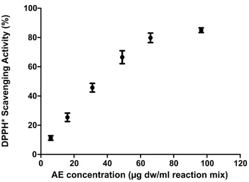
As reported inFig 1,C. officinarum AE had a DPPH dose-dependent radical scavenging
activity, showing a percentage of DPPH radical inhibition ranging from 11.26± 1.54% at 5,95μg dw/ml assay mix to 85.0 ± 1.6% at 96.5 μg dw/ml assay mix. The linearity range was 5.95–49μg dw /ml, with IC50value of 35.46μg/ ml.
HPLC analysis
50μl of C. officinarum AE was analyzed by HPLC, equipped as described in “Materials and methods” section. The products were identified in the HPLC chromatogram by comparison
with analytical standards. As reported inFig 2, the main peak of the chromatogram (Retention Time 8.97 min) was chlorogenic Acid.
Effect of
C. officinarum AE on human enterocytes
Using Caco-2 cells which express morphological and biological characteristics of human intes-tinal enterocytes [45], no harmful effects ofC. officinarum AE were oberved. Mild toxicity was
observed only at the highest dose (250μg dw /ml: 97.4±7.5%; 500 μg dw /ml: 98.7 ±6.5%;1000 μg dw /ml: 76.6 ±5.2%�; results represent the mean± SD from three separate
experiments, each performed in quadruplicate;�p < 0.05 versus untreated cells).
Fig 1. DPPH dose-dependent radical scavenging activity ofC. officinarum AE. Results are the mean ± SD of more
than 3 different experiments. The median of each sample differs from the others (p<0.05) as inferred by the Kruskal Wallis test followed by Dunn’s post test.
https://doi.org/10.1371/journal.pone.0218734.g001
Fig 2. Chromatogram ofC. officinarum AE. From the obtained chromatogram the major peak was identified as Chlorogenic Acid.
Spectrophotometric crystallization measurements
The number of crystals formed was measured through absorbance at 620nm (A620nm) used as
an estimate of turbidity. In this assay system, A620nmis a good measure of particle
concentra-tion per unit volume, so that an increase in A620nmmainly reflects an increase in particle
num-ber as a function of time. Therefore, the maximum slope of increase in A620nm, termed the
slope of nucleation (SN) and determined by linear regression analysis, mainly represents the
maximum rate of formation of new particles and thus crystal nucleation.
The time-course measurements of A620nmin the control experiment followed the typical
curve as previously reported [26,46].
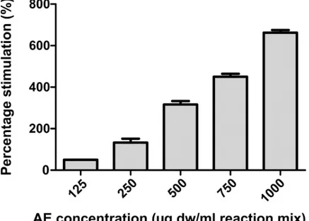
As shown inFig 3,C. officinarum AE displayed a stimulatory activity on CaOx
crystalliza-tion; the stimulation percentage of nucleation produced by AE, calculated as [(SN AE- SNc) /
SNc]× 100 (where “c” stands for control) increased with increasing concentrations of the
extract in a dose-dependent manner from 125 to 1000μg AE dw/ml.
In the control experiments, after a maximum increase of A620had been reached, a
progres-sive decrease of absorbance was observed despite continuous stirring, due to crystal aggrega-tion [46] while, in the presence ofC. officinarum AE this decrease was not observed. Crystal
aggregation in the control sample and the total inhibition of this phenomenon, due to the pres-ence ofC. officinarum AE, were clearly shown by ESEM analyses (S1 Fig).
Light microscopy observations
CaOx crystallization in the presence and absence ofC. officinarum AE was also analysed by
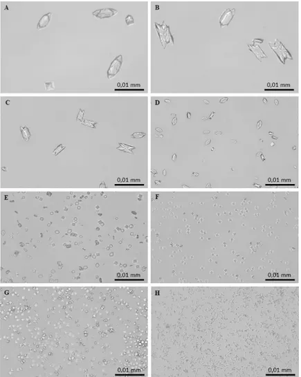
light microscopy as reported in “Materials and methods” section. In the control system, the micrographs obtained after 24 h of incubation showed the formation of two types of CaOx crystals: abundant COM crystals with monoclinic prismatic shapes or in twin form, and only a few COD crystals with typical bipyramidal shapes (Fig 4A and 4B). Both COM and COD crys-tals were large, with COM showing a roughly orthorhombic structure with highly facetted
Fig 3. Percentage of stimulation of nucleation produced byC. officinarum AE. Results are the mean ± SD of more
than 3 different experiments. The median of each sample differs from the others (p<0.05) as inferred by the Kruskal Wallis test followed by Dunn’s post test.
{100} (Fig 4A) and {010} faces (Fig 4B). The addition ofC. officinarum extract to the reaction
mix results in a modification in structure, number, and size of crystals produced. Already at an AE concentration of 0.9–2μg dw/ml (Fig 4C) a slightly higher number of particles formed, most of which were still COM. These COM crystals were smaller, and almost halved in num-ber compared to controls; however, {100} and {010} twin planes highly facetted due to the presence of macrosteps, and smaller, more irregular shapes suggested a kinetic of growth in progress. As the concentration of the extract was increased up to 15–30μg dw/ml (Fig 4D), COM crystals become more numerous and smaller, thinner and more regularly shaped, with an evident depression on the smooth {100} face, while COD crystals were still rare and medium-sized. As shown inFig 4E, in the presence of 60–125μg dw/ml COD crystals, still medium-sized, become more numerous whereas COM crystals are much more numerous, smaller, thinner, with rounded edges, sometimes dumbbell-shaped or crescent-shaped, proba-bly due to the deep depression on the smooth {100} face. At greater AE concentrations of up to 130–200μg dw/ml (Fig 4F), crystal numbers increase significantly and are almost all medium-sized and small COD crystals with only occasional COM ones. Higher concentrations ofC. officinarum AE from 500 to 1000 μg dw/ml (Fig 4G and 4H) showed the exclusive presence of
Fig 4. Light micrographs of CaOx crystals grown in the absence and presence ofC. officinarum AE increasing
concentrations. (A) and (B) Controls; (C) AE, 2μg dw/ml; (D) AE, 30 μg dw/ml; (E) AE, 125 μg dw/ml; (F) AE, 200 μg dw/ml; (G) AE, 500μg dw/ml; (H) AE, 1000 μg dw/ml. Magnification was 400x for all panels.
the COD form. The number of COD crystals formed increased with increasing concentrations of the extract in a dose-dependent manner and, simultaneously, a progressive decrease in the size of COD was observed, the highest concentrations leading to the formation of large num-bers of minute crystals.
XRPD analysis
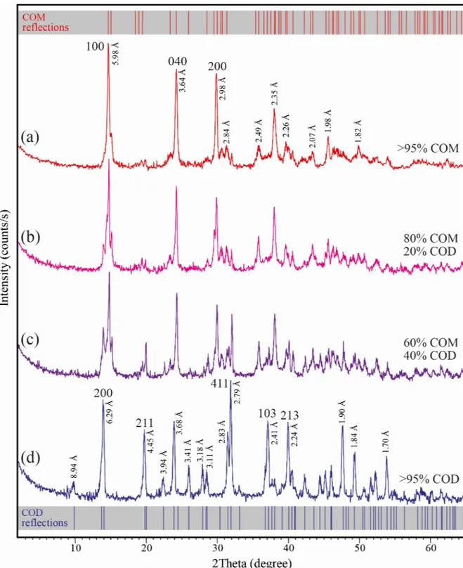
XRD measurements revealed the crystallization of different types and amounts of CaOx crys-tals produced in the presence and absence ofC. officinarum AE. Representative XRD
diffrac-tion patterns of CaOx crystals grown in the absence (a) and in the presence of different concentrations (b, c, d) ofC. officinarum AE are shown inFig 5. The main reflections of CaOx crystals in the absence ofC. officinarum AE (Fig 5A) are located at 2 theta 14.81, 24.41 and 29.98˚, attributable to {100}, {040} and {200} planes of calcium oxalate monohydrate (COM, monoclinic, space groupP21/c), respectively. For CaOx crystals obtained in the presence of
1000μg dw/ml C. officinarum AE (Fig 5D) the strong reflections at 14.21, 19.95, 32.05, 37.31 and 40.20˚ are assigned to {200}, {211}, {411}, {103}, and {213} planes of calcium oxalate dihy-drate (COD, tetragonal, space groupI4/m), respectively. The diffraction patterns (b) and (c) of
Fig 5are related to crystals obtained in the presence of 200 and 500μg dw/ml C. officinarum AE, respectively, and show the presence of both COM and COD crystals. Semi-quantitative estimations using relative peak heights/area proportions indicate that COM crystals (80 vol.%) prevail over the COD crystals (20 vol.%) in the presence of 200μg dw/ml C. officinarum AE (Fig 5B), while they tend to become equivalent (60 vol.% of COM, and 40 vol.% of COD) at increasing amounts (500μg dw/ml) of C. officinarum AE (Fig 5C), with only COD crystals being detected at 1000μg dw/ml C. officinarum AE (Fig 5D).
ESEM analysis
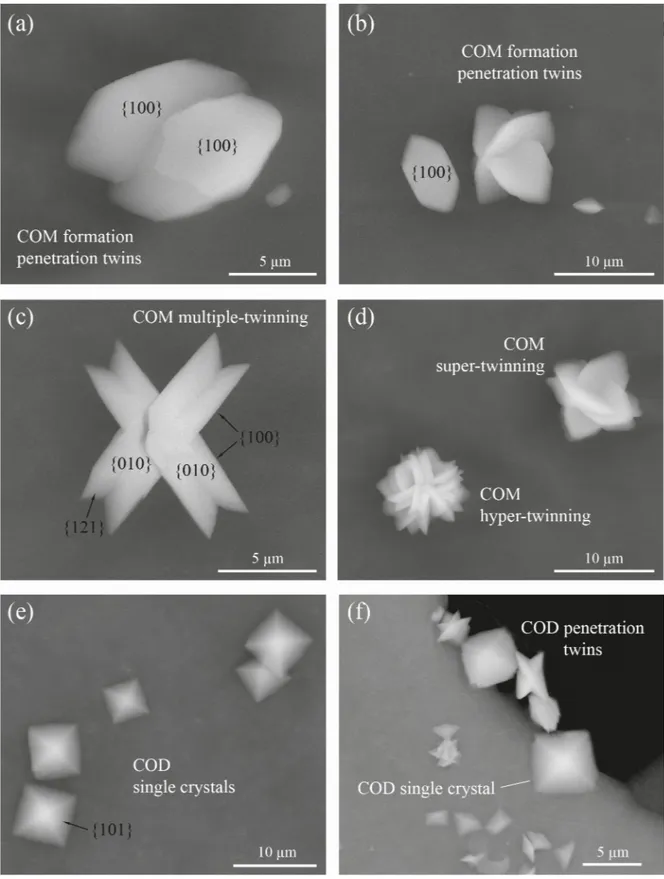
Crystal phases and morphologies of precipitates are described using crystallographic charac-teristics (habits, angles between crystal faces, crystal symmetries, etc.) available in the literature [47–49]. The morphology of the crystals demonstrated clearly that the various habits (shapes and forms) were closely related to the hydration state of the calcium oxalate evaluated from the XRD data. According to the ESEM observations (Fig 6), almost all CaOx crystals precipitated in the absence ofC. officinarum AE are calcium oxalate monohydrate (COM) single crystals
having a tabular habit. They mainly present the face forms pinacoid {100}, {010} and prism. The pinacoids {100} and {010} are generally the predominant forms and have the shape of a rhomboid and parallelogram, respectively. The average size of these COM single crystals is 8.2μm in length ({001} dimension), 4.4 μm in width ({010} dimension), and are generally 1–2μm in height ({100} dimension). Among these main morphological types, there are also transition forms and penetration twins. The penetration twins formed were nucleated from {010} and {100} faces (Fig 6A and 6B). These crystals are well facetted with {100}, {010} and {121} faces developed. A transformation from penetration to multiple-twinning, super-twin-ning and rarely to hyper-twinned aggregates is also observed (Fig 6C and 6D). The crystal hab-its of the super/hyper twins tend to exhibit rounded {100}/{121} edges (Fig 6D). Calcium oxalate dihydrate (COD) crystals are very rare in the absence ofC. officinarum AE, while the
trihydrate (COT) or amorphous phases were not detected.
In contrast, a significant change in the crystal morphology occurred in the presence of increasing concentrations ofC. officinarum AE (Fig 6E500μg dw/ml.;Fig 6F1000μg dw/ml of AE). The total volume of precipitated COM drastically decreases until it disappears, while COD crystals become the dominant to exclusive species. The typical forms of COD crystals are shown inFig 6E and 6F. They have the classical crystal habit of COD, represented by single,
Fig 5. XRPD diffraction diagrams of calcium oxalate monohydrate (COM) and dihydrate (COD) crystals. The crystals were grown in
the absence and presence ofC. officinarum AE: (a) COM crystals obtained in absence of AE.; (b, c) COM and COD crystals obtained in the presence of AE 200μg dw/ml (b) and 500 μg dw/ml (c); (d) COD crystals obtained in the presence of AE 1000 μg dw/ml. D values and indices of the main reflections are reported. The standard diffraction spectra of COM (upper grey bar, reference code 00-016-0379) and COD (lower grey bar, reference code 01-075-1314) crystals are also shown for comparison.
Fig 6. Scanning electron micrographs of various morphological types of COM and COD obtained in the absence (a-d) or presence (e, f) of different concentrations ofC. officinarum AE. (a) Twinned COM crystals viewed from a [1] direction. (b) Single and twinned COM crystals. (c) Double-twinned COM crystals viewed a [10] direction. (d) Super- and hyper-twinned COM crystals, forming flower-like aggregates. (e) Single tetragonal bipyramidal COD crystal in the presence of 500μg dw/ml AE; (f) Single, tetragonal COD crystals and small penetration twins of flat COD crystals in the presence of 1000μg dw/ml AE.
tetragonal bipyramids dominated by the {101} planes with average dimensions of about 6.5μm side length and about 1 μm side length along the c axis of the crystals (Fig 6E). All these bipyramids are characterized by the absence of prismatic {100} faces, giving the crystals a very flat form. In the higher magnification ESEM images (Fig 6F), the formation of local penetra-tion twins is evident amongst the smaller, flat tetragonal bipyramids.
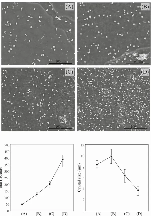
Observing the ESEM images of different CaOx precipitates obtained in the absence and in the presence of different concentrations ofC. officinarum AE (Fig 7), a progressive decrease in the size of the crystals accompanied by an increase in their number clearly emerges. The aver-age size of crystals (COM) in the absence ofC. officinarum AE is 8.2 μm (length). The crystal
size (COD) slightly increases in the presence of 200μg dw/ml of C. officinarum AE (average length of 9.93μm), while a notable size-reduction occurs on passing from 500 μg dw/ml. (aver-age length 6.43μm) to 1000 μg dw/ml (average length of 3.69 μm) of AE. This crystal-size reduction is accompanied by a considerable increase in the total number of crystals per unit area, which passes to an average of 47 crystals/unit area in the absence ofC. officinarum AE to
393 crystals/unit area in the presence of the maximum concentration of AE (1000μg dw/ml.).
AFM (Atomic Force Microscopy) analysis
The topographic images of the samples showed an unambiguous decrease in size of the layered particles, on varying the amount ofC. officinarum AE from 1 μg dw/ml to 1000 μg dw/ml
(Fig 8).
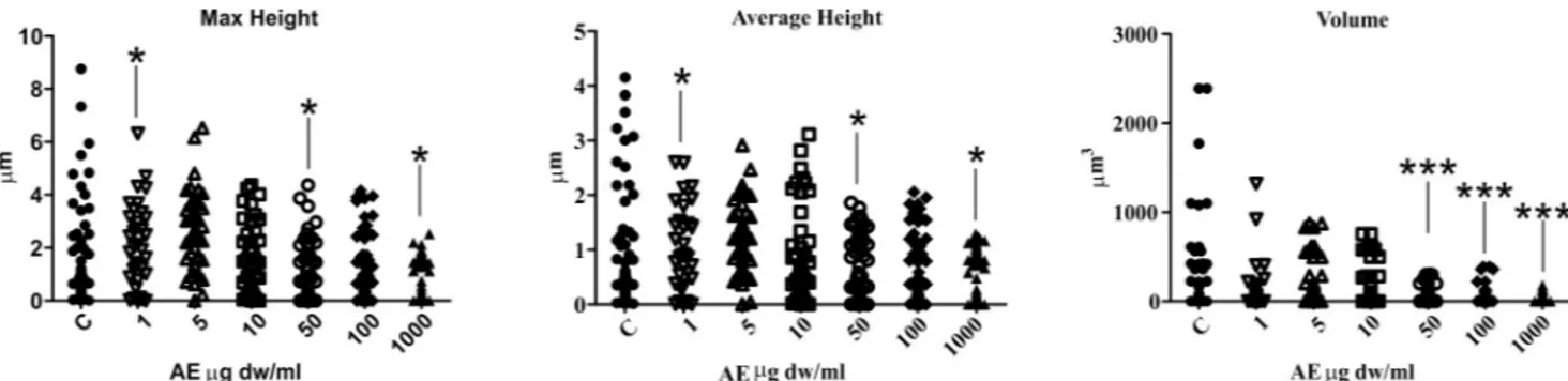
Measurement of maximum and average heights and crystal volume, showed that addition of the AE alters the shape and size of the CaOx particles. In fact, all the distributions of descrip-tor values moved down in a dose-dependent manner as illustrated inFig 9. The analysis of maximum and average height parameters reported that only the samples treated with 1μg dw/ ml, 50μg dw/ml and 1000 μg dw/ml of AE significantly differ from the control sample, while the volume analysis revealed a significant statistical difference between the control sample and the 50μg dw/ml, 100 μg dw/ml and 1000 μg dw/ml AE treated samples. The maximum and average heights depend upon the orientation of the CaOx crystals and, for this reason, their differences from the control sample are less than those of the volume data which provided more consistent results.
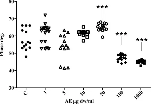
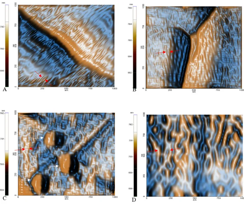
The phase signal analysis also revealed a significant statistical differences between the con-trol and the 50μg dw/ml, 100 μg dw/ml and 1000 μg dw/ml AE-treated CaOx solutions (Fig 10). This shows that the chemico-physical properties of the surfaces are altered when the AE is present. This could be caused by adhesion of AE component(s) to the surface of the crystals, thus changing the surface chemical properties.
InFig 11representative phase images of three samples show the absence of globular struc-tures on the surface of the control sample (Fig 11A), while they are observed in the 50μg dw/ mlC. officinarum AE sample increasing in size in the 1000 μg dw/ml sample (Fig 11B and 11C).
The strange occurrence is the dissimilar phase variation among the 50μg AE sample (increased phase outcome compared to control) and the 100μg and 1000 μg AE samples (strong reduction in phase signal compared to control) as shown inFig 10. We presume that this phenomenon depends not only on the possible surface adhesion ofC. officinarum AE
component(s), but also on its influence on crystal growth direction, thus exposing different surface chemical groups which may alter the phase signal. In fact, by amplitude signal investi-gation it is possible to finely investigate the nanometric and sub-nanometric organization of surfaces [50], and we were able to denote an altered pattern of CaOx substructures dependent onC. officinarum AE concentration (Fig 12). Probably at 50μg dw/ml the phase signal is
Fig 7. Scanning electron micrographs (upper) and morphometric / numerousness data (lower) of the CaOx crystals obtained in the absence or presence of different concentrations ofC. officinarum AE. (A) Control; (B) 200 μg dw/ml; (C)
500μg dw/ml.; (D) 1000 μg dw/ml of AE. https://doi.org/10.1371/journal.pone.0218734.g007
mainly influenced by altered CaOx substructures orientation, while at higher concentrations adsorption and interaction ofC. officinarum AE component(s) with the crystal surfaces
becomes dominant.
Influence of crystal surface adhesion component(s) on CaOx crystallization
In order to verify the hypothesis that the specific binding of crystal surface component(s) (sol-ubilized as described in Materials and methods section) influenced the modification of CaOx crystallization as observed with the wholeC. officinarum AE, light microscopy observations
were carried out.
The CaOx crystallization micrographs obtained after 24 h of incubation in the absence and presence of these component(s) at increasing concentrations showed that they induced modi-fications of structure, number and size of crystals when compared with the controls (Fig 13A and 13B). Already at a concentration of 1.2μg dw/ml (Fig 13C) it was possible to verify a slight rounding of COM angles; this trend accentuated on increasing the concentration to 2.3μg dw/ ml (Fig 13D) and at 4.7μg dw/ml (Fig 13E and 13F), crystals were almost all medium-large COD. However, although rarely, some rounded COM crystals, quite thick and in twin form, were still observed. Higher concentrations of solubilized component(s) from 9.4μg dw/ml (Fig 13G) up to 18.7μg dw/ml (Fig 13H) were associated with an exclusive presence of the COD forms. These COD crystals progressively increased in number with a corresponding decrease in size, in a dose-dependent manner.
Discussion
Nephrolithiasis continues to be a global problem of public health given that it afflicts 1–20% of the adult world population. CaOx is the main component of renal stones, and CaOx crystal precipitation along the urinary tract, dependent on supersaturation, represents the primary condition for the stone formation [26]. Because crystal retention has also been shown to be a critical factor in stone generation [6–11], the interference with CaOx crystallization and reten-tion processes could provide a useful therapeutic approach to preventing and controlling recurrent stone formation.
Increasing amounts of evidence [33,34] have suggested the beneficial effects ofC. offici-narum Aqueous Extract (AE) in the treatment of kidney stones but, in spite of this, little is
known about the actual mechanism ofC. officinarum antilithiatic action.
Fig 8. Representative topographic images of CaOx crystals with increasingC. officinarum AE concentration. (A) and (B)
Control samples, (C) 1μg dw/ml AE, (D) 5μg dw/ml AE, (E) 10μg dw/ml AE, (F) 50μg dw/ml AE, (G) 100 μg dw/ml AE and (H) 1000μg dw/ml AE.
https://doi.org/10.1371/journal.pone.0218734.g008
Fig 9. Scatter plots of maximum height, average height and volume obtained by AFM imaging. Dunnett’s multiple comparison test was employed to
In addition, besides a condition of urine supersaturation and the occurrence of a favourable environment, many steps take place in the formation of a stone, and different antiurolithiatic plants exert their effects at different stages of urolithiasis. In particular, many plant extracts carry out a clear antilithiatic activity by protecting against the occurrence of oxidative stress [51–53]. One explanation is related to the formation of COM papillary calculi which account for 13% of urinary stones, as opposed to COM calculi formed in renal cavities, which consti-tute 16% of renal stones. COM papillary calculi development is linked to initial subepithelial calcification of renal papilla by hydroxyapatite deposition which depends on pre-existing injury involving reactive oxygen species and oxidative stress [51,53]. Phenolic compounds
Fig 10. Scatter plots of phase data obtained by phase AFM imaging. Dunnett’s multiple comparison test was
employed to verify the statistical differences among the samples and only comparison with control are reported.���P «
0.01.
https://doi.org/10.1371/journal.pone.0218734.g010
Fig 11. Surface pattern of three phase images by AFM. The figure shows the probable effects of AE component(s) on the CaOx crystal surface. In
contrast to control sample (A), in the presence of 50μg dw/ml C. officinarum AE (B) small globular shapes are present increasing in size in the presence of 1000μg dw/ml C. officinarum AE (C).
such as flavonoids, phenolic acids and tannins, are considered to be mainly responsible for the antioxidant capacity of plants.
Our results confirm thatC. officinarum AE is also a rich source of antioxidants, with an
important antioxidant activity evaluated by DPPH•radical scavenging assay (Fig 1). The spec-trophotometric determination of the total phenolic and flavonoid content, as well as the HPLC analysis (Fig 2), indicate thatC. officinarum aqueous extract includes elevated levels of
pheno-lic constituents so that a contribution of these phytochemicals onC. officinarum AE
antilithia-tic activity cannot be excluded.
Our data demonstrate a major effect ofC. officinarum aqueous extract on in vitro induced
calcium oxalate crystallization kinetics and crystal morphology, with a significant impact on the retention step, pointing to a critical role in renal stone formation and/or elimination.
Fig 12. Colour enhanced representative images of FF transformed amplitude signals. Red arrows indicate the nanometric domains of the CaOx crystals free of AE
adsorption. Is possible to observe a variation of surface structures dependent onC. officinarum AE increasing concentrations, that probably influence the crystal growth. (A) control sample, (B) 50μg, (C) 100 μg and (D) 1000 μg dw/ml C. officinarum AE respectively. In (C) white arrows indicate probable AE component(s) adsorption. https://doi.org/10.1371/journal.pone.0218734.g012
More specifically, in the spectrophotometric analysis, the increase in A620nmwith increasing
concentrations of the extract suggested a dose-related increase in the number of crystals, because of the heavy dependence of A620nmon new crystal generation, that is, the nucleation
process itself (Fig 3). However, to a lesser degree, A620nmis also related to crystal size [26,46].
Indeed, the light microscopy observations showed that the addition ofC. officinarum AE to
the crystallization system did not merely inhibit CaOx crystal precipitation, but actually induced a greater number of crystals with increasing doses of extract, although the crystals produced were progressively smaller than those in the control samples, in a dose-dependent manner (Fig 4). Also, crystal aggregation, suggested by the spectrophotometric measurements and observed in the ESEM analysis in control samples was inhibited in the presence ofC. offici-narum AE (S1 Fig). These effects would already be sufficient to provide a significant advantage in the prevention of lithiasis, inhibiting crystal growth and aggregation, with consequent inhi-bition of calculus formation, making it easier for subsequent elimination of very small dis-persed crystals through the urinary tract. However, we have obtained additional important results in the role ofC. officinarum AE in lithiasis prevention. Firstly, our microscopic analysis
verified thatC. officinarum AE modifies COM crystal morphology: over a range of 60–120 μg Fig 13. Light micrographs of CaOx crystals grown in the absence and presence ofC. officinarum solubilized
component(s) in increasing concentrations. (A) and (B) Controls; (C) 1.2μg dw/ml; (D) 2.3 μg dw/ml; (E) and (F) 4.7μg dw/ml; (G) 9.4 μg dw/ml; (H) 18.7 μg dw/ml. Magnification was 400x for all panels.
dw/ml, the AE induced the formation of COM crystals characterised by a concave depression in the {100} surface and rounded interfacial angles, promoting a COM transition from hexagonal to spherical shape, in a way that recalls earlier reports with citrate and Mg2+[54], as well as with various other plant extracts includingTerminalia arjuna and cystone [55]. Also an AE of Phyl-lantus niruri, an Euphorbiaceae family plant widely consumed in Brazilian folk medicine and
extensively studied for its beneficial effects in the treatment of urolithiasis [56], when tested by our experimental protocol, induced similar shape modifications in CaOx monohydrate crystals (S2 Fig). In particular, regarding citrate, a well-known inhibitor of calcium oxalate stone forma-tion, Shang et al. [57] showed that orally administrated potassium citrate to patients with CaOx calculi led to the occurrence of depressions on the surfaces of urinary COM crystals, which con-sequently appeared concave with rounded blunt edges, while their size decreased.
These rounded COM crystals are reported to be thermodynamically less stable and exhibit decreased affinity for cell renal membranes and lower adhesion to these cells than hexagonal COM crystals [55]. These shape-modified COM crystals can be more easily excreted through the urinary tract, thus preventing kidney stone generation.
Secondly, more interestingly, our observations by light microscopy and ESEM (Figs6and 7) highlighted thatC. officinarum AE at concentration of 120–500 μg dw/ml promoted the
for-mation of calcium oxalate dihydrate rather than monohydrate crystals, so that finally, between 500–1000μg dw/ml, exclusively COD were observed and these crystals were progressively smaller, in a dose-dependent manner. COD crystals are less likely to adhere to and be retained by renal cell surfaces than COM crystals, and therefore cause less injury to the tubular epithe-lial cells. Significant decreases in COM crystals with a concomitant increase of COD crystals were also shown in our observation in the presence ofPhillantys niruri aqueous extract (S2 Fig). Also Shang et al [57] observed that citrate converted COM to exclusively COD crystals and attributed this event to complexation of Ca2+ions with citrate to form a chelate with a great solubility. This chelation could slowly dissolve the crystals leading to the emergence of concave depressions. The dissolved Ca2+ions are continuously re-deposited on the surface of CaOx crystals and this continuous dissolution-deposition could cause COM morphological changes and their conversion to COD.
Our additional observations by AFM (Figs8–10) further confirmed theC. officinarum AE
dose-dependent effect on morphometric parameters and on total number of COM and COD crystals described utilizing light microscopy and ESEM. Moreover, on the basis of these results from AFM analyses we were able to demonstrate that, contrary to the control samples, the crystals obtained in the presence ofC. officinarum AE presented on their surfaces small
globu-lar shapes which became globu-larger in a dose-dependent manner (Fig 11). These data strongly sug-gested the adsorption ofC. officinarum AE component(s) on the crystal surface as a possible
factor leading to the crystal modifications described. However, the dissimilar phase variation among the sample 50μg dw/ml AE and the samples 100 μg and 1000 μg dw/ml AE suggests thatC. officinarum AE not only performs the effects described above through its component
interaction (a mechanism that seems to be dominant at higher concentrations), but also imme-diately, already at lower concentrations, seems to shift particular surface chemical groups so modifying the phase signal (Fig 10); it could result in affecting the crystal growth direction, as can be reasonably inferred from the remodelled nanometric and sub-nanometric texture of CaOx crystal surfaces (Fig 12).
These results are consistent with findings from previous AFM studies of other authors [58– 59] concerning the effects on CaOx crystal growth of specific modulator adsorption on crystal surfaces. Based on AFM imaging evaluation, Guo et al. [58] verified that the adsorption of anionic polymers such as poly(aspartate) (polyD) and poly(glutamate) (polyE) on different COM crystal surfaces via their functional groups (aspartate and glutamate residues,
respectively) differently modified the mechanism through which COM crystal growth seems take place. Moreover, De Yoreo et al. [59] by AFM analyses verified the pivotal role of specific interactions between crystal and growth modulators as citrate and osteopontin (OPN).
With regard to the presence of presumed AE active components adsorbed on the crystal surface (Fig 11) we decided to isolate them applying a solubilisation procedure of COD crystals on which these component(s) were more visible. Such a strategy allowed us to obtain a sample with an activity very similar to that of the AE. The CaOx crystallization micrographs obtained after 24 h of incubation in the absence and presence of increasing concentrations of these com-ponent(s) showed, in fact, that they induced modifications of structure, number and size of COM crystals from typical forms, through more rounded COM crystals, to the complete con-version to COD crystals which increased in number and progressively decreased in size, in a dose-dependent manner (Fig 13).
In conclusion, in thisin vitro study we showed that C. officinarum AE, in addition to having
high antioxidant power, is a potent inhibitor of COM growth and aggregation. Furthermore, it is highly effective in stimulating crystal nucleation, inducing a significant increase in the num-ber with a corresponding evident reduction in the size of COM crystals, which become increasingly thinner, rounded and concave; these smaller and shape-modified COM crystals are considered less adherent and more easily excreted through the urinary tract, thus prevent-ing kidney stone generation. What is more,C. officinarum. AE over a low threshold of
concen-tration promotes the formation of calcium oxalate dihydrate rather than monohydrate, further reducing the size of COD crystals in a dose-dependent manner. Because the COD crystals have the least adsorptive capability, the results reported in this paper may strongly support a protecting effect ofC. officinarum AE on the formation of kidney stones. Our ability to verify
the adsorption ofC. officinarum active component(s) on the surfaces of modified COM and of
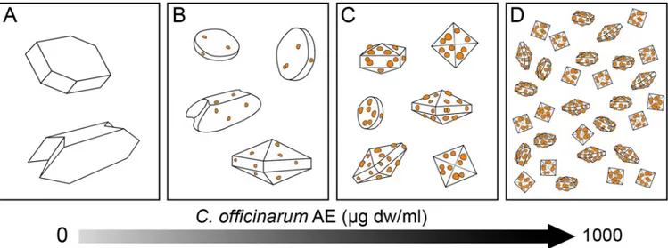
COD crystals produced in the presence of rising AE concentrations suggested a hypothetic trigger factor that may direct the crystal modifications towards COD forms as shown inFig 14.
Based on these data, together with no significant toxicity on a human differentiated CaCo cell line, widely used as a model of the intestinal epithelial barrier [41],C. officinarum. aqueous
extract could represent an attractive natural alternative therapy for urolithiasis.
Fig 14. Schematic illustration of hypothetic model for morphological changes of CaOx crystals induced by increasing doses ofC. officinarum AE. The
diagram is indicative of the progressive adhesion of active components fromC. officinurum on crystal surfaces as a hypothetic trigger factor directing the crystal modifications from COM towards COD forms. (A): Control; (B-D) progressively increasing doses ofC. officinurum AE.
In the very near future we intend to identify and characterize such crystal growth modula-tor(s) and for this purpose, we have already started further extensive investigations to help us to finally shed light on the underlying mechanisms for the positive effects and benefits ofC. officinarum in the treatment of urolithiasis.
Supporting information
S1 Fig. Scanning electron micrographs of CaOx crystals. Crystals were obtained in the
absence (A) or presence (B) of 1000μg dw/ml of C. officinarum AE. Evidence of crystal aggre-gation are usually observed in the control sample (A) while they are completely absent in the presence of the extract (B).
(TIF)
S2 Fig. CaOx crystallization at increasing concentrations ofPhyllantus niruri AE. A)
Con-trol; B) 2μg dw/ml; C) 15 μg dw/ml D) 30 μg dw/ml E) 125 μg dw/ml; F) 250 μg dw/ml; G) 500μg dw/ml; H) 1000 μg dw/ml. Magnification was 400x for all panels.
(TIF)
Acknowledgments
The authors thank dr. Roberta Saltarelli for polyphenol analyses, dr. Elena Antonini for ORAC analyses and prof. Paolino Ninfali for critical revision of the manuscript.
Author Contributions
Conceptualization: Roberta De Bellis, Maria Piera Piacentini, Maria Assunta Meli, Michele
Mattioli, Laura Chiarantini.
Data curation: Roberta De Bellis, Maria Piera Piacentini, Maria Assunta Meli, Michele
Mat-tioli, Michele Menotta, Michele Mari, Letizia Palomba, Donatella Desideri, Laura Chiarantini.
Formal analysis: Maria Piera Piacentini, Michele Mattioli.
Investigation: Roberta De Bellis, Maria Piera Piacentini, Maria Assunta Meli, Michele
Mat-tioli, Michele Menotta, Michele Mari, Laura Valentini, Letizia Palomba, Laura Chiarantini.
Methodology: Maria Assunta Meli, Michele Menotta, Michele Mari, Laura Valentini. Supervision: Laura Chiarantini.
Writing – original draft: Roberta De Bellis, Maria Piera Piacentini, Laura Chiarantini. Writing – review & editing: Roberta De Bellis, Maria Piera Piacentini, Michele Mattioli,
Leti-zia Palomba, Donatella Desideri, Laura Chiarantini.
References
1. Prezioso D, Illiano E, Piccinocchi G, Cricelli C, Piccinocchi R, Saita A, et al. Urolithiasis in Italy: an epide-miological study. Arch Ital Urol Androl. 2014; 86:99–102.https://doi.org/10.4081/aiua.2014.2.99PMID: 25017588
2. Ahmed S, Hasan M, Mahamood ZA. Antiurolithiatic plants: Formulations used in different countries and cultures. Pak J Pharm Sci. 2016; 29(6):2129–2139. PMID:28375136
3. Tiselius HG, Hallin A, Lindba¨ ck B. Crystallisation properties in stone forming and normal subjects’ urine diluted using a standardised procedure to match the composition of urine in the distal part of the distal tubule and the middle part of the collecting duct. Urol Res. 2001; 29:75–82. PMID:11396732
4. Khan SR. Role of renal epithelial cells in the initiation of calcium oxalate stones. Nephron Exp Nephrol. 2004; 98:e55–e60.https://doi.org/10.1159/000080257PMID:15499208
5. Lieske JC, Toback FG. Renal cell-urinary crystal interactions. Curr Opin Nephrol Hypertens.2000; 9:349–355. PMID:10926170
6. Khaskhali MH, Byer KJ, Khan SR. The effect of calcium on calcium oxalate monohydrate crystal-induced renal epithelial injury. Urol Res. 2009; 37:1–6.https://doi.org/10.1007/s00240-008-0160-6 PMID:19005647
7. Scheid CR, Cao LC, Honeyman T, Jonassen JA. How elevated oxalate can promote kidney stone dis-ease: changes at the surface and in the cytosol of renal cells that promote crystal adherence and growth. Front Biosci. 2004; 9: 797–808. PMID:14766409
8. Khan SR. Calcium oxalate crystal interaction with renal tubular epithelium, mechanism of crystal adhe-sion and its impact on stone development. Urol Res. 1995; 23:71–79. PMID:7676537
9. Wang B-H, Wu B-H, Liu J, Yao W-M, Xia D, Li L, et al. Analysis of altered microRNA expression profiles in proximal renal tubular cells in response to calcium oxalate monohydrate crystal adhesion: implica-tions for kidney stone disease PLoS ONE. 2014; 7(9):1–8.
10. Khan A, Byer K, Khan SR. Exposure of Madin–Darby canine kidney (MDCK) cells to oxalate and cal-cium oxalate crystals activates nicotinamide adenine dinucleotide phosphate (NADPH)-oxidase. Urol. 2014; 83(2):510.e1–510.e7.
11. Ma M-C, Chen Y-S, Huang H-S. Erythrocyte oxidative stress in patients with calcium oxalate stones cor-relates with stone size and renal tubular damage. Urol. 2014; 83(2):510.e9–510.e17.
12. Bhandari A, Koul S, Sekhon A, Pramanik SK, Chaturvedi LS, Huang M, et al. Effects of oxalate on HK-2 cells, a line of proximal tubular epithelial cells from normal human kidney. J Urol. 2002; 168:253–259. PMID:12050552
13. Schepers MS, van Ballegooijen ES, Bangma CH, Verkoelen CF. Crystals cause acute necrotic cell death in renal proximal tubule cells, but not in collecting tubule cells. Kidney Int. 2005; 68:1543–1553. https://doi.org/10.1111/j.1523-1755.2005.00566.xPMID:16164631
14. Thongboonkerd V, Semangoen T, Chutipongtanate S. Factors determining types and morphologies of calcium oxalate crystals: molar concentrations, buffering, pH, stirring and temperature. Clin Chim Acta. 2006; 367(1–2):120–131.https://doi.org/10.1016/j.cca.2005.11.033PMID:16458875
15. Schroder FH. Association of calcium oxalate monohydrate crystals with MDCK cells. Kidney Int. 1995; 48:129–138. PMID:7564069
16. Aggarwal A, Tandon S, Singla SK, Tandon C. Diminution of oxalate induced renal tubular epithelial cell injury and inhibition of calcium oxalate crystallization in vitro by aqueous extract of Tribulus terrestris. Int Braz J Urol. 2010; 36:480–489.https://doi.org/10.1590/s1677-55382010000400011PMID:20815954
17. Sheng X, Ward MD, Wesson JA. Crystal surface adhesion explains the pathological activity of calcium oxalate hydrates in kidney stone formation. J Am Soc Nephrol. 2005; 16:1904–1908.https://doi.org/10. 1681/ASN.2005040400PMID:15930089
18. Bouatia M, Benzeid H, Idrissi MOB, Benramdane L, Draoui M. In vitro effect of acetylsalicylic acid on calcium oxalate crystallization: an approach to antilithiasis. Int J Pharm Pharmaceut Sci. 2015; 7 (3):329–331.
19. Spivacow FR, Negri AL, Polonsky A, Del Valle EE. Long-term treatment of renal lithiasis with potassium citrate. Urol. 2010; 76(6):1346–9.https://doi.org/10.1016/j.urology.2010.02.029PMID:20399488
20. Prasad KVSRG, Sujatha D, Bharathi K. Herbal drugs in urolithiasis: a review. Pharmacog Rev. 2007; 1 (1):175–8.
21. Choubey A, Parasar A, Choubey A, Iyer D, Pawar RS, Patil UK. Potential of medicinal plants in kidney, gall and urinary stones. Int J Drug Dev Res. 2010; 2:431–447.
22. Basavaraj DR, Biyani CS, Browning AJ, Cartledge JJ. The Role of urinary kidney stone inhibitors and promoters in the pathogenesis of calcium containing renal stones. EAU-EBU 2007; 5:126–36.
23. Tiwari A, Soni V, Londhe V, Bhandarkar A, Bandawane D, Nipate S. An overview on potent indigenous herbs for urinary tract infirmity: urolithiasis. Asian J Pharm Clin Res. 2012; 5:7–12.
24. Chitme HR, Alok S, Jain SK, Sabharwal M. Herbal treatment for urinary stones. IJPSR. 2010; 1:24–31.
25. Bijarnia RK, Kaur T, Singla SK, Tandon C. A novel calcium oxalate crystal growth inhibitory protein from the seeds of Dolichos biflorus (L.). Protein J 2009; 28:161–168. https://doi.org/10.1007/s10930-009-9179-yPMID:19488841
26. Mittal A. Tandon S. Singla SK, Tandon C. In vitro inhibition of calcium oxalate crystallization and crystal adherence to renal tubular epithelial cells by Terminalia arjuna. Urolith. 2016; 44:117–125.
27. Newman DJ, Cragg GM. Natural products as sources of new drugs over the last 25 years. J Nat Prod. 2007; 70:461–77.https://doi.org/10.1021/np068054vPMID:17309302
28. Di Sanzo P, De Martino L, Mancini E, De Feo V. Medicinal and useful plants in the tradition of Rotonda, Pollino National Park, Southern Italy. J Ethnobiol Ethnomed. 2013; 9:19–28.https://doi.org/10.1186/ 1746-4269-9-19PMID:23522331
29. Zˇ ivkovićS, IvkovićM, SkorićM. Sˇ iler B, DmitrovićS, FilipovićB, et al. Phytochemical characterization and antioxidant potential of rustyback fern (Asplenium ceterach L.) Lekovite Sirovine. 2017; 37:15–20.
30. Karadeniz A, C¸ inbilgel I, Gu¨n SŞ, C¸ etin A. Antioxidant activity of some Turkish medicinal plants. Nat Prod Res. 2015; 29(24):2308–2312.https://doi.org/10.1080/14786419.2015.1005618PMID: 25649168
31. Carrio´ E, Vallès J. Ethnobotany of medicinal plants used in Eastern Mallorca (Balearic Islands, Mediter-ranean Sea). J Ethnopharmacol. 2012; 141:1021–1040. PMID:22783553
32. Viegi L, Pieroni A, Guarrera PM, Vangelisti R. A review of plants used in folk veterinary medicine in Italy as basis for a databan. J Ethnopharmacol. 2003; 89:221–244. PMID:14611886
33. Guarrera PM, Leporatti ML. Ethnobotanical remarks on Central and Southern Italy. J Ethnobiol Eth-nomed. 2007; 3:23–33.https://doi.org/10.1186/1746-4269-3-23PMID:17537240
34. Pieroni A, Quave C, Nebel S, Heinrich M. Ethnopharmacy of the ethnic Albanians (Arbe¨ reshe¨) of north-ern Basilicata Italy. Fitoterapia. 2002; 73:217–241. PMID:12048017
35. Singleton VL, Orthofer R, Lamuela-Ravento´s RM. Analysis of total phenols and other oxidation sub-strates and antioxidants by means of folin-ciocalteu reagent. Meth Enzymol 1999; 299:152–178.
36. Saltarelli R, Ceccaroli P, Buffalini M, Vallorani L, Casadei L, Zambonelli A, Iotti M, Badalyan S, Stocchi V. Biochemical characterization and antioxidant and antiproliferative activities of different Ganoderma collections. J Mol Microbiol Biotechnol. 2015; 25(1):16–25.https://doi.org/10.1159/000369212PMID: 25662590
37. Bag GC, Grihanajali DP, Bhaigyavati Th. Assessment of total flavonoid content and antioxidant activity of methanolic rhizome extract of three hedychium species of manipur valley. Int J Pharm Sci Rev Res. 2014; 28:154–159.
38. Ninfali P, Mea G, Giorgini S, Rocchi M, Bacchiocca M. Antioxidant capacity of vegetables species and dressings relevant to nutrition. Br J Nutr 2005; 93:257–266. PMID:15788119
39. Saltarelli R, Ceccaroli P, Iotti M, Zambonelli A, Buffalini M, Casadei L, et al. Biochemical characterisa-tion and antioxidant activity of mycelium of Ganoderma lucidum from Central Italy. Food Chem. 2009; 116:143–151.
40. Bradford MM. A rapid and sensitive method for the quantitation of microgram quantities of protein utiliz-ing the principle of protein-dye bindutiliz-ing. Anal Biochem. 1976; 72:248–54. PMID:942051
41. Tunisi L, Forte N, Ferna´ndez-Rilo AC, Mavaro I, Capasso R, D’Angelo L, et al. Orexin-A prevents lipo-polysaccharide-induced neuroinflammation at the level of the intestinal barrier. Front Endocrinol. 2019; 10(219):1–8.
42. Palomba L, Amadori A, Cantoni O. Early release of arachidonic acid prevents an otherwise immediate formation of toxic levels of peroxynitrite in astrocytes stimulated with lipopolysaccharide/interferon-γ. J Neurochem. 2007; 103:904–913.https://doi.org/10.1111/j.1471-4159.2007.04793.xPMID:17666049
43. Mittal A, Tandon S, Singla SK, Tandon C. Mechanistic insights into the antilithiatic proteins from
Termi-nalia arjuna: a proteomic approach in urolithiasis. PlosOne. 2016;20; 11(9):1–33.
44. Kulaksizoğlu S, Sofikerim M, Cevik C. In vitro effect of lemon and orange juices on calcium oxalate crys-tallization. Int Urol Nephrol. 2008; 40:589–594.https://doi.org/10.1007/s11255-007-9256-0PMID: 17721827
45. van Breemen RB, Li Y. Caco-2 cell permeability assays to measure drug absorption. Expert Opin Drug Metab Toxicol. 2005; 1:175–185.https://doi.org/10.1517/17425255.1.2.175PMID:16922635
46. Hess B, Jordi S, Zipperle L, Ettinger E, Giovanoli R. Citrate determines calcium oxalate crystallization kinetics and crystal morphology-studies in the presence of Tamm-Horsfall protein of a healthy subject and a severely recurrent calcium stone former. Nephrol Dial Transplant. 2000; 15(3):366–374.https:// doi.org/10.1093/ndt/15.3.366PMID:10692522
47. Tazzoli V, Domeneghetti C. The crystal-structures of whewellite and weddellite–reexamination and comparison. Am Mineral 1980; 65:327–334.
48. Grohe B, Rogers KA, Goldberg HA, Hunter GK. Crystallization kinetics of calcium oxalate hydrates studied by scanning confocal interference microscopy. J Cryst Growth 2006; 295:148–157.
49. Grohe B, Taller A, Vincent PL, Tieu LD, Rogers KA, Heiss A, et al. Crystallization of calcium oxalates is controlled by molecular hydrophilicity and specific polyanion–crystal interactions. Langmuir 2009; 25:11635–11646.https://doi.org/10.1021/la901145dPMID:19725562
50. Scho¨nherr H, Paraschiv V, Zapotoczny S, Crego-Calama M, Timmerman P, Frank CW, et al. Unravel-ing the nanostructure of supramolecular assemblies of hydrogen-bonded rosettes on graphite: An
atomic force microscopy study. PNAS 2002; 99(8):5024–5027.https://doi.org/10.1073/pnas. 072685399PMID:11929980
51. Itoh Y, Yasui T, Okada A, Tozawa K, Hayashi Y, Kohri K. Preventive effects of green tea on renal stone formation and the role of oxidative stress in nephrolithiasis. J Urol. 2005; 173(1):271–275.https://doi. org/10.1097/01.ju.0000141311.51003.87PMID:15592095
52. Grases F, Prieto RM, Gomila I, Sanchis P, Costa-Bauza´ A. Phytotherapy and renal stones: the role of antioxidants. A pilot study in Wistar rats. Urol Res. 2009; 37(1):35–40. https://doi.org/10.1007/s00240-008-0165-1PMID:19066877
53. Grases F, Prieto RM, Fernandez-Cabot RA, Costa-Bauza´ A, Tur F, Torres JJ. Effects of polyphenols from grape seeds on renal lithiasis. Oxid Med Cell Longev. 2015;813737.https://doi.org/10.1155/2015/ 813737PMID:25883748
54. Guerra A, Meschi T, Allegri F, Prati B, Nouvenne A, Fiaccadori E, et al. Concentrated urine and diluted urine: the effects of citrate and magnesium on the crystallization of calcium oxalate induced in vitro by an oxalate load. Urol Res. 2006; 34:359–64.https://doi.org/10.1007/s00240-006-0067-zPMID: 16953377
55. Mittal A, Tandon S, Singla SK, Tandon C. In vitro studies reveal antiurolithic effect of Terminalia arjuna using quantitative morphological information from computerized microscopy. Int Braz J Urol. 2015; 41 (5):935–944.https://doi.org/10.1590/S1677-5538.IBJU.2014.0547PMID:26689519
56. Barros ME, Schor N, Boim MA. Effects of an aqueous extract from Phyllanthus niruri on calcium oxalate crystallization in vitro. Urol Res. 2003; 30:374–379.https://doi.org/10.1007/s00240-002-0285-yPMID: 12599017
57. Shang Y-F, Xu M, Zhang G-N, Ouyang J-M. Concave urinary crystallines: direct evidence of calcium oxalate crystals dissolution by citrate in vivo. Bioinorg Chem Appl. 2013; 2013:637617.https://doi.org/ 10.1155/2013/637617PMID:24363634
58. Guo S, Ward MD, Wesson JA. Direct visualization of calcium oxalate monohydrate crystallization and dissolution with atomic force microscopy and the role of polymeric additives. Langmuir 2002; 18:4284– 4291.
59. De Yoreo JJ, Qiu SR, Hoyer JR. Molecular modulation of calcium oxalate crystallization. Am J Physiol Renal Physiol. 2006; 291:F1123–F1132.https://doi.org/10.1152/ajprenal.00136.2006PMID:17082348