Università degli Studi del Piemonte Orientale
“Amedeo Avogadro”
Dipartimento di Scienze del Farmaco
Dottorato di Ricerca in Biotecnologie Farmaceutiche ed
Alimentari
XXVI ciclo A. A. 2010-2013
EXPRESSION AND DISTINCT FUNCTIONAL ROLES
OF AKT ISOFORMS IN MALIGNANT PLEURAL
MESOTHELIOMA CELLS
Ester Borroni
Supervised by Prof. Laura Moro
Contents
Chapter 1
3Introduction
1- Malignant Mesothelioma 1.1. Aetiology of Malignant Mesothelioma
1.2. MPM classification
1.3. Diagnosis, Staging and Prognosis
1.4. Therapy
2- AKT 2.1. AKT Family: Isoforms and Roles 2.2. PI3K/AKT/mTOR Pathway 2.3. Down Stream Effectors of AKT 2.4. Inactivation of AKT 2.5. Post-translational Modifications of AKT 2.6. AKT in Cancer 2.7. AKT Inhibitors
Chapter 2
55Outline of the thesis
Chapter 3
59 Targeting different AKT isoforms in Malignant MesotheliomaChapter 4
81Hsc70 (Heat Shock Cognate Protein 70) and AKT interaction
Chapter 5
95 XX Conclusions3
Chapter 1
Introduction
1 - MALIGNANT MESOTHELIOMA
1.1. Aetiology of Malignant Mesothelioma
Malignant Mesothelioma (MM) is a highly aggressive cancer arising from the mesothelial lining of the pleura, peritoneum and pericardium. Among these, Malignant Pleural Mesothelioma (MPM), that represents approximately 90% of cases, is the most common primary tumor of the pleura. MPM has a very poor prognosis; it typically spreads and invades locally, even though distant metastases to the contralateral lung, peritoneum, bone or liver can occur (1).
The median survival is less than 12 months, depending on the stage of the disease at the time of diagnosis (2). MPM is associated with exposure to asbestos. Asbestos is the generic term for a group of naturally occurring, fibrous minerals with high tensile strength and resistance to heat, chemicals, and electricity. Six minerals, defined as "asbestos", are divided in two classes: the serpentine and the amphibole. Chrysotile (white asbestos), is the only one member of the serpentine class, is made up of curled fibers and accounts for approximately 95% of all asbestos used worldwide. The amphiboles’ group can be distinguished for its friable fibers and includes amosite (brown asbestos) and crocidolite (blue asbestos), extensively used in industries (3).
Between the 1940s and 1979 in the United States and Europe, asbestos was widely used in the shipbuilding and construction industries thanks to its
4
fire-resistant properties (4). The incidence of asbestos-related diseases has been rising worldwide since 1970, due to the exposure during the past and it is predicted to increase further in coming decades, especially in developing countries where asbestos has not yet been banned. The mean latency period between the time of initial exposure and diagnosis is about 30 years and ranges from 20 to 50 years; moreover, the disease latency has an inverse relationship with duration of exposure (5).
The first hypothesis about a link between cancer and asbestos, was proposed by Wagner et al. (6) in 1870, who described crocidolite asbestos exposure in 33 cases of mesothelioma in South Africa’s North Western Cape, while further confirmations came during the following years when McDonald found a proportional mortality ratio for exposed subjects.
Currently rates of MPM are rising and estimates indicate that the incidence of MPM will peak within the next 10-15 years. Although the use of asbestos has been banned in many countries around the world, production of and potentially hazardous exposure to asbestos is still present with locally high incidences. Today a new man-made material, carbon nanotubes (CNTs), has arisen as a concern; CNTs may display ´asbestos-like´ pathogenicity with mesothelioma induction potential (7).
Exposure mapping within countries reveals high regional variability in incidence and mortality: for instance, in Italy, significant municipal clusters of disease have been identified close to asbestos industries, shipyards, oil refineries and petrochemical industries (8); similarly in the UK, the highest mesothelioma mortality rates were recorded in areas with a history of ship building, such as Barrow-in-Furness, Plymouth, Portsmouth, Tyneside and Southampton (9). The correlation between amphibole, asbestos exposure and MPM development is generally accepted and crocidolite is considered the most oncogenic type of asbestos. Even if the relative oncogenicity of the specific asbestos fibers type is controversial, the evidence has been
5
considered sufficient by the World Health Organization (WHO) to conclude that all types of asbestos cause cancer in humans (10).
Asbestos fibers can be inhaled deeply and trapped in the lower lung where mechanically cause pleural irritation and induce an inflammatory response as consequence of repeated scratching of mesothelial surface. Moreover, phagocytosed fibers can initiate a cascade of events that includes activation of c-Myc and c-Jun oncogenes, binding with growth factor receptors (EGFRs) (11,12), or alternatively, mechanically interference with the mitotic process of the cell cycle by disrupting the mitotic spindle, with consequent chromosomal abnormalities and aneuploidy (13). Finally, another proposal is that asbestos can also induce reactive oxygen species (ROS) or reactive nitrogen species (RNS) (resulting in DNA damage), cytokines, growth factors -such as transforming growth factor-β (TGF-β)- as well as transcription factors -such as nuclear factor kappa B (NF-kB) and activator protein-1 (AP-1) (14, 15).
1.2. MPM classification
According to the WHO classification, based on the histological analysis of tissue obtained by biopsies, MPM is divided in three different classes: epithelioid, sarcomatoid and biphasic.
Epithelioid
Around 60% of all mesothelioma cases comprises mesothelial cells with epithelial-like shape arranged in tubulo-papillary or trabecular formations and represents the most common histotype characterized by a better prognosis (16).The most frequent patterns encountered are tubulopapillary, adenomatoid (microglandular) and sheet-like, while less common patterns include small cells, clear cells and deciduoid. In particular, the tubule papillary form shows varying combinations of tubules with connective tissue
6
cores and clefts. Most epithelioid mesotheliomas are singularly bland in appearance and mitoses are scarce; usually a pattern predominates but often-different ones are commonly found in the same tumor, as much as sheets and nets of cells are frequently seen in association with other patterns (17). The fibrous stroma of epitheloid mesotheliomas can show varying degrees of cellularity from hyalinised acellular to highly cellular, merging with sarcomatoid; furthermore, these tumors may be difficult to distinguish from biphasic mesotheliomas.
Sarcomatoid
Sarcomatoid histotype is associated with less differentiated and more aggressive tumors in which more than 90% of the cells assume a spindle morphology and are arranged in fascicles or sheets, showing many mitotic figures, necrosis, and cytological atypia. Cells frequently tend to be irregular rather than uniform and overlap one other: their pattern often looks like fibrosarcoma, but marked anaplasia and uncommon multinucleate tumor cells make it more similar to malignant fibrous histiocytoma (18).
Biphasic
The biphasic histotype contains both epithelioid and sarcomatoid patterns and each component should represent at least 10% of the tumor to warrant the term biphasic. Cells of the biphasic variety form groups rather than displaying a uniform mixture (19).
1.3. Diagnosis, Staging and Prognosis
Malignant mesotheliomas are not ordinarily graded.
Patients with MPM commonly present with dyspnoea, chest wall pain and pleural effusion and diagnosis is often made at an advanced stage of the disease, so that in untreated patients median survival is less than one year.
7
Difficulty in diagnosing the disease, especially in its early stages, prevented the development of a generally accepted stage-related approach. In addition, MPM is a heterogeneous disease with a variable clinical course: the majority of patients (80%) are diagnosed in stage III/IV and these patients are not candidates for surgical cure (20). Thus, systemic therapy is the only treatment option for the majority of this kind of patients, but poor performance status and the low chemo and radio-sensitivity of the tumor result in a poor prognosis. For these reasons, advances in the understanding of the molecular biology of MPM is needed to establish diagnostic, therapeutic and prevention methods and allow the identification of promising new candidates for targeted treatments.
Knowledge of the biological basis of MPM progression and response may facilitate a personalized treatment approach involving early identification of poor prognostic indicators that will reduce the heterogeneity of the clinical response (1, 18).
One contributing factor for the diagnosis of malignant mesothelioma is the possibility to differentiate it from adenocarcinoma, a cancer originating in glandular tissue. The most important differential is metastatic or locally invasive (from lung or chest wall) tumor that covers the pleural surface, even if various localized tumors also exist in the pleura and some mimic mesothelioma microscopically. For this reason, knowledge of the distribution of tumor, whether obtained from radiographic studies, the operator’s description of the findings at thoracotomy or thoracoscopy, or from a resected specimen, is crucial to make a proper diagnosis. However, beyond the distinction between epithelioid and sarcomatoid forms, these histopathologic features do not always correlate well with prognosis (17).
8
Staging
Several staging systems for mesothelioma have been used over the years, almost exclusively dealing with primary pleural mesothelioma. Peritoneal mesothelioma does not have its own staging system. The oldest staging system used for pleural mesothelioma is the Butchart system; it is still commonly used in some parts of the world (13). This method is based on a simple description of the extent of the disease without considering the histologic subtype: pleural contained (Stage I), chest wall or mediastinal invasion (Stage II), peritoneal or diaphragmatic penetration (Stage III), or distant metastases (Stage IV). Meanwhile, the Brigham system tries to define surgical resectability and lymph node involvement. Stage I disease is resectable without nodal spread while stage II is resectable with lymph node involvement. Stage III involves the chest wall, heart, diaphragm, or abdominal cavity, with or without lymph node involvement: stage III tumors are considered unresectable. Stage IV disease is distant metastases. This system is not utilized at present. The most practical and most commonly used system is the tumor-node-metastasis system developed by the International Mesothelioma Interest Group. This system is the currently accepted standard adopted by the American Joint Committee on Cancer (21, 22). Most patients present with advanced disease and are considered unresectable.
Histopathology and Survival
The prognosis for MPM is significantly affected by histopathologic variant. Most studies have found a clear survival benefit for patients with epithelioid subtype. A recently published study on 312 patients undergoing surgical therapy for MPM evaluated the effect of histology on survival (23). Patients with epithelioid subtype had the longest median survival of 15.3 months, and 1-year and 2-year survival of 63% and 32% respectively. Patients with
9
sarcomatoid subtype had a median survival of 5 months, and 1-year and 2-year survival of 4% and 0%, respectively. The biphasic subtype had intermediate survival rates. Multiple other studies have demonstrated extremely poor survival for patients with sarcomatoid-type MPM regardless of medical or surgical therapy, and most centres do not offer surgery to these patients (24).
1.4. Therapy
Mesothelioma is difficult to treat because many people are not diagnosed until their disease is advanced and the decisions about treatments are based on different tasks: where the tumor arose and its stage, the grade of malignancy, its location, whether it is resectable or not, symptoms, general health conditions of the patient and age (25). Treatment options are limited and the results with conventional therapies have been rather disappointing to this date. Moreover, chemotherapy is the only evidence-based treatment for mesothelioma patients in good clinical condition, with an increase in median survival of only 2 months: so finding out new different approaches to afford this malignancy became necessary.
Actually the therapy for mesothelioma is represented by surgery, radiotherapy, chemotherapy and the so-called “trimodality therapy” that consists in combination of chemotherapic drugs following surgery (26).
1.4.1. Surgery and Radiation Surgery
Surgery for mesothelioma could be done essentially for two reasons: to try to cure cancer or to relieve pain and other symptoms carried on by the tumor. The curative surgery is an option for patients who are living with good health conditions because the cancer has not spread a lot and it could be removed completely also because it is thought to be localized. However
10
sometimes surgeons are not able to remove all of the cancer, some cells remain and can grow normally, divide allowing the cancer to come back after surgery. Otherwise, palliative surgery may be an option if the tumor has already spread beyond where it started and complete removal is not possible, or if the patient is too ill for a more extensive operation (27). The type of surgery done depends mainly on the location of the tumor, the stage of the cancer, the subtype of mesothelioma and other factors, such as patient overall health, also because chemotherapy and radiation represent additional therapies that are used in combination with surgery.
- Pleural mesothelioma surgery
There are two types of surgical treatments that may be offered to patients with pleural mesothelioma: pleurectomy/decortication (P/D) and extrapleural pneumonectomy (EEP). People with early stage or localized (stage I) pleural mesothelioma are undergone to P/D with removal of both the parietal pleura and visceral pleura together with the tumor, while the lung is not removed. In more advanced stages, a P/D can also be done to help control fluid build-up in the chest, improving breathing and relieve symptoms, such as chest pain caused by the tumor. Patients in stage I or II, and some with stage III pleural mesothelioma are treated with extrapleural pneumonectomy (EPP), only if the tumor is considered operable, is localized and has not spread into the diaphragm – this typically occurs in patients with resectable mesothelioma of the epithelioid type whose cancer has not reached the lymph nodes(28).
- Peritoneal mesothelioma surgery
Surgery is not often possible for people with peritoneal mesothelioma, but in people who are elegible candidates for surgery this treatment is often done to help relieve symptoms or to remove the tumor from the abdomen wall and organs; unfortunately as with pleural mesothelioma, these tumors often have spread too far to allow a complete removal (29). Essentially,
11
people with peritoneal mesothelioma may have a peritonectomy, in which the membrane that lines the abdomen (peritoneum) is removed, sometimes involving also other structures; or an omentectomy, a surgery that removes the layer of fatty tissue that covers the contents of the abdomen, when mesothelioma spread beyond the peritoneum (30).
Radiation
The pattern of spread of malignant pleural mesothelioma (MPM) poses unique challenges to a radiation oncologist. At first, patients are treated with conventional radiation techniques, when radiotherapy administered as adjuvant therapy following extrapleural pneumonectomy (EPP) or pleurectomy/decortication (P/D) (31). Anyway, with conventional radiotherapy there may be radiation underdosing near regions that are blocked: this has the potential to lead to increased risk of local failure in approximately 10-15% of patients. More recently, complex intensity-modulated radiation therapy (IMRT) techniques for the treatment of MPM have been explored, with early outcomes suggesting acceptable safety in appropriately selected patients. Multiple subsequent studies incorporated this technique into a multimodality approach combining chemotherapy, EPP and hemithoracic radiation (32).
1.4.2. Chemotherapy and Multimodal Therapy
Treatment modalities for MPM patients depend on age, performance status and disease stage at onset. It is now generally accepted that the future of cancer therapy lies in the combination of therapies with different mechanisms of action; actually the therapy for MPM is represented by surgery and radiotherapy, chemotherapy and the so-called “trimodality therapy” that consists in combination of chemotherapic drugs following surgery.
12
Chemotherapy
A phase 3 trial of combined therapy using pemetrexed and cisplatin has shown major efficacy compared with cisplatin monotherapy and consequently, current approved first line therapy now include cisplatin and antifolate agent, such as pemetrexed or raltitrexed (33).
Several other cytotoxic agents with definite activity in mesothelioma have recently been evaluated, including gemcitabine, vinorelbine and the antifolates pemetrexed and raltitrexed. Vinorelbine has been shown to have activity in a small phase II study; similar results have been achieved with vinflunine, a new semi-synthetic fluorinated vinca alkaloid. In contrast, taxanes, such as paclitaxel and docetaxel, showed very low or no activity. Platinum analogues, doxorubicin and some antimetabolites (methotrexate, raltitrexed, pemetrexed) have shown modest efficacy as single-agent. The combination of antifolates and cisplatin was proven to prolong survival over cisplatin alone in Phase III studies, while in other phase II studies, the combination of pemetrexed and gemcitabine was also effective as a first-line chemotherapy for patients with MPM (34).
Recently a study on 409 patients with MPM was conducted assigning them randomly to one of three treatments for 12 weeks: active symptom control (ASC) alone, ASC plus MVP (mitomycin vinblastine, and cisplatin), or ASC plus vinorelbine. The median survival was 7.6 months in the ASC alone group and 8.5 months in the ASC plus chemotherapy group. Considering that ASC group included treatments with steroids, analgesic drugs, bronchodilators or palliative radiotherapy, the addition of chemotherapy to ASC did not offered significant benefits in terms of the overall survival or quality of life (35).
Chemotherapies consisting of doxorubicin, doxorubicin plus cyclophosphamide, oxaliplatin-raltitrexed or ZD 0473 (a platinum analogue) appeared ineffective. Some interesting response rates were noted with
13
pemetrexed alone, or the combination of carboplatin and pemetrexed and of cisplatin, irinotecan and mitomycine C, used as second line therapy (36). Weekly vinorelbine administration demonstrated useful clinical activity in the second-line treatment of MPM, resulting in a median overall survival of 9.6 months. Gemcitabine and vinorelbinein pemetrexed-pretreated patients with MPM resulted in a 10.9-month median survival time. Moreover, retreatment of pemetrexed-pretreated patients appeared effective, with 1-year survival rates of 71.0% for retreated patients versus 18.8% without retreatment.
In patients receiving second-line chemotherapy after the phase 3 trial of pemetrexed plus cisplatin versus cisplatin alone, a positive impact on survival was evident. MPM patients receiving second-line therapy with pemetrexed exhibited significant anti-tumor activity, improved disease control rates, and longer progression-free survival than patients receiving the best supportive care alone, although survival was not improved (37, 38).
- Immunotherapy
Immunotherapy is one of the more recent approaches to cancer therapy: it represents a new class of agents in the treatment of mesothelioma and it is based on the generally-accepted hypothesis that the immune system is the best tool humans have for fighting disease. This therapy is sometimes used by itself to treat cancer, but it is most often used in combination with traditional treatments like radiation, chemotherapy, and surgery in order to enhance their effects. One of the possible benefits of immunotherapy is that it has the potential not to be as toxic as radiation or chemotherapy, and less invasive than surgery.
In the case of cancer, the immune system alone often fails to effectively fight the tumor; normally the immune system does not recognize tumor cells because they are derived from the body's own cells, so the body “thinks” of
14
the tumor as “self” and creates a phenomenon known as “tumor tolerance”. Otherwise the immune system may also recognize cancer cells, but the response is not strong enough to destroy the cancer (because the response is insufficient in amount, it does not occur in the right place in the body, or it does not send the right “immune cells”). Moreover, the tumor has the ability to defend itself and cancer cells may secrete substances that keep the immune system in check. Thus, in the case of cancer, the immune system needs something to afford it more efficiently (39).
The purpose of cancer immunotherapy is to promote (activating or intensifying) the antitumor effects of the immune system of the patient; several approaches for immunotherapy have been developed over the years and some are in various stages of pre-clinical research. Immunotherapy can be divided in two main categories, passive and active immunotherapy. Passive immunotherapy uses in-vitro produced effectors that are able to influence tumor cell growth: the most common form is based on the monoclonal antibody therapy, which consists in humanized monoclonal antibodies targeting cells directly or indirectly, which efficacy may be often enhanced by linking a toxin (e.g., radionucleotides or anticancer drugs) (40). Active immunotherapy approaches aim at inducing or boosting immune effector cells in vivo against tumor cells, through the administration of immune mediators capable of activating the immune system (i.e. cytokines capable recruiting specific immune cells for enhance antitumor immunity, as IL-2, IL-12, IL-15, TNF-α, GM-CSF). These cytokines can be used as single agent or in combination with other immunotherapeutic strategies (41).
- Molecular-Targeted Therapy
Epidermal growth factor receptor (EGFR), platelet-derived growth factor receptor (PDGFR), vascular endothelial growth factor receptor (VEGFR), SRC, hepatocyte growth factor (HGF), transforming growth factor (TGF),
15
insulin like growth factor I receptor (IGF-1R), mammalian target of rapamycin (mTOR), histone deacetylases (HDAC), and the proteasome have been investigated and can be targeted in MPM treatment (42, 43). Several clinical studies have been conducted. Phase 2 clinical trials based on EGFR tyrosine kinase inhibitor (erlotinib and gefitinib) therapy have shown limited or no activity in MPM; probably, the lack of EGFR mutations that confer sensitivity to TK-inhibitors in NSCLC could also explain the resistance to EGFR tyrosine kinase inhibitors in this cancer.
The phase 2 studies with the anti-EGFR monoclonal antibody, cetuximab, with platinum/pemetrexed are ongoing. In three phase 2 trials involving imatinib as a PDGFR inhibitor, no objective response was obtained; moreover, a combination of imatinib with gemcitabine, cisplatin/gemcitabine, or cisplatin/pemetrexed has also been investigated without good outcomes (44,45). Bevacizumab, a recombinant human anti-VEGF monoclonal antibody that blocks the binding of anti-VEGF to its receptors, is under evaluation in a double-blind, randomized phase II trial in combination with cisplatin and gemcitabine.
Other novel agents under investigation include sorafenib, an inhibitor of VEGFR-2, PDGFR-b, and B-Raf tyrosine kinase; in a phase I trial, vorinostat, a histone deacetylase inhibitor, has produced objective responses in 20% of MPM patients, and a phase III double-blind, placebo-controlled trial is under way (46).
Multimodal Management of MPM
Failure of single-modality treatments to increase survival in MPM patients has led to a variety of multimodality approaches.
The typical curative surgical treatment is extrapleural pneumonectomy (EPP) that involves a total en-bloc resection of the lung, pleura, pericardium, and diaphragm, while pleurectomy/decortication (P/D) involves
16
resection of the parietal and visceral pleura, and pericardium. This cytoreductive surgery has been added to systemic and/or intrapleural chemotherapy and to external-beam or intraoperative RT, with the main aim of improving local control. Early studies of neoadjuvant chemotherapy followed by EPP and hemithoracic RT have been published, and confirmatory trials on patients are ongoing.
Several studies applying gemcitabine and cisplatin as an induction chemotherapy in combination with EPP and radiation in a combined-modality approach for resectable MPM have been also reported.
Patients completing trimodality therapy displayed a median survival of 29.1 months and a 2-year survival rate of 61.2%.
Patients who underwent trimodality therapy involving EPP and adjuvant chemoradiotherapy displayed a median overall survival of 13– 23.9 months. It was concluded that selected patients with MPM may benefit from EPP, particularly when combined with induction or adjuvant chemotherapy and adjuvant radiotherapy. Due to its toxicity, is difficult to administer EPP as adjuvant chemoradiotherapy; thus, a trimodality therapy consisting of induction chemotherapy and EPP followed by radiotherapy was preferred. Finally, surgery may remove macroscopic diseases and multimodality therapy with radiation and chemotherapy should be included to eradicate residual microscopic disease (47, 48).
2 - AKT
2.1. AKT Family: Isoforms and Roles
AKT/PKB (Protein Kinase B) regulates many cellular functions such as cell growth and proliferation, cell survival, apoptosis, energy metabolism, migration and resistance to anticancer therapeutics (49). Despite the
17
growing amount of researches demonstrating the existence of isoform-specific regulation, many papers still draw generalized conclusions about AKT functions without considering the unique function of each AKT isoform. AKT family consists of three isoforms, AKT1, AKT2 and AKT3 (also known as PKBα, PKBβ and PKBγ, respectively) (50). Although these three isoforms are encoded by separate genes, they share a common NH2-terminal pleckstrin homology (PH) domain (of 110 amino acids), a catalytic (kinase) domain in the middle (of about 260 amino acids) separated by a hinge region of 39 amino acids (“Linker”) and a C-terminal regulatory domain (with an identity in amino sequence of about 80%), which contains an hydrophobic motif, characteristic of the cAMP-dependent protein kinase A/protein kinase G/protein C (AGC) superfamily of protein kinases (51). They are differentially expressed in various tissues: AKT1 is most abundant in brain, heart, and lung; AKT2 is predominantly expressed in skeletal muscle and embryonic brown fat, while AKT3 is mainly expressed in kidney, brain and embryonic heart, although it is also present in adipose tissue, mammal glands and lungs.
AKT isoform-specific knockout mice suggest that different AKT kinases are not completely overlapping and that isoform-specific signaling contributes to the diversity of their biological function.
- AKT1 knockout mice are smaller than littermate controls and show increased rates of apoptosis in some tissues, reflecting its role in cell survival (52).
- AKT2 null mice develop type 2 diabetes and impaired glucose utilization, suggesting its importance in the insulin receptor-signaling pathway (53).
- The precise role of AKT3 is less understood: AKT3 is a key modulator of several tumors like glioma, ovarian cancer and melanoma in which an increase of active this isoform promotes
18
tumorigenesis by decreasing apoptosis; moreover, mice lacking AKT3 show impaired brain development (54).
2.2. PI3K/AKT/mTOR Pathway
AKT activity is modulated downstream of phosphatidylinositol 3 (PI3) kinase via a multistep process that starts in response to various growth factors and cytokines such as plateled-derived growth factor receptor (PDGF-R), epidermal growth factor (EGF) and insulin-like growth factor1 (IGF-1). In the absence of growth factor stimulation in quiescent cells, all three isoforms of the AKT kinases are catalytically inactive (55).
The PI3Ks constitute a family of intracellular lipid kinases that phosphorylate the 3’-hydroxyl group of phosphatidylinositols (PIs) and phosphoinositides. Based on structure and their specificity for substrates, PI3Ks are classified into three groups (classes I, II and III).
Class I PI3Ks are the primary lipid kinases that generate PIP3 from PIP2, and are subdivided into two classes (a, activated by growth factor TK-receptor, and b, activated by G protein-coupled receptors); class II binds clathrin and are while both (class II and III) use PI to generate PI-3-P (56). After ligand-induced activation of specific receptors, a phosphorylated Y residue on receptor serves as docking site for regulatory p85 sub-unity of PI3K that recruits p110 sub-unity to the complex. PI3K activated localizes to the cytoplasmic face of the plasma membrane, where it converts PIP2 (phosphatidylinositol 4,5-bisphosphate) to PIP3 (phosphatidylinositol 3,4,5-trisphosphate), which promotes membrane localization of molecules such as AKT and 3-phosphoinositide- dependent kinase 1 (PDK1) by binding to their PH domain.
The C-terminal PH domain of PDK1 binds phospholipids, keeping it constitutively localized at the plasma membrane; upon GF stimulation AKT interacts with these phospholipids, causing its translocation to the inner
19
membrane, where PDK1 is located. The interaction of the AKT PH domain with 3’- phosphoinositides makes conformational changes in AKT with the exposure of its main phosphorylation sites, T308 and S473 (57).
Direct homodimerization of the two PH domains between AKT and PDK1 might phosphorylate Thr-308 in AKT, which stabilizes the activation loop in an active conformation and renders Ser473 phosphorylation by mTORC2 (mammalian target of rapamycin complex 2), resulting in full activation of the kinase. However, other molecules, including integrin-linked kinase (ILK) and mitogen-activated protein kinase-activated protein kinase-2 (MAPK-APK2), mTOR and AKT itself have also been described to phosphorylate Ser473 (58). Full activation of AKT is a multistep process that leads to AKT phosphorylation on two sites, Thr308 (for human AKT1) and Ser473 (for AKT1).
In particular, to optimize the kinase activity of all AKT isoforms, the concomitant phosphorylation of threonine and serine residues is required: curiously, they are these two aminoacids are positioned in marginally different locations in the three isoforms.
For instance, the most essential regulatory amino acid residues are threonine 308 and serine 473 in AKT1, whereas the amino acid residues are threonine 309 and serine 474 in AKT2, while in the case of AKT3, the regulatory residues are threonine 305 and serine 472 (59).
AKT can be activated in a PI3K-independent manner: for example, agonists of the PKA pathway, such as cAMP elevating agents, can activate AKT: calcium binds to calmodulin, and the Ca2+/calmodulin complex activates the calcium/calmodulin dependent kinase kinase (CaMKK), which directly phosphorylates T-308. Finally, some non-kinase interactors such as Hsp90/Cdc37 (molecular chaperone and co-chaperone), Hsp27, Tcl1 (T-cell leukemia family), Grb10 (growth factor receptor binding protein10), Ft1 and many others can positively regulate AKT catalytic activity (60).
20
2.3. Downstream effectors of AKT
AKT activation is primarily implicated in cell survival and cell growth through regulation of several substrates. Among these substrates there are:
- regulators of cell survival or death, such as BAD, caspase-9, ASK1 (apoptosis signal-regulating kinase 1), forkhead box O transcription factors (FoxOs), Bim1, FasL, inhibitor of nuclear factor-kB kinase (IKK-NFkB), and p53;
- regulators of angiogenesis, such as mTOR and hypoxia-inducible factor-1 (HIF-1);
- regulators of cell metabolism, such as glucose transporter 1 (Glut1), GSK3, and a Ras homologue enriched in brain (RheB).
AKT also regulates cell growth acting on tuberous sclerosis complexes 1 and 2 (TSC1/2), mTOR, elongation-initiation factor 4E binding protein-1 (4E-BP1) and mTOR pathway; it also modulates cell cycle progression through direct actions on the p21 and p27 (CDK inhibitors) and indirect effects on the levels of cyclin D1 and p53 (61).
2.4. Inactivation of AKT
AKT cascade can be blocked by cellular inhibitors as the phosphatase and tensin homolog (PTEN) and INPP4B, which directly antagonize PI3K functions, dephosphorylating PIP3 and revoking its downstream events. PTEN has been also demonstrated to be one of the most commonly altered genes in human malignancies. In contrast, gain-of-function AKT mutations are uncommon, but frequently occur at residue 17, within the PH domain (E17K-Lys 17 alters the electrostatic interactions of the pocket and forms new hydrogen bonds with a phosphoinositide ligand). This mutation activates AKT leading its localization to the plasma membrane, stimulates downstream signaling, transforms cells and induces leukemia in mice, indicating a direct role of AKT in cancer; moreover, the E17K mutation
21
does not change AKTs sensitivity to ATP-competitive inhibitors, but it modify change its sensitivity to the allosteric ones (62).
Negative regulation of PI3K/AKT pathways is mainly accomplished by PTEN (phosphatase and tensin homologue deleted). It was originally identified as a tumor-suppressor gene and was found frequently mutated in a wide variety of solid tumors, including endometrial, breast, prostate carcinomas, and glioblastomas. As a dual lipid and protein phosphatase, the primary physiological target of PTEN is considered to be the PI3K/AKT pathway.
PTEN specifically catalyses dephosphorylation of the 3-phosphate of the inositol ring in PIP3, resulting in the biphosphate product PIP2 and inhibition of AKT activity. Inactivating mutations or loss of PTEN expression leads to a permanent increase in the basal level of PI3K/AKT signaling, generally resulting in increased cell proliferation and resistance to apoptosis. Recently, a family of novel protein phosphatases, namely, PHLPP (PH domain and leucine-rich repeat protein phosphatase), have been identified as important regulators of AKT kinases and protein kinase C (PKC). Two isoforms, PHLPP1 and PHLPP2, directly dephosphorylate Ser473 and therefore inactivate AKT. It has been shown that PHLPP differentially terminates AKT signaling through particular regulation of AKT isoforms. PHLPP2 dephosphorylates AKT1 and AKT3, whereas PHLPP1 is specific for AKT2. PHLPP expression is commonly lost in cancer, including colon, breast, ovarian, Wilms tumors, and prostate cancer, while its co-deletion, together with PTEN is strongly associated with metastatic prostate cancer, suggesting the role of PHLPP as a tumor suppressor (63).
2.5. Post-translational Modifications of AKT
A part phosphorylation other post-translational modifications has been described to modify AKT activity/stability.
22
AKT acetylation
The level of protein acetylation is regulated by enzymes that increase or decrease acetylation. When the balance between these mechanisms is altered, it results in protein hyperacetylation or deacetylation. As phosphorylation, the reversible acetylation of cellular proteins plays an important role in cellular processes; whereas different amino acids (i.e., threonine, serine, and tyrosine) can be phosphorylated, acetylation occurs only on lysine residues (64, 65).
Deacetylases are enzymes that remove acetyl groups: they can be divided in four classes based on their structural and functional similarities, and among these, the major family is represented by sirtuin enzymes, that consist in seven components designated as SIRT1 through SIRT7, exhibiting different tissue distributions and subcellular localizations.
Activity of AKT requires tight control, as evidenced by the multiple means by which the protein is regulated: phosphorylation, ubiquitination and binding to phospholipids.
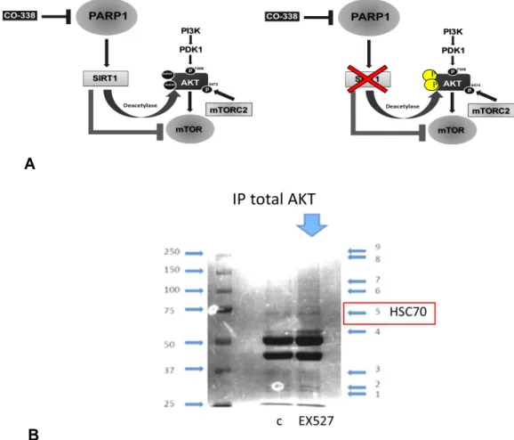
Recently, Sundaresan et al. reported the regulation of AKT and PDK1 by reversible acetylation. Their work showed that p300 and pCAF acetylate AKT and PDK1, while SIRT1 deacetylates them. The acetylation regulated the ability of AKT to bind PIP3, offering a new sight of AKT regulation through reversible acetylation: AKT was acetylated in murine cells in a manner inversely related to its activation. The kinase was associated with the histone deacetylase SIRT1 and deacetylation enhanced its ability to be activated. In contrast, activation of AKT after growth factor stimulation was inhibited in cells lacking SIRT1 (66, 67); moreover, cells effects on cardiac hypertrophy in mice lacking or overexpressing SIRT1 were associated with altered regulation of AKT. Thus, acetylation of AKT (regulated by SIRT1) appears to be an important aspect of control of different biological activities of the enzyme (68). Another elegant work conducted by Ramakrishnan and
23
colleagues, focused the attention on SIRT2, the primary cytoplasmic sirtuin, to which it have been attributed tumor suppressor functions and a role in maintaining genome integrity and in programmed necrosis. There is substantial crosstalk between the insulin-PI3K-AKT-metabolic pathways and sirtuins: SIRT1 and SIRT2 can deacetylate and regulate the function of FoxO transcription factors, which are direct AKT targets.
In particular, they showed that the main sirtuin that binds and regulates AKT activation in insulin-responsive cells is SIRT2 rather than SIRT1. While AKT associates with both SIRT1 and SIRT2 in cells with constitutive PI3K activation, it exclusively binds SIRT2 in cells with normal regulation of the PI3K-AKT pathway. The SIRT2-AKT binding is induced by glucose and nutrient deprivation and PI3K inhibition, while insulin treatment induces the dissociation of the complex.
Finally, the study identifies SIRT2 as a new positive AKT regulator that potentiates insulin responsiveness in normal cells, while both SIRT1 and SIRT2 play a role in maintaining AKT activation in cancer cells with constitutive PI3K activation; all these observations lead to considerate that SIRT2 activators could be useful in the treatment of obesity-associated metabolic syndrome and type-2 diabetes, while SIRT1/2 inhibitors may have therapeutic use in cancers with constitutive PI3K-AKT pathway activation (69).
Ubiquitin-dependent AKT degradation
The ubiquitin proteasome system (UPS) regulates many cellular functions such as cell cycle, growth and cell polarity, through degradation of some proteins. The UPS acts involving the sequential activation of three enzymes: ubiquitin activating enzymes (E1), ubiquitin-conjugating enzymes (E2), and ubiquitin ligases (E3), which determine the specificity of substrates; activation of E1, E2, and E3 results then in the conjugation of
24
ubiquitin to the lysine residues of target proteins, that are tagged with poly-ubiquitin and degraded by the proteasome complex (70, 71, 72).
It is demonstrated that E3 ligases are involved in AKT degradation by UPS: Kim et al. reported that mannitol-induced degradation of AKT can be blocked by IGF-1, while in other studies reported that deprivation of vascular endothelial growth factor (VEGF) or blockade of the VEGF cascade with the tyrosine kinase inhibitor PTK787/ZK222584 resulted in a specific decrease of AKT protein level and the subsequent cellular stimulation with VEGF rescued AKT stability in endothelial cells (73, 74). In addition, the mTOR inhibitor also neutralized the VEGF-protective effect in proteolysis-dependent reduction of AKT protein, suggesting that this signaling is involved in VEGF-protected AKT degradation (75). Moreover, mTORC2 is generally rapamycin insensitive, while the rapamycin-sensitive mTORC1 lies downstream of AKT; other groups reported that selective AKT degradation by the UPS in dendrites is required for generating neuronal polarity under physiological conditions, and those findings indicate that local protein degradation of AKT mediated by the UPS is important in determining neuronal polarity (76).
Caspase-mediated AKT cleavage
Differently from the UPS mediated AKT degradation, the mechanism by which AKT protein is cleaved by caspases is not completely clear. As a critical survival factor downstream of the receptor protein tyrosine kinases, AKT-transmitted survival signals have protective effects against apoptosis induced by a variety of stimuli. AKT protein is degraded when cells undergo apoptosis. Rokudai et al described that after incubation with active caspase, AKT was cleaved at three sites to produce 40- and 44-kDa fragments (77). As expected, the loss of the C-terminal domain of the AKT protein reduced its kinase activity; overexpression of AKT fragment with
25
deletion of the N-terminal domain or deletion of the C-terminal domain increased cell sensitivity to apoptosis-inducing stimuli, indicating that caspase-dependent cleavage of anti-apoptotic AKT turns off survival signals and increases apoptotic cell death (78). In interleukin-3 (IL-3)-dependent 32D cells, Xu et al discovered that cytokine withdrawal resulted in AKT degradation by caspases as well: in this study, the authors identified the Asp462 residue of AKT1 as the primary cleavage site for caspase-3. Mutation of this site (AKT1-D462N) prevented caspase cleavage. Similar to previews description of C-terminal deletion fragment, the AKT truncation mutant mimicking the caspase cleavage product lost its kinase activity, functioning as a dominant negative mutant and promoting cell death. These results showed that the balance between AKT and caspase activity controls cell survival in 32D cells: moreover, upon survival factor withdrawal, caspases are able to render AKT inactive, inhibiting AKT pathway to allow apoptosis to occur (79).
SUMOylation and AKT
SUMO (Small Ubiquitin-like modifier) conjugation or “SUMOylation” is a post-translational modification (PTM), transient and reversible, that consists in the attachment of SUMO to specific lysine residues of target proteins, mainly nuclear proteins. SUMOylation affects activity, structure and sub-cellular localization of the target proteins, promotes radical changes and plays a critical role in various cellular processes, including transcription, DNA repair/replication and cell cycle progression. Moreover, SUMOylation is involved in human disease such as neurodegenerative disorders (associated with huntingtin, ataxin-1, tau, PARK-7 or alpha-synuclein) and has been associated with cancer development, due to its cancer-related targets such as p53, pRB and Mdm2 (82).
26
SUMO conjugation shows some similarities with the ubiquitin pathway: SUMO needs to be cleaved to expose the glycine-glycine (GG) motif in its c-terminal (and this is performed by SUMO-specific proteases); mature SUMO is activated by the heterodimer SAE I/SAE II(E1), than it is transferred to the Ubc9 (E2) and finally Ubc9 transfers SUMO to the lysine in the target protein. While no role in proteolytic targeting has been observed, recently, SUMOylation has been demonstrated to function as a secondary signal that mediates the ubiquitin-dependent degradation (83). During years, most of the studies about the regulation of AKT activity have been focused on its phosphorylation, and in a second moment, acetylation and ubiquitynation have been considered as regulatory modifications of this protein. Recently, Risso and colleagues described AKT as target protein of SUMOylation: in particular, SUMOylation occurs at Lys276 and Lys301 within the kinase domain of AKT and both residues are crucial for SUMO modification. They also demonstrated that different post-translational modifications don’t overlap, in fact diminished sumoylation of AKT didn’t prevent its phosphorylation at T308 or S473, and inhibition of PI3K did not alter basal (or Ubc9-stimulated) SUMO conjugation (84).
However, downstream activities of AKT are drastically impaired in the SUMO-deficient mutant, indicating that this PTM does not regulate the activation of the kinase, but it might regulate its interaction with downstream targets. Moreover, considering the role of AKT in the balancing between cell survival and apoptosis they demonstrated that SUMO conjugation to AKT is not only relevant for cell cycle progression at the G1/S transition, but has relevance also for the production of different mRNA splice variants, associated with cell proliferation and survival (83).
Finally, in another work, Li et al. found that this PTM influences AKT activity in cell proliferation and tumor development: they showed that diminishing
27
SUMO conjugation to the cancer-associated mutant AKT1 E17K reduces its oncogenic capacity (84).
Heat shock proteins and AKT stability
Molecular chaperones are a group of proteins involved in the maintenance of other “clients” proteins in folded and active conformations. The term is usually related to proteins that play a role in protein folding and refolding and derived from a large family of genes, originally known as heat shock protein (HSP) genes; within this family we can find HSPA (Hsp70-Hsc70), HSPB (small HSP), HSPD (Hsp60), HSPC (Hsp90) and HSPH (large HSP) (86, 87).
These proteins conduct the folding of much of the proteome, with the formation of proteins (or complexes) able to direct several functions in the cell. Some of these proteins are also expressed at high levels after proteotoxic stresses such as exposure to high temperatures, heavy metals, alcohols or chemical agents: thus, they are mostly recognized as heat shock proteins.
As a result of different proteotoxic stresses levels of unfolded, aggregated and ubiquitinated proteins increase and cells respond to insults through abundant synthesis of HSPs. These proteins are also known to be involved in cell survival after stress in two way: both through direct chaperoning of misfolded proteins, as well as inhibition of programmed cell death. Moreover, alterations in molecular chaperones regulation are also associated with human diseases (88).
Among molecule chaperones, Hsp90 is one of most important proteins for the cancer cell survival. It contains a unique nucleotide binding-domain at its amino terminal pocket that binds ATP and has ATP hydrolysing activity. Hsp90 forms the basis of a super-chaperone machine that promotes the
28
proper folding of client proteins so that they can respond to a stimulus or bind a ligand. ATP hydrolysis and ADP/ATP nucleotide exchange drive the cycling of the Hsp-90–based chaperone machine: in fact, the half-life of a client protein depends by the period that it remains in association with the complex Hsp-90/Hsp-70, because during this time, the client protein is susceptible to ubiquitination and delivery to the proteasome where it is degraded (89, 90, 91). Client proteins of Hsp90 consist in various key components of multiple signaling pathways active in cancer cells to maintain growth and/or survival, and among these, PI3K/AKT/mTOR (92, 93). Hsp90 also stabilizes AKT and prevents AKT from PP2A-mediated inactivation. When cells are exposed to heat shock, AKT is activated: the heat shock-induced AKT activation is PI3K dependent, as blocking PI3K leads to rapid dephosphorylation of AKT and detachment of AKT from Hsp90. This event increases AKT sensitivity to dephosphorylation mediated by PPA and subsequent degradation (94). Moreover, blockade of the AKT-HSP-90 complex formation by the specific inhibitor 17-AAG (a geldanamycin analogue that is undergoing clinical testing in malignant melanoma and breast and prostate cancers) induces AKT dephosphorylation and degradation. In addition, 17-AAG can block the intrinsic ATPase activity of Hsp90 and subsequently block the formation of a multi-chaperone complex, including the AKT-Hsp90 complex. However, it is not yet clear in which patho-physiological conditions the ubiquitin proteasome pathway will recycle or degrade Hsp90-bound AKT (95).
2.6. AKT in cancers
The AKT/PKB (protein kinase B) kinases, which include AKT1, AKT2, and AKT3, are known to play an important role as intermediates of signaling pathways that regulate cellular processes as proliferation, survival, glucose metabolism, genome stability and neo-vascularization.
29
When deregulated they can contribute in development of cancer: moreover, AKT has been found mutated or hyperactivated in various solid tumors and haematological malignancies, underlining its crucial role in cancer progression, where AKT activation correlates with advanced disease or poor prognosis. Some studies evidenced that approximately 40% of breast and ovarian cancers and more than 50% of prostate carcinomas had increased AKT1 activity, while in other studies, the activation of the AKT2 kinase was observed in 30–40% of pancreatic and ovarian cancers. Further, elevated AKT3 activity has been reported in oestrogen receptor-deficient breast cancer and androgen-insensitive prostate cancer cell lines, suggesting that also AKT3 may contribute to the aggressiveness of these carcinomas (96).
2.6.1. AKT in malignant mesothelioma
As in many other cancers, the PI3K/AKT pathway plays a critical role for the cell cycle progression in human MPM cells and it has been reported that inhibition of the PI3K activity leads to significant cell cycle arrest and suppression of proliferation of different MPM cell lines.
Activation of AKT, as described by Altomare et al. was observed in 65% of MPM specimens but no frequent genetic alterations were found for PI3-K/AKT activation in mesothelioma cells (97).
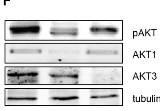
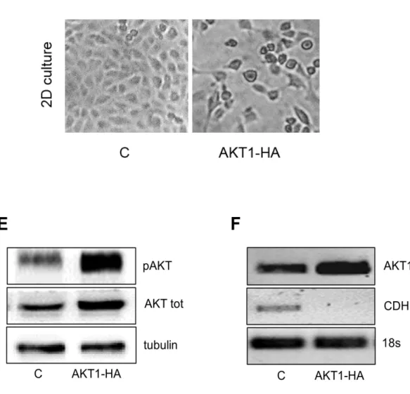
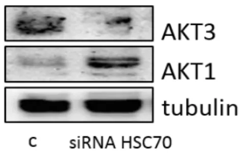
Recently our lab has firstly described that mesothelioma cells express the two isoforms of AKT, AKT1 and AKT3, while they completely lack of AKT2. The role and the relevance of AKT kinases in MPM are described, but no data are available on AKT1/3 expression and function in this kind of tumor. The PI3K/AKT signaling pathway, aberrantly active and involved in MPM cell cycle progression and chemo-resistance, represents a novel good therapeutic target. Various studies have also demonstrated that growth of tumor cells with elevated AKT activity is more sensitive to targeted
30
disruption of AKT signaling; moreover, these evidences suggest that targeting AKT activity sensitizes MPM cells to conventional drugs such as cisplatin, and that sensitivity after AKT inhibition might be more effective in cells with an elevated AKT activity. In a work recently published by our group, AKT was investigated as a relevant target of perifosine; it has been described that perifosine inhibits both AKT1 and AKT3 phosphorylation and activation in cells growing exponentially in serum-containing medium and after growth factor stimulation (98).
2.6.2. AKT in ovarian cancer
The involvement of AKT in cancer was firstly described in 1992 when Cheng et al. showed the hyperactivation of AKT2 in ovarian cancer cell lines. Ovarian cancer is the fifth most common cause of death due to cancer in women; therapy is successful at first, but the recurrence of the disease makes the rate of survival low. In ovarian cancer, the PI3K/AKT/mTOR pathway is frequently mutated or amplified, underlining its role in progression, cell proliferation, migration and chemoresistance of the tumor (99).
Different studies report the expression of AKT in ovarian cancer: in particular, AKT1 and AKT2 were found activated and/or over-expressed, while AKT3 was detected up-regulated in over 20% of primary ovarian tumors and involved in the regulation of the G2/M transition.
Tang et al. reported that AKT1 is frequently activated in ovarian cancer, while AKT2 has been shown to be amplified and overexpressed in human ovarian carcinoma cell lines and primary ovarian tumors, as described by Bellacosa and colleagues. Moreover, amplification of AKT2 was particularly frequent in undifferentiated ovarian tumors, suggesting a correlation between AKT alterations and tumor aggressiveness (100).
31
The main biological functions of AKT activation are anti-apoptotic and pro-proliferative in cancer cells; proliferation and invasion are also affected when AKT is directly targeted as well: silencing of AKT1 isoform reduces proliferation and invasion of OVCAR-3 cells, while targeting the AKT2 isoform has been shown to increase the activation of apoptosis (101).
2.6.3. AKT in prostate cancer
The PI3K/AKT/mTOR pathway has a pivotal role in prostate cancer and is estimated to be up-regulated in 30-50% of the cases, often associated with PTEN loss. Furthermore, phospho-AKT levels were also significantly greater in high-grade prostate tumors if compared to low/intermediate grade tumors, as different studies showed (102). In fact, according to the Gleason score, a grading assigned to a prostate tumor with values between 2 and 10, when lowest numbers indicate slow-growing tumor and the highest ones indicate an aggressive tumor, phospho-AKT was detected mostly in tumor samples with Gleason score ≥8 (103). Levels of phospho-AKT were significantly increased in cancer cells relative to normal prostate epithelium and benign prostatic hyperplasia. In normal tissue AKT isoforms are present in different percentage, with the prevalence of AKT2 and AKT3, while changes in expression and activation of AKT have been reported in prostate cancer. Recent studies showed that silencing of AKT2 led to an increasing of migration, allowing epithelial-mesenchymal transition (EMT); on the contrary, silencing of other two isoforms did not evolve in such consequences. Moreover, down-regulation of AKT1 and AKT2, but not AKT3, induced activation of cell surface β1-integrins and enhanced adhesion, migration and invasion; while silencing of AKT1 and AKT2 resulted in increased focal adhesion size (104).
32
2.6.4. AKT in lung cancer
Lung cancer is one of the most common human cancers and non-small cell lung cancer (NSCLC) represents around 80% of deaths caused by all primary lung cancers. Due to diagnosis at late stages of the disease and despite advances in surgery and other treatments, the 5-year rate of survival for patients with NSCLC is only 15%. The annual incidence worldwide is more than 1.3 million lung cancer cases, while more than 1.1 million lung cancer deaths per year are estimated (105). Researches based on molecular cancer biology and biochemical alterations evidenced that deregulation of some pathways involved in cellular proliferation and survival are responsible for cancer progression and among these, a pivotal role is played by the phosphoinositide-3-kinase/protein kinase B (AKT) signaling pathway.
NSCLC cells express all the three AKT isoforms; in accordance with results published by different groups, there is a prevalence of AKT1 activation in all NSCLC subtypes. Regarding AKT1 and AKT3 activation, no significant difference between expression in tumor cores and control cores was observed, whereas PTEN and PI3K showed a significant difference in expression; in addition, high expression of PI3K was observed in 29% of all tumor core (106). Recently new findings studies have reported a lack of association between activated AKT and survival or clinic-pathological variables such as stage of disease and metastasis. In 102 NSCLC cases, Tang et al. found that patients with concomitant p-AKT Ser473 expression and loss of PTEN had a significantly reduced 5-year survival rate, while other research groups have reported the prevalence of phosphorylated AKT and its relevance to prognosis in NSCLC. Moreover, others studies observed high non-phosphorylated AKT2 expression in epithelial cells and considered it a positive prognosticator (107).
33
2.6.5. AKT in breast cancer
Breast cancer is the most common malignancy among women, and it is the illness most frequently diagnosed in developed countries. Mutations in a number of genes are now known to cause susceptibility to breast cancer. In high-risk families, the most significant of these are the BRCA1 and BRCA2 genes, while the human epidermal growth factor receptor 2 gene (HER2) is overexpressed and/or amplified in ∼15% of breast cancer patients and has been identified as a marker of poor prognosis (108). Other genes are involved in the genesis of breast cancer as well; these are tumor suppressor genes as p53 and PTEN: in particular, mutations in components of PI3K pathway and AKT hyper-activation are found in 70% of breast cancers. Studies in transgenic mice have shown that AKT promotes mammary tumor progression by increasing cell survival. The majority of the currently available in vivo data agree with the overall model whereby AKT1 and AKT2 kinases play opposite roles in breast cancer migration, invasion and metastatic dissemination (events that ultimately determine the clinical outcome): as several studies reported, AKT1 functions as an inhibitor of migration and invasion in breast, while AKT2 acts in a positive manner (109). Moreover, AKT activation is associated with poor outcome in endocrine-treated breast cancer, whereas high levels of cytoplasmic AKT2 are associated with an improved overall survival. AKT signaling pathway regulates also IKKε, an inducible kinase and is amplified and overexpressed in breast, and in particular AKT2, but not the AKT1 or AKT3 isoform, is responsible for IKKε induction (110).
2.6.6. AKT in skin cancer
Malignant melanoma is a devastating tumor of the skin, with poor prognosis for patients (cause metastasis spread early) and resistant to all current treatments including chemo-, immuno-, or radiation therapy. In melanoma,
34
both the Ras/Raf/MEK/ERK (MAPK) and the PI3K/AKT (AKT) signaling pathways are constitutively activated and represent a crucial point for cancer development and progression; in particular, PTEN deletion leads to AKT activation that promote cell survival and can results in downstream loss of differentiation or senescence, through Raf-ERK cascades.
Members of the PI3K and AKT3 signaling cascades have been implicated in initiation, progression, invasive, and drug resistance phenotypes of melanomas. Increased phospho-AKT expression in melanoma is associated with tumor progression and shorter survival: recently, has been also described that AKT3 in early melanocytic lesions can phosphorylate
V600E
BRAF reducing its activity and promoting proliferation to overcome the senescence block (111).
Furthermore, the discovery of an AKT3 mutation in melanoma underlined the critical role of this isoform. Stahl et al (2004) described that AKT3 protein, but not AKT1 or AKT2, was increased in melanoma cell lines when compared to normal melanocytes, while other studies reported that PTEN loss cooperates in increasing melanoma cell and non-transformed melanocytes capabilities of invasion and migration, by enhancing AKT2 activity and through E-Cadherin down-regulation. Interestingly, targeting PTEN differentially regulates AKT3- mediated cell survival and AKT2-mediated metastasis in melanomas (112).
Expression of AKT3 has been detected elevated in cell lines derived from primary melanoma tumors at the radial and vertical stages of cell growth compared to normal melanocytes; on the contrary in the same samples, no significant changes were observed in the levels of AKT1 and AKT2, indicating that AKT3 expression is predominant in melanoma development. Although a central role of the AKT3 isoform in melanoma maturation is well established, this evidence still remain controversial: a recent report using biopsy of melanoma found AKT2 as a predominantly activated isoform in
35
melanomas, while a different study demonstrated that loss of PTEN promoted melanoma cell metastasis via activating AKT2 but not AKT1 or AKT3 (113, 114).
2.6.7. AKT in thyroid cancer
The Cowden’s syndrome, an autosomal dominant disorder characterized by the onset of various type of cancer affecting breast, colon, skin and thyroid, has been connected to the inactivation of the tumor suppressor PTEN, a phosphatase that regulates negatively the PI3K signaling, with the consequent constitutively activation of AKT. Although these observations, PTEN inactivation resulted to be sporadic in thyroid cancer, while AKT might be a central regulator of many thyroid oncogenes.
In normal thyroid cells AKT activation is the major mediator of cell growth and inhibition of apoptosis; overexpression of an active form of AKT1 in thyroid cells results in serum-independent growth and leads to resistance to cell death, although it seems insufficient to transform the thyroid cells: this evidence suggests that additional signaling pathways are needed for developing thyroid cancer. Other recent study demonstrated that AKT signaling is enhanced in human thyroid cancer, in particular that AKT1 and AKT2 protein levels are increased in follicular thyroid cancer if compared with normal thyroid tissue as well as the level of phospho-AKT. Finally, among the isoforms, AKT2 results having an important role in the pathogenesis and/or progression of thyroid cancer (115).
2.6.8. AKT in colon cancer
Colorectal cancer (CRC) represents one of the most common malignancies in the world; it undergoes a multistage carcinogenesis pathway from adenomatous polyps to carcinoma and despite advances in chemotherapy, this tumor remains a major cause of death. Genetic events that lead to
36
neoplastic processes include alterations in tumor suppressor genes that in normal conditions regulate cell cycle progression, or somatic mutations in the TGFb receptor and the K-ras oncogene (116).
Activation of AKT signaling and impaired expression of phosphatase and tensin homolog (PTEN) has been reported in 60-70% of human CRC and leads to inhibition of the pro-apoptotic GSK3b, with resulting increase in the levels of the anti-apoptotic beta catenin protein. AKT is up regulated in 57% of colon cancer, rate markedly higher than in other malignancies; moreover, AKT can act synergically with RAS and RAF cascades, both critical in colorectal carcinogenesis, and has been shown to impact also on WNT signaling, to regulate expression of the anti-apoptotic COX2 protein and to modulate cellular motility enhanced by prostaglandin E2. Although mutations in AKT genes are not frequently found in CRC, a somatic missense mutation of AKT1 (E17K) in the PH domain has been described and resulted in constitutive association of AKT1 with the plasma membrane, with its prolonged activation and mTOR deregulation. Finally, AKT proto-oncogene is overexpressed in cancer suggesting that its overexpression may be crucial in earliest stages of carcinogenesis process (117, 118).
2.6.9. AKT in glioma
Malignant glioma is the most common primary brain tumor in adults; it originates from the supportive cells of the brain (glial cells) and carries a poor prognosis. The median survival of patients with high-grade glioma (World Health Organization [WHO] grade III or IV) is 10-30 months despite multimodal treatment with surgery, radiation, and chemotherapy.
Malignant gliomas develop because of the consequent accumulation of alterations in genes regulating cell proliferation, differentiation, and apoptosis. The amplification and mutation of tyrosine kinase receptors (i.e.
37
EGFR) activates several signaling pathways including the PI3K/AKT pathway that delivers major survival signals to human malignant glioma. AKT is activated in 70% of gliomas and is usually associated with PTEN mutations. Gliomas express all the AKT isoforms in different ratios; according to the WHO classification, the expression of AKT1 protein was not associated with the pathological glioma grade, while AKT2 expression was notably higher in tissues from grade III and IV than in normal tissues. On the contrary, AKT3 protein diminished with increasing glioma grades (119).
Moreover, AKT3 was found down-regulated in malignant glioma cell lines overexpressing AKT2. In a recent study, Mure et al. observed the induction of apoptosis via the activation of caspase-9 and caspase-3 in glioma cell lines, after silencing of AKT2 or AKT3, while the combined knock-down of AKT2 and AKT3 effectively inhibited the growth of malignant cells, demonstrating the pivotal role of the two isoforms in the biology of this cancer (120).
2.7. AKT Inhibitors
Based on the evidence that AKT pathway has a pivotal role in various cellular functions and is one of the most frequently activated pathway in several type of cancers, the advent of targeted agents represents a new possibility to overcome the plateau achieved by current chemotherapic treatments.
The three isoenzymes of AKT are not functionally identical, but their sequence is still similar to other kinases of the AGC family (>90% homology among AKT isoforms and >50% with PKA-α and PKC-α). Moreover, when considering the homology between ATP-binding site in AKT1 and AKT2 we reach almost the 100% (96% of homology with AKT2/3 and more than 70% with PKA-α / PKC-α). All these observations underline how the