PhD in Clinical & Experimental Medicine
UNIVERSITÀ DEGLI STUDI DEL PIEMONTE ORIENTALE
Dipartimento di Medicina Traslazionale
Corso di Dottorato di Ricerca in Medicina Clinica e Sperimentale
Ciclo XXVIII
New strategies for tissue regeneration
SSD BIO/16
Coordinatore Tutor
Prof.ssa Marisa Gariglio Prof. Mario Cannas
Dottorando Dott.ssa Alessia Borrone
Table of contents
Pag.Abbreviations 1
1. General introduction 3
2. Aim of the project 8
3. Cartilage regeneration 10
3.1 Introduction 10
3.1.1 Cartilage tissue 12
3.1.2 Articular cartilage injury 15
3.1.3 Cartilage repair and regeneration 16
3.1.4 Stem cells 18
3.2 Materials and Methods 20
3.2.1 Materials 20
3.2.2 Chondrogenic organ culture model 21
3.2.3 Mechanical evaluations 22
3.2.4 Histology and immunohistochemistry 22
3.2.5 Glycosaminoglycan and collagen quantification 23
3.2.6 Real-time PCR 24
3.2.7 Isolation and culture of human Chondrocytes (hChs) 24
3.2.8 Effect of lipoaspirate on hCh proliferation and matrix production 25
3.2.9 Cell outgrowth from lipoaspirate 26
3.2.10 Cell phenotype characterization with flow cytometry analysis 27
3.2.11 Cell differentiation capacity 28
3.2.12 Transduction and Examination of Lenti-GFP lipoaspirates 29
3.2.13 Evaluation of the repair of damaged cartilage 29
3.2.14 Statistical analysis 30
3.3 Results and Discussion 31
3.3.1 Chondrogenicity of lipoaspirate 31
3.3.2 Effect of lipoaspirate on chondrocytes 34
3.3.3 Cell outgrowth from lipoaspirates: quantification, phenotype, differentiation capacity, and
cartilage repopulation. 35
4. Bone regeneration 43
4.1 Introduction 43
4.1.1 Bone tissue 44
4.1.2 Bone healing 46
4.1.3 Bone regeneration 48
4.1.4 Cellular morphological changes and Rho pathway 50
4.1.5 Lysophosphatidic acid (LPA) 53
4.2 Materials and methods 58
4.2.1 Cell culture and reagents 58
4.2.2 Isolation of collagen from rat tails 59
4.2.3 Silver Staining in acrylamide gels 60
4.2.4 Collagen matrix contraction 60
4.2.5 Fluorescence and confocal microscopy 61
4.2.5 Osteoblast proliferation and differentiation 62
4.2.6 Migration test 63
4.2.7 In vitro culture model of bone fracture 63
4.2.8 Mechanical properties 64
4.2.9 In vivo experiments 65
4.2.10 µ-CT 66
4.2.11 Statistical Analysis 67
4.3 Results and Discussion 68
4.3.1 Collagen characterization 68
4.3.2 Gel contraction 70
4.3.3 Human osteoblasts evidenced a Rho mediated actin cytoskeleton modifications 74 4.3.4 Human osteoblasts proliferation and differentiation in response to LPA 76
4.3.5 Human osteoblasts migration in response to LPA 77
4.3.6 LPA and D3 co-operate to expedite bone regeneration 78
4.3.7 In vivo experiment 81
4.4 Conclusions 83
5. References 86
6. List of Publications 101
1
Abbreviations
ACI: Autologous Chondrocyte Implantation ALP: Alkaline Phosphatase
ASC: Adipose Derived Stem Cells ATP: Adenosine Triphosphate ATX: Autotaxin
BCA: Bicinchoninic Acid
BCIP: 5-Bromo-4-Chloro-3-Indolyl Phosphate BM-MSCs: Bone Marrow Mesenchymal Stem Cells BSA: Bovine Serum Albumin
CAMP: Ciclic Adenosine Monophosphate DAPI: 4', 6-diamino-2-fenilindolo cloridrato
DMEM/F12: Dulbecco’s Modified Eagle Medium: Nutrient Mixture F-12 DMEM: Dulbecco’s Modified Eagle Medium
DMEM-HG: Dulbecco’s Modified Eagle Medium: High Glucose DMEM-LG: Dulbecco’s Modified Eagle Medium: Low Glucose ECM: Extracellular Matrix
EGF: Epidermal Growth Factor
ERK: Extracellular Signal Regulated Kinase FACS: Fluorescence-activated cell-sorting analysis FAFA: Bovine Serum Albumin Fatty Acid Free FBS: Fetal Bovine Serum
FGF: Fibroblast Growth Factors GAG: Glycosaminoglycans GDP: Guanosine Diphosphate GFP: Green Fluorescence Protein GPCR: G-Protein-Coupled Receptors GTP: Guanosine Triphosphate
2
hMSC: Human Mesenchymal Stem Cell hOB: Human Osteoblast
HSA: Human Serum Albumin
IMDM: Iscove’s Modified Duolbecco’s Medium
ITS: (1 mg/ml insulina, 0,55 mg/ml transferrina umana, 0,5 mg/ml selenito di sodio, 50 mg/ml albumina sierica e 470 μg/ml acido linoleico)
LIMK: LIM Kinase
LPA: Lysophosphatidic Acid LPLs: Lysophospholipids
MACI: Matrix-induced Autologous Chondrocyte Implantation MAPK: Mitogen Activated Protein Kinase
MLC: Myosin Light Chain MMA: Methylmethacrylate MSCs: Mesenchymal Stem Cells PBS: Phosphate Buffered Saline PI3K: Phosphatidylinositide 3-Kinase PKC: Protein Kinase C
PRP: Platelet Rich Plasma PS: Penicillin Streptomycin
PSF: Penicillina Streptomicina Fungizone PTH: Paratormone
RM: Regenerative medicine RNA: Ribonucleic Acid
ROCK: Rho-Associated Protein Kinase
SDS-PAGE: Sodium Dodecyl Sulphate - Polyacrylamide Gel Electrophoresis SVF: Vascular Stromal Fraction
TGF-β: Transforming Growth Factor beta TNF-α: Tumor Necrosis Factor alpha VEGF: Vascular Endotheliar Growth Factor WNT: Wingless-Int
3
1. General introduction
In the last years, tissue or organ transplantation changed reconstructive surgery field, improving the functionality of transplanted organ or tissue. These improvements were reached thanks to the understanding of the biochemical mechanisms that rule immune response of the host. Some limitations, such as the limited number of donors, the possibility of chronic rejection (that requires immunosuppressive therapies), the limited amount of suitable tissue for autotransplant and partial recovery of tissue functionality, has stimulated researcher to develop alternative strategies. Tissue engineering and regenerative medicine emerge in this context, using mostly biomaterials to repair or substitute damaged tissues. The idea of tissue regeneration is to reconstruct lost tissue or tissue function by a regenerative process. This is possible by:
1. Tissue Engineering that involve transplantation of cells in matrices. 2. Cell Transplantation that consist in injection of isolated cells. 3. Cell Homing trough an activation of resident cells.
The biomaterials used as scaffolds have to facilitate or even regulate cell processes as proliferation, migration and matrix synthesis and have to reinforce the defect (35). They should maintain the shape of the defect until the reconstruction process is finished. Additionally, the biomaterial should prevent the infiltration of surrounding tissue that may interfere with the tissue regeneration process. In some cases the biomaterials serve as a delivery vehicle for growth factors and genes that support cell processes for tissue reconstruction. It is within the framework of recent technological advances in cellular and molecular biology, engineering, materials science and especially nanotechnology that tissue engineering is
4
emerging to help fulfill a long-sought dream of using tissue-like materials for the replacement of tissue defects.
1.
Tissue engineering can be considered basically as a three step process (36-37),(Fig. 1):a. Cells of the desired phenotype are isolated from a small biopsy from the patient and amplified in number by means of classic cell culture techniques.
b. After the desired cell number is reached, the seeding of these cells into a scaffold is needed. The biomaterial gives the cells a temporary structure in which they further proliferate and differentiate in order to build functional organ-like units.
c. A maturation of the cell-biomaterial construct into a functional tissue mass is proceeded.
Biomaterials are grouped as synthetic (metallic, ceramic or polymeric surfaces) or natural materials, eventually biodegradable.
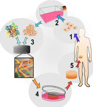
Figure 1: The tissue engineering cycle, using autologous cells. 1-A small number of cells are removed
from the body. 2-Cell proliferation. 3-Cells are seeded onto porous scaffolds. 4-Theseeded scaffolds are placed in culture to further increase cell number. 5-The regenerated tissue is implanted into the site of damage. (http://www.centropede.com/UKSB2006/ePoster/images/background/TE_model_large.jpg).
5
2.
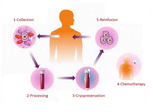
Cell transplantation consists in the collection of cells from patients or donor, processing ofthe cells, and the reinjection of cells in the patient (Figure 2).
Figure 2: The autologous transplant process. 1-Collection: stem cells are collected from the patient’s bone marrow
or blood. 2- Processing: Blood or bone marrow is processed in the laboratory to purify and concentrate the stem cells. 3-Cryopreservation: blood or bone marrow is frozen to preserve it. 4-Chemotherapy: high dose chemotherapy and/or radiation therapy in given to the patient. 5-Reinfusion: thawed stem cells are reinfused into the patient. (http://biomed.brown.edu/Courses/BI108/BI108_2007_Groups/group03/sources.html).
The cells can be mature cells or stem cells, like mesenchymal stem cells (MSCs). Stem cell transplantation is a generic term covering several different techniques. For allogeneic transplants, hemopoietic stem cells are taken from the bone marrow, peripheral blood, umbilical cord blood or adipose tissue of a healthy donor matched for HLA type, who may be a family member or an unrelated volunteer. For autologous transplants, usually, stem cells are taken from patients'own bone marrow or adipose tissue. Interestinghy, adipose tissue is readily available in large quantities and can be obtained through less invasive procedures; it contains up to 2% of MSCs (ASC) compared with only 0.02% in bone marrow (BM-MSC). Stem cells have been used routinely for more than three decades to repair tissues and organs damaged by injury
6
or disease. The prospect of exploiting stem cells more widely in regenerative medicine was encouraged by the growing evidence that various adult cells retained greater versatility than had been suspected to date (38). The aim is to employ stem cells as a source of appropriately differentiated cells to replace those lost through physical, chemical or ischaemic injury, or as a result of degenerative disease.
3.
The other possibility in regenerative medicine is cell homing by endogenous cells suppliedfrom the defect area, without the necessity of cell transplantation (Figure 3).
Figure 3:Homing and mobilization of native cells for bone regeneration. Homing factors are directly delivered to the defect site. Native cells may be mobilized by the administration of a stimulation factor .
Most insights (39) in the mechanisms underlying migration and homing are from studies that evaluated the migration into inflamed tissue of leukocyte (40), hematopoietic stem cells (HSCs) (41) and metastatic cancer cells (42).
7
A significant body of literature also exists related to the mechanism of MSCs migration towards target tissues and the role of cell surface receptors and molecules in the process. The role of activated endothelial cells in migration of MSCs is being also extensively studied. The migration of cells from the side of defect, together with an appropriate scaffold which can facilitate cell migration, could be important, for example, to speed-up bone healing and bone regeneration. Different approaches have been used to deliver homing factors to the fracture site. In scaffold-based tissue regeneration strategies, homing factors can be covalently bound or absorbed to the scaffold. Drug delivery systems such as hydrogels, microspheres, and nanoparticles have been used on their own or in combination with scaffolds and/or biomaterials. Of note, the carrier material has a significant impact on the release profile of the homing factors.
8
2. Aim of the project
My PhD project is divided in two parts, focusing on the development of new strategies for orthopedic tissue regeneration. In particular, the first part is about cartilage regeneration using human lipoaspirate as autologous injectable active scaffold for one-step repair of cartilage defects, the second part is about bone regeneration, through an injectable medicated graft substitute active on bone tissue regeneration.
I.
Cartilage regeneration
Research on mesenchymal stem cells from adipose tissue (ASC) shows promising results for cell-based therapy in cartilage lesions: in these studies cells have been isolated, expanded, and differentiated in vitro, before transplantation into the damaged cartilage, or onto materials used as scaffolds to deliver cells to the impaired area. The present study employed in vitro assays to investigate the potential of intra-articular injection of micro-fragmented lipoaspirate, as a one-step repair strategy; it aimed to determine whether adipose tissue can act as a scaffold for cells naturally present at their anatomical site. Cultured clusters of lipoaspirate showed a spontaneous outgrowth of cells with mesenchymal phenotype and with multi-lineage differentiation potential. Transduction of lipoaspirate clusters by lentiviral vectors expressing GFP underlined the propensity of the outgrown cells to repopulate fragments of damaged cartilage. On the basis of the results, which showed an induction of proliferation and extracellular matrix (ECM) production of human primary chondrocytes, it was hypothesized that lipoaspirate may play a paracrine role. Moreover, the structure of a floating culture of lipoaspirate, treated for three weeks with chondrogenic growth factors, changed: tissue with a high fat component was replaced by a tissue with a lower fat component and connective tissue rich in glycosaminoglycan (GAG)
9
and in collagen type I, increasing the mechanical strength of the tissue. From these promising in vitro results, it may be speculated that an injectable autologous biologically-active scaffold (lipoaspirate), employed intra-articularly, may: 1) become a fibrous tissue that provides mechanical support for the load on the damaged cartilage; 2) induce host chondrocytes to proliferate and produce ECM; 3) provide cells at the site of injury, which could regenerate or repair the damaged or missing cartilage.
II.
Bone regeneration
With the aim to obtain an injectable medicated scaffold, which speeds bone formation in sinus lift augmentation, in bony void and in fracture repair, we have developed a three-dimensional (3D) jelly collagen containing Lysophosphatidic acid (LPA) and 1a,25-Dihydroxyvitamin D3 (1,25D3) using soluble native collagen prepared from rat tail tendons. We have demonstrated with an in vitro 3D culture model of bone fracture an osteoblasts’Rho-kinase mediated contraction of the collagen that causes an approach of human bone trabecular fragments with the formation of new union tissue within 3 weeks of organ culture. The contraction was faster in LPA medicated collagen while 1,25D3 enhanced the mineralization of the new formed tissue that showed also increased tensile strength. LPA was shown to modulate gel contraction rate not only mechanically, working in cytoskeleton reorganization, but also osteoconductively evidencing activity on proliferation, differentiation and migration of human primary osteoblasts (hOB). When LPA was used in combination with 1,25D3 a synergism on hOB’s activity in term of alkaline phosphatase and mineralization was seen. On the basis of these data, collagen can be considered as an injectable natural scaffold that allows the migration of cells from the side of bone defect and its enrichment with LPA and 1,25D3 could be used in vivo to accelerate bone growth and fracture healing.
10
3. Cartilage regeneration
3.1 Introduction
The treatment of degenerative joints is becoming a very frequent necessity. Increased life expectancy and inappropriate medical treatments (e.g. removal of the meniscus) are leading to an increase of chondropathies and osteoarthritis, which also occurs at a relatively young age. Chondrocytes are the parenchymal cells of cartilage, and have limited regenerative capacity; thus after traumatic injury, the cartilage undergoes degenerative changes, which may be irreversible (18). The more superficial lesions tend to progress towards degeneration (29); those that penetrate the subchondral bone progress towards the formation of fibro-cartilage tissue. This differs from normal hyaline cartilage in both biomechanical and biochemical properties, and frequently undergoes subsequent degeneration (30). Any cartilage lesion should therefore be considered as the beginning of a chronic degenerative disease, with little chance of cure, due to the poor regenerative capacity of this type of cell (7).
Modern treatments for damaged cartilage and for the prevention of osteoarthritis include the intra-articular administration of hyaluronan (15), treatment with platelet rich plasma (PRP) (3), bone-marrow stimulation techniques (subchondral drilling, abrasion, micro-fracture) (24), osteochondral grafting (mosaicplasty) (23), autologous chondrocyte implantation (ACI), and matrix-assisted autologous chondrocyte implantation (MACI) with autologous chondrocytes cultured on collagen membranes prior to re-implantation (11). Chondrocyte-based therapy has demonstrated promising clinical results, but the procedure requires an invasive protocol, in which autologous cartilage is harvested from the patient at a healthy anatomical site. This produces damage to the normal cartilage at that site leading to pathological changes. It is
11
therefore evident that to avoid the disadvantages arising from the use of autologous chondrocytes, alternative cell sources must be investigated. Mesenchymal stem cells (MSCs) obtained from adult tissues of different origins (umbilical cord blood, adipose tissue, bone marrow, synovial membranes, periosteum and muscle) have recently been investigated, and their chondrogenic potential assessed and compared (28,34,14). Bone marrow and adipose tissue are the most readily available sources of MSCs. Moreover, adipose tissue is readily available in large quantities and can be obtained through less invasive procedures; of the many cell types contained in adipose tissue, MSCs (ASCs) comprise up to 2%, whereas only 0.02% of cells in bone marrow are MSCs (BM-MSCs) (20). MSCs have shown promising results for cartilage repair (31); however, in the studies published to date, cells were isolated from the originating tissue, then expanded and differentiated in vitro, prior to transplantation into the damaged cartilage or seeding in materials used as scaffolds to deliver cells to the injured area. No optimal stimulation regimen has yet been proposed to differentiate stem cells into chondrocytes prior to implantation; further difficulty lies in the large number of processing steps (enzymatic digestion, cell expansion and differentiation), which increases the translational barriers.
With the aim of avoiding excessive handling of stem cells and side effects derived from the scaffolds used for cell-based cartilage repair, this in vitro study investigated whether the intra-articular injection of autologous lipoaspirate might be an alternative cell-based therapy in treating cartilage diseases and in preventing osteoarthritis. Lipoaspirate is a natural scaffold rich in stem cells which, once injected into the intra-articular space, might give rise to different biological responses. To determine whether lipoaspirate can lead to the outgrowth of cells to the damaged cartilage, thus repopulating and repairing it, and whether lipoaspirate may have a paracrine effect on resident chondrocytes, we studied in vitro: 1) the ability of resident cells in
12
lipoaspirate to grow out from adipose tissue; 2) the phenotype of these outgrowing cells, together with their differentiation potential and their capability of repopulating an organ culture of human cartilage; 3) the effect of the lipoaspirate on the proliferation rate of human chondrocyte, and on their production of cartilaginous matrix. Furthermore, with the aim of determining whether lipoaspirate clusters might differentiate into a different tissue at the intra-articular site, thus providing intra-intra-articular mechanical reinforcement, the chondrogenic differentiation of cells not extracted from lipoaspirate, but simply at their own natural anatomical site, was examined.
3.1.1 Cartilage tissue
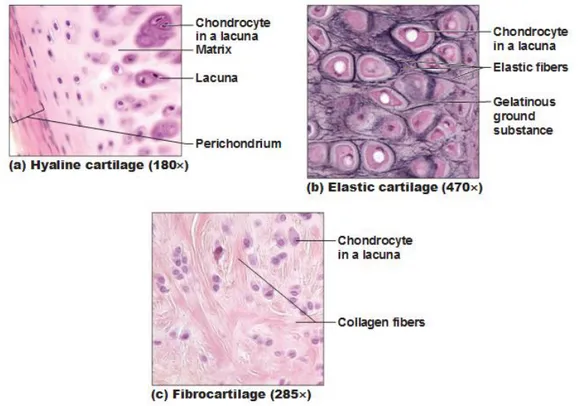
Cartilage is a connettive tissue consisting of a dense matrix of collagene fibres and elastic fibres embedded in a rubbery ground substance. The matrix is produced by cells called chondroblast, which became embedded in the matrix as chondrocytes. They occur, either singly or in group , within spaces called lacunae in the matrix. The surface of most of the cartilage in the body is surrounded by a membrane of dense irregular connective tissue called perichondrium. Cartilage doesn’t contain blood vessels or nerves, except in the perichondrium. There are three different type of cartilage that have slightly different structures and functions. They are hyaline cartilage, fibrocartilage and elastic cartilage (Figure4).
13
Figure 4:Types of Cartilage. (a) Hyaline cartilage provides support with some flexibility. (b)Elastic cartilage provides firm but elastic support. (c) Fibrocartilage provides some compressibility and can absorb pressure. September 27, 2011 By Antranik.
Hyaline cartilage is semi-transparent and appears bluish-white in colour. It is extremely strong, but very flexible and elastic. Hyaline cartilage consists of living cells, chondrocytes, which are situated far apart in fluid-filled spaces, the lacunae. There is an extensive amount of rubbery matrix between the cells and the matrix contains a number of collagenous fibres. Hyaline cartilage occurs in trachea, the larynx, the tip of the nose, in the connection between the ribs and the breastbone and also the ends of bone where they form joints. Temporary cartilage in mammalian embryos also consists of hyaline cartilage.
Fibrocartilage is an extremely tough tissue. The orientation of the bundles depends upon the stresses acting on the cartilage. The collagenous bundles take up a direction parallel to the cartilage. Fibrocartilage is found as discs between the vertebrae between the pubic bones in front
14
of the pelvic girdle and around the edges of the articular cavities such as the glenoid cavity in the shoulder joint.
Elastic cartilage is similar to hyaline cartilage, but in addition to the collagenous fibres, the matrix of the elastic also contains an abundant network of branched yellow elastic fibres. They run through the matrix in all directions. This type of cartilage is found in the lobe of the ear, the epiglottis and in parts of the larynx.
In humans, chondrocytes represent only about 1% of the volume of hyaline cartilage but are essential since are these cells that replace degraded matrix molecules, in order to maintain the correct size and mechanical properties of the tissue (43). Thus, microscopically, the cells’ endoplasmic reticulum and Golgi apparatus are prominent. In addition, many contain lipid and glycogen stores and secretory vesicles. Some chondrocytes have cilia that extend from the cell into the ECM and are believed to play a role in sensing the mechanical environment of the cell, since chondrocytes are known to modify matrix properties in response to loading (44).
Chondrocytes originate from MSCs found in the bone marrow of mature individuals. During embryogenesis, the MSCs start to differentiate into chondrocytes and secrete a cartilaginous matrix. During this time, the cells continue to divide. They pass through various lineage states and eventually the chondrocytes in the central zone, located next to what will soon be bone, enter the final stage of development and become hypertrophic chondrocytes, producing proteins that are important in the calcification of the matrix. Other chondrocytes (on the periphery) secrete collagen and matrix molecules in the right proportions to produce hyaline cartilage. Mature articular chondrocytes, unable to proliferate, appear rounded and are completely encased in matrix (44,45).
15
3.1.2 Articular cartilage injury
Articular cartilage has very limited capability to heal. Cartilage disruption results in progressive loss of joint motion, pain and disability, decreasing the quality of life of the affected person (46). Globally, the number and severity of cases are on ascending trend, increasing the burden of disease and taking a high toll on healthcare resources (47). Articular cartilage covers bone surfaces within a joint, functioning as a shock absorber and facilitates motion. Its structure is adapted to functioning under high compression, tensile and shear stress. This ultra specialized tissue is aneural, avascular and alymphatic with low cellularity, high extracellular matrix content and a stratified cell-fiber distribution. The nourishment of a relatively low cell population is provided by nutrient diffusion from subchondral bone vessels and synovial fluid. The same structural particularities, which are the basis of the remarkable biomechanical endurance, are considered to be the principal cause of the incapacity to heal after trauma and degeneration (48). There are three main types of cartilage injury: matrix disruption, partial thickness defects, and full thickness defects. Matrix disruption occurs from blunt trauma, such as dashboard injuries in automobile accidents. The ECM is damaged, but if the injury is not extreme, the remaining viable chondrocytes will increase their synthetic activity to repair the tissue. Partial thickness defects demonstrate disruption of the cartilage surface (fissures, etc.) but this does not extend to the subchondral bone. Immediately following the injury, nearby cells begin to proliferate, but for reasons that remain unclear, cellular attempts to fill the defect cease before it is repaired. Full thickness defects arise from damage that transverses the entire cartilage thickness and penetrates the subchondral bone. In this case, the defect is filled with a fibrin clot and a classic wound healing response ensues. With this type of injury, unlike the others, there is access to a population of progenitor cells from the bone marrow which can migrate to fill the defect (49,
16
50). These cells usually cause replacement of the fibrin clot with tissue intermediate between hyaline and fibrocartilage. This tissue is usually less stiff and more permeable than native cartilage, which could contribute to its eventual degradation over a period of months (50). Several factors may explain why cartilage demonstrates very little capability for self-repair after injury. Chondrocytes are not required to proliferate to maintain cartilage, as in other tissues (i.e. skin), and mature chondrocytes have a relatively low metabolic activity (44). Except in the case of full-thickness defects, there is no direct access to progenitor cells (such as are found in bone marrow) and the resident chondrocytes may be impeded by the ECM from migrating to fill the defect (49,51). In addition, the proteoglycans in the ECM can prevent cell adhesion, further undermining the native repair process (51).
3.1.3 Cartilage repair and regeneration
The attempt to cure the cartilage injury remains still frustrating to modern orthopedic surgery (52). Different methods of grafting make use of autologous or allogeneic cartilaginous or non-cartilaginous tissues to fill joint surface defects (53). Procedures that stimulates subchondral bone, such as subchondral drilling, microfracture, and abrasion arthroplasty are used to create controlled microlesions within the osseous tissue underlying the defect. The blood clot produced by such procedures is rich in bone marrow progenitors and is thought to induce cartilage repair (54). These interventions are capable to generate short term satisfactory results reducing pain and restoring mobility. Their major drawback is the poor quality of the repair tissue. The fibrous cartilage does not reproduce the structural organization and the mechanical endurance of the native hyaline joint surface. Such repair is prone to progressive clinical and morphological deterioration followed by the generalized joint failure (55). Regenerative medicine (RM) is
17
introducing the concept of complete structural and functional restoration of tissues, organs and systems made possible by the use of breakthrough discoveries in the field of molecular cell and developmental biology (56). It is sought that using RM specific tools, complete structural regeneration and functional rehabilitation of affected joint would be possible. Biological joint restoration and complete re growth of hyaline cartilage could address both traumatic defects as well as tissue deterioration. To this end, a wealth of basic and translational research is currently going on. Specific RM strategies, cell therapy, tissue engineering or gene therapy are in different stages of preclinical testing or clinical applicability. Introduced in the early 1990’s by a Swedish group, autologus chondrocyte implantation (ACI) uses a cell suspension of autologus adult cells to treat focal cartilage defects (57). A development of ACI, the matrix assisted chondrocyte implantation/transplantation (MACI/MACT), can be considered a form of cartilage engineering (Figure 5).
Figure 5: Major principles for the regenerative treatment of cartilage lesions.a) During ACI, chondrocytes are harvested from a small cartilage biopsy sample, cultured and implanted as a cell suspension into a cartilage defect. To prevent a leakage of cells, the defect is covered by a periosteal flap or collagen sheet. b) During MACI, isolated and culture-expanded chondrocytes are seeded into matrices. Besides other advantages, such as homogeneous cell distribution, initial mechanical stability and simplified surgical handling, over ACI, such grafts can be fixed onto subchondral bone, which, therefore, enables a stable fixation in diffuse defects. (Nature Reviews Rheumatology 8, 493-498 (August 2012)).
18
With this procedure, cells are delivered to the cartilage defect by means of a three-dimensional matrix thereby improving their retention and enhancing their biological activity at implantation site (58). Currently several scaffolds and methods of cell seeding are commercially available (59, 60). However, the clinical advancement of adult cell based therapy is limited by technical inconveniences, cell source related obstacles as well as by the availability and affordability of the procedure. Having an increased proliferative and differentiation potential, MSCs are looked upon as a suitable cell source for cartilage regeneration. Case series of succesful use of MSC to treat cartilage defects are reported (61). Several clinical trials using autologus or allogeneic MSCs for cartilage regeneration are going on (62).
3.1.4 Stem cells
Stem cells have been used routinely for more than three decades to repair tissues and organs damaged by injury or disease. The prospect of exploiting stem cells more widely in regenerative medicine was encouraged by the growing evidence that various adult cells retained greater versatility than had been suspected hitherto (38). The aim is to employ stem cells as a source of appropriately differentiated cells to replace those lost through physical, chemical or ischaemic injury, or as a result of degenerative disease.
The ideal stem cell for use in functional tissue engineering needs to be abundantly available, harvested with minimal morbidity, differentiated reliably down various pathways and able to be transplanted safely and efficaciously. Stem cells exist in an undifferentiated state, and exhibit both the capacity to self-renew, and the capability to differentiate into more than one type of cell (73). Although embryonic stem cells seem to exhibit unlimited differentiation potential both
19
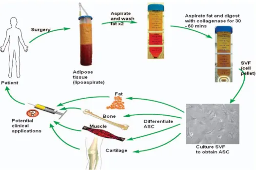
not generally available in current medical practice or research. Stem cells from adult tissue, on the other hand, suffer from few such restrictions. Multipotent stem cells can be isolated from various mesenchymal tissue sources in adults, most commonly bone marrow. The harvest of BMSC has practical constraints. These include pain at the harvest site, and harvest of only a small volume of bone marrow (and therefore a small number of stem cells), meaning that they are likely to require ex vivo expansion of the cells to obtain clinically significant cell numbers. Umbilical cord blood (UCB) from the placenta of term infants also contains mesenchymal stem cells. The use of UCB has been limited by the practical difficulties in obtaining and isolating the mesenchymal stem cells at the time of birth and ensuring adequate long-term storage for autologous use, and the fact that their differentiation potential seems to be lower than that of BMSC (63). Adipose tissue is derived from embryonic mesoderm. As MSCs, the isolated cells are capable of forming bone, cartilage and muscle, as well as fat (64-68) (Figure 6). These stem cells have been variously termed pre-adipocytes, stromal cells, processed lipoaspirate cells, multipotent adipose-derived stem cells, and adipose-derived adult stem cells. A recent consensus reached by investigators at the 2004 conference of the International Fat Applied Technology Society has settled on the term ‘adipose-derived stem cells’ (ASC) (69). Abundant numbers of ASC can be derived from lipoaspirate, the waste product of liposuction surgery. The yield compares favourably with a bone marrow aspirate (70). Compared with BMSC, ASC are more easily cultured and grow more rapidly (71, 72). They can also be cultured for longer than BMSC before becoming senescent (63). All of these qualities make ASC a useful source of mesenchymal stem cells.
20
Figure 5: Summary of cycle of human adipose-derived stem cell (ASC) isolation and differentiation for clinical usage. SVF, stromal vascular cell fraction.
3.2 Materials and Methods
3.2.1 Materials
Lipoaspirates were obtained from five healthy female patients (age range 30-45) undergoing an elective liposuction from a single anatomical site (abdominal subcutaneous fat tissue) at the IMAGE Institute (Milan, Italy). The ethical committee of the University of Milan approved the design of the study. Exclusion criteria were: body mass index (BMI) >30, diabetes, hypertension, and nicotine or alcoholic abuse. Written informed consent, specifying that residual material destined to be disposed of could be used for research, was signed by each participant before the biological materials were removed, in agreement with Rec(2006)4 of the Committee of Ministers Council of Europe on research on biological materials of human origin. After a preliminary infiltration of 400ml of saline solution, with adrenaline 2g/ml (S.A.L.F. Spa., Bergamo, Italy) as a vasoconstrictor and lidocaine 0.02% (AstraZeneca, Luton, UK) as an
21
anesthetic, the aspiration was performed using a 10cc syringe with a Luer-Lok tip (BD Medical, VWR Int., Milano, Italy) connected to a disposable 19 cm blunt cannula (3mm OD), with 5 oval holes (1x2mm). From each of five patients, 210 ml of lipoaspirate were obtained and processed as follows, giving four batches of lipoaspirate. 10 ml were washed with 25 ml phosphate buffered saline (PBS) (Sigma-Aldrich, Milan, Italy) on a 250 m Nitex filter (Dutscher Scientific, Essex, UK) and used as control (CTR); 50 ml were centrifuged for 3 minutes at 1500 x g (22) and 10 ml used as Coleman (Col); 50 ml were processed with a commercial device (PureGraft Cytori Therapeutics, San Diego, CA, USA) according to the manufacturer’s instructions (25) and 10 ml used (PurGr); 100 ml were processed with a commercial device (Lipogems, Lipogems Int. S.p.A., Milano, Italy) that washes and micronized lipoaspirate (4) following the manufacturer’s instructions and 10 ml used (LipoG).
3.2.2 Chondrogenic organ culture model
500 l of each batch of lipoaspirate were cultured as free-floating aggregates, in a conical polypropylene centrifuge tube (EuroClone, Milan, Italy) in 4 ml of chondrogenic medium containing 1 ml Dulbecco’s Modified Eagle Medium (DMEM–high glucose Sigma-Aldrich), ascorbate-2-phosphate (100 M, Sigma), dexamethasone (10−7 M, Sigma) and 1% Insulin-Transferrin_Selenite (ITS+1 media supplement, Sigma) for 3 weeks at 37°C in a humidified atmosphere containing 95% air and 5% CO2. The tubes were gently shaken daily, to allow the culture media to cover the sample, which otherwise tended to float. Half of the culture medium was changed twice a week with the addition of fibroblast growth factor 2 (FGF2) and transforming growth factor beta 2 (TGF2) found in previous research to be chondrogenic on bone marrow stem cells (6). Replicate aggregates from each batch of lipoaspirate were
22
processed for the following determinations: histological and immunohistological evaluation, mechanical evaluations, biochemical studies, RNA extraction, and RT–PCR analysis. Before processing, digital images were taken of each lipoaspirate organ culture using a digital camera, on samples treated or not for three weeks with the chGF.
3.2.3 Mechanical evaluations
The compression properties of untreated and chGF-treated lipoaspirates were tested using an Electro Force BioDynamic Test Instrument (Bose, TA Instruments, Eden Prairie, MN, USA). Samples of 2 ± 0.5 mm3 were positioned inside the instrument’s bioreactor chamber between two pistons. Tests were performed with a constant crosshead speed of 0.05 mm/sec. The instrument showed the real-time displacement of the piston, and the force acting on the sample, producing a force/displacement curve. Compressive strength and Young modulus were then calculated, using the surface area of the sample, initial length of the sample, compressed length, and force at breaking point. All samples were tested in triplicate, and data are presented as mean ± standard deviation (SD).
3.2.4 Histology and immunohistochemistry
After 21 days of culture, neo-formed tissues were rinsed in PBS and fixed overnight in 10% buffered formalin (Diapath S.r.l., Martinengo, Italy) at 4°C. The fixed tissues were dehydrated by treatment with alcohol, embedded in paraffin, and sectioned to a thickness of 7 m. For histological evaluation, sections were deparaffinized using xylene, rehydrated, and stained with Hematoxylin/Eosin (H&E Sigma-Aldrich) or Toluidine Blue (Sigma-Aldrich) to detect proteoglycan, or with Sirius Red F3BA (Chroma, Stuttgart, Germany) to detect collagen.
23
Alternate sections were used for immunohistochemistry to detect vessels: rehydrated sections were treated with 0.5% H2O2 (Sigma-Aldrich) to inactivate endogenous peroxidases, incubated in 1 mg/ml pepsin (Sigma-Aldrich) in 0.5 M acetic acid (Sigma-Aldrich) for 2 h at 37°C, blocked with 1.5% goat serum (Vector Laboratories, Burlingame, CA, USA) in PBS for 1 h at room temperature and incubated for 1 h at room temperature with anti Von Willebrand factor 25
g/ml (DAKO, Glostrup, Denmark) and FITC-conjugated secondary antibody 10 g/ml (Vector Laboratories).
3.2.5 Glycosaminoglycan and collagen quantification
2 mg of weighed tissue were taken after 21 days of culture, washed with PBS, and digested with 200 l papain (1 g/ml in 50 mM NaH2PO4, pH 6.5, containing 2 mM N-acetyl cysteine and 2 mM EDTA, all from Sigma-Aldrich) for 16 h at 65°C. Glycosaminoglycan were quantified by reaction with 1.9-dimethylmethylene blue (16 g/ml DMMB, Sigma-Aldrich) (12) using chondroitin sulphate (Sigma-Aldrich) as a standard. Measurements were made with a microplate reader (BS 1000 Spectracount, Packard BioScience Company, Meriden CT, USA) at a wavelength of 530 nm. The results are reported as mean ± SD (n = 3) g sGAG content/mg of the tissue and as % increase versus untreated sample (21 days’ culture in basal medium). Collagen was quantified according to Tullberg-Renert & Jundt (32). Briefly, 2 mg of tissue were extracted overnight at 4°C in 0.5 ml of acetic acid 0.2%, air dried and stained with 1 ml Sirius Red dye (Sigma-Aldrich) reagent (1mg/ml in saturated aqueous picric acid, Sigma-Aldrich) for 1 h. The dye solution was then removed, and the tissue was washed with 0.01 N hydrochloric acid (Sigma-Aldrich) to remove all non-bound dye; the stained material was dissolved in 0.2– 0.3 ml 0.1 N sodium hydroxide for 30 min at room temperature. The optical density (OD) was
24
measured with a BioRad microplate reader 3550 (BioRad, Milan, Italy) at 550 nm against 0.1 N sodium hydroxide as blank. The results are reported as means SD (n=3) g collagen/ml and as % increase versus the untreated sample (21 days of culture in basal medium).
3.2.6 Real-time PCR
Total cellular RNA was extracted using a Quiagen QIAsshredder kit (Qiagen, Dusseldorf, Germany) and quantified using a RiboGreen RNA quantification assay (Molecular Probes, Invitrogen, Milan, Italy). 200 ng were reverse transcribed (RT) using a High-Capacity cDNA Archive Kit (Applied Biosystems, Foster City, CA, USA). The RT thermal profile was 25°C for 10 min, 37°C for 120 min, 85°C for 5 min and kept at 4°C. Primers and probe were provided by TaqMan gene expression assay kit (Applied Biosystems) and were added to the reaction mixture according to the manufacturer’s directions: SOX-9 (Hs01001343_g1), COL2A1 (Hs00264051_m1), COL1A1 (Hs00164004_m1), GADPH (Hs02758991_g1). Amplification reactions were performed with SSOFast Probes Supermix with ROX (BioRad, Milan, Italy). mRNA levels were measured by real-time RT-PCR based on TaqMan methodology, using CFX96 Real-Time System C 1000 Thermal Cycler (Bio Rad, Milan, Italy). Real-time data analysis was run with BioRad CFX Manager 2.1 software. Target gene expression was normalized to GADPH mRNA expression. Relative differential gene expression was calculated (27).
3.2.7 Isolation and culture of human Chondrocytes (hChs)
Normal human articular cartilage was obtained from the femoral or radial heads of three male subjects with displaced fractures. The mean age of the group was 33 years (range: 20–41).
25
Written consent was signed by each participant before biological materials were removed according to Rec(2006)4 of the Committee of Ministers Council of Europe and used after local ethics committee approval. Immediately after surgery performed at the Traumatology division of the Major Hospital of Novara, healthy cartilage was taken under sterile conditions, washed with PBS supplemented with 50 U/ml Penicillin, Streptomycin 50 g/ml and 250 ng/ml Fungizone (PSF, all reagents from Sigma-Aldrich) and cut into pieces of approximately 2 x 2 mm, before enzymatic digestion, as follows and according to Archer et al (1): pieces were placed for 30 min at 37°C, 5% CO2 in DMEM/F12 (EuroClone) containing Trypsin (0.25% w/v, Sigma-Aldrich) and PSF (100-120 rpm shaking); the supernatant was discarded and the cartilage fragments were further digested in DMEM/F12 with PSF containing 0.8 mg/ml collagenase II (Sigma-Aldrich) for 4 hours at 37°C, 5% CO2 (100-120 rpm shaking). The digested tissue was then allowed to settle and the supernatant containing cells was centrifuged at 1500g for 5 minutes. The cell pellet was washed with PBS-PSF and re-suspended in growth medium: DMEM/F12 supplemented with PSF, glutamine 200mM (EuroClone), ascorbic acid 50g/ml (Sigma-Aldrich), FBS 2% (EuroClone), TGF 1ng/ml (Sigma-Aldrich), FGF2 1ng/ml (Sigma-Aldrich), IST+ 10g/ml (Sigma-Aldrich), seeded in monolayer in culture plates at low density (4000 cell/cm2) and incubated in a CO2 incubator at 5% CO2, 37°C.
3.2.8 Effect of lipoaspirate on hCh proliferation and matrix production
Chondrocytes were used for the experiments within the third expansion treatment. They were plated at a density of 103 cells/well in a 96-multiwell plate and hChs were cultured in basal culture medium (DMEM/F12 supplemented with antibiotics, glutamine ascorbic acid, FBS 2%, TGF 1ng/ml, FGF2 1ng/ml IST+ 10g/ml as described for hChs culture and abbreviated CTR)
26
were compared to cells cultured in lipoconditioned culture medium made up of basal culture medium supplemented with 15% of lipoaspirate culture medium that was obtained as follows: 1 ml of lipoaspirate corresponding to 850±20 mg of tissue, was cultured in 10 ml of basal medium in a CO2 incubator at 5% CO2, 37°C for 4 days; upon this treatment, extracellular proteins were released into the medium. Chondrocytes were cultured in basal or in lipoconditioned culture medium for 1, 3, 6 and 12 days; after each incubation time cell proliferation and cartilaginous matrix production were assayed.
For the proliferation assay, an ATP quantification Kit (ViaLight, Cambrex Profarmaco, Milan, Italy) was used following the manufacturer’s protocol; briefly, cells were lysed with cell lysis reagent, supplied with the kit, and treated with ATP monitoring reagent, which utilizes luciferase and was supplied with the kit. The light produced was measured by a luminometer (PerkinElmer) and expressed as relative luminescence units (RLUs) (9). For the cartilaginous matrix production assay 0.1 ml of each standard or culture medium to be tested were added to 0.1 ml DMMB dye in a 96 microplate wells; they were read in a micoplate reader at A525.
3.2.9 Cell outgrowth from lipoaspirate
The outgrowth of cells from Col and LipoG clusters was studied by quantifying, on the bottom of the well, the number of cells that had grown out from 50 mg of tissue/well, when the lipoaspirate was cultured in floating conditions in a 24 multiwell dish, from time 0 to 18 days. The outgrowth study was done also in a 3D matrix, as follows: to 50 l of collagen solution, obtained from rat tail tendon (10), a cluster of lipoaspirate was added, prior to gel formation, which was achieved within 1 hour of incubation at 37°C in culture medium (DMEM with 10% FBS, 100 U/ml penicillin, 100 μg/ml streptomycin and 2 mM L-glutamine all from
Sigma-27
Aldrich). The kinetics of cell outgrowth, and the number of cells/mg tissue, were evaluated by quantifying 4',6-diamidino-2-phenylindole (DAPI, Sigma-Aldrich)-positive cells. Briefly, 3D collagen gels were fixed in formalin 4% for 1 hour at room temperature, and stained for 10 minutes with DAPI 300nM (Molecular Probes), before counting nuclear positivity by fluorescence microscopy (Leica Microsystems, Milan, Italy).
3.2.10 Cell phenotype characterization with flow cytometry analysis
To determine whether the mechanical and enzymatic treatments, normally used to isolate stem cells from adipose tissue for regenerative medicine applications, may be avoided, the phenotype of cells obtained by outgrowth from the clusters was compared to that of cells obtained as follows. After digestion of lipoaspirate with type I collagenase 1mg/ml in PBS (Sigma-Aldrich) at 37°C for 30-60 min, the stromal-vascular fraction (SVF) containing ASCs was obtained through centrifugation for 10 min at 450 x g. Cell pellets were re-suspended in red blood cell lysis buffer (2.06 g/L Tris base pH 7.2, Sigma-Aldrich and 7.49 g/L NH4Cl Sigma-Aldrich) and incubated at RT for 10 min, to remove any remaining erythrocytes. Pellets were collected and filtered sequentially through 100- and 40-m cell strainers (VWR Int., Milano, Italy) to remove undigested tissue. The pellets were then washed and the cells re-suspended in DMEM-Ham’s F-12 medium (vol/vol 1:1) supplemented with 10% FBS, 100 U/ml penicillin, 100 μg/ml streptomycin and 2 mM L-glutamine, and plated in a 75 cm2 tissue culture flask coated with collagen (Thermo Scientific, Waltham, MA, USA). ASCs were self-selected out of the SVF, based on their adherent phenotype, during subsequent tissue culture passages, after which they were maintained in a humidified atmosphere with 5% CO2 in an incubator at 37°C. Cells were incubated with 1 g/106 cells of fluorescent antibodies for 40 min at 4°C in the dark. The
28
antibodies used were: anti-CD146 (10μg/ml), anti-CD105 (10μg/ml), anti-CD90 (10μg/ml), anti-CD73 (5μg/ml), anti-CD45 (5μg/ml), anti-CD44 (3.5μg/ml), anti-CD34 (7.5μg/ml), and anti-CD31 (2.5μg/ml) (all from BioLegend, San Diego, CA, USA). After washing, cells were analyzed on a flow cytometer (FACSAria, BD Biosciences, San Jose, CA, USA) by collecting 10,000 events, and the data were analyzed using the FACSDiva Software (BD Biosciences).
3.2.11 Cell differentiation capacity
To confirm the mesenchymal activity of those cells that grew out naturally from the lipoaspirate clusters, while under culture conditions, their in vitro differentiation capacity was studied according to Noel et al (26). Briefly, 10 x 103 cells/cm2 were cultured in DMEM-low glucose supplemented with 10% FBS, 0.5 mM isobutyl-methyl xanthine (IBMX, Sigma-Aldrich), 200 μM indomethacin, 1 μM dexamethasone, and 10 μg/ml insulin (all from Sigma-Aldrich); after 2 weeks they were stained with fresh oil red-O solution (Sigma-Aldrich) for adipogenesis; after 3 weeks of culture in DMEM-low glucose supplemented with 10% FBS, 10 mM β-glycerophosphate, 0.2 mM ascorbic acid, and 10 nM dexamethasone (all from Sigma-Aldrich), for osteogenic differentiation, cells were stained with Calcein solution (Sigma-Aldrich) to evidence mineralization (16) and with 5-Bromo-4-chloro-3-Indolyl phosphate (BCIP, Sigma-Aldrich) to evidence alkaline phosphatase activity (8). To demonstrate chondrogenic differentiation, pellets of 5 x 10⁵ cells were cultured for 3 weeks in chondrogenic medium (Lonza, Cologne, Germany), formalin-fixed, embedded in paraffin, and immunostained for Type II collagen by incubation with rabbit polyclonal anti-collagen type II primary antibody 1
g/ml (Biogenesis, Oxford, UK) and with secondary antibody conjugated to peroxidase (Vector Laboratories).
29
3.2.12 Transduction and Examination of Lenti-GFP lipoaspirates
High-titer VSV-pseudotyped lentiviral vectors (LV) were produced in human embryonic kidney 293T cells (ATCC® CRL-3216) by transient transfection with the lentiviral transfer vector construct pCCLsimPPT.PGK.GFP.WPRE (Lenti-GFP) and purified by ultra-centrifugation, as described by Follenzi et al (13). Expression titers for green fluorescent protein (GFP)-expressing vectors were determined on 293T cells by fluorescence-activated cell-sorting (FACS) analysis (FACSCalibur; BD Biosciences Immunocytometry Systems, San Jose, CA, USA) and was between 5 × 10⁹ and 1010 transducing units (TU)/ml. Clusters of lipoaspirate were transduced by Lenti-GFP into serum-free DMEM for 24 hours at 37°C at several concentrations of LV particles (108-106 TU/ml), and the most efficient concentration was selected after 48 hours by scoring GFP-positive cells using fluorescence microscopy. The transduction efficiency was determined by counting GFP-positive and -negative cells in at least 10 representative high-power fields against DAPI (DAPI; D-1306; Molecular Probes).
3.2.13 Evaluation of the repair of damaged cartilage
GFP-LipoG clusters were co-cultured with an organ culture of healthy or mechanically damaged cartilage, obtained from head of the femur of one of the three fracture cases, and cultured in a 24-well plate in DMEM/F12 supplemented with PSF, glutamine (200mM), ascorbic acid (50g/ml), FBS 2%, TGF (1ng/ml), FGF2 (1ng/ml), and IST+ (10g/ml). At 1, 3, 6, 12 and 24 days after the beginning of the culture, fluorescence microscopy was used to detect whether GFP-positive cells had migrated out from the lipoaspirate, to populate the cultured cartilage. To determine whether cells originating from the lipoaspirate (GFP positive cells) that had repopulated the damaged cartilage, had differentiated into chondrocytes, SOX-9-Texas Red
30
(Santa Cruz Biotechnology, Heidelberg, Germany) immunostaining was applied. The repopulated cartilage was formalin fixed and after 1 h incubation in 5% goat serum and 0.2% Triton X-100 (Sigma-Aldrich) in PBS it was incubated 1 h at room temperature with a rabbit polyclonal antibody for SOX-9 2g/ml (Santa Cruz Biotechnology, Heidelberg, Germany) and a Texas Red conjugated secondary antibody 10g/ml (Vector Laboratories).
3.2.14 Statistical analysis
The data were analyzed statistically using the SPSS for Windows software package (Statistical Package for Social Science, IBM, Armonk, NY, USA). Multiple comparisons of data were performed using one-way analysis of variance (ANOVA) and Bonferroni post hoc tests were applied to evaluate differences in mechanical properties, biochemical results, real-time PCR, and FACS. The p value was obtained from the ANOVA table; the conventional p≤0.05 level was considered to reflect statistical significance.
31
3.3 Results and Discussion
3.3.1 Chondrogenicity of lipoaspirate
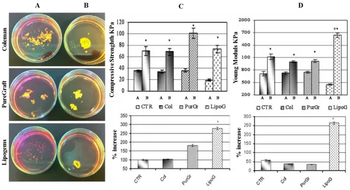
Morphological, mechanical, biochemical and molecular characterization showed that treatment of the lipoaspirate organ culture with the combined chondrogenic growth factors, induced the formation of a tissue completely different from the starting material and from that treated for 3 weeks in basal medium. Comparing the four lipoaspirates obtained with the four techniques described above, the washed lipoaspirate and that obtained with Col technology showed similar behavior, while that obtained with the LipoG technique produced the most significant changes in cellular organization, in mechanical properties of the new-formed tissue, and also in its microscopic and molecular aspects. From the macroscopic image (Fig. 6 A and B) it can be seen that the structure of all lipoaspirates changed after 3 weeks of treatment with chondrogenic factors; the compactness of the tissue had increased in all cases. However, the Col and PurGr lipoaspirates started from a tissue with a more compact texture than that of the LipoG batch; before treatment, the latter was characterized by individual isolated micronized fat globules of 0.40.1 mm2 whereas Lipoaspirate, Col and PurGr batches contained clusters of 1.80.9 mm2. The new-formed tissues also showed statistically-relevant differences in mechanical properties, measured under compression, using a Bose Electoforce instrument. The aggregation of the starting tissue was particularly evident in the case of the LipoG which generated a compact structure, with about 270% increase compared to the starting value in both tensile strength (Fig. 6C) and Young modulus (Fig. 6D). In Figures 6C and 6D are indicated with A samples before
the 3 weeks of culture in the chondrogenic model and with B samples after the 3 weeks of culture
32
Figure 6: Macroscopic results (A before; B after 3 weeks’ culture with chondrogenic factors). C and D show mechanical results, respectively as Compressive Strength and Young Modulus. The upper bar graph shows the results of samples before treatment (A) and after 3 weeks’ culture in the chondrogenic model (B); the lower graph shows the % increase following treatment. Results are expressed as mean ± SD of two experiments for each patient (n=10). *p<0.05 sample B compared to sample A. °p<0.05 compared to the other samples.
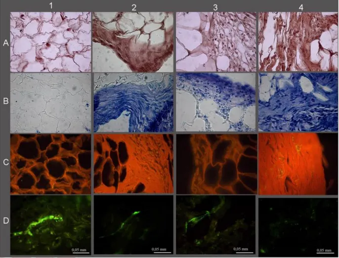
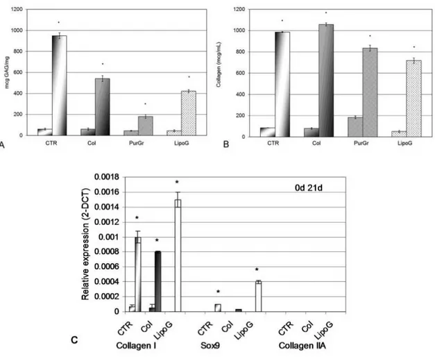
The histological and immunohistological analysis also showed considerable tissue modification (Fig. 7) with a reduction of the fatty component in favor of a connective tissue rich in GAGs (Fig. 7B), in collagen (Fig. 7C), and with a reduced presence of blood vessels (Fig. 7D). Comparing the histology of the lipoaspirates obtained with the four techniques tested the LipoG batch (Panel 4, Fig. 7) underwent the most significant changes in cellular organization, with only a few areas of adipose tissue, the remainder being almost completely replaced by collagen and GAGs. These data were confirmed by the biochemical quantifications, shown in Figure 8A for GAG and Figure 8B for collagen. Since the PurGr batch of lipoaspirate gave the poorest performance in the preliminary experiments for the remainder of the study only the CTR, Col and LipoG batches were employed. Real-time PCR showed that, before treatment, all three
33
lipoaspirates expressed similarly low levels of collagen type I, whereas none expressed mRNA for collagen type II, nor for Sox9, at 40 cycles of PCR reaction. After three weeks’ treatment in chondrogenic culture medium, the mRNA of lipoaspirate clusters was altered in a similar direction, with an increase of gene expression of collagen type I, and a small increase of gene expression of Sox9 (Fig. 8C). The increase in Sox9 gene expression was particularly evident in the LipoG batch. No collagen type II was detected.
Figure 7: Histological and histochemical results; A-Hematoxylin/Eosin; B-Toluidine Blue; C-Sirius Red; D-VonWillebrand. 1- CTR lipoaspirate before chondrogenic treatment; 2, 3, and 4- respectively Col, PurGr, and LipoG batches after 3 weeks’ treatment with chondrogenic factors.
34
Figure 8: Biochemical quantification of GAG expressed as mg GAG per mg tissue (Figure 8A) and of
total collagen expressed as absolute value in µg/mL (Figure 8B). Expression of chondrocytic markers in lipoaspirate tissue extracts normalized with GAPDH and reported as relative differential gene expression (Figure 8C). For each sample, the reported values are from samples before (first column) and after (second column) chondrogenic treatment (21 days’ culture in chGF enriched chondrogenic medium) *p<0.05 compared to the sample before chondrogenic treatment. The results are given as means ± SD (n = 3).
3.3.2 Effect of lipoaspirate on chondrocytes
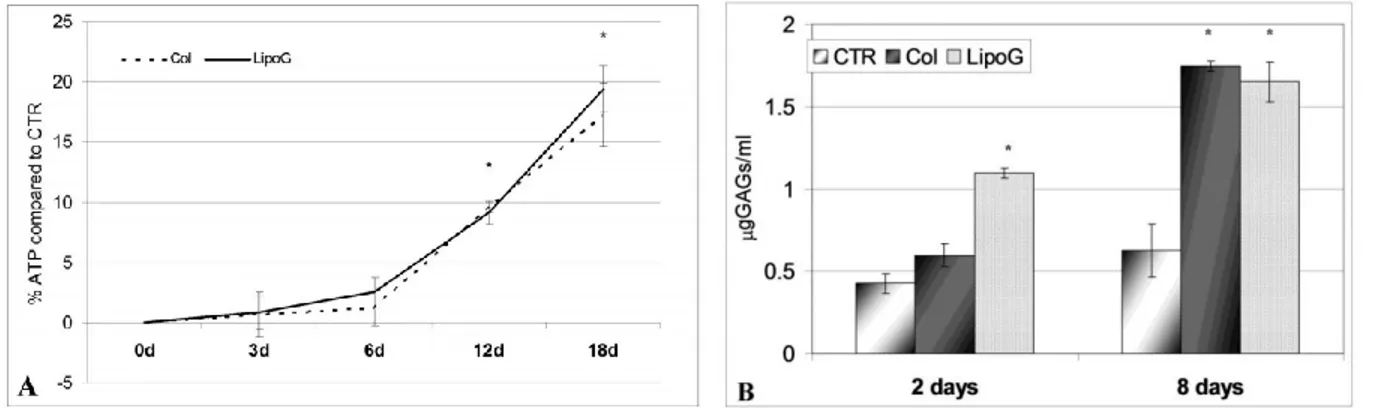
Culture medium from both Col and LipoG lipoaspirates was found to induce chondrocyte proliferation and ECM production to the same extent. As shown in Fig. 9A, already 3 days after the addition of 15% lipoaspirate culture medium, a statistically-significant increase in ATP levels occurred in both cases compared to the CTR. This increase was more evident at longer incubation times, reaching a plateau at 18 days of culture, with 80% more ATP in chondrocytes treated with 15% culture medium of the lipoaspirate and LipoG compared to CTR that were
35
chondrocytes treated for the same period of time with 15% of the same culture medium used for lipoaspirate culture. Moreover, as shown in Fig. 9B, culture medium from the Col and LipoG batches activated ECM synthesis of chondrocytes, as shown by increased levels of GAGs.
Figure 9: Proliferation (Figure 9A) and GAG production (Figure 9B) of human primary chondrocytes cultured with 15% of culture medium from lipoaspirates. Results in Figure 9A are expressed as mean ± SD relative to control cells (n=3); in Figure 9B results are reported as mean ± SD (n=3) in µg/ml of sulfated glycosaminoglycans quantified in culture medium of chondrocytes cultured for 2 days and 8 days in basal medium (CTR) and in culture media supplemented with 15% medium from lipoaspirates (Col and LipoG). *p<0.05 compared to CTR.
3.3.3 Cell outgrowth from lipoaspirates: quantification, phenotype,
differentiation capacity, and cartilage repopulation.
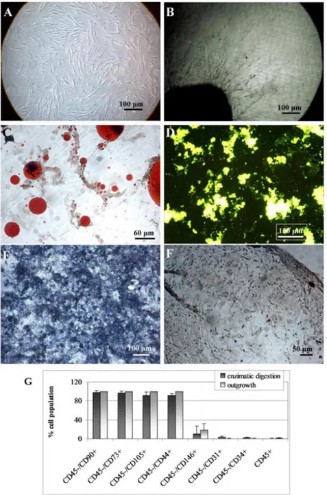
The next stage of the research showed that the LipoG batch of lipoaspirate, when transferred into tissue culture, without any processing began to leave cells out from the tissue clusters after 2-3 days. Cells attached to the plastic of the tissue-culture wells and reached 70-80% confluence in 7-12 days. In Fig. 10 are reported representative images of cells grown out from the clusters: Fig. 10A showed cells at the bottom of the plastic wells which had grown out from the clusters when cultured in floating conditions; Fig. 10B showed cell outgrowth in the 3D collagen culture model. Both lipoaspirates (Col and LipoG) showed good cell outgrowth from the clusters; the
36
only difference was that, from the LipoG batch, outgrowth was faster than from the Col batch; further from the same weight of lipoaspirate, the number of cells from LipoG was higher than that from the Col batch. Of the two culture models studied, cell outgrowth was faster in the 3D collagen matrix than it was from the floating culture. Cell outgrowth was already visible within 2 days from time 0 in the 3D culture model, whereas no cells were visible before 4-5 days in the floating culture.
Figure 10: Cell outgrowth from clusters: representative images at phase contrast microscopy of cells outgrown from clusters in the monolayer culture model (Figure 10A) and in the 3D collagen matrix (Figure 10B). Figures 10C-F show representative microscopic images of four separate experiments of the multi-lineage differentiation capacity of the cells outgrowing from lipoaspirate. Adipogenic differentiation (Figure 10C) was revealed by Oil Red-O staining for neutral lipids. Osteogenic differentiation was evidenced by the formation of mineralized matrix as shown in green by Calcein staining in Figure 10D and ALP activity in Figure 10E. Chondrogenic differentiation was revealed by immune-histochemical stain for collagen II (Figure 10F). The bar-graph (Figure 10G) shows the immune-phenotyping flow cytometry analysis of cells expanded from the enzymatically digested lipoaspirate, and of cells outgrowing from cultured lipoaspirate. Data are expressed as means ± SD (n=3) of the percent of positive cells for the indicated markers.
37
FACS analyses (Fig. 10G) showed that cells obtained by outgrowth from the clusters expressed typical mesenchymal markers (CD90, CD73, CD105 and CD44) at high percentages, close to 100%, and also CD146 positive cells index of pericytes population Fig. 10 bar graph. No statistically-significant difference was found versus cells obtained by enzymatic digestion of the lipoaspirate. Furthermore, cells outgrown from the lipoaspirates were found to have the developmental potential of hMSCs (Fig. 10C-F). Adipogenic differentiation showed a progressive loss of the fibroblastoid-like shape, and the production of cytoplasm lipid vacuoles (Fig. 10C). Osteogenic differentiation was revealed at the first week of induction by morphological changes (from fibroblastoid-like to trapezoidal cells) and, at the end of the induction period, by the formation of mineralized matrix, as shown in Fig. 10D, and of ALP-positive cells (Fig. 10E). Chondrogenic induction revealed abundant extracellular matrix positive to human type II collagen antibody (Fig. 10F).
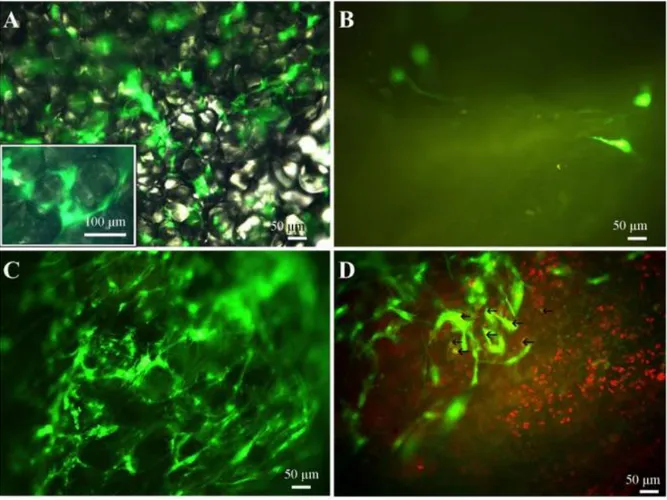
LVs were used to transduce cultured clusters of lipoaspirate. Transduction was visualized by GFP expression, and a very high transduction efficiency was demonstrated as shown by direct fluorescence (Fig. 11A). Transduction was not limited to the surface of the clusters, but was also evident in the inner part. Cell counts, performed with nuclear counterstaining, revealed that a mean of 62% ± 4% of cells of the cluster were GFP-positive, and no cell loss was detected (p=0.94 for comparison with control lipoaspirates, n=3). Moreover, 49±20% of cells outgrown from the lipoaspirate clusters were GFP-positive.
When GFP-transduced clusters were placed in an organ culture of healthy or mechanically-damaged cartilage, it was found that, within a few days, positive cells grew out from the clusters; moreover they only repopulated the damaged cartilage (Fig. 11C) and not its healthy counterpart (Fig. 11B). Among GFP-positive cells, only 10% showed SOX9 co-localization
38
indicating that, in the organ culture model used, only 10% of cells outgrowing from the lipoaspirate differentiated into chondrocytes (Fig. 11D).
Figure 11: Lentiviral GFP transduction of the clusters is shown in Figure 11A. No GFP-positive cells growing out from GFP-transduced clusters are visible upon a healthy cartilage fragment (Figure 11B): a mechanically-damaged cartilage fragment was well repopulated (Figure 11C). Few of the cells that repopulated the damaged cartilage differentiated to chondrocytes (GFP-positive and SOX9-positive, yellow highlighted with arrows in Figure 11D).
3.4 Conclusions
The use of ASCs in regenerative medicine is a rapidly-growing area of research. There is some
in vitro evidence of success using these cells in osteochondral defect repair (17), and they have
also recently been used successfully as a therapeutic tool in treating OA (5). ASCs have been available commercially for veterinary use since 2003, although little has been published
39
documenting their clinical efficacy; such reports might minimize the gap between human and veterinary markets, accelerating the introduction of stem-cell-based cartilage repair for human diseases. Beneficial effects of ASCs have been reported in treating OA in some experimental animal models (19,33). However, these studies used a tissue-engineering approach, and a wide variety of biodegradable scaffolds were used to assist chondrogenic differentiation.
To our knowledge, use of an autologous biological tissue, naturally rich in stem cells, implanted in a different anatomical site from where it was harvested (i.e. autologous transplantation of subcutaneous fat to intra-articular sites) has not been previously attempted. The present study has shown that it is possible to avoid stem-cell isolation, expansion, and differentiation for several days in vitro, before autologous implantation with scaffold or scaffold-free approaches. We have demonstrated that micro-fragmented lipoaspirate clusters give rise to spontaneous cell outgrowth both when cultured in floating conditions and in the 3D collagen matrix. Although adipose tissue contains adipose-derived stem cells, fibroblasts, endothelial cells, hematogenous cells, pericytes, adipocytes, and pre-adipocytes, it was found that cells obtained naturally exhibit a mesenchymal phenotype with stem cell surface expression markers similar to those obtained through enzymatic extraction; they also have the capacity for multilineage differentiation, showing the classical commitment to osteogenic, chondrogenic and adipogenic lineages. These data show that micronized lipoaspirate, acting as a natural scaffold for mesenchymal cells that are simply trapped in the stromal vascular portion, may be considered a source of autologous multipotent undifferentiated cells. Since it requires no processing steps before use, the risk of infection, or that of retaining enzymatic residues from tissue digestion during cell isolation can be avoided. This also reduces costs associated with a long period of cell expansion and differentiation (12-21 days) with residual of growth supplements, as well as the side effects associated with the natural or synthetic scaffolds used for cell-based cartilage repair. Despite
40
these promising results, however, the physiological role of native adipose-derived stromal/stem cells in vivo is not fully understood, and more histological studies must be performed before it may be deemed safe for clinical use. It will be necessary to identify in situ the expression of a wide range of markers; these may well include more than those studied to analyze culture-expanded preparations (2). The use of GFP-transduced lipoaspirates demonstrated, in vitro, that cells outgrown from lipoaspirate can repopulate an organ culture of damaged cartilage. It may therefore be hypothesized that, when intra-articular injection of human lipoaspirate enters clinical trials, it will give rise to cells having mesenchymal potential to repopulate the lesion. Moreover, there appears to be a possibility that native stem-like cells arising from lipoaspirate, may have a better physiological phenotype and higher regenerative potential than cells obtained with the isolation technique and culture conditions generally used for ASC preparation prior to transplantation.
In addition to these data, the finding that lipoaspirate culture medium induced human primary chondrocyte proliferation and ECM synthesis reinforces the promise of such preparations, comprising micronized fat, becoming a therapeutic tool for cartilage regeneration. The paracrine and endocrine secretions of adipose tissue have recently been described (19); fat might not be considered only as an energy storage tissue, but rather as an active tissue involved in the metabolism (21), in immunomodulatory activities, and in providing an extensive peri-vascular reservoir of multipotent, undifferentiated cell populations, involved in homeostasis and regeneration. As a large micro-vascular bed, fat appears to provide an ideal pool of undifferentiated cells in close proximity to peri-vascular access routes for the mobilization and relocation demands of injured or diseased structures. It is characterized by different cell types (including adipocytes, pre-adipocytes, vascular cells, fibroblasts, pericytes, stem cells, and immune cells) that, by secreting a range of cytokines, growth factors, chemokines, and