A
A
l
l
m
m
a
a
M
M
a
a
t
t
e
e
r
r
S
S
t
t
u
u
d
d
i
i
o
o
r
r
u
u
m
m
–
–
U
U
n
n
i
i
v
v
e
e
r
r
s
s
i
i
t
t
à
à
d
d
e
e
g
g
l
l
i
i
S
S
t
t
u
u
d
d
i
i
d
d
i
i
B
B
o
o
l
l
o
o
g
g
n
n
a
a
Scuola di Dottorato in Scienze Mediche e Chirurgiche
Dottorato di Ricerca in Scienze Biomediche – PFDR Scienze
Morfologiche Umane e Molecolari
Ciclo XXVI
Settore Concorsuale di afferenza 05/H2
Settore Scientifico disciplinare BIO/17
Tesi di Dottorato:
MUSCULOSKELETAL
TISSUE
REGENERATION
BY
HUMAN
NON-EMBRYONIC
STEM
CELLS
Presentata da: Tutor:
D
OTT.
SSAA
LESSANDRAP
ISCIOTTAC
HIAR.
MOP
ROF.
A
NTOD
EP
OLCoordinatore:
C
HIAR.
MOP
ROF.
L
UCIOC
OCCO1
TABLE OF CONTENTS
Table of contents………..page 1
Abstract……….………3
Introduction………..6
Tissue Engineering………...7
Cell sources………..……….7
-
Primary cells……….……….7
-
Stem cells……….……...8
-
Mesenchymal stem cells………..…14
Dental Pulp Stem Cells (DPSCs)………...17
-
Dental pulp embryogenesis……….19
- DPSCs characterization
……….21
Amniotic Fluid Stem Cells (AFSCs)………...23
-
AFSCs isolation………...………25
-
AFSCs characterization………...25
Bone regeneration………..……….27
-
Bone composition………...27
-
Osteoblastogenesis regulation and bone related proteins expression...29
-
Tissue engineering as a novel approach for bone healing………....32
2
Skeletal muscle regeneration……….……….40
-
Structure of skeletal muscle……….40
-
Muscle development (myogenesis)………41
-
Molecular mechanisms modulating satellite cells: quiescence, activation, replenishment………...43
-
Muscular dystrophy………...46
Materials and methods………...49
Results……….73
Discussion……….102
1.
Human DPSCs and AFSCs for bone tissue regeneration...102
2.
Human serum is a suitable substitute for FCS to expand and differentiate human DPSC………....105
3.
Skeletal muscle regeneration by human DPSCs and AFSCs………….…...…109
Conclusions………...…113
3
ABSTRACT
The aim of the thesis is to investigate the regenerative potential of alternative sources of stem cells, derived from human dental pulp (hDPSCs) and amniotic fluid (hAFSCs) respectively, and, specifically to evaluate their capability to be committed towards the osteogenic and myogenic lineages, with the long-time objective to apply these stem cells to translational strategies in regenerative medicine for the repair of bone and skeletal muscle tissues. So far orthopaedic surgery, based on the use of conventional prosthesis and engineering biomaterials, does not represent a definitive solution in the treatment of some diseases, because the implanted material either does not offer the best histo-integration and does not replace the function of the original tissue.
The tissue engineering approach could be therefore a promising tool to restore bone defects and deficiencies that currently are surgically treated through the application of artificial permanent implants. In particular, the in vitro bone production by stem cells may represent a radical breakthrough in the treatment of congenital or acquired pathologies and secondary traumas characterized by critical bone mass defects, which yet do not have any medical or surgical solution. Therefore, hDPSCs and hAFSCs were seeded and pre-differentiated on different scaffolds to test their capability to subsequently reach the osteogenic differentiation in vivo, in order to recover critical size bone defects.
Equine collagen, silk fibroin and P(d,l)LA were utilized to determine the best scaffold for hDPSCs and hAFSCs to undergo the effective osteogenic differentiation in vivo, to repair surgically operated parietal bone defects in rats. Results obtained in a recent study on ectopic implants allowed to identify fibroin scaffolds as a promising tool for osteogenic differentiation of human stem cells.
4
This study demonstrated that fibroin scaffold promotes mature bone formation and defect correction when combined to both hDPSCs and hAFSCs, in particular, with a higher bone amount produced by hAFSC-seeded scaffolds.
Since the ultimate goal of cell-based therapy is to utilize new stem cell sources for clinical applications with minimal safety concerns, this study has investigated a culture condition that might allow human DPSCs to be used for human cell therapy in compliance with good manufacturing practices (GMPs). This study demonstrated that human serum (HS) is an appropriate supplement for the in vitro expansion of human DPSCs, since its addition to culture medium produced a good proliferation rate in comparison with the one evaluated after culturing the cells with foetal calf serum (FCS). As a matter of fact, human serum appeared to be an adequate additive to the osteogenic medium in order to achieve the expression of bone related proteins and induce the mineralization of the extracellular matrix by hDPSCs, in vitro. Also, hDPSCs pre-differentiated towards the osteogenic lineage with the addition of HS showed a huge contribution in regenerating of the critical size bone defects in vivo.
Similarly, cell-based therapy with hDPSCs and hAFSCs could represent an alternative therapeutic approach for patients with musculoskeletal traumas or diseases. This thesis investigated the ability of these stem cells to undergo the myogenic commitment in vitro by using different protocols, either when co-cultured with murine myoblasts (C2C12) and when differentiated alone after the demethylating treatment with 5-aza-2’-deoxycytidine. Human DPSCs and AFSCs, indeed, expressed under both these conditions regulatory factors and markers – such as myogenin and myosin - that are typical of the myogenesis process. Furthermore, after being pre-differentiated by means of demethylation, with or without the addition of conditioned media from differentiated C2C12, hDPSCs and
5
hAFSCs were tested for the capability to regenerate the damaged skeletal muscle of SCID/mdx mice, animal model of Duchenne Muscular Dystrophy (DMD). When injected into dystrophic muscles of SCID/mdx mice, pre-differentiated hDPSCs and hAFSCs were able to recover the skeletal muscle tissue, and more interestingly to restore dystrophin expression. These observations, altogether, suggest the eventual applicability of human DPSCs and AFSCs to translational strategies, in order to enhance the repair of injured skeletal muscle in DMD patients.
6
Introduction
In Greek mythology, the Moirai also known in English as the Fates, and in Latin as the Parcae - euphemistically the "sparing ones" - were white-robed figures, personifying the inescapable destiny. Daughters of Zeus, they were three: Clotho (the spinner), who spun the thread the life from her distaff onto her spindle, Lachesis (the allotter), who measured the thread of life allotted to each person, and Atropos (the inexorable or inevitable), who was the cutter of the thread of life. Thus, they controlled the metaphorical thread of life of every mortal from birth to death.
In medical research stem cells – whose definition derives from the Latin “stamen” – can be therefore metaphorically considered as the thread of life, namely the starting point from which the human being comes from.
Actually, they have been demonstrated to carry out a fundamental role in maintaining, healing, regenerating and aging processes, therefore representing an adequate tool to be applied in regenerative therapies.
7
Tissue Engineering
The primary goal of tissue engineering scientists is to optimize cell isolation, proliferation and differentiation of stem cells and to develop scaffolds and/or delivery systems that enhance the coordinated growth of three-dimensional tissues. The most used basic strategy consists in harvesting cells from a patient, expand them in cell culture, and seed them onto a scaffold that provides a biomechanical environment that leads the formation of the required tissue upon the production of a certain extracellular matrix by stem cells. The tissue can be grown on a scaffold that will completely resorb as the new tissue grows, so that only the new tissue will be implanted, or that will be gradually reabsorbed after implantation. Therefore, the tissue engineered complex must be able to survive, restore normal function, and integrate with the surrounding tissues (Polak and Bishop, 2006).
Cell sources
As a primary factor to achieve successful results in tissue engineering it is desirable and necessary to obtain a sufficient number of cells that show constantly an adequate phenotype and the ability to perform their specific biological functions, i.e. the correct organization of the extracellular matrix, the secretion of signaling molecules, and the interaction with the surrounding cells and tissues. Taken altogether, these functions are meant to enhance the repair (Polak and Bishop, 2006).
Primary Cells
These cells are tissue specific, are generally harvested from explant samples collected by surgical procedure. Primary cells are the most desirable with regard to immunological
8
compatibility, but they are differentiated and post-mitotic. When cultured ex vivo a de-differentiation of the primary cells themselves as well as the development of an inappropriate phenotype might be observed.
Moreover, functionality and proliferation rates tend to lower, and for the isolation of certain cell types, i.e. spinal cord neurons, the harvesting of primary cells is not recommended.
These aspects have triggered the pursuit of alternative cell sources for tissue engineering approaches and stem cells actually provide solutions to some problems related to the use of primary cells (Polak and Bishop, 2006).
Stem Cells
Stem cells can be described as undifferentiated cells that are characterized by three fundamental abilities: proliferation, self-renewal, and differentiation towards multiple cell lineages (Polak and Bishop, 2006).
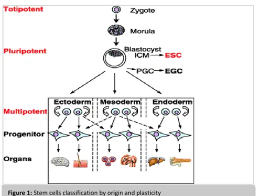
They can be isolated from different sources, and with this regard are classified as embryonic stem cells (ESCs), foetal stem cells (FSCs), and adult stem cells (ASCs), whose range of differentiation potential may vary. Indeed, a more useful classification can be based on stem cells plasticity (Figure 1):
Totipotent are the most plastic ones and differentiate into any cell type, including placenta (i.e. the zygote and morula at 8-cells stage)
Pluripotent are the second most plastic stem cells after the totipotent ones, can differentiate into all cells, except for the totipotent and placental cells
9
Progenitor cells may differentiate into only one or a limited number of cell types, therefore are the least plastic cells
Embryonic stem cells (ESCs) are the most plastic source available in tissue engineering.
They were first described when they were isolated from the inner cell mass of a mouse blastocyst and then expanded in vitro (Evans and Kaufman, 1981; 3 Martin GR, 1981). ESCs have actually been demonstrated to be pluripotent, namely able to differentiate into all derivatives of the three primary germ layers: ectoderm, endoderm, and mesoderm(Nagy A et al, 1990; Bradley A et al, 1984; Xu RH et al, 2002). The murine ESCs were able to maintain an undifferentiated state while proliferating, and to differentiate towards all the mature somatic phenotypes, when stimulated with the appropriate signals. The initial isolation of murine ESC lines provided an easy model system to analyze the processes occurring during early development and cellular differentiation. These findings have also
10
led the way to tissue engineering applications with similar pluripotent cells obtained from human blastocyst. As a matter of fact, human ESCs, were first derived in 1998 for infertility purposes (Thomson JA et al, 1998; Reubinoff BE et al, 2000); they show various discrete differences from murine ESCs, in fact they proliferate more slowly and most of them form flat, instead of spherical, colonies. Also, they can be dissociated in single cells more easily than their murine counterparts (Laslett AL et al, 2003).
Because of their plasticity and potentially unlimited capacity for self-renewal, ESCs cell therapies have been proposed for regenerative medicine and tissue replacement in case of injury or disease. Diseases that might eventually be treated by ESCs cells include several blood and immune-system related genetic diseases, cancers, and disorders, type I diabetes, Parkinson's, blindness and spinal cord injuries. Nevertheless - beside the existing ethical controversy related to the use of embryonic stem cells - there is a huge concern with the possible transplantation of ESC into patients as therapies, because of the related possible formation of tumors, including teratoma (Knoepfler PS, 2009). Therefore, investigations have been carried out in order to find alternative solutions to ES cells. Takahashi and Yamanaka published a milestone manuscript in 2006 that defined a specific set of transcription factors capable of reverting differentiated cells back to a pluripotent status, thus creating induced pluripotent stem cells (iPS cells) (Takahashi and Yamanaka, 2006): four key transcription factors - Oct4, Sox2, Klf4, and c-Myc - identified by screening 24 preselected mouse embryonic stem cell–specific factors were sufficient to reprogram adult mouse fibroblasts into embryonic stem cell–like iPS cells (Takahashi and Yamanaka, 2006). The same combination of transcription factors was demonstrated to be sufficient for the pluripotent induction of human cells as well (Takahashi K et al, 2007). Considering the convenience and reproducibility of generating iPS cells, experts have
11
raised the hope that iPS cells might accomplish much of the promise of human ESCs in regenerative medicine (Pera MF, 2008). Nonetheless, findings from different research groups have demonstrated that iPS cells injected into immunodeficient mice give rise to teratomas comprising all three embryonic germ layers, as similarly observed with ESCs. A further potential complication related to the use of iPS cells is the use of retroviral and lentiviral vectors to activate the necessary reprogramming transcription factors, in fact, even though much progress has been made in generating integration-free murine iPS cells, the safety of iPS cells needs to be rigorously tested, since all essential reprogramming factors are oncogenes, and their overexpression has been linked with cancers (Liu SV, 2008).
Foetal stem cells (FSCs) are the mesenchymal stem cells derived from the fetus. Recently,
they have been isolated from foetal blood and raised the possibility of using autologous cell to treat fetuses in utero (Campagnoli C et al, 2001).
The mesenchymal population obtained from foetal blood is characterized by adherent cells that divide upon 40 passages in culture and are able to differentiate towards the chondrogenic and osteogenic lineages, but also can form oligodendrocytes and hematopoietic cells. Furthermore, they are a unique source of stem cells since they can engraft into multiple organs and differentiate in a tissue-specific manner (Polak and Bishop, 2006).
Adult stem cells (or postnatal somatic cells) are undifferentiated cells found among
differentiated cells in a tissue or organ that can renew itself. These stem cells are located in a variety of tissues, including bone marrow, brain, liver, skin, and blood (Presnell SC et al,
12
2002). Their principal role is to maintain and repair the tissue they derive from. Although in a first moment they were considered to own a very limited differentiation potential, there has been much evidence that they show a considerable degree of plasticity, not without some debate about it (Hawley and Sobieski, 2002; Holden and Vogel, 2002; Verfaillie CM et al, 2002; Poulsom R et al, 2002; Raff M, 2003). Unlike ESCs, the application of adult stem cells in research and therapy is not considered to be controversial, since they do not require the destruction of embryos and can be accessibly obtained from adult tissue samples.
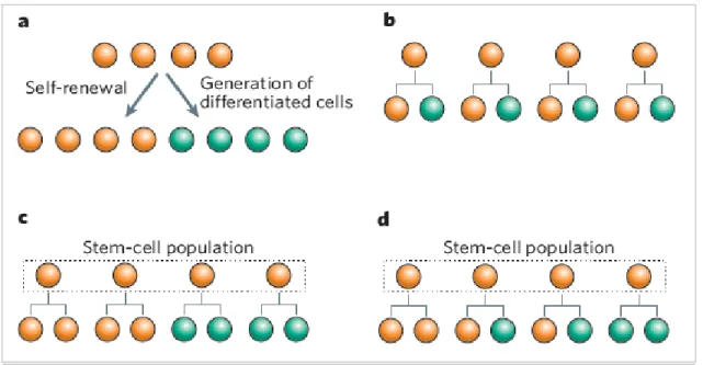
In comparison with ESCs and FSCs, adult stem cells generally exhibit a lower differentiation potential and yield a more limited number of distinct progenitor cells. However, adult stem cells can significantly undergo proliferation and differentiation into more mature and tissue-specific cell types, according to changes in the specialized microenvironment where they reside - the niche - and the stem cell behavior is regulated through direct contact and paracrine signaling (Fuchs E et al, 2004; Niemann C, 2006; Wilson and Trumpp, 2006; Morrison and Spradling, 2008; Jones and Wagers, 2008). As mentioned above, stem cells are defined by their ability to produce more stem cells and also cells that differentiate. Stem cells can accomplish these two tasks by means of asymmetric cell division (Figure 2b), whereby each stem cell divides to generate one daughter with a stem-cell fate (self-renewal) and one daughter that differentiates (Betschinger and Knoblich, 2004; Clevers H, 2005; Doe and Bowerman, 2001; Yamashita YM et al, 2005). However, a limit of this division type is that stem cells would be unable to expand in number. Thus, asymmetric cell divisions cannot be the only explanation. Stem cells must have further self-renewal strategies that allow a dynamic control of their numbers: in fact, with symmetric divisions they can self-renew and produce differentiated
13
progeny (Figure 2c). Symmetric divisions are defined as the generation of daughter cells that are intended to acquire the same fate. Although the idea that stem cells can divide symmetrically may sound like a contradiction, stem cells are defined by their “potential” to generate more stem cells and differentiated daughters, rather than by their production of a stem cell and a differentiated daughter at each division. Therefore, when thought as a population, a pool of stem cells with identical developmental potential may generate only stem-cell daughters in some divisions and only differentiated daughters in others (Figure 2d). Broadly, stem cells can rely either completely on symmetric divisions or on a combination of symmetric and asymmetric divisions (Morrison and Kimble, 2006).
Figure 2: Stem cells (orange) must accomplish the dual task of self-renewal and generation of differentiated
cells (green); b–d: possible stem-cell strategies that maintain a balance of stem cells and differentiated progeny. b: asymmetric cell division; c: symmetric cell division; d: combination of cell divisions: each stem cell can divide either symmetrically or asymmetrically (Morrison and Kimble, 2006).
14
Mesenchymal Stem Cells
Mesenchymal stem cells (MSCs) can be found in bone marrow and in many other tissues. Bone marrow is a mesoderm derived tissue, basically constituted by elements from the stroma and the hematopoietic system, and is present in the interior of bones (Arai F et al, 2004).
For long time, it has been known that the bone marrow contains two types of stem cells, the hematopoietic stem cells (HSCs) – committed to differentiate towards mature blood cells – and the more undifferentiated stromal mesenchymal cells (MCs).
The identification of the mesenchymal stem cell has so far depended on in vitro culture systems, which have provided very heterogeneous information on MSCs.
They were identified through a combination of poorly defined physical, phenotypic, and functional properties. The first direct evidence that non-hematopoietic, mesenchymal precursor cells were present in the bone marrow derived from the research performed during the late 1960s by Friedenstein and colleagues: bone marrow samples incubated in tissue culture flasks gave rise to a fraction of adherent cells within a few days, and soon after individual aggregates of 2 to 4 fibroblasts were observed, which could differentiate into cells able to form small deposits of bone or cartilage (Friedenstein AJ et al, 1976). These cells were called “colony forming unit-fibroblasts” or CFU-F. During the 1980s, several studies showed that cells isolated by the Friedenstein method were multipotent and able to differentiate towards osteoblasts, chondroblasts, adipocytes, and also myoblasts (Prockop DJ, 1997). The formation of CFU-F has been considered peculiar of mesenchymal stem cells, although a direct relationship between the two has not been clearly established, likely because of the high heterogeneity in morphology, cell size and differentiation potential observed among species and between colonies (Javazon EH et al,
15 2004). Caplan demonstrated that bone and cartilage turnover was mediated by MSCs, and the surrounding conditions were critical to inducing MSC differentiation (Caplan AI, 1991). Later, the multilineage differentiation capability of MSCs was definitively demonstrated by Pittenger (Pittenger MF et al, 1999).
The defining features of mesenchymal stem cells are inconsistent among scientists; to address this issue, the Mesenchymal and Tissue Stem Cell Committee of International Society for Cellular Therapy (ISCT) has recently proposed a set of standards to define human MSCs either for laboratory-based scientific investigations and for preclinical studies. First, MSCs must be plastic-adherent when maintained in standard culture conditions using tissue culture flasks. Second, ≥ 95% of the MSCs population must express CD105 (endoglin), CD73 (ecto-5’-nucleotidase) and CD90 (Thy-1), as determined by flow cytometry. Further, no more than 2% of these cells may express CD45 (pan-leucocyte marker), CD34 (hematopoietic progenitor and endothelial cell marker), CD14 or CD11b (monocyte and macrophage markers), CD79α or CD19 (B cell markers) and HLA-DR (marker of stimulated MSCs). Third, the cells must be able to differentiate towards osteoblasts, adipocytes and chondroblasts, under the standard differentiating conditions in
vitro (Dominici M et al, 2006).
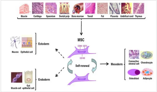
MSCs embryologically derive from two major sources, neural crest and mesoderm, however these cells have been identified in not only mesoderm-derived but also endoderm- and ectoderm-derived tissues. Examples of tissues where MSCs have been characterized include, but are not limited to: mesodermal - bone marrow, trabecular bone, synovium,
cartilage, fat, muscle, and tonsil; endodermal – thymus; ectodermal - skin, hair follicle, dura mater, and dental pulp; prenatal and perinatal tissues - umbilical cord, umbilical cord blood, amniotic fluid and placenta (Kuhn and Tuan, 2010).
16 Although still controversial, literature exists demonstrating the potential of MSCs to differentiate along non-mesodermal lineages, such as ectodermal skin and neurons and endodermal hepatocytes (Figure 3). MSCs are still correctly defined as “multipotent” rather than “pluripotent” despite these reports of their potential capability to differentiate into derivatives of all three germ layers.
Most of the adult sources, including large volumes of normal bone marrow, are relatively difficult to access as a tissue source for the isolation of MSCs. Conversely, birth-associated tissues, including placenta, are readily and widely available. However, the bone marrow appears to be the most commonly exploited source of MSCs for most preclinical and clinical studies.
Figure 3: Identification and characterization of mesenchymal stem cells. Cells with MSC-like characteristics have
been isolated and from several adult tissues. The most common phenotypical markers are CD90, CD73, and CD105. MSCs have the capability to self-renew and exhibit multilineage potential into many mesodermal cell types. Other studies have described the potential of MSCs to differentiate into ectodermal and endodermal lineages.
17 It is necessary to highlight that although MSCs can be isolated from these different tissues and can develop into bone, cartilage, or fat, they are distinctive and reflects aspects peculiar to their tissue of origin: as a matter of fact, human pellets of MSCs from bone marrow have shown to be able to make cartilage extracellular matrix through chemical exposition to TGF-β (Johnstone B et al, 1998), while mesenchymal progenitors from adult fat require both TGF-β and BMP-6 in order to produce cartilage (Yoo JU et al, 1998; Estes BT et al, 2006).
Dental pulp stem cells (DPSCs)
Multipotent adult stem cells, as MSCs, are present in all mature tissues of the human body and are thought to reside in a specific area of each tissue where they may remain quiescent (non-dividing) for many years until they are activated by disease or tissue injury. MSC-like stem cells have been isolated – according to the standard criteria of mesenchymal stem cells illustrated above (Friedenstein AJ et al, 1976; Prockop DJ, 1997; Caplan AI, 1991; Pittenger MF et al, 1999; Dominici M et al, 2006) - from different tissue sources including bone marrow, adipose tissue, bone, periosteum, synovium of the joints, skeletal muscle, skin, pericytes of blood vessels, peripheral blood, periodontal ligament, umbilical cord, and dental pulp of permanent and deciduous teeth (Mizuno H et al, 2012).
Lately, human dental pulp has been attracting interest for its ease of collection from permanent and deciduous teeth through minimally invasive and safe procedure.
Dental pulp is a loose vascular connective tissue, surrounded by dentin (Figure 4) and consisting of a heterogeneous population of cells: the potential preodontoblasts, fibroblasts, stromal cells, endothelial and perivascular cells, neural cells and others (Pierdomenico L et
18 To date, multiple stem/progenitor cells have been isolated from human dental pulp, characterized, and classified. The postnatal stem cells first isolated from dental pulp were termed “dental pulp stem cells” (DPSCs) (Gronthos S et al, 2000). Besides this group, three more types of dental cell populations were isolated and characterized: stem cells from exfoliated deciduous teeth (SHEDs) (Miura M et al, 2003), periodontal ligament stem cells (PDLSCs), dental follicle progenitor cells (DFPCs) (Morsczeck C et al, 2005) and stem cells from apical papilla (SCAPs) (Sonoyama W et al, 2008).
In particular, stem cells from human dental pulp of impacted third molars (also known as “wisdom teeth”) show a greater advantage for the ease and low invasivity of recruitment, respect to the mesenchymal stem cells derived from bone marrow, adipose tissue, peripheral blood, and umbilical cord blood.
Figure 4: The main structures of the tooth. Dental pulp is part
of the center of the tooth, is contained within the pulp chamber and harbors vessels and nerves.
19 DPSCs are an attractive alternative source of mesenchymal stem cells since they show a wide panel of favorable properties:
- Easy surgical access and low invasiveness for the patient: the extraction of one or more molars, in healthy individuals, is a widespread and routinary procedure among the population.
- Very low morbidity of the anatomical site after the collection of the pulp.
- High efficiency of the extraction procedure of the stem cells from the pulp tissue. - Minimal tooth processing may be required for banking of samples with no
immediate plans for DPSC expansion and use, which subsequently would limit costs and favor the clinical banking of these cells.
- Cells expanded in culture can be preserved and stored for at least six months, and likely longer, at −85°C with regards to the qualitative ability to differentiate in at least a tri-lineage manner. Therefore, they can be cryopreserved, retaining multi-differentiation potential (Graziano A et al, 2008).
- No teratomas have been observed after in vivo transplantation.
- Mesenchymal stem cells from the dental tissue have been shown to possess immunosuppressive properties. These findings have elicited further interest in these cells as alternative cell sources able to modulate alloreactivity and tissue regeneration, following transplantation into human leukocyte antigen-mismatched donors (Wada N et al, 2013).
Dental pulp embryogenesis
In vertebrate embryos, a group of cells, called neural crest (NC) cells, separate from the neural tube and migrate to re-aggregate with other cells. In the developing embryo, almost
20
all organs, glands, and tissues - such as craniofacial skeleton, cornea, teeth/dentin, thyroid gland, thymus, cardiac septa, adrenal gland, melanocyte, autonomic nerve, sensory nerve, and Schwann cells - have these basic cells (Crane and Trainor, 2006; Alberts B et al, 2008; Lee G et al, 2010). The contribution from NC cells in building of the head of vertebrates has been considered to be a turning point in the evolution of the vertebrates (Gans and Northcutt, 1983). Although NC cells are of ectodermal origin, it has been suggested to call them “mesectoderm” or “ectomesenchyme”, since they undergo “mesenchymalization.” This property is important with regard to mesenchymal stem cells, since their origin is the mesenchyme, which is derived from the mesodermal germ layer. On the other hand, along with the cranial skeleton and other tissues of head and neck, odontoblasts and tooth papillae are derived from mesectoderm, or ectomesenchyme (Le Douarin NM et al, 2004). Oral ectomesenchymal and ectodermal interactions phylogenetically precede the origin of odontogenesis (Moss ML, 1969).
During the sixth week of embryogenesis, the ectoderm covering the stomodeum starts to proliferate, giving rise to the dental laminae. Reciprocal interactions between ectoderm and mesoderm layers lead to placode formation. One of these thick, ovoid ectodermal structures develops into tooth germs, where cells, belonging to the neural crest, will differentiate into the dental germ, containing both dental papilla and follicle. Therefore, dental pulp is made of ecto-mesenchymal components, containing neural crest-derived cells, which display plasticity and multipotential capabilities (Sinanan AC et al, 2004). Pulp is externally separated from dentin by odontoblasts and by Hohl’s subodontoblastic cells, that are pre-odontoblasts (Goldberg and Smith, 2004). Adjacent to this layer, the pulp is rich in collagen fibers and poor in cells. Then, another, more internal layer, contains progenitor cells and undifferentiated cells, some of which are considered stem cells (Jo YY
21 et al, 2007). From this layer, undifferentiated cells migrate to various districts where they
can differentiate under different stimuli and make new differentiated cells and tissues. The final, innermost layer is the core of the pulp; this area comprises the vascular plexus and nerves. Up to more recent discoveries (Gronthos S et al, 2000; D’Aquino R et al, 2007), researchers hypothesized that DPSCs were present in this layer (Fitzgerald M et al, 1990). Actually, only undifferentiated perivascular cells can be found in it.
The third molar tooth germ begins development around the sixth year of life. Until this time, embryonic tissues of dental lamina remain quiescent and undifferentiated within the jaw of the child. Even though crown mineralization begins during the eighth year of life, often third molar roots are still incomplete at the age of 18. This means that the structure of those teeth is still immature at this age, and a conspicuous pool of undifferentiated cells, resident within the “cell rich zone” of the dental germ pulp, are needed for development.
DPSCs characterization
During the characterization of these newly identified dental stem cells, certain aspects of their properties have been compared with those of bone marrow mesenchymal stem cells (BMMSCs).
Differences have been noted between the dental stem cell populations and BMMSCs. One important feature of pulp cells is their odontoblastic differentiation potential. Human pulp cells can be induced in vitro to differentiate into cells of odontoblastic phenotype, characterized by polarized cell bodies and accumulation of mineralized nodules (Tsukamoto Y et al, 1992; About I et al, 2000; Couble ML et al, 2000). DPSCs isolated with enzyme treatment of pulp tissues form CFU-Fs with various characteristics (Gronthos S et al, 2000; Huang GT et al, 2006a). Colonies show different cell densities, suggesting
22
that each cell clone may have a different growth rate, as reported for BMMSCs (Gronthos S et al, 2002). Within the same colony, different cell morphologies and sizes may be observed also. If seeded onto dentin, some DPSCs convert into odontoblast-like cells with a polarized cell body and a cell process extending into the existing dentinal tubules (Huang GT et al, 2006a; Huang GT et al, 2006b). In addition to their dentinogenic potential, subpopulations of hDPSCs also possess adipogenic and neurogenic differentiation capabilities, by exhibiting adipocyte- and neuronal-like cell morphologies and expressing respective gene markers (Gronthos S et al, 2002). More recently, DPSCs were also found to undergo osteogenic, chondrogenic and myogenic differentiation in vitro (Laino G et al, 2005; Zhang W et al, 2006; D’Aquino R et al, 2007).
Current evidence suggests that biochemical pathways involved in the differentiation of DPSCs into functional odontoblasts are similar to differentiation pathways of BMMSCs into osteoblasts (Shi S et al, 2001): odontoblasts and osteoblasts express similar mineralized matrix proteins, such as dentin matrix protein 1, fibronectin, collagen type I, alkaline phosphatase, osteonectin, osteopontin, bone sialoprotein, and osteocalcin (Nakashima M et al, 1994; Shiba H et al, 1998; Kuo MY et al, 1992; Tsukamoto Y et al, 1992; Buurma and Rutherford, 1999). Therefore, DPSCs actually share a similar pattern of protein expression with BMMSCs in vitro.
To date, dental-tissue-derived stem/progenitor cells have been used for tissue engineering studies in large animals to assess their potential in pre-clinical applications (Sonoyama W
23
Amniotic fluid stem cells (AFSCs)
Since decades, cells from cord blood, amniotic fluid, and the chorion were used in perinatal medicine mainly for invasive diagnostic purposes, such as detection of foetal infections, rare metabolic diseases, and mainly foetal karyotyping. With the introduction of the use of umbilical cord stem cells for the treatment of haematologic diseases many years ago, a new future for the use of perinatal cells has begun, namely the use of perinatal (amniotic, placental, chorionic, umbilical) stem cells for therapeutic purposes.
In normal pregnancies, amniotic cells can be collected by amniocentesis, between 16th and 18th week of gestation. In routine amniocentesis 16-20 ml of amniotic fluid are commonly sampled (Figure 5).
Figure 5: amniotic fluid aspiration during amniocentesis at 16-18 weeks of
24
Amniotic fluid contains heterogenous population of cells, which are contributed mainly from the foetal skin, and possibly from placenta as well. Amniotic fluid-derived stem cells (AFSCs) have shown physical characteristics of both embryonic and adult stem cells. According to their morphological and growth characteristics, amniotic fluid cells can be classified into three types: epithelioid, amniotic-fluid specific, and fibroblastoid (Milunsky A, 1979).
The first evidence of human amniotic fluid containing stem cells came from Prusa et al. in 2003. They identified Oct-4 expression in human amniotic fluid and proposed that human amniotic fluid might be a new source for pluripotent-like stem cells without raising any ethical debates associated with human embryonic stem cells (Prusa AR et al, 2003). In the same year, In’t Anker et al. demonstrated that human amniotic fluid was an abundant source of foetal MSCs, and these cells exhibited a phenotype and multilineage differentiation potential that appears to be similar to that of BMMSCs. They suggested that these AF-derived MSCs could be used in co-transplantation together with umbilical cord blood derived hematopoietic stem cells (In’t Anker PS et al, 2003). In 2004, Tsai MS et al. successfully isolated AF-derived MSCs using a new two-stage culture protocol, and their study showed that these cells could be expanded rapidly and maintained the ability to differentiate towards multiple cell types, such as adipocytes, osteoblasts and neurons in
vitro (Tsai MS et al, 2004). Following, in 2007, Atala and colleagues isolated the c-kit
(CD117) positive population by immunoselection, from second trimester amniotic fluid samples and demonstrated that AFSCs are “broad-spectrum multipotent” (that is, pluripotent) - similarly to embryonic stem cells - and can differentiate into adipogenic, osteogenic, myogenic, endothelial, neurogenic, and hepatic lineages, including the representatives of all three germ layers (De Coppi P et al, 2007).
25
AFSCs isolation
Currently, there are three main techniques for the isolation of AFSCs from human amniotic fluid. The first one is the direct culture from native amniotic fluid obtained from amniocentesis specimens, as described by In’t Anker et al. (In’t Anker PS et al, 2003). The second one is the immunoselection, through the use of magnetic beads, of the c-kit positive cell population from amniocentesis samples, which allowed to sort cells that are easily expandable in culture as stable lines (De Coppi P et al, 2007). The third one is a two-stage culture protocol performed by Tsai et al. using non-adhering amniotic fluid cells (NA-AFCs) of the primary culture to isolate AFSCs without interfering with the ordinary procedure of foetal karyotyping (Tsai MS et al, 2004): in this protocol, NA-AFCs are collected from the supernatant of primary amniocytes in culture (first stage) in serum free Chang’s medium (Chang and Jones, 1985), then these NA-AFCs are plated for the expansion of AFSCs after completing the foetal chromosome analysis (second stage). The main difference of this protocol, compared to the other two, is that it isolates the non-adhering cells which had been left without any added nutrition for 7 to 10 days, a condition similar to serum deprivation that might enhance the expression of some embryonic genes by the stem cells (Pochampally RR et al, 1994).
AFSCs characterization
The AFSCs are positive for HLA I antigens (HLA-ABC), and negative for HLA II antigens (HLA-DR, DP, DQ).
AFSCs are negative for markers of the hematopoietic lineage (CD45) and of hematopoietic stem cells (CD34, CD133). Nevertheless, AFSCs also show phenotypic characteristics similar to those of MSCs derived from other sources, such as umbilical cord blood,
26
placenta and first-trimester foetal tissues (blood, liver and bone marrow), which are positive for SH2, SH3, SH4, CD29, CD44, and CD166, and are negative for CD10, CD11b, CD14, CD34, CD117 and EMA (Colter DC et al, 2001; Pittenger MF et al, 1999; Young HE et al, 2001). Most importantly, AFSCs show the expression of Oct-4 mRNA and Oct-4 protein, a transcription factor expressed by embryonic cancer cells, embryonic stem cells, and embryonic germ cells, thus reflecting a key role in the maintenance of pluripotency of mammalian stem cells, in vitro and in vivo (Cogle CR et al, 2003; Henderson JK et al, 2002; Jiang Y et al, 2002).
Therefore, it is clear that human amniotic fluid contains a subpopulation of stem cells characterized by a high potential, that arise from embryonic and extra-embryonic tissues during the process of foetal development and growth.
The AFSCs can be expanded rapidly and can maintain the capability to differentiate towards multiple cell types in vitro. As mentioned above, their expression of the transcription factor Oct4 suggests that they represent an intermediate stage between pluripotent ESCs and lineage-restricted adult stem cells (De Coppi P et al, 2007).
It was observed indeed that AFSCs are able to differentiate along adipogenic, osteogenic, myogenic, endothelial, neurogenic and hepatic pathways. The acquisition of lineage-specific functionality was shown by AFSCs differentiated in vitro toward neurons, osteoblasts and hepatocytes. Unlike ESCs, AFSCs do not form tumors in vivo (De Coppi P
et al, 2007).
Based on these findings, amniotic fluid may provide a promising alternative source for investigation of human “broadly multipotent” stem cells without raising the ethical controversies correlated with embryonic stem cells.
27
Bone regeneration
Bone composition
Bone is a dynamic, highly vascularized connective tissue with the unique capacity to heal and remodel, depending on the line of stress (Jayakumar and Di Silvio, 2010). Bone is constituted by cells and an extracellular mineralized matrix within which the majority of cells are contained. Osteoblasts, along with osteocytes, osteoclasts, cells of the neurovascular supply, and cells of the periosteum, endosteum, and marrow, constitute the cellular component of bone (Jayakumar and Di Silvio, 2010).
Osteoblasts are cells originally derived from osteoprogenitor cells of mesenchymal origin
located in bone marrow and connective tissues. They are responsible for forming bone by generating, secreting, depositing, and mineralizing bone matrix. Osteoblasts contain active cytoskeletal proteins for maintaining of their structural integrity as well as to facilitate motility and attachment to surfaces. Their contact interactions with the surrounding osteoblasts and osteocytes at intercellular gap junctions are achieved by the extensions created by their plasma membranes. Osteoblasts primarily synthesize and secrete organic matrix, predominantly collagen type I and trace quantities of collagen type V as monomers that undergo extracellular configuration as triple helical procollagen. They produce a variety of other factors and glycoproteins and also express genetic markers.
Osteoblasts deposit a premineralized bone matrix, osteoid, and subsequently facilitate its mineralization. The mineralization process involves supersaturation of extra-cellular fluids at local zones and increased osteoblastic alkaline phosphatase (ALP) activity, which raises local calcium and phosphate concentrations. Osteoblasts also produce osteocalcin, which binds calcium, further concentrating local calcium levels.
28 Osteocytes are essential bone maintenance cells derived from osteoblasts and are
surrounded by newly formed osteoid matrix; they are spread within matrix, helping to preserve it, but are not as active as osteoblasts in the generation of osteoid and mineralized matrix.
Osteoclasts are the primary cells involved in bone resorption, by causing demineralization.
They are specialized in local removal of bone during growth and during remodeling of osteons and bone surfaces. Osteoblastic formation and osteoclastic resorption are two sides of a finely “coupled” process in normal bone.
Premineralized matrix laid down by osteoblasts occurs in the early stages of bone formation. Osteoblasts produce most of the organic components of matrix, including collagenous and non-collagenous matrix components. Collagen synthesized by osteoblasts polymerizes from tropocollagen, at extracellular level. The molecular structure of collagen in bone is more covalently crosslinked than in other connective tissues, and crosslinking progresses with maturation, making the configuration stronger. Non-collagenous matrix components include proteoglycans, matrix proteins, growth factors, and cytokines. These complex macromolecules attach to collagen fibers and bone crystals. Among the non-collagenous matrix proteins are osteocalcin, osteonectin, osteopontin, and thrombospondin. Osteocalcin is required for the bone mineralization, by binding calcium and hydroxyapatite, and is a specific marker of bone formation. Osteonectin is a phosphorylated protein which binds to collagen and hydroxyapatite, and may be a factor in cell adhesion and initiating the crystallization of hydroxyapatite. Osteopontin and thrombospondin are bone sialoproteins and bind to integrins on osteoclasts, mediating their adhesion to bone surfaces before taking resorptive action.
29
Undifferentiated MSCs form osteoprogenitor cells which subsequently form osteoblasts in low-strain and high-oxygen-tension environments. Osteoprogenitor cells are also located in periosteum, endosteum, and Haversian canals and are in a standby state, ready for a stimulus to proliferate and differentiate into osteoblasts before forming bone (Jayakumar and Di Silvio, 2010).
Osteoblastogenesis regulation and bone related proteins expression
Bone formation is a tightly regulated process which is characterized by a sequence of events starting by the commitment of osteoprogenitor cells, their differentiation into pre-osteoblasts and in mature pre-osteoblasts whose function is to synthesize the bone matrix that becomes progressively mineralized. Osteoblast commitment, differentiation and function are all governed by several transcription factors, resulting in expression of phenotypic genes and acquisition of the osteoblast phenotype (91 Marie, 2008).
Runx2 (CBFA1) has been identified as the major transcription factor controlling osteoblast
commitment and differentiation. It is a member of the Runt family of transcription factors, is expressed by mesenchymal cells at the onset of skeletal development and is present in osteoblasts throughout their differentiation (Marie PJ, 2008). Molecular studies and genetic manipulation of Runx2 in vivo indicated that the expression of Runx2 is both necessary and sufficient for mesenchymal cell differentiation towards the osteoblast lineage (Karsenty and Wagner, 2002; Komori T, 2006). As a member of the Runx family of transcription factors, Runx2 operates in bone lineage cells by binding to the Runx consensus sequence (PuACCPuCA), first named the osteoblast specific element.
The Runx regulatory element can be found in the promoter of all major osteoblast genes controlling their expression, including type I collagen alpha 1 chain, osteopontin, bone
30
sialoprotein and osteocalcin, resulting in the establishment of an osteoblast phenotype. In addition, Runx2 was found to negatively control osteoblast proliferation by acting on the cell cycle (Pratap J et al, 2003).
Recent studies indicate that Runx2 interacts with several regulatory proteins within the nuclear architecture, resulting in activation or repression of genes which control the program of osteoblast proliferation and differentiation (Stein GS et al, 2004). This indicates that Runx2 can control osteoblastogenesis through multiple mechanisms.
Molecular and genetic studies revealed that Osterix (Osx) is a zinc finger transcription factor specifically expressed by osteoblasts which is important for osteoblast differentiation. Osx-deficient mice show absence of osteoblasts and defective bone formation (Nakashima K et al, 2002). However, Runx2 is expressed in Osx-deficient mice, indicating that Osx acts downstream of Runx2.
Osx transcription is positively governed by Runx2 (Celil AB et al, 2005) and acts by directing pre-osteoblasts to immature osteoblasts. Although little is known on the mechanisms of action of Osx, this protein was found to form a complex with the nuclear factor of activated T-cells (NFAT), resulting in activation of COLIA1 promoter activity (Koga T et al, 2005). Accordingly, constitutive activation of NFAT activates the Wnt signaling pathway, bone formation and bone mass (Winslow MM et al, 2006).
Significantly, Osx modulate the expression of important osteoblast proteins such as OPN, OCN, bone sialoproteins and Col I. Osteopontin (OPN) - also known as secreted phosphoprotein-1 (SPP1), urinary stone phosphoprotein, uropontin, and early T-cell activator (ETA-1) - is a highly conserved multifunctional phosphorylated glycoprotein which is expressed in many mineralized and soft tissues including bone, dentin, elastin, muscle, tumors, and in body fluids (milk, inner ear, and urine). It is also a major
31
component of the calcium carbonate–containing eggshell (Gericke A et al, 2005). In bone, OPN is produced by osteoblastic cells at various stages of differentiation, by differentiated osteoblasts and osteocytes, and also by osteoclasts. Studies on the temporal expression of OPN during the formation of bone in vitro and during the formation of intramembranous and endochondral bone in vivo, have revealed a pattern in which OPN is produced early in the osteogenic differentiation, in immature osteoblasts (McKee and Nanci, 1995), and then it is progressively down-regulated according to its role of mineralization inhibitor during the bone development (Hunter GK et al, 1996).
Osteocalcin (OCN) is the major bone-specific protein, produced by mature osteoblasts,
and it is expressed only post-proliferative phase, during the differentiation of normal diploid osteoblasts, when mineralization of extracellular matrix occurs. OCN is the most abundant non-collagenous protein that constitutes 1-2% of the total bone protein or 20% of the non-collagenous protein, and is the seventh most abundant protein of the human body. The biological function of OCN has been correlated with bone turnover, and it has been used as a biochemical marker for the clinical evaluation of bone metabolism. Transcription of the OCN gene is controlled by modulatory organized basal regulatory sequences and hormone responsive enhancer elements: these include the TATA box, which contain sequences for glucocorticoid receptor binding.
Interestingly, many growth factors were found to control osteoblastogenesis by modulating transcription factors expression or activity. Notably, bone morphogenetic protein (BMP)-2 promotes Runx2 expression in mesenchymal osteoprogenitors and osteoblastic cells (Lee KS et al, 2000; Lee MH et al, 2003a; Haÿ E et al, 2004). BMP2 also promotes Osx and Dlx5 expression in osteoblastic cells (Miyama et al, 1999; Lee MH et al, 2003b). Although transforming growth factor – β (TGF-β) was found to inhibit Runx2 activity in vitro (Kang
32
JS et al, 2005), TGF-β promotes bone formation in vivo by increasing Runx2 and decreasing PPARγ2 expression (Ahdjoudj S et al, 2002). Another anabolic factor, insulin-like growth factor-1, promotes Osx in osteoblastic cells (Celil and Campbell, 2005). The anabolic factor fibroblast growth factor-2 (FGF-2) increases Runx2 phosphorylation and activity (Kim HJ et al, 2003). Consistently, activation of fibroblast growth factor receptor 2 (FGFR2) expression or signaling results in increased Runx2 expression and enhanced osteoblast differentiation (Guenou H et al, 2005; Chen L et al, 2003; Tanimoto Y et al, 2004).
Tissue engineering as a novel approach for bone healing
The majority of bone lesions, such as fractures, heal well under standard conservative or surgical therapy. Severe bone defects can be treated through bone grafting, distraction osteogenesis, or the use of bone-replacing materials.
The basis of the understanding of bone regeneration are some fundamental principles. Osteoinduction is the ability to induce bone formation by attracting and stimulating bone forming cells of the recipient. This phenomenon features in most of bone healing processes, and osteoinductive materials provide a biological stimulus for induction, recruitment, stimulation, and differentiation of primitive, undifferentiated, and pluripotent stromal cells into osteoblasts or preosteoblasts, the initial cellular phase of a bone-forming lineage. Osteoinductive materials include autografts, demineralized bone matrix, and specific BMPs that naturally form bone within the skeleton as well as extra-skeletally (Hotz and Herr, 1994).
Osteoconduction describes the capacity of a material to guide bone-forming tissue from the periphery to a given defect. Osteoblasts from the edges of the defect that is being
33
grafted use the bone graft material as a framework upon which to spread and generate new bone, by taking advantage of the composition, shape, and surface texture of the graft material. Examples of exclusively osteoconductive biomaterials are hydroxyapatites (Cornell and Lane, 1998).
Osteogenic materials are defined as those that contain living cells and are capable to form bone.
Osseointegration is the process of achieving stable direct anchorage and contact between bone and implant. This process was first described by Brånemark as “direct structural and functional connection between ordered, living bone and the surface of a load-carrying implant” (Brånemark PI, 1983).
It has also been defined at the histological level as the direct anchorage of an implant by formation of bone tissue around the implant without the growth of fibrous tissue at the bone–implant interface.
Nowadays, despite the current therapeutic measures, large bone defects, due to traumas, infections or cancer, represent a primary problem in orthopaedic, trauma and craniofacial surgery. Autologous bone grafting is currently considered as the ‘gold standard’ for osteogenic bone replacement (118 Gazdag AR et al, 1995). It is highly efficient in filling defects, even under problematic conditions. However, the use of autologous bone grafts is limited by a significant donor-site morbidity, and by the need for vascularization which increases with the amount of bone transplanted. Moreover, bleeding, haematoma, infection, and chronic pain are common complications of bone-graft harvest, and occur in up to 30% of such procedures (Hill NM et al, 1999; Seiler and Johnson, 2000).
Allogenic bone can also be used for the repair of bone defects when autologous bone grafts are not available (Gazdag AR et al, 1995). The initial biomechanical properties resemble
34
the autologous bone grafts, but the allogenic bone is not osteogenic and, therefore, is less efficient in bone formation than the autologous bone.
Lastly, synthetic materials are mainly used as bone cements, in fact they are widely used for the fixation of total joint prosthesis and for craniofacial bone defects. Nevertheless, these materials are neither degraded nor integrated over time, hence long-term results are not always satisfying.
Bone tissue engineering is a state-of-the-art science designed to overcome the issues related to the conventional approaches. A successful bone tissue engineering requires a well coordinated interaction between cells, growth factors, and scaffold biomaterials in order to produce a skeletal tissue environment that is biologically and mechanically consistent with the original. MSCs have been widely used in experimental bone tissue engineering and currently hold promise for fracture repair and treatment of critical mass bone defects, osteoporosis, tumours, osteonecrosis (Panetta NJ et al, 2009; Lee K et al, 2009; Gordeladze JO et al, 2009; Clines GA, 2010; Bernstein P et al, 2009; Abdallah and Kassem, 2008; Oreffo and Triffitt, 1999).
In particular, in vitro expanded MSCs are commonly combined with a scaffold complex in order to be implanted at a given site, and the choice of the scaffold type itself relies on the site of application.
While materials intended for implantation were in the past designed to be “bio-inert”, the focus has been recently shifted toward the design of deliberately ‘bioactive’ materials that integrate with biological molecules or cells and regenerate tissues (Langer and Vacanti, 1993; Hench and Polak, 2002). In the case of bone, optimal materials should preferably be both osteoinductive, osteoconductive and capable of osseointegration.
35
Many bone substitute materials intended to replace the need for autologous or allogeneic bone have been evaluated over the last two decades. In general, they consist of either bioactive ceramics, bioactive glasses, biological or synthetic polymers, and composites of these (Hench and Polak, 2002; Kretlow and Mikos, 2007; Liu and Czernuszka, 2007). The ideal basic premise, if following the tissue engineering paradigm, is that the materials will be resorbed and replaced over time by, and in tune with, the body’s own newly regenerated biological tissue (Langer and Vacanti, 1993).
36
Substitutes for foetal bovine serum as supplements to culture
media
Recently, researchers and clinicians have addressed great interest on the identification of the optimal conditions for cell culture medium. The development of techniques to grow in
vitro human mesenchymal stem cells, and the deeper understanding of cell differentiation
pathways have expanded the horizon of prospective therapeutical applications. The level of concern about potential contamination and damage to cells/tissues depends on how and how much they have been handled and manipulated. When stem cell-based products involve more than minimal manipulation - such as expansion or differentiation - the cells are usually grown in in vitro culture, and the standard culture medium contains bovine serum. This common process, which involves the use of non-human serum, may be the source of possible contamination or immune reaction to foreign proteins, such as xenogenic bovine serum antigens, underlining the necessity for a more careful evaluation of culture media.
Foetal bovine or calf serum (FBS and FCS, respectively) are the most widely used growth supplements for cell culture, primarily because of their high levels of growth stimulatory factors and low levels of growth inhibitory factors. At the moment, most isolation and expansion protocols for clinical scale production of stem cells use culture media frequently supplemented with FCS, which may contain potentially harmful xenogeneic compounds. In particular, bovine serum proteins may be internalized in stem cells stimulating immunogenicity (Martin MJ et al, 2005; Spees JL et al, 2004); consequently, immunological reactions may occur due to the bovine protein attachment to cells in culture that act as antigenic substrates once transplanted (Heiskanen A et al, 2007). A wide variety
37
of polypeptides contained in bovine serum may attach to cells, inducing significant metabolic/morphologic changes; for example, bone marrow derived mesenchymal progenitor cells show a different behavior in culture performed in the presence of FCS with or without heat inactivation (Bruinink A et al, 2004); a significant antibody response to bovine lipoproteins is observed using autologous T cells grown in FCS-supplemented medium (Tuschong L et al, 2002), linked to the observation that FCS contains a low density lipoprotein, which, in turn, binds to its human receptor (Haylett and Moore, 2003). These evidences allow to assume that FCS is unsuitable for the culture of stem cells to be used in cellular therapy, suggesting that cell culture medium composition should not include animal products (Dimarakis and Levicar, 2006; Sekiya I et al, 2002).
Several studies are in progress in order to define safer culture conditions for stem cells, indeed it is believed that the serum replacement medium should ideally be replaced by one containing only human proteins, hormones and growth factors.
Cultivation in serum-free conditions has demonstrated contradictory results, and no breakthroughs have yet been reached. In particular, some studies revealed that human MSCs from bone marrow and umbilical cord blood were efficiently ex vivo expanded in serum-free medium (Liu CH et al, 2007; Pochampally RR et al, 2004). Nevertheless, additional studies demonstrated that serum-free media cannot promote human MSCs growth without the addition of cytokines and/or growth factors (Gronthos and Simmons, 1995; Kuznetsov SA et al, 1997), likely due to the fact that serum induces intracellular calcium oscillations, which are vital to stem cell proliferation and differentiation (Foreman MA et al, 2006).
Using human blood derivatives should eliminate or reduce the risk of secondary effects due to FCS constituents. Various human plasma derivatives have been purposed as
38
alternatives to FCS to supply nutrients, adhesion and growth factors. These alternative supplements include autologous or allogenic human serum, human plasma, human platelet lysates and their released factors (Schallmoser K et al, 2007; Stute N et al, 2004; Kocaoemer A et al, 2007).
In the last years platelet-rich plasma was largely used to grow and differentiate MSC but its role in MSC osteogenic differentiation is debated (Feng Y et al, 2010; Gruber R et al, 2006; Tomoyasu A et al, 2007). Human platelet lysate and platelet-rich plasma have been shown to be suitable culture supplements to replace FCS, for the expansion and in vitro osteogenic differentiation of hDPSCs (Lee UL et al, 2011; Govindasamy V et al, 2011). Apart from human plasma, in which have been identified factors that might be relevant for the differentiation of adipocyte precursor cells (e.g., growth hormone and insulin) (Sypniewska G et al, 1987), many studies have focused the attention on the use of autologous serum. Several papers evidenced that autologous serum/plasma preserves the differentiation capacity, promotes a greater cell amplification, and enhances motility of stem cells, improving cellular and genetic therapies (Shahdadfar A et al, 2005; Oreffo RO
et al, 1997; Yamamoto N et al, 2003; Stute N et al, 2004; Lin HT et al, 2005; Kobayashi T et al, 2005; Gregory CA et al, 2006; Mizuno N et al, 2006); evidence in favour of using
autologous serum exists for the expansion of human bone marrow MSCs, indeed it results as effective as supplementing the culture medium with FCS, and cells appear transcriptionally more stable over many cell doublings (Shahdadfar A et al, 2005). On the other hand, autologous serum might be a scarcely available or qualitatively affected source, particularly in the case it must be taken from some classes of patients, such as children, elderly, anaemic and with ongoing inflammatory processes individuals (Tateishi K et al, 2008).
39
Therefore, allogenic human serum, potentially from blood type AB healthy donors, might be investigated as a preferable alternative to autologous serum for the expansion and differentiation of human MSCs.
40
Skeletal muscle regeneration
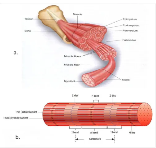
Structure of skeletal muscle
Skeletal muscles are composed of multinucleated myofibers organized into fascicles that are grouped together to form individual muscles. Individual myofibers develop by end-to-end fusion of mononucleate myoblasts to form a multinucleate syncytium. Each mononucleate cell is a contractile unit known as sarcomere, bounded by Z discs, that is, structures anchoring the actin filaments into each end of the sarcomere. The sarcomere shortens when the actin filaments slide along the thick myosin filaments toward the middle of the sarcomere (Ham and Cormack, 1987; Alberts B et al, 2008). Each myofiber is wrapped by endomysium, multiple myofibers are organized into fascicles surrounded by perimysia, and the fascicles collectively constitute the muscle, which is surrounded by an epimysium (Figure 6).