Alma Mater Studiorum - Università degli Studi di Bologna
Dottorato di Ricerca in Biochimica
CICLO XX
Coordinatore Prof. Giorgio Lenaz
Identification of Drosophila heart-specific
Cis-Regulatory Modules under Hox control
Tesi di Dottorato della Dott.ssa Maria Florencia TEVY
Relatore: Dr.ssa Maria CAPOVILLA
SUMMARY
Cardiac morphogenesis is a complex process governed by evolutionarily conserved transcription factors and signaling molecules. The Drosophila cardiac tube is linear, made of 52 pairs of cardiomyocytes (CMs), which express specific transcription factor genes that have human homologues implicated in Congenital Heart Diseases (CHDs) (NKX2-5, GATA4 and TBX5). The Drosophila cardiac tube is linear and composed of a rostral portion named aorta and a caudal one called heart, distinguished by morphological and functional differences controlled by
Hox genes, key regulators of axial patterning. Overexpression and inactivation of the Hox gene abdominal-A (abd-A), which is expressed exclusively in the heart, revealed that abd-A controls
heart identity. The aim of our work is to isolate the heart-specific cis-regulatory sequences of
abd-A direct target genes, the realizator genes granting heart identity. In each segment of the
heart, four pairs of cardiomyocytes (CMs) express tinman (tin), homologous to NKX2-5, and acquire strong contractile and automatic rhythmic activities. By tyramide amplified FISH, we found that seven genes, encoding ion channels, pumps or transporters, are specifically expressed in the Tin-CMs of the heart. We initially used online available tools to identify their heart-specific cis-regutatory modules by looking for Conserved Non-coding Sequences containing clusters of binding sites for various cardiac transcription factors, including Hox proteins. Based on these data we generated several reporter gene constructs and transgenic embryos, but none of them showed reporter gene expression in the heart. In order to identify additional abd-A target genes, we performed microarray experiments comparing the transcriptomes of aorta versus heart and identified 144 genes overexpressed in the heart. In order to find the heart-specific cis-regulatory regions of these target genes we developed a new bioinformatic approach where prediction is based on pattern matching and ordered statistics. We first retrieved Conserved Non-coding Sequences from the alignment between the D.melanogaster and D.pseudobscura genomes. We scored for combinations of conserved occurrences of ABD-A, ABD-B, TIN, PNR, dMEF2, MADS box, T-box and E-box sites and we ranked these results based on two independent strategies. On one hand we ranked the putative cis-regulatory sequences according to best scored ABD-A biding sites, on the other hand we scored according to conservation of binding sites. We integrated and ranked again the two lists obtained independently to produce a final rank. We generated nGFP reporter construct flies for in vivo validation. We identified three 1kb-long heart-specific enhancers. By in vivo and in vitro experiments we are determining whether they are direct abd-A targets, demonstrating the role of a Hox gene in the realization of heart identity. The identified abd-A direct target genes may be targets also of the NKX2-5, GATA4 and/or TBX5 homologues tin, pannier and Doc genes, respectively. The identification of sequences coregulated by a Hox protein and the homologues of transcription factors causing CHDs, will provide a mean to test whether these factors function as Hox cofactors granting cardiac specificity to Hox proteins, increasing our knowledge on the molecular mechanisms underlying CHDs. Finally, it may be investigated whether these Hox targets are involved in CHDs.
Homeotic transformations
The mutant fly with four wings and no halteres was first identified by Calvin Bridges in Thomas Hunt Morgan laboratory in 1915 (Figure 1).
Figure 1.
This now famous little "monster" was a starting point for Edward Lewis in his research on homeotic transformations. In 1978, Lewis published his work on the bithorax complex (Lewis, 1978; Lewis; 1998). In this paper he reviewed a series of Drosophila mutations that affect the thoracic and abdominal segmental identities. These mutations were called “homeotic” (from the greek homoios or “similar”) because one single mutation transforms a whole segment into another (towards an anterior one). Remarkably, the different homeotic mutations mapped to the third chromosome in an order that corresponds to the anterior to posterior order of the segments in which they act. These collinear genes responsible for the anterior to posterior patterning along the body axis are now called Hox genes, because they share a 180bp sequence called “homeobox”, which encodes a 60 amino acids DNA-binding domain called “homeodomain”. Still more astonishing was the fact that these genes are evolutionarily conserved in function and organization from Hydra to Homo. In the genome of Drosophila, there are two clusters called Antennapedia and bithorax, both located on chromosome III. Mammals possess 39 Hox genes ordered in four clusters that reflect their temporal order of expression during embryogenesis and along the anterior-posterior (AP) axis (Figure 2). This means that the genetic control mechanisms of body planning have been preserved roughly unchanged through 650 million years of evolution. (reviewed in Lawrence and Morata, 1994; Carroll, 1995; Carroll, 2005; Maeda & Karch, 2006; Iimura & Pourquie, 2007).
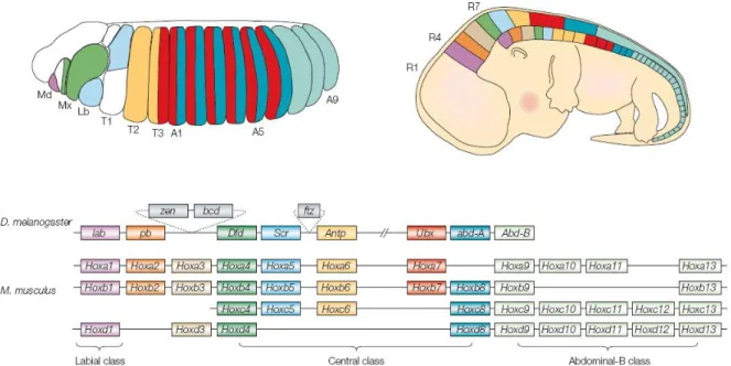
Figure 2 (modified from Pearson et al., 2005). Hox expression and genomic organization in
Drosophila melanogaster and Mus musculus. The diagram on the left shows a stage 13 Drosophila embryo colored to indicate the approximate domains of expression for all Drosophila Hox genes except proboscipedia (pb), which is not expressed in the embryo. The
segments are labeled: Md, mandibular; Mx, maxillary; Lb, labial; T1-T3, thoracic segments; A1-A9, abdominal segments. The diagram on the right shows a mouse embryo at embryonic day 12.5, with the approximate Hox expression domains depicted on the head to tail axis. The positions of the hindbrain rhombomeres are labeled R1, R4, R7. In both diagrams, the domains of expression of the Hox transcripts are color-coded as the corresponding genes in the Hox cluster diagram below. The diagrams below the embryos show the Hox clusters in the genomes of Drosophila and mouse with the genes in the order in which they are found in the chromosome. Genes are colored to differentiate between Hox paralogous members. Genes that are orthologous between clusters and specie are labeled in the same color. The position of three non-Hox homeodomain genes zen, bcd and ftz are shown in the fly Hox cluster in grey boxes. Gene abbreviations: lab, labial; pb, proboscipedia; zen, zerknullt; bcd, bicoid; Dfd, Deformed;
Scr, Sex combed reduced; ftz, fushi tarazu; Antp, Antennapedia; Ubx, Ultrabithorax; abd-A, abdominal-A; Abd-B, Abdominal-B. Anterior is to the left and dorsal to the top.
Hox genes in morphogenesis
The homeotic function is the trademark by which Hox genes became known. Nevertheless, the function of Hox genes is that of regulating morphogenesis and organogenesis. This is the function we have to study if we want to answer the question of how differences arise from sets of genes so widely shared.
Hox genes encode homeodomain transcription factors (reviewed in McGinnis, 1994).
One target of Hox genes are Hox genes themselves. To control organogenesis, Hox proteins act at a local cellular level within the segment they specify and regulate groups of target genes. The
Hox protein function here is to get integrated into an enhanceosome at a given moment of
development and confer AP positional information to this enhanceosome (Castelli-Gair Hombría & Lovegrove, 2003). Garcia Bellido (1977) coined the term “realizator genes” to describe these sets of genes subordinated to Hox control that mediate segment-specific morphogenesis.
Hox target genes
As Capovilla et al. stated in their 1994 paper: “Understanding how genes of the HOM/Hox family control morphogenesis requires the identification and characterization of their target genes [...]. Yet very little is known about the nature of these target genes and their roles in segment-specific morphogenesis.”
There are characteristics inherent to Hox genes that make the identification and characterization of their target realizators difficult. To start with, almost all Hox proteins bind in
vitro to a short core consensus 5'-TAAT-3' sequence. This sequence occurs in the genome
approximatively every 1 kilobase. In vitro promiscuous binding is very common and in consequence, of all the in vitro detected binding sites most have no in vivo significance (Ekker et
al., 1991, 1992). Second, Hox protein action may not be cell autonomous, acting through
signaling cascades which can in turn lead to effects outside the Hox expression domain. This is the case where the Hox gene abd-A activates only the target gene rhomboid (rho) in the C1 cell lineage to promote the differentiation of oenocytes in the dorsal ectoderm (Brodu et al., 2002). At another level, in some tissues and organs, Hox genes have overlapping expression domains and cross-regulate each other, therefore the target may require inputs from multiple Hox genes. For example, the activities of the Hox genes Ubx and abd-A are integrated at the decapentaplegic (dpp) visceral mesoderm enhancer, where UBX acts as an activator and ABD-A as a repressor by binding to the same sites in the sequence (Capovilla et al., 1994; Capovilla and Botas, 1998). UBX and ABD-A instead appear to act both as repressors of the limb promoting target gene
Distal-less (Dll) apparently also binding through the same HOX binding sites (Gebelein et al.,
2004). Moreover, the control region of the putative target may be far from the transcription unit of the target gene, and thus difficult to find.
In spite of all these experimental difficulties, a few Hox target genes (direct or indirect) are known, although the exact number is still a matter of debate. A direct target provides compelling evidence that a specific Hox protein is binding to a specific enhancer in vivo (Mahaffey, 2005; Pearson et al., 2005). The definitive demonstration that a given gene is a direct target of a given HOX is the mutation of the HOX binding sites of its enhancer towards BICOID (BCD) binding sites. Bcd is not a Hox gene and its protein binds to a different consensus site than Hox proteins. HOX can be made to bind to this BCD site by changing only one amino acid in their homeodomain (Treisman et al., 1992; Schier & Gehring, 1992). So, if BCD sites are introduced in place of the HOX sites and the enhancer looses activity, and if a compensatory mutation in the Hox protein, allowing it to bind to the mutant BCD site, restores the activity of the enhancer, we are sure that that HOX transcription factor binds in vivo to the enhancer sequence of the given gene. Nevertheless, such rigorous test has only been performed in four cases: to demonstrate that dpp is a direct target of Ubx (Capovilla et al., 1994) and of abd-A (Capovilla and Botas, 1998), that apterous is a direct target of Antp (Capovilla et al., 2001), and that the intronic sequence of Hoxa-4 is regulated by Ubx when assayed in Drosophila (Haerry & Gehring, 1997).
Moreover, most of the known targets are either transcription factors or signaling molecules, therefore not true realizators. On the other hand, we still have little or no knowledge of targets regulated at the same time and in the same place by the same Hox protein. Finding groups of co-regulated direct target realizator genes within similar cell types during development will help us assess the question of how is it that a Hox gene triggers specification, differentiation and organogenesis. From a broader point of view, Hox-dependent regulatory networks will help us to understand how these master genes create cell diversity in order to make serial structures different.
HOX specificity
There are only eight Hox genes in Drosophila and probably thousands of realizator genes in order to make and organ. How do Hox proteins selectively regulate such broad spectrum of target genes? The segments in Drosophila share plenty similarities so one could think that similar sets of target genes are shared by different Hox proteins or by different combinations of
Hox proteins. But if we think the number of target genes in a co-regulated set is variable from
one in the precursors of Keilin’s organs (Vachon et al., 1992) or the oenocytes (Brodu et al., 2002) to possible more than a hundred in the halteres (Akam, 1998; Weatherbee et al., 1998), we then realize such explanation does not account for so many clear morphological differences that are Hox-dependent.
On the other hand, at the protein level, Hox proteins bind to DNA through the homeodomain. The three alpha helices that conform the homeodomain are shared by all these transcription factors (Gehring et al.,1994, Mann, 1995). Moreover, Hox proteins (except for ABD-B) have indistinguishable in vitro binding sites. Then, how is HOX functional specificity achieved in vivo? How do individual Hox proteins selectively activate or repress target gene expression?
HOX specificity appears to depend on several non-exclusive mechanisms. One possibility is that the binding of a cofactor to a Hox protein confers the selectivity (and higher affinity of the Hox protein for its target). Unfortunately, only a couple of HOX cofactors constituted by extradenticle (exd) and Homothorax (Hth), the homologs of mammalian PBX and MEIS proteins respectively (Moens & Selleri, 2006), have been identified to date (Chan et al., 1994; Mann & Affolter, 1998; Ryoo et al., 1999). On the other hand, many processes involving the function of Hox genes have been found to be exd-independent (Pinsonneault et al., 1997; Galant et al., 2002). The failure of traditional genetic and biochemical screens to identify new HOX cofactors suggests that Hox genes might not always function exactly how we previously expected. This view is reinforced by the recent and unforeseen finding that Hox and segment-polarity genes cooperate to control Dll expression. Thus, another possibility is that Hox proteins collaborate (cooperate and/or recruit sets of activators and repressors) with other transcription factors to control gene expression. Collaboration does not imply a physical interaction between the transcription factors integrated in the enhanceosome (Walsh & Carroll, 2007). In other words, a combination of different proteins including one or more HOX would regulate transcription. In fact, there is an expanding pool of transcription factors that are putative collaborators in the modulation of Hox target genes. Some evidence comes from the collaboration between UBX and SMADs in the selective repression of the target gene spalt (sal) in the Drosophila haltere disc (Walsh & Carroll, 2007). Furthermore, new insights on target selection come from the way a Hox protein binds to the DNA structure. Recent experiments suggest that Hox proteins recognize “generic” binding sites through the homeodomain-major groove interactions while the N-terminal arm and the linker residues would select among these sites by “reading” the structure and electrostatic potential in the minor groove (Joshi et al., 2007).
A new model for old questions
From all the mentioned characteristics of Hox genes and Hox proteins it is evident that their functions are difficult to analyze. Since the 1980s new techniques have been developed and with them new questions regarding Hox genes have been raised. Instead, the number of model systems to assess Hox function and HOX specificity has remained mainly the same and new ones need to be introduced. Cardiogenesis in Drosophila constitutes such a system.
The Drosophila cardiac tube
The embryonic cardiac system of Drosophila is formed by a simple linear tubular structure at the dorsal midline below the epidermis of the embryo and it is called “dorsal vessel” or “cardiac tube” (Campos-Ortega & Hartenstein, 1997). Just like the vertebrate primitive cardiac tube, the
Drosophila dorsal vessel presents AP polarity (Lo & Frasch, 2003). The anterior portion of the
tube is called “aorta” and the posterior one “heart” (Figure 3). The heart is the autonomous beating organ (Wessells et al., 2004) that pumps the haemolymph (“insect blood”) in a posterior to anterior manner (Rizki, 1978), comparable to the direction of blood flow in the vertebrate primitive cardiac tube (Forouhar et al., 2006). In the fly open circulatory system, the haemolymph reenters the heart through valve-like cells named ostia (Rizki, 1978).
Figure 3 (modified from Campos-Ortega & Hartenstein, 1997). Dorsal view of the cardiac tube of an embryo at the end of embryogenesis. The dorsal vessel extends from segment T1 (not shown in the diagram) to segment A7. Alary muscles (al) attach the dorsal vessel to the apodemes. The dorsal vessel, consisting of the heart posteriorly (middle of segment A4 to segment A7) and the aorta anteriorly (segment T1 to middle of segment A4), is formed by cardiomyocytes (ca), in association with a bilateral row of pericardial cells (pc). In its anterior portion, the cardiac tube is flanked by the lymph glands (lg) and the ring gland (rg). Abbreviations: brain, br; supraoesophageal commissure, sec; posterior spiracle, sp; thoracic segment, t; abdominal segment, a.
The formation of the cardiac tube
The dorsal vessel, like the somatic and visceral musculature and the fat body, has mesodermal origin. The mesoderm is derived from the most ventral cells of the blastoderm (Campos-Ortega & Hartenstein, 1997). The cells of the mesoderm express twist (twi) and later snail (sna), both encoding transcription factors. twi, in turn, activates tinman (tin), the gene encoding a homeodomain transcription factor of the NK class (reviewed in Ruiz-Gomez, 1998). At gastrulation, these cells invaginate along the ventral furrow and spread dorsally to form a monolayer beneath the ectoderm. During dorsal migration important regulatory events that will ultimately lead to specification of heart progenitors are triggered (Frasch, 1999). The cardioblasts become specified thanks to inductive signals that come from the neighbouring ectoderm (Zaffran & Frasch, 2002), which comprehend the cascades of the signaling factors dpp and wingless (wg), homologous to the vertebrate BMP and WNT molecules in vertebrates, respectively. Upon spreading of the mesoderm beneath the ectoderm, the mesoderm utilizes the pattern information from the ectoderm to acquire its distinct dorsal-ventral polarity. dpp
transmits dorsal positional information from the ectoderm to the mesoderm (Frasch, 1995; Lockwood & Bodmer, 2002). A consequence of dpp function is the restriction of tin expression to the dorsal mesoderm (Frasch, 1999) and possibly the induction of pannier (pnr), encoding a Zinc finger transcription factor (Klinedinst & Bodmer, 2003). As development proceeds, wg plays a key role in segmental regulation in cooperation with sloppy paired (slp) (Lee & Frasch, 2000).
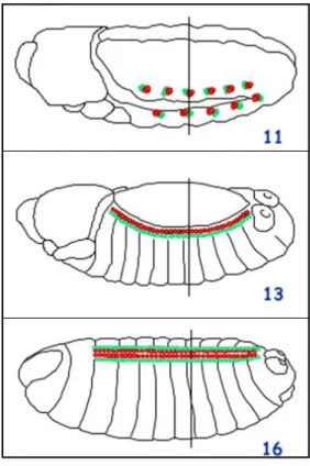
The cells from the dorsal-most edge of the mesoderm form the so-called mesodermal crests on either side of the embryonic germ band. The heart progenitors derive from segmentally restricted clusters of cells from the dorsal mesodermal crests (Campos-Ortega & Hartenstein, 1997) (Figure 4). The presumptive heart progenitors rearrange and the clusters come into contact. These cells undergo a mesenchymal-epithelial transition and as a result of these movements, two rows of cells on either side of the embryo are formed: a dorsal row of cardioblasts and a ventrally adjacent row of pericardial cells (Fremion et al., 1999) (Figure 4).
Figure 4 (from Campos-Ortega & Hartenstein, 1997). Embryonic heart development. (A) Stage 11 embryo showing segmentally restricted clusters of cells from the dorsal mesoderm, which will give rise to the heart progenitors. (B) Stage 13 embryo where the heart progenitors rearrange and the clusters come into contact after mesenchymal-epithelial transition. Two rows of cells on either side of the embryo are formed: a dorsal row of cardioblasts and a ventrally adjacent row of pericardial cells. (C) Stage 17 embryo where the two rows of cardioblasts meet and fuse at the dorsal midline to form the dorsal vessel, which becomes a hollow tube.
During germ band elongation the two rows of cardioblasts meet and fuse at the dorsal midline to form the dorsal vessel, which becomes a hollow tube extending from the first thoracic segment (T1) to seventh abdominal segment (A7) (Figures 3 and 4). At this moment, upon dorsal closure, all the differentiation programs have already been triggered so that the cardioblasts become cardiomyocytes. Part of these differentiation programs are activated by tin and pnr (Gajewski et al., 2001). Two differentiation transcription factors activated by tin and pnr
are muscle enhancer factor 2 (mef2) (Gajewski et al., 2001) and the basic helix loop helix transcription factor dHand (Han & Olson, 2005).
The mature cardiac tube is constituted by 52 pairs of cardiomyocytes, which become connected by adherent junctions. The dorsal vessel becomes surrounded in the thorax by the lymph glands (of mesodermal origin) and the ring gland (of ectodermal origin) and also by loosely arranged, non-myogenic pericardial cells (thought to act as stationary macrophages) (Rizki, 1978). The cardiac tube is attached to the dorsal ectoderm by seven pairs of alary muscles (Figure 3). As Drosophila is a holometabolous insect, this morphogenetic patrimony will remain the same until metamorphosis when all the structures will be eliminated or remodeled (Monier et
al., 2005).
Anterior-posterior organization of the dorsal vessel
Like the primitive heart tube in vertebrates, the mature cardiac tube of Drosophila presents morphological and functional axial differentiation. Anterior to posterior polarity is manifested not only in the subdivision of the dorsal vessel into the two domains, aorta and heart (intersegmental polarity), but also, within each segment that forms the cardiac tube (intrasegmental polarity) (Ponzielli et al., 2002; Lo et al., 2002).
Rostral-caudal polarity can be observed within each segment that forms the cardiac tube. Each segment is composed of six pairs of cardiomyocytes. The four most anterior pairs of cells of each segment that forms the cardiac tube express the gene encoding the homeodomain transcription factor tin (Bodmer, 1993). In addition to tin, the two most anterior pairs of cells express the transcription factor ladybird (lb) (Jagla et al., 1997). The two most posterior pairs of cells within each segment express the gene encoding the transcription factor seven up (svp), the
Drosophila homolog of the COUP-TF orphan receptors (Lo & Frasch, 2001).
Moreover, as mention previously, the entire dorsal vessel presents two distinct domains: an anterior one (from segment T1 to middle of segment A4) termed “aorta” and a posterior one (from middle of segment A4 to segments A7) called “heart” (Rizki, 1978) (Figure 3). The heart portion of the cardiac tube presents a wider diameter and lumen compared to the aorta (Ponzielli
et al., 2002). The three pairs of svp-expressing cells of the heart differentiate into valve-like cells
called ostia, which allow the inflow of haemolymph into the cavity (Lo & Frasch, 2001). The
tin-expressing cardioblasts of the heart differentiate into strong contractile beating
cardiomyocytes, which propel this hemolymph, through the aorta, into an open circulatory system in a caudal to rostral direction (Molina & Cripps, 2001). Thus, the aorta constitutes the outflow tract of the fly cardiac tube. The SVP- positive cells of the aorta do not differentiate into functional ostia and the TIN-positive cells of this domain differentiate into cardiomyocytes that are smaller and poorly contractile compared to those of the heart (Ponzielli et al., 2002).
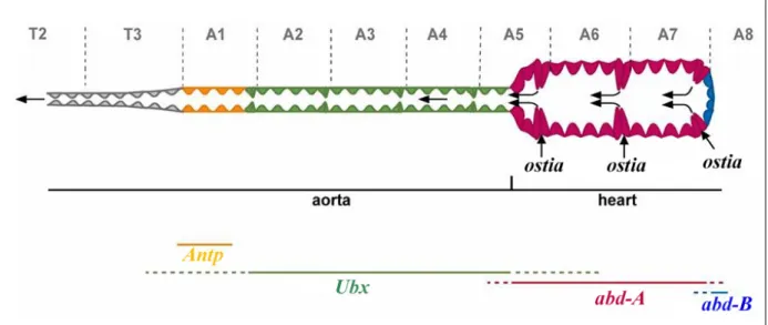
The correlation between Hox expression domains and axial polarity of the dorsal vessel prompted several laboratories to study the possible involvement of Hox genes in cardiac diversity. Four Hox genes were found to be expressed in the cardiac tube. Antp is expressed in the anterior portion of the aorta, Ubx in the posterior portion of the aorta, and abd-A is expressed in all the cells of the heart except in the last four cells where Abd-B is expressed (Figure 5).
Figure 5 (from Ponzielli et al., 2002). Rostral-caudal polarity in the Drosophila cardiac tube. The diagram shows the expression of the Hox gene Antp and the three genes of the bithorax complex (Ubx, abd-A and Abd-B) along the AP axis of the dorsal vessel. Anterior is to the left. The black arrows indicate the direction of the haemolymph flow.
In the quintuple mutant Scr-, Antp-, Ubx-, abd-A-, abd-B-, all cardioblasts adopt anterior thoracic cardiac cell identity (i.e., cardiomyoblasts that express Antp) and thus no posterior aorta cardiomyocytes and heart cardiomyocytes differentiate. Antp affects svp expression. Loss of
Antp leads to a loss of svp expression in the first pair of abdominal cardiomyocytes, while
ectopic Antp expression activates svp expression ectopically in the thorax (Perrin et al., 2004).
Abd-B is known to be a repressor of myogenesis (Michelson, 1994). Early overexpression
of Abd-B in the mesoderm inhibits cardiogenesis. Consistently, in loss of function experiments for this gene, the cardiac tube is formed by 116 cells instead of the wild-type number 104 (Lo et
al., 2002).
The double mutant Ubx-, abd-A- embryos phenocopy the quintuple mutant, that is, there is no cell diversity along the AP axis of the cardiac tube, indicating the crucial requirement of these two genes in the control of thoracic versus abdominal cell identity during early stages of cardiac development (Ponzielli et al., 2002).
In Ubx loss of function embryos, the cells of the posterior aorta (segments A1 to middle of segment A4), where Ubx is normally expressed, have some defects in polarization and pericardial cells of this domain are disorganized. On the other hand, the heart of Ubx loss of function embryos is similar to that of wild-type embryos. Ectopic expression of Ubx impairs morphology of the heart cardioblasts and the ostia do not become functional (Ponzielli et al., 2002).
In abd-A loss of function embryos, the cells of the heart are transformed into posterior aorta-like cells (i.e., those cells which express Ubx) and thus ostia function and heartbeat are not observed in living embryos (Figure 6). Ectopic expression of abd-A transforms the aorta morphologically and physiologically into heart (Ponzielli et al., 2002; Figure 6). Thus, abd-A controls heart identity.
Figure 6 (from Ponzielli et al., 2002). Control of heart identity by abd-A. (A) Expression of
Hox genes in a wild-type embryo. (B) Transformation of the heart into aorta in an abd-A-
embryo. (C) Transformation of the aorta into heart in an embryo overexpressing abd-A (UAS>abd-A).
How does abd-A control heart identity?
Hox genes encode transcription factors, so in order to control morphogenesis they regulate
downstream effector genes. These effector genes can be other transcription factors, signaling molecules or realizator genes. In order to find out which are the Hox target realizator genes and in order to determine if these are direct or indirect targets we need to find and study the cis-regulatory sequence of such genes.
Three genes, Ndae1, Ih and Ork1 have been found to be expressed in the TIN-positive cells of the heart portion of the cardiac tube (Perrin et al., 2004; Monier et al., 2007). These encode channel proteins: Ndae1 encodes a Na+ driven anion exchanger belonging to the NBC family (Romero et al., 2000), Ih encodes a Na+/K+ hyperpolarization activated channel belonging to the HCN family, and Ork1 a two pore domain K+ channel (Perrin et al., 2004; Monier et al., 2007). The latter one, is involved in heart rate and heartbeat activities in the fly (Lalevée et al., 2006) and Ih possibly generates the cardiac pacemaker If or Ih currents (Occor et al., 2007).
Thus, these genes constitute part of the realizators of the heart. Moreover, these genes have concomitant expression to that of abd-A in wild-type as well as in gain and loss of function experiments (Perrin et al., 2004). To find the heart cis-regulatory sequences of these putative
abd-A target realizator genes constitutes the main aim of our project. The importance of
understanding how realizator genes are regulated in normal heart development will help us understand the mechanisms underlying Congenital Heart Diseases (CHDs) From these experiments we will also gain insight on Hox specificity during development.
A
B
The late role of abd-A and Ubx in cardiogenesis
There is only indirect evidence, from loss and gain of function experiments, for the role of abd-A as an activator of putative target genes such as Ndae1, Ih and Ork1. The possible role of Ubx as a repressor in late cardiogenesis comes from the observation of a slight loss of heart expression of
Ndae1 (Perrin et al., 2004), Ih and Ork1 (Bruno Monier, Laurent Perrin and Michel Sémériva,
unpublished) upon ectopic expression of Ubx. Therefore, in such embryos the loss of expression of the target genes in the heart may be due to the forced posterior expression of the repressor. On the other hand, in the same embryos, the target genes may be activated by normal presence of
abd-A. In wild-type embryos, abd-A normally represses Ubx. In abd-A- embryos, Ubx is de-repressed posteriorly, and therefore the lack of expression of the target gene in these mutant embryos could be due either to repression of Ubx or to lack of activation by abd-A. Given the fact that abd-A and Ubx are involved in lineage specification at earlier stages of cardiogenesis (Perrin et al., 2004), it is not possible to asses the expression of putative target genes in double mutant Ubx-, abd-A- embryos, since these embryos lack cardiomyocyte diversity and all the cells of the cardiac tube resemble those cells of the anterior aorta that normally express Antp (Perrin et
al., 2004).
We designed one experiment to resolve this issue. UAS>abd-AHx are flies that carry an inducible mutated hexapeptide variant of abd-A (Merabet et al., 2003) capable of inducing normal lineage choice in early cardiogenesis and repressing Ubx in the heart. Such protein variant does not lead to ectopic expression of the putative target gene Ih (BM, LP and MS, unpublished). We propose here to study in vivo the late role of abd-A and Ubx in the activation/repression of realizator genes by assaying expression in embryos which bear the
UAS>abd-AHx transgene in a double mutant Ubx-, abd-A- background. Such embryos will lack
Ubx and abd-A expression but will have appropriate early lineage specification and thereby
abdominal cardiomyocytes will be formed. Nevertheless, the abdominal cardiomyocytes will acquire posterior aorta morphology. A lack of Ih expression in these embryos indicates a late role of abd-A as an activator, since in these double mutant embryos the repressor function of Ubx is absent.
A core network of transcription factors to develop a heart
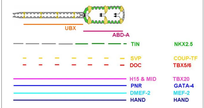
Although abd-A controls heart identity, it does not account alone for the regulation of a myriad of target genes that are needed to form a heart and that are also expressed in other tissues or organs. Indeed, heart development is governed by a core network of evolutionarily conserved transcription factors (Olson, 2006; summarized in Figure 7).
The first gene to play a role in mesodermal subdivisions encodes the homeodomain transcription factor TIN (Bodmer, 1993). In tin mutants the heart fails to form (Bodmer, 1993; Azpiazu & Frasch, 1993), but on the other hand, overexpression of tin does not cause ectopic heart induction (Yin & Frasch, 1998; Lockwood & Bodmer, 2002). Nkx2-5 is the vertebrate homologue of tin, but it does not rescue completely the phenotype of tin mutants (Park et al., 1998; Ranganayaculu et al., 1998). Nevertheless, the repression function of the protein is equivalent in late stages of development (Zaffran et al., 2006) meaning that some of their functions during heart development have diverged while others have been conserved.
pnr (Ramain et al., 1993; Winick et al.,1993), encoding the ortholog of the vertebrate
Zn-finger GATA-4 transcription factor (Gajewski et al., 1999), is expressed only in the cardiogenic region during mesoderm differentiation where it mediates dpp signaling and acts in concert with
tin (Klinedinst & Bodmer, 2003). In pnr mutant embryos, tin expression is dramatically reduced
in the clusters that correspond to the cardiac precursors (Klinedinst & Bodmer, 2003). Ectopic expression of pnr can only activate early mesodermal ectopic expression of tin, meaning that pnr is insufficient to maintain tin expression later on (Klinedinst & Bodmer, 2003). Like NKX 2-5
and GATA-4 in vertebrates, TIN and PNR act synergistically (Durocher et al., 1997) to activate differentiation genes like D-mef2 (Gajewski et al., 2001) and dHand (Han & Olson, 2005).
Possibly the oldest of these core heart transcription factors is the MADS-box protein MEF2 (Olson, 2006). Vertebrates have three different mef2 genes (Potthoff & Olson, 2007) while there is only one in Drosophila. The single mef2 gene in Drosophila (D-mef2) is expressed in all muscle lineages (Nguyen et al., 1994; Lilly et al., 1994), thus it is expressed in all the cardiomyocytes. D-mef2 mutant embryos have proper heart specification but cardioblasts fail to differentiate and contractile protein genes fail to be activated (Lilly et al., 1995). Recently, it has been demonstrated that D-mef2 activates its targets in a temporal dose-dependent manner (Elgar
et al., 2008).
dHAND is a basic helix-loop-helix transcription factor that, like D-mef2, is a direct target of tin and pnr during cardioblast differentiation (Han & Olson, 2005). dHand is expressed in all the cardiomyocytes, pericardial cells as well as in the haematopoietic precursors of the lymph glands (Kolsch & Paululat, 2002; Han & Olson, 2005). dHand mutant embryos display abnormal cardiac morphology and a deficient number of pericardial cells is observed (Han et al., 2006). HAND factors are also involved in vertebrate cardiogenesis (Yamagishi et al., 2001). Moreover, human Hand genes rescue Drosophila dHand mutant embryos, suggesting an evolutionarily conserved role of these factors in mammalian cardiogenesis (Han et al., 2006).
The TBX family of transcription factors is also necessary to build a heart. There are eight T-box genes in the Drosophila genome. Five of them, Dorsocross 1-3 (Doc) (Reim et al., 2003, 2005), H15 and midline (mid) (Miskolczi-McCallum et al., 2005; Qian et al., 2005; Reim et al., 2005) are expressed in the cardiac tube. The three Doc genes are expressed in the svp-expressing cells of the heart (Reim & Frasch, 2005) while H15 and mid are expressed in all the cardiomyocytes of the cardiac tube (Miskolczi-McCallum et al., 2005; Qian et al., 2005; Reim et
al., 2005). Doc2 and Doc3 mutants display severe defects in cardiac cell specification as shown
by the absence of multiple markers for the cardioblast population (Reim & Frasch, 2005). Doc interacts with tin and pnr in cardiac cell specification (Reim & Frasch, 2005). Ectopic expression of Doc in conjunction with tin and pnr results in cardioblast differentiation, suggesting these three factors act synergistically during dorsal vessel development (Reim & Frasch, 2005), just like the vertebrate TBX20 directly interacts with NKX2-5 and GATA factors (Stennard et al., 2003).
Figure 7. Cardiac expression of transcription factors involved in cardiac development. (A) Diagram representing the cardiac tube divided in aorta and heart. Below are shown the domains of expression of Ubx and abd-A. (B) The colored horizontal bars indicate the domains of expression in the cardiac tube of the transcription factors indicated to their right. On the right column are indicated the vertebrate homologs of the fly genes.
All these transcription factors involved in heart development bind to DNA through characteristic binding sites. The binding site of a given transcription factor can be represented in the form of a Positional Weight Matrix (PWM). A PWM is a matrix that is created from a collection of in vitro footprints of a given RNA or DNA-binding protein. Such biding sites are aligned, and a score is assigned to the probability of occurrence of a certain base in a certain position (Hertz & Stormo, 2002). In this way, binding sites that slightly diverge from the consensus sequences can be also represented. Due to the sequencing of Drosophila genomes and the availability of information of the sequence of binding sites of many transcription factors, it is possible to assess through bioinformatic methods the presence of putative in vivo binding sites of transcription factors in a given DNA sequence (Stormo, 2000; Osada et al., 2004). In the past, in
vitro inmunoprecipitation experiments were used to look for putative in vivo binding sites of a
transcription factor to the DNA sequence of a putative target gene. For example, the visceral mesoderm enhancer of dpp was found by inmunoprecipitating a genomic fragment containing the enhancer with the Hox protein Ubx (Capovilla et al., 1994). Bioinformatic tools allow to assess the presence of a number of transcription factor binding sites simultaneously and moreover to assess their spatial distribution in order to determine if such transcription factor binding sites form a cluster which could stand in vivo for a cis-regulatory motif of a given gene (Halfon et al., 2002; Markstein et al., 2002; Grad et al., 2004; Berman et al., 2004). Using bioinformatic tools, the binding sites of the mentioned evolutionarily conserved transcription factors in the core network can be found in the sequence of the heart realizator genes.
In first instance, we analyzed through bioinformatics if the binding sites of these conserved transcription factors are present in the putative cis-regulatory sequences of the putative abd-A target genes expressed in the heart portion of the cardiac tube. We looked for conserved clusters of binding sites of the mentioned transcription factors that might be forming
part of the enhanceosome regulating realizator gene expression in the heart. On second place, we are currently studying which of these transcription factors might cooperate or collaborate in vivo with abd-A in the regulation of the putative Hox heart targets in order to realize a heart with its morphological and physiological distinct cell types. As some of the human homologues of these transcription factors are involved in congenital heart diseases (in particular, NKX2-5, TBX5 and GATA4) the identification of the cis-regulatory sequences of their target genes may help elucidate what are the molecular abnormalities of the altered proteins in the patients and help device eventual therapeutic strategies.
A. Bioinformatics
The bioinformatic approaches applied in this project have been developed through time as more informatic tools evolved from 2004 to present. All bioinformatic procedures applied here are the product of a collaboration with experts in this research area (David Martin and Bruno Zeitouni at the IBDML of CNRS in Marseille, France; Stein Aerts at the Laboratory of Neurogenetics, Dept. of Molecular and Developmental Genetics, University of Leuven, Belgium).
1 - First bioinformatic approach based on available online tools
Most methods used up to 2004 identified cis-regulatory sequences from interspecies sequence comparison. They identified Conserved Non coding Sequences (CNSs), operationally defined as islands of non coding sequence with relatively high conservation flanked by regions of low conservation and assumed that this conservation reflected regulatory function (Bergman et al, 2002). The benchmark set by the new toolkits developed in 2004 was that of searching for conserved clusters of known transcription factor binding sites within conserved regions of the
Drosophila genome.
The sequence of a given transcription factor binding site is expressed as a Positional Weight Matrix (PWM). The source of the PWMs in this boinformatic approach is FlyReg/pollard [http://rana.lbl.gov/~dan/matrices.html]. These PWMs were created by Dan Pollard (Eisen’s Lab) from a DNAseI footprint database. The PWMs used from this database are those of the following proteins: DMEF-2, TIN, GATA, TBX, UBX. We used the UBX PWM instead of the ABD-A one because the former was more precise (i.e., was created from a larger set of DNAseI footprints) and in any case UBX and ABD-A bind in vitro to the same sequences (Capovilla et al., 1994).
For the transcription factor SVP, which was absent in the mentioned database, and no footprints were available in literature, we created a PWM de novo from a few published binding sites of the SVP vertebrate homolog COUP-TF.
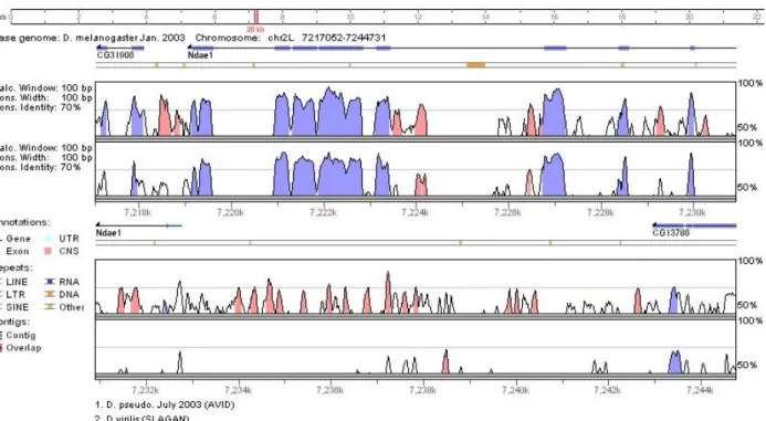
In 2004, Berman et al. published the eCIS-ANALYST toolkit, which searches in the
Drosophila melanogaster and Drosophila pseudoobscura genomes conserved clusters of
Transcription Factor (TF) Binding Sites (BSs). More information about how this tool works can be found at http://rana.lbl.gov/cis-analyst/. At the same time, were published the VISTA tools (Frazer et al., 2004), which are a comprehensive suite of programs and databases for comparative analysis of genomic sequences. More information about this toolkit can be found at
http://genome.lbl.gov/vista/index.shtml. We used the VISTA Browser to retrieve CNSs between
the gene of interest in the D. melanogaster genome and its orthologue in the D. pseudoobscura genome sequence and to visualize these CNSs in an alignment. We also used rVISTA (Loots et
al., 2002) to search for conserved clusters of TF BSs.
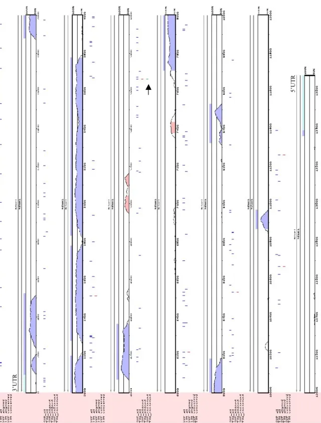
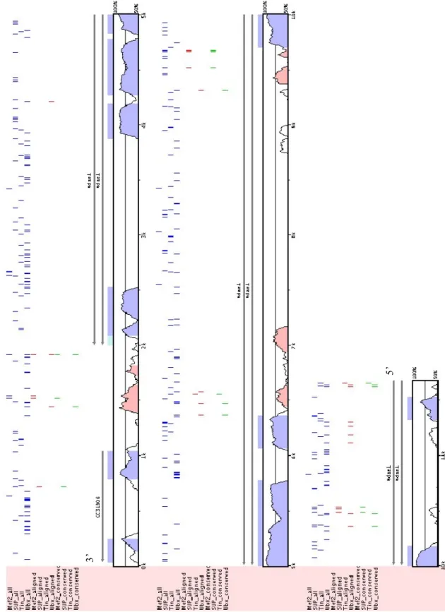
A third approach, based always on the same principle, was the one we set up in collaboration with Dr. David Martin (unpublished). This method applied two strategies. One strategy was to look first throughout the D. melanogaster gene of interest for TF BSs using PWMs. We took the genomic sequence including the query gene from the previous Computed Gene (CG) to the next one. In a second step, we aligned this sequence to the orthologue in D.
pseudobscura, using pre-computed global alignments. We then tested if the TF BS score fell into
CNSs. A second strategy of this same approach was designed to circumvent the problem that a few conserved bases inside a non-conserved region of the genome might be missed in the general alignment. We manually introduced in the previously aligned sequence all TF BSs found previously. This allowed us to distinguish aligned sites from conserved sites. We also allowed the program to find TF BSs that slightly deviated from the consensus (i.e., we changed the p-value during the scoring step). Moreover, with this method we could analyze different percentages of sequence identity (i.e., 50% conservation and 70% conservation).
merged and compared in order to give the selected regions the priority to be cloned. The overlapping was made taking the query gene sequence aligned (either to the D. pseudobscura or the D. virilis orthologue) and manually drawing all the different outputs. We did not use any statistical methods.
2 - Second bioinformatic approach based on a novel pattern matching based
method
Throughout the last three years, our collaborators in Dr. Semeriva's lab found seven genes to be differentially expressed in the heart portion of the cardiac tube, through a candidate gene approach (Monier et al., 2007). Moreover, recently, they undertook a genome-wide approach to gain more knowledge on the heart transcriptome. They performed microarray experiments comparing the larval heart transcriptome with that of the aorta. With this experiment, 144 genes were found to be overexpressed in the heart with respect to the aorta. These constituted a new set of putative downstream target genes under specific abd-A regulation. Following these new results came the necessity to improve our bioinformatic methodology for heart cis-regulatory discovery.
Summary
A pattern matching based approach was used to search for motifs with PWMs of known TF BSs in CNSs. This approach consists of two strategies, which we named A and B.
In strategy A, we first searched for the CNSs with the best multiple high-scoring ABD-A BS. We then looked for clusters of BS of heart TFs except ABD-A. The same two searches were applied to the CNSs of D. pseudoobscura. We then integrated these results and produced a first scoring ranking which is after refined in a final ranking according to the presence of at least three high scoring TF BSs besides that of ABD-A. We called this last ranking list “TopA”.
Instead in strategy B, we first searched the best CNSs with clusters of BSs of heart TFs (ABD-A included). We did this separately for the D. melanogaster and D. pseudoobscura CNSs. We kept the CNSs with the best motif conservation score from alignments. Then, we integrated the results for CNSs of both species and produced a ranking which we called “TopB” list.
We posteriorly overlapped and ranked the lists TopA and TopB to produce a final list containing the best 20 scores that we called “Top20”. For these best 20 putative Cis-Regulatory Modules (CRM) we used Toucan to draw inside each of them the predicted binding sites.
Our new approach in detail
We used a pattern matching based approach to search for motifs with PWMs of known TF BSs in CNSs. This approach consists of two strategies, which we named A and B. Our first gene dataset from which we started off to validate the approach was composed of 7 genes, expressed in the heart and not in the aorta, which were found by candidate gene approach (Na+ driven anion exchanger 1: Ndae1; Ca++ channel protein β subunit: Ca-β; Ih channel: Ih; Calcium
ATPase at 60A: CaP60A; Open rectifier K+ channel 1: Ork1; seizure: sei; painless: pain) and 2
genes (Dro-myosupressin: Dms and CG15537) found in the microarray data analysis to have the highest fold change expression in the heart compared to the aorta. The expression of these latter two genes was further corroborated through fluorescent in situ hybridization.
This is our first gene set: CG4675 (Ndae1) CG6320 (Ca-β) CG8585 (Ih) CG3725 (Ca-P60A) CG1615 (Ork1) CG3182 (sei) CG15860 (pain) CG15537 (CG15537) CG6440 (Dms)
We then decided to cover all the intergenic region to avoid the problem of missing regulatory elements distantly positioned with respect to the transcription unit of the gene. We delimited the regions containing the query gene plus 20kb upstream and 20kb downstream the transcription unit. We aligned these sequences to their corresponding orthologues in the D. pseudobscura genome using SLAGAN and we retrieved all CNSs using CNScan (Bruno Zeitouni, unpublished). The CNSs retrieved match the following parameters:
- 65% identity, 100bp windows - not exons
- not UTR
We obtained 386 CNSs. We then decided to extend the CNSs with flanking sequence so that they are minimally 300bp long because it is generally acknowledged that an enhancer element is around 300-1000 bp. For example, the cardiac tube enhancer of dHand is 300 bp (Han & Olson, 2005) as the dSur cardiac tube enhancer (Akasaka et al., 2006; Hendren et al., 2007).
Since VISTA only returns the D. melanogaster sequences of the CNSs, the D.
pseudoobscura sequences are obtained from the UCSC “net” alignments
(http://genome.ucsc.edu/index.html?org=D.+melanogaster)
In strategy A, we first searched for multiple high-scoring ABD-A BSs within the CNSs.
For doing this, we used MotifLocator
(http://homes.esat.kuleuven.be/~thijs/help/help_motiflocator.html) with the ABD-A PWM from
FlyReg/pollard (http://rana.lbl.gov/~dan/matrices.html) using a threshold of 0.8. From the output we retained the top 3 scoring hits for each CNS. We then scored each CNS according to the formula: ! cnsscore PMWscore 1 3
"
CNSs were then ranked accordingly. The same scoring method is used to score and rank the D. pseudobscura CNSs found for these genes. We integrated these ranks using order statistics as described in Aerts et al. (2006). The second step of strategy A was to find clusters of BSs of TFs known to be involved in heart development. Below are listed the selected TFs and the source from where the corresponding PWM was taken. The website addresses are cited at the end of the list.
ABD-B (from Pollard matrices)
NKX25.01 (high affinity, from TRANSFAC): homologous to the D. melanogaster TIN NKX25.02 (low affinity, from TRANSFAC): homologous to the D. melanogaster TIN GATA (from TRANSFAC): orthologous to the D. melanogaster PNR
MEF2A (MADS box, from Jaspar): homologous to the D. melanogaster D-MEF2 SRF (MADS box, from ClusterBuster ): homolog to the D. melanogaster D-MEF2 TBX core motif "GGTGT" (from Gosh et al., 2001): same binding site than the
D. melanogaster TBX factors H15, MID and DOC (Frasch lab, unpublished).
EBOX (CANNTG): orthologous to the D. melanogaster dHAND
*TRANSFAC: http://www.biobase-international.com/pages/index.php?id=transfac
*Jaspar: http://jaspar.genereg.net/
* ClusterBuster: http://zlab.bu.edu/cluster-buster/cbust.html
We then used ClusterBuster setting the gap parameter at 20 and the motif threshold equal to 6 in order that predicted motifs will have relative high specificity and thus reduce the number of false positives. We ranked all CNSs of D. melanogaster according to the ClusterBuster score. We treated D. pseudobscura CNSs in the same way and finally we integrated these rankings using order statistics. We then integrated the ABD-A ranking obtained in first place to that obtained with ClusterBuster ranking and again using order statistics, we obtained an overall ranking from these previous two. We then selected those sequences with clusters with binding sites for a minimum 3 different PWMs excluding ABD-A and from these we selected the top 30 best scored in the overall ranking. We called these 30 predictions “TopA”.
In strategy B, we first looked for clusters of binding sites for all the above mentioned PWMs, including ABD-A. For this, again, we used ClusterBuster, but we ranked them according to the CNS score as defined in strategy A. We treated the CNSs obtained from D. pseudobscura in the same way. Once more, we integrated these rankings using order statistics. In a second step we looked at the evolutionary conservation of the binding sites in a cluster. We retrieved all the binding sites found in D. melanogaster CNSs. From a LAGAN alignment of each CNS we retrieved all the aligned sequence in D. pseudobscura and looked for the binding sites of D.
melanogaster in these aligned sequences. If the D. pseudobscura aligned site was of the same
length as that of D. melanogaster, we counted the number of identical nucleotides. The sum of all conserved base pairs within all binding sites of a cluster constituted our conservation score. We ranked separately all the CNSs of D. melanogaster and D. pseudobscura according to this conservation score and we integrated the obtained rankings with order statistics. In the same way as we had done in strategy A, we selected those CNSs with clusters of binding sites containing a minimum of three different PWMs including ABD-A and from these we selected the top 30 best scored in the overall ranking. We called these 30 predictions “TopB”.
We then proceeded to integrate the “TopA” and “TopB” lists according to order statistics thus creating a final “Top20” list with the best 20 putative CRMs. We used Toucan
(www.esat.kuleuven.be/~saerts/software/toucan.html) to draw inside each of these 20 CNSs the
binding sites predicted to be in each CRM.
For in vivo validation of these CRMs we arbitrarily selected further parameters as follows:
1) distance of the putative CRM to the transcription start site 2) quality and quantity of the TF BSs found in the CRM:
- number of ABD-A sites (the more the better) - at least a TIN site and a MEF2 site
- spatial configuration of the BS inside the cluster 3) GO annotations
For cloning of the predicted CRMs, whenever possible, CRMs belonging to same gene were cloned into the same construct in order to reduce the amount of transgenic flies and increase the number of predictions validated. Further details of the transgenic reporter constructs selected for
in vivo validation are explained in “Results”.
Application of this novel approach using as dataset co-regulated genes differentially overexpressed in the heart found through microarray experiments:
After in vivo validation of this “Top20”, we applied the same strategy using as initial dataset those genes found to be differentially expressed in heart with respect to the aorta through the microarray experiments. The initial dataset contains 144 significant differentially expressed genes ranked according to their expression Fold-Change (FC) in the heart with respect to the aorta. Dms and CG15537 are first in the FC ranking. From this new enlarged dataset we obtained a list of 40 CRMs, which we called “Top40”. Further details of the transgenic reporter constructs selected for in vivo validation are explained in “Results”.
B. Cloning
The molecular biology techniques described below have been adjourned from “Molecular Cloning” (Sambrook et al., 1989).
To obtain the fragments to be cloned, either restriction or polymerase chain reaction (PCR) methods were used according to the availability of restriction sites present in the fragment of interest.
1 - Vectors used
The vectors used for this project are:
Blue/white selection vectors
pBluescript KS+ (Stratagene) pUC19
Transformation reporter vectors CHAB (Capovilla et al.,1994) pStinger (Barolo et al., 2004)
pH-Stinger (Barolo et al., 2004)
2 - Polymerase Chain Reaction (PCR)
Primer design
For amplification by PCR, primers were designed using the Invitrogen online tool “OligoPerfect Designer” (http://www.invitrogen.com/content.cfm?pageid=9716).
To each primer, we added a restriction site not present in the fragment to be amplified, but present in the polylinker of the vector, to enable ligation of such fragment to the vector.
Afterwards, primers were checked for primer-dimers and secondary structure using the online tool “Oligo Calc” (http://www.basic.northwestern.edu/biotools/oligocalc.html).
PCR reactions
The DNA polymerase used in all reactions is KOD XL polymerase (Novagen), a proofreading enzyme isolated from the extreme thermophile Thermococcus kodakaraensis KOD1, which possesses superior processivity and fidelity that enables faster and more accurate PCR amplification than that which can be achieved with conventional enzymes, including Pfu DNA polymerase.
The thermocycler used in every case is Px2 Thermal Cycler (Thermo Scientific).
3 - Restriction Reactions
Restrictions with the appropriate restriction enzyme/s were carried out using one unit of enzyme for each microgram of DNA in the restriction buffer recommended by the company in a final volume according to the amount of DNA to be cut. Each restriction was carried out at 37oC for at least one hour.
4 - Purification of vector and insert
Restricted vectors were always purified from TAE 1% agarose gel using the QIAquick Gel Extraction Kit (QIAGEN). The same procedure was utilized for inserts when we had not obtained a unique PCR product. Instead, inserts where only the desired PCR product had been obtained where restricted and purified using the QIAquick Gel Extraction Kit without loading them on gel.
5 - Ligation
The nanograms of insert to put in the ligation reaction are the product of the proportion in size of insert to vector by the nanograms of vector by the excess number of molecules of insert with respect to vector, according to the following formula:
(kb of insert : kb of vector) x ng of vector x 5-10 (excess number of molecules) = ng of insert The mixture with the appropriate amounts of insert and vector was heated up at 65oC for 20 minutes. An equal volume containing a mix of the T4 ligase and buffer (Takara Ligation kit) was added to the mixture. This final ligation mixture was incubated for at least one hour at 18oC.
6 - Transformation
The plasmids constructed were transformed into Calcium-competent cells of the Escherichia coli DH5α strain lacking ampicillin resistance.
Bacterial cells were incubated on ice with the DNA of interest for 30 minutes. Transformation was performed by heatshock at 42oC for 1 minute and subsequent incubation on ice for 5 minutes. Recovery of the cells and resistance expression were allowed by shaking the cells in 1ml of Luria Broth (LB) for 20 minutes.
Recovered cells were plated on LB agar plates containing 100 µg/ml of ampicillin.
7 - Minipreps
Screening of the colonies was done using Qiagen miniprep protocol (http://www1.qiagen.com), without column purification.
8 - Midipreps
Amplification of the quantity of the plasmid of interest was done growing 50ml of plasmid-containing bacterial culture and purifying the DNA with the QIAgen tip100 midiprep kit. In every case, the DNA was resuspended in 100 µl of TE (10mM Tris-HCl pH 7.5, 1mM EDTA).
9 - Glycerol stocks
Long-term conservation of the plasmid of interest at -80oC was achieved by adding 1 ml of 50% sterile glycerol to 0.5 ml of midiprep cell culture.
C. Transgenesis
1 - Preparation of DNA for injection
The midiprep DNA to be injected was centrifuged for 20 minutes to eliminate any insoluble particles that might have remained, which could clog the needle. The supernatant was transferred to a new tube and quantified with the spectophotometer.
The plasmids to be injected bear a transformation vector containing a P element and were co-precipitated with the so called “helper plasmid” (pIChIID2-3, Ken Irvine, personal communication) bearing the transposase necessary for the integration of the P element and thus of the construct of interest into the Drosophila genome. The construct to be injected was mixed with helper plasmid in a ratio 6:1 (12 ug +2 ug) and co-precipitated in a final volume of 50 µl by addition of 0.3M NaOAc and 2.5 volumes of absolute ethanol.
After centrifugation the pellet was washed in 70% ethanol for 3 minutes and resuspended in 20 ml of injection buffer (5mM KCl, 0.1mM NaPO4, pH 6.8). The DNA was recentrifuged,
transferred to a clean new tube and only 2 µl were loaded into a Femptoneedle.
2 - Egg laying and preparation of the embryos to be injected
Egg laying
The flies were put to lay in cages on a molasses agar medium (see Appendix). Flies were submitted to a normal photoperiod, thus eggs were usually laid from the beginning of the afternoon until late in the evening.
As the transformation vectors used carry the mini white gene, the strain of flies used for egg laying were mutant for this gene. This strain is w1118 carrying a deletion of the whole
endogenous white gene.
Dechorionation, dissection and alignment of the embryos
Embryos were recovered from the molasses agar plate with a brush and washed with milliQ water. Dechorionation of the embryos was done using 40% bleach for 1 minute, followed by washing with milliQ water.
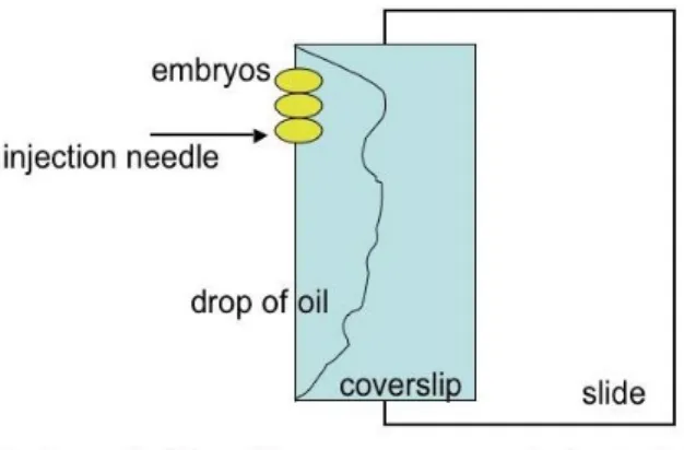
After dechorionation, embryos were aligned on a piece of agar plate with the same anterior to posterior direction for 7-10 minutes, since embryos must be injected before blastoderm cellularization, a developmental stage that begins 45-50 minutes after eggs are laid at 22°C. Then embryos were transferred to a double-sticky tape on a coverslip. The embryos aligned on the coverslip were put to dessicate for 4 minutes in a chamber containing silica gel. After dessication, embryos were covered with Halocarbon oil, and the coverslip was placed on a slide under a NikonSMZ 645 microscope, as shown in figure 8 below.
The injector used was an Eppendorf Femtojet Easy.
Figure 8. Diagram of embryos to be injected. The posterior part of the embryo, where the needle is introduced is towards the anterior part of the slide.
Treatment of the embryos after injection
The slide with the injected embryos covered with oil was placed on a molasses agar plate and covered with a lid to keep the necessary humidity. The humid chambers were placed at 18oC until the eclosion of the larvae 48 hours later.
Isolation of transformants
First instar larvae were collected from the humid chamber and out of the oil to be placed in a food vial containing a drop of fresh yeast. The vial was placed at 25oC. At this temperature, after 10 to 12 days the new adults emerged from the pupae and were crossed individually to flies of the strain w1118 in order to evidence in the next generation the presence of the transgene bearing
the w+ gene and thus the transformants (colored eyed flies) obtained from injections.
The sucessive treatment given to transformants is described on this same chapter “Materials and Methods” under the subtitle “Fly Genetics”.
D. Fly Genetics
Summary of symbols ♀ : female ♂: male w-: white strain w-1118 Hmz: homozygote Htz: heterozygote MASS: mass cross R!: recombination event Gn: generation+: wild-type chromosome
Notes:
* The chromosomes of Drosophila are written in the folllowing order: first the X, next the second (II) and last the third (III). Each pair of chromosomes is separated from the other with a
semicolon while the homologous are separated by a slash. Different alleles in the same homologous are separated by a comma.
* Drosophila males are achiasmatic.
* In every cross and throughout the generations the genotype of interest and to be selected is highlighted in yellow.
1 - Mapping of transformants
In order to map in which chromosome the transgene landed we crossed a colored eyes male to virgin females of the double balancer line SM5/Xa/TM3. SM5 and TM3 are balancer chromosomes of the second and third chromosome, respectively. They avoid meiotic recombination because they bear inversions. Xa is a translocation between the second and third chromosomes with a breakpoint in the gene apterous. SM5 is characterized by the dominant marker Curly of Oster (CyO) whose phenotype is Curly wings while TM3 is characterized by the dominant marker Stubble (Sb) whose phenotype is short bristles. In G1, males with colored eyes (due to the mini w- present in the P-element) and with both balancers will be selected to cross to
w- females. In this cross the balancer chromosomes will segregate independently from each other and the P-element will segregate opposite to the homolog where it is inserted.
G0 ♀ w-/w-; SM5/Xa/TM3 x ♂ w; P?/+
G1 ♂ w/ w-/ Y; SM5/P?/ TM3 x ♀ w-/w-; +/+; +/+
G2 Possible genotypes and phenotypes obtained depending on chromosome linkage: w- / w- or Y; P/ +; +/ TM3: colored eye flies with Stubble bristles
w- / w- or Y; P/ +; +/ +: colored eye flies
w- / w- or Y; +/ SM5; +/ TM3: white eye flies with Curly wings and Stubble bristles w- / w- or Y; +/ SM5; +/ +: white eye flies with Curly wings
or
w- / w- or Y; +/ SM5; P/+: colored eye flies with Curly wings w- / w- or Y; +/ +; P/ +: colored eye flies
w- / w- or Y; +/ SM5; +/ TM3: white eye flies with Curly wings and Stubble bristles
w- / w- or Y; +/ +; +/TM3: white eye flies with Stubble bristles or
P/ w-; +/+; +/+: colored eye females w-/Y; +/+; +/+: white eye males
P on the III P on the II P on the X
2 - Making balanced stocks of transformants
In order to generate balanced stocks of each transgene, virgin females with genotype w-/w-;
SM5/P?/TM3 (obtained in the G1 of the mapping cross described above) are crossed to males of
the same genotype (their brothers) if the P-element insertion maps to the second or third chromosome. Instead, if no color eye males where obtained in the G1 of the “mapping cross”, the virgin females of the mentioned genotype are crossed to males bearing an X balancer chromosome (FM7i).
A) If the P-element insertion is on the II chromosome:
G0 ♀♀ w-/w-; P/SM5; +/TM3 x ♂♂ w w-/w-; P/SM5; +/TM3 G1 (STOCK LINE) w-/w-; P/ SM5; +/TM3 or w-/w-; P/ P ; +/TM3
B) If the P-element insertion is on the III chromosome:
G0 ♀♀ w- /w-; +/SM5; P/TM3 x ♂♂ w/w-; +/SM5; P/TM3 G1 (STOCK LINE) w- /w-; +/ SM5; P/TM3 or w- /w-; +/ SM5; P/P
C) If the P- element insertion is on the X chromosome:
FM7i is a w- balancer X chromosome which bears the dominant marker Bar whose phenotype is “bar” shape eyes.
G0 ♀♀ P/w-; +/SM5; +/TM3 x ♂♂ FM7i/Y; +/+; +/+ G1 ♀♀ P/FM7i x ♂♂ P/Y