Alma Mater Studiorum
Alma Mater Studiorum –– Università di Bologna
Università di Bologna
DOTTORATO DI RICERCA
Biodiversità ed Evoluzione
XX Ciclo
Sezione disciplinare di afferenza: BIO/06 Anatomia Comparata e Citologia
Mechanism of colonization in the Mediterranean-sea.
Asperarca nodulosa: morphological and genetic analysis
Presentata da
Michela Kuan
Coordinatore Relatore Prof. Prof.
Giovanni Cristofolini Bruno Sabelli
Esame finale anno 2008
1. Introduction
1.1 Asperarca nodulosa, species description and geographic range
1.2 Biogeography of the Mediterranean basin
1.3 Eastern Mediterranean peculiarity: currents and salinity
1.4 Modes of Development
1.5 Morphology details
1.6 Molecular markers
2. Material and methods
2.1 Sampling area
2.2 Methods of sampling
2.3 Kind of investigation
3. Results and discussion
3.1 Morphological analysis
3.2 Hystological investigation
3.3 Diversity by longitudinal gradient
3.4 Genetic study
4. New colonization in the Mediterranean-sea from alien species:
Anadara demiri
5. Conclusion
1. Introduction
The genetic variability is considered a basin of information related to the discontinuity of the multiple variables of time and space of the environment. Unquestionably in the great depths of the sea the habitat seems to present the most stable parameters on the globe; the factors influencing the marine environment, such as temperature, light, zoo and phyto-plancton remain pratically unchanged beyond 100m. (Sanders et al., 1965; Sanders, 1969; Sandler & Hessler, 1969).
On basing of these considerations, the notion that the living fauna presents a low molecular variability at high intensities (200-1000m.) is spread. In reality, a high rate of polymorphism was noticed in precedent studies ( Gooch & Schopf, 1970); demonstrating how the ecology and the potential rates of necessary bathyal species still necessitate study even now.
My thesis is focused on the study of molecular variability in the Mediterranean populations of Asperarca nodulosa (Muller, 1776) (Arcidae-Bivalvia). The investigation was carried out in order to analyze the gene flow between differentes populations and therefore the capacity of larval dispersal in such an environment.
The analysis was performed on specimens originating from oceanographic cruises the whole Mediterranean Sea (13 collection stations) between 400 and 600 meters depth.
The Mediterranean sea is a basin with a malacofauna easily datable, having a successive origin up to the Messinian extinction. It is therefore interesting to quantify the genetic variability of bathyal species to understand the sensibility of spatial variations in a habitat with costly environmental parameters, and the consequent capacity of adaptation and the necessary lapse in time of their expansion into the Meditteranean from West to Est.
Such a research was performed via different genetic approaches, morphological (analysis of the shell larvae) and histological (to test the eventual hermaphroditism and the possibility of incubation inside gills) approach.
In order to have a more general description of the colonization mechanism in the Mediterranean, it was investigated the introductory mechanism of the alien costal species Anadara demiri, that would be able to be entered passively into the Mediterranean thanks to the bilge waters of the oil tankers from the Atlantic shipping or , actively, across the Suez channel.
1.1
Species description: Asperarca nodulosa (Müller, 1776)
Phylum: Mollusca Class: Bivalvia Subcalss: Pteriomorphia Order: Arcoida Family: ArcidaeSpecies: Asperarca nodulosa
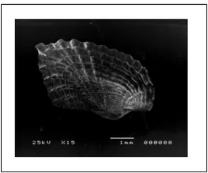
Fig. 1.1.1/ 1.1.2 – Asperarca nodulosa
The Arcidae, commonly known as ark shells, living in nearly all marine environments, are most common in the intertidal and shallow sublittoral zones. This species has a heavy, elongate, inequilateral shell, with a well-defined radial sculpture; clear and concentric growth lines intersecting the radial ones. The shell surface is covered by a thick and layered periostracum.
The taxodont hinge comprises numerous small, straight teeth, arranged transversely along the hinge line and the broad ligament are elongate with the umbones well separated. The shell is equivalve, squarely shaped and ivory, dark yellow colored.
The anatomy present a large, flat mantle without siphons, lobes are attached to shell valves along the pallian line, peripheral to wich is the mantle edge.
Asperarca nodulosa lives as byssate nestlers in corals, rocks and stones so the byssus is long and well development and, consequently, the food is little and not grown.
The geographic range is wide, the species is distributed from North-Atlantic area to middle-Atlantic and in all the Mediterranean water basin from the infralittoral (70m.) to the abyssal depth (4000m.).
1.2
Biogeography of the Mediterranean basin
Between 7 and 4 Ma, Alpine deformations were particularly marked in the Mediterranean area and the geography of this region was affected by tremendous changes related to the Messinian Salinity Crisis (Hsü et alii, 1978). Sediment samples from below the deep seafloor of the Mediterranean Sea, which include evaporite minerals, soils, and fossil plants, show that about 5.9 million years ago in the late Miocene period the precursors of the modern Strait of Gibraltar closed tight and the Mediterranean Sea evaporated into a deep dry basin trough several cycles including a pre-evaporitic phase, punctuated by high frequency anoxic event.
The consequences over the biota were the extermination of the basin’s marine organism.
Fig. 1.2.1 – Mediterranean sea during the “Messinian Salinity crisis”
The re-flooding and, therefore, the re-colonization of the entire Mediterranean sea was a rapid event (Ryan, Hsu et al., 1973; McKenzie et al., 1999; Iaccarino et al., 1999; Fortuin and Krijgsman, 2003) taking place at the beginning of the Pliocene, if not already during the latest Messinian (Steffahn and Michlzik, 2000).
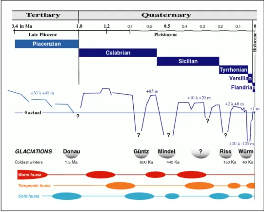
During the Plaisancian transgression, the warm climate (approximately 4°C above the present) was relatively stable; warm to subtropical sea-waters
predominated. This allowed the entrance through the strait of Gibraltar of an Atlantic fauna from the Ibero-Moroccan region that resulted in the first restocking of the Mediterranean Sea. Then, at about 2.1-2 Ma, the climate became temperate to cold. The change favored the entrance of a great number of species from the temperate regions of the Atlantic Ocean. An "Atlantico-Mediterranean" stock was established and became dominant in the Mediterranean fauna. Although the Plio-Pleistocene boundary remains controversial, it may be defined by the arrival of these boreal species in the Mediterranean consequent on climatic and oceanographic changes during the regression at the end of the Plaisancian. The temperature of surface waters decreased several degrees with changes in the direction of the prevailing wind during the Donau glaciation
Thereafter, during the Quaternary (Pleistocene to Present) a succession of remarkable climatic fluctuations took place, produced by successive glacial and interglacial periods. The bathymetry of the Mediterranean was similar to that of today, the Strait of Gibraltar was silled at about 300 m and variations in sea levels were produced by the glacio-eustatic processes of the World Ocean.
During the cold climates of the glacial periods, large biocenosis developed on the outer part of the continental shelf and along the continental slope. They consisted mainly of circalittoral and bathyal species, generally of a temperate to boreal origin. Their introduction from the Atlantic was favoured by the incoming bottom-current in the strait of Gibraltar. On the other hand, during the interglacial periods with a temperate to warm climate (representing only about 10 % of an entire cycle of glaciation: 90 to 125 Ka), the coastal current entering the Mediterranean allowed the colonisation of subtropical species from the Senegalese Province especially those in the littoral waters, while the outgoing bottom-current made the arrival of deep-sea species difficult
During the Calabrian (Donau glaciation), the Sicilian (Mindel glaciation) and the Tyrrhenian (Riss glaciation), the Pliocene stock of subtropical species disappeared and a stock of boreal species (sometimes called the "Celtic" fauna) was added in successive waves to the previous Atlantico-Mediterranean stock of temperate origin from the beginning of the Donau glaciation, that included the few surviving representatives of the paleo-Mediterranean epoch. These new arrivals took place in a temperate climate with well marked seasons. Surface water temperatures ranged between 9-10°C in winter to 19-20°C in summer. Some of these boreal species still live in the northeastern Atlantic Ocean, among them is the bivalves Asperarca nodulosa, still living in the Mediterranean-sea. It was at the end of the Sicilian and at the beginning of the Tyrrhenian (Riss glaciation) that the bathyal and abyssal biocenoses developed and colonized the Mediterranean.
During the Riss-Würm interglaciation, sea level was 2 to 8 m above the present level; temperate-warm species arrived, mainly from the Senegalese province, among them an important stock of molluscs no longer present.
During the last glaciation, the Würm (approximate duration from 70,000 to 18,000 years BP), sea level dropped 100 to 120 m below the present level. Living conditions for the bathyal and abyssal biocenoses were very harsh and entrance for Atlantic deep-sea species was limited.
In the Eastern Mediterranean, large quantities of organic and terrigenous material (partly of aeolian origin) sank to the sea floor so that all free oxygen in the stagnant bottom waters was consumed. This anoxia developed in the Bathyal zone (600-1,000 m) during an interglacial period, and at that time prevented development of a colonisation there, which in turn led to faunal extinction below this limit thus hindering deeper colonisations. Recolonisation occurred during the glacial period (Galil and Goren, 1992). However, the higher temperature and salinity of the eastern basin prevented the penetration and propagation of
stenothermal and stenohaline species from the western Circalittoral and Bathyal zones despite a favourable bottom-current over the Siculo-Tunisian sill.
Deep-water colonization is confined essentially to the Bathyal zone and the introduction and development of new elements took place mainly during the glacial periods when abiotic conditions, i.e. temperature, salinity, nutritional input, water currents, etc., favoured colonisation by "cold-temperate" species: their successful arrival depended on their characteristics and the ecological requirements in relation to available environments.
Fig. 1.2.2 - Summary of the principal characteristics of a Quaternary glaciation cycle (glacial and interglacial period) in the Mediterranean Sea
1.3 Eastern Mediterranean peculiarity: currents and salinity
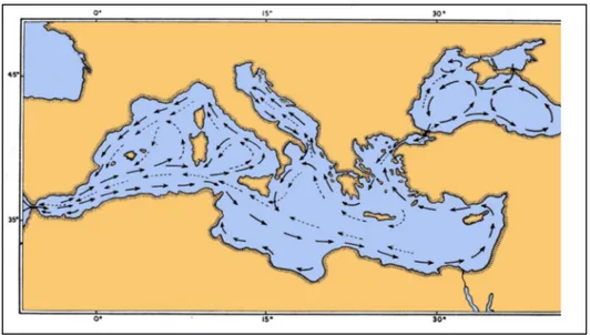
The Mediterranean Sea is a mid-latitude semi-enclosed sea, or almost isolated oceanic system. Many processes which are fundamental to the general circulation of the world ocean also occur within the Mediterranean, either identically or analogously.
The Mediterranean Sea exchanges water, salt, heat, and other properties with the North Atlantic Ocean. The North Atlantic is known to play an important role in the global thermohaline circulation, as the major site of deep- and bottom-water formation for the global thermohaline cell (conveyor belt) which encompasses the Atlantic, Southern, Indian, and Pacific Oceans. The salty water of Mediterranean origin may affect water formation processes and variabilities and even the stability of the global thermohaline equilibrium state.
The Mediterranean Sea is composed of two nearly equal size basins, connected by the Strait of Sicily. The Adriatic extends northward between Italy and the Balkans, communicating with the eastern Mediterranean basin through the Strait of Otranto. The Aegean lies between Greece and Turkey, connected to the eastern basin through the several straits of the Grecian Island. The Mediterranean circulation is forced by water exchange through the various straits, by wind stress, and by buoyancy Sux at the surface due to freshwater and heat Suxes.
The new picture of the general circulation in the Mediterranean Sea which is emerging is complex, and composed of three predominant and interacting spatial scales: basin scale (including the thermohaline circulation), sub-basin scale, and mesoscale. Complexity and scales arise from the multiple driving forces, from strong topographic and coastal influences, and from internal dynamical processes. There exist: free and boundary currents and jets which bifurcate, meander and grow and shed ring vortices; permanent and recurrent subbasin scale cyclonic and anticyclonic gyres; and small but energetic mesoscale eddies.
Fig. 1.3.1 – Mediterranean sea currents
In the Mediterranean basin exist tree crucial sills, that limits the diffusion of species by currents; in fact between Sicily and Tunisian , Puglia and Rodhes and at the entrance of Black sea; currents bifurcate and grow in ring vortices, making gyres.
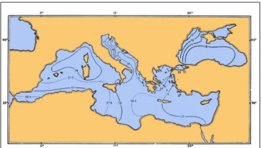
An other parameter peculiar for the basin is the ample range of salinity from West-Mediterranean to East-Mediterranean sea.
Salinity increases from 36.5‰, near the Gibraltar Strait (reproducing the Atlantic salinity conditions between Iberian and North-Africa zone), to 39.0‰ at the East coast, reproducing the Red-sea condition; consequently, the Levant basin shows an actual capacity of receiving and sustaining shallow-water benthic species with tropical or subtropical affinity, whilst such capacity is not shared by the rest of the Mediterranean.
The salinity mother basin of the Mediterranean-sea, particularly in its western part, acts as a biogeographic barrier for its easternmost Levant sub-basin. The East part of the basin is in fact characterised by a significantly reduced diversity compared to the rest of the Mediterranean sea.
1.4 Modes of Development
An important difference between the inhabitants of the deep sea (or marine environments in general) and of terrestrial environments is that marine animals have the potential for much wider dispersal and, concomitantly, gene flow among separated population.
Dispersal might be accomplished by a variety of means and during different stages of the cycle, either step by step, passively, drifting by the slow currents of the abyss, or actively by swimming.
Three general types of larvae are recognized: direct developing, lecitotrohic, and planktotrophic. Direct development, with the young released as miniature adults, is characterized by small and numerous eggs. Lecitotrophy (yolk eating) describes a free-swimming, nonfeeding larva that is different from adult and is pelagic for a short period before metamorphosing into a miniature adult.
The third mode of development is the planktotrophic one, in this case the first larval stage, trocophore, transforms into a veliger. In the veliger the byssus develops, which may act as a drogue to assist in larval flotation in the planktonic stage. Later it will secure the metamorphosed juvenile to the substratum and sustain attachment in adults. The trochophore protoconch remains, and enlarges into an extensively ciliated bilobed velum that provides propulsion and also assists in food gathering, while the shell, foot and rudimentary ctenidia are forming.
In the settled juvenile the velum is lost, its remnant forming the lips of the mouth.
A A B Fig. 1.3.1
A- larval anatomy with lecitotrophic development B- veliger ciliatum
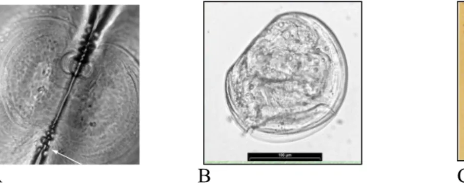
Modes of development are reflected into the shell’s sculpture. In fact the shell of species with plankyotrophic development is formed in three successive stages: prodissoconch I is secreted by the shell gland of the embryo, prodissoconch II is formed by the mantle of the planktonic veliger; after metamorphosis, the teleoconch is formed by the mantle of the adult. On the contary the shell of species with lecitotrophic development consist of only two successive stages: prodissoconch and teleoconch.
A B C
Fig. 1.3.2 A-Prodissococnch II, showing teeth at hinge extremities (44X) Fig. 1.3.2 B-Larvae with lecitotrophic development
Fig. 1.3.2 C-Larvae with pelagic stage
What’s more, the veliger (feeding larvae) grows in size in the plankton; during this time the embrionic shell continues to grow in a larval prodissoconch II and may become ornamented with sculpture which is unlike that of the adult.
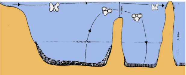
Planktotrophy is especially important in achieving long-distance dispersal, as is required in shallow water for recruitment of decimated populations in new environments. A pelagic larvae would be able to cross sills or arrive far from the parental area.
Fig. 1.3.3 Plackotrophic larvae dispersal ability
1.5 Morphology
Brooding of larvae is a mode of reproduction which has evolved in many species within a variety of higher taxa (Webber, 1997; Mackie, 1984; Brahmachary, 1989), and brooding mechanisms varies considerably. Among brooding bivalve molluscs, both freshwater and marine, the larvae (embryos) are contained in the mantle cavity of the female, either in the suprabranchial or the infrabrachial chamber, but always in association with the gill.
The larvae are sometimes retained in the interlamellar spaces of both demibranchs or of the inner or outer demibranchs only; alternatively, they may be confined to brood sacs, marsupial, mucous masses, or other specialized structures (Ockelmann, 1964; Solis, 1967; Franz, 1973; Mackie et al., 1974; Heard, 1977; Mackie, 1984; Tankersley and Dimock, 1992, 1993). Most authors have concluded that when the larvae are brooded in specialized structures; they are restricted to those structures (Morton, 1977; Kabat, 1985; Asson-Batres, 1988; Richard et al, 1991; Tankerseley and Dimok, 1992) and show little or no motility until the release period (Mackie, 1984). Several species posses brood masses, often confined by a membrane, and in other the embryos is attached to the demibranches with byssal threads or fixed to gill papillae (Heard, 19977; Bartlett, 1979; Richardson, 1979; Kabat, 1985; Asson-Batres, 1988; Russel and Huelsenbeck, 1989). Conversely, in species that brood the larvae between the demibranches, the larvae have been presumed to move within the mantle cavity of the mother, unattached to the material tissues. In some cases brooding is sequential, i.e., not all eggs are fertilized at the same time, and embryos are gradually displaced ventrally in the mantle cavity as they mature (Kabat, 1985; Russel and Huelsenbeck, 1989).
Animals showing a larva’s incubation in gill filaments or in the mantle is possible to deduce the type of larval development, in fact, in case of the presence of larvae in these body-cavity the mollusc has a direct or a lecitotrophic development.
1.6 Molecular markers
ITS
Internal transcribed spacer (ITSs) are sequences located in the eukaryotic ribosomal DNA (rDNA) between the 18S and 5.8S rRNA genes (ITS1), and between the 5.8S and 28S rRNA genes (ITS2). Using primers annealing in the 18S abd 28S rRNA genes, both spacer plus the 5.8S rRNA gene can be easily amplified by PCR and this stretch of rDNA is often called the ITS region (Fig.1.6.1).
Fig.1.6.1 ITS regions
Internal transcribed spacer rDNA can be used to infer the phylogenetic relationships within of species (Audeberta F. et al.,2000) and have proved to be useful to distinguish species (Wael M. Lotfy et al., 2004).
Additionally the ribosomal DNA ITS has been shown to be a useful genetic marker for species identification and phylogenetic reconstruction (Preston 1910). Moreover ITS studies have uncovered significant intra-specific genetic variability suggesting the presence of cryptic species complexes (Tuan R.& Dos Santos P. 2007).
Studies carried out in various organism have shown that the transcript of these spacer provides structural elements and secondary structures required for accurate and efficient pre-rRNA processing ( Hadjiolova et al. 1994; van Nues et al. 1994, 1995).
The ITS sequences show more sequence divergence than their flaking coding regions ( Hillis and Dixon 1991), and they are often used to distinguish related species (Guillamon et al. 1998; Proft et al. 1999; D’Amelio et al. 2000) and to infer phylogenetic relationships from population to families and even higher taxonomic levels (e.g, Gonzalez et al. 1990; Vogler and DeSalle 1994; Coleman and Vacquier 2002).
RAPD:
Random amplified polymorphic DNA analysis is a technique for rapidly detecting genomic polymorphisms (Williams, J. G. et al, 1990), utilizing a single short oligonucleotide primer of arbitrary sequence in a polymerase chain reaction. The PCR is carried out under low stringency conditions to generate a reproducible array of strain-specific products that are subsequently analyzed by gel electrophoresis. This kind of analysis has been used in numerous applications, including gene mapping, detection of strain diversity, population analysis, epidemiology, and the demonstration of phylogenetic and taxonomic relationships (Welsh, J. et al., 1995). RAPD analysis has become a widely used technique because it enables the quick detection of polymorphisms at a number of different loci using only nanogram quantities of genomic DNA. Furthermore, RAPD analysis can be carried out on organism for which there is little or no information concerning genomic sequences or organization, thus making it possible to analyze polymorphism for virtually any organism from which relatively pure genomic DNA can be isolated.
The random amplified polymorphic DNA (RAPD) can contribute to the discrimination of species (D. Rollinson, et al., 1998); especially used to analyze species-specific status in species with ambiguous morphology (Marin, S. A. et al., 2007).
RAPD a fast and useful method of genome marking that is useful for different studies e.g., parentage, mating patterns, taxonomy of sibling species and intra-specific population genetic structures (N. Mikhailova and K. Johannesso, 2004 – RAPD analysis of Littorina).
These arbitrary primers were used, also, because requires lee tissue (especially with juvenile snails) than other protein electrophoresis, are less laborious then
1995 – Random amplified polymorphic DNA markers reveal cross-fertilisation in Biomphalaria glabrata. Pulmonata. Basommatophora).
Patwary (1994) first used RAPD technique in bivalve mollusc for analyzing genetic divergence in scallop Placopecten magellanicus and distinguished different allele frequencies in different population. Wilding et al. (1998) analyzed genetic differentiation of morphotypes of periwinkle Littorina saxatilis from two locations of England. They considered the diverse RAPD patterns as evidence of separate gene pools. Liu et al. (1998a, 1998b) analyzed RAPD polymorphism of oyster species in Northern China, finding the genetic distances between three species Crassostrea talienwhanesis, C. plicatula and C. gigas and the intraspecific distance between four geographic populations of C. talienwhanesis.
2. Material and methods
2.1 Sampling area
Asperaca nodulosa:
The present study is based on tree populations of Asperarca nodulosa collected from depths of 450-550m in the Mediterranean basin.
The first was sampled from the West part of the sea between Mallorca and Minorca islands; the second one came from the centre of the basin, in particular near isola del Giglio, and the last one includes sample from the East part in middle-Greece water (schedule 2.1); therefore the analysis was performed on specimens originating from the entire basin to fully understand the flow between the varied populations.
Fig 2.1- Sampling area
St.name St.num Date Depht Location Point
Corti 29 28/12/2003 448,00m NW Capraia 43°50.8527N Triangul. dredge
Corti 109 04/01/2004 547,00m TiberinoSMT 41°50.8527N Rock
dredge
Cobas 96 16/04/04 572,00m E Menorca 04°23.6296E Dredge
Cobas 98 16/04/04 469,00m E mellorca 03°49.4389E Dredge
Cobas 107 18/04/04 557,00m NE menorca 04°.23.6253E Dredge
Geco 1 30/08/2006 445.00 m N Gavhos 24°05.8000’1 Grab
Geco 5 2/08/2006 626,00 m W Crete 23°18.4666’0 Dredge
Geco 12 4/08/2006 510,00 m SE Crete 21°28.3775 grab
Anadara demiri
:The second investigation was based on tree different species sampled from the Adriatic-sea (Mediterranean basin) and from the Atlantic Ocean.
Anadara transversa (Say, 1822)
1. Martha's Vineyard at Sengenkontacket Pond, Edgartown, Massachusetts USA:
41° 23' N 70° 30' W, agosto 2007, (adults), collected by K. Czaja 2. St. Augustine Inlet, St. Johns County, Florida 29° 54' 30" N 81° 17' 05", 7 agosto 2007, collected by M. Krisberg (juveniles)
Anadara demiri (Piani, 1981)
1. Porto Garibaldi (Ferrara), fishing-boats harbour, collected by F. Favero 2. Porto Garibaldi
LAT xx°xx.xx' WGS84 44° 39'69" LON xx°xx.xx' WGS84 12°17'28"
Collected by C. Mazziotti, ARPA Emilia-Romagna, Struttura Oceanografica Daphne
Anadara inaequivalvis (Bruguière, 1789) Porto Garibaldi
LAT xx°xx.xx' WGS84 44° 39'69" LON xx°xx.xx' WGS84 12°17'28"
Collected by C. Mazziotti, ARPA Emilia-Romagna, Struttura Oceanografica Daphne
2.2 Methods of sampling
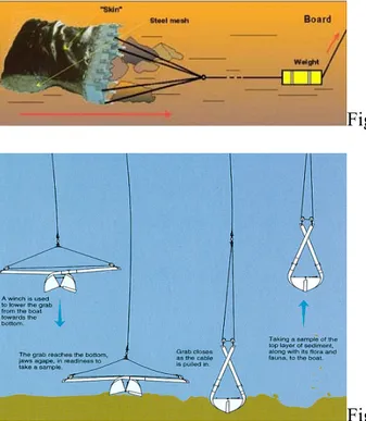
The samples were collected aboard the research vessel Urania during three different oceanographic cruise. All samples were taken using a dredge (Fig.2.2.1) or grab (2.2.2) let down at a depth between 400m. and 600m. The close packed sediment, so obtained, was washed trough four sieves with different granulometry.
Fig 2.2.1 Dredge operation
Fig. 2.2.2.Grab operation
Where possible, shells were selected from each sample spanning the entire range of adult and sub-adult sizes, so that the analysis include the natural range of shapes and allometries expressed by the taxon at the localities. The samples were preserved in alcohol 100%.
2.3 Kind of investigation
Samples were shared in tree group for different kind of analysis.
2.3.1 Morphological analysis:
After having taken off the animal by a Lysis buffer, the two valvas were separated, cleaned with ultra-sound machine, and, washed with ultra-pure water. Each valva was put on a stub to be photographed at S.E.M., looking for micro-sculpture of prodissococh in comparison with micro-micro-sculpture of the adult shell and the lines between teleoconch and prodissoconch I and II.
2.3.2 Hystological investigation:
Adults (7to28mm length) from differente populations were included in paraffin (schedule 2.3.2) and cut with microtome; the sections were stained with a double coloration by ematossilina and eosina to make out citoplasmatic and nuclear regions. The hystological investigation was performed in order to search for possible larval incubations in gills or mantle cavity and hermaphrodism.
Water 120° Alcool 70° 240’ Alcool 80° 240’ Alcool 90° 240’ Alcool 100° Over night Alcool 100° 120’ Bio-clear(x2) 40’ paraffin(x2) 45’ Over-night
Double coloration Ematossilina
Eosina
2.3.3 Genetic analysis:
Genetic information (DNA) is organized in the chromosomes that are contained in a cell nucleus (nuclear DNA), and in mitochondria (mitochondrial DNA or mtDNA). The nuclear DNA is made up of coding sequences, functional domains regulating the protein synthesis, and non-coding DNA that exist in families of repeat sequences.
Mitochondrial DNA is a circular double helix made up of 15.000-20.000 nucleotides, depending on the species (Hartl & Clark, 1993). It is replicated, independently from cell and DNA nuclear replication, each time the mitochondria divide; The different types of mtDNA that are originated from mutations and that are present in populations are called “mitochondrial haplotypes”.
Analysis
Genetic analysis was made focusing on comparing populations in the three sampling area.
At the beginning DNA was extracted from tissues using the phenol/chlorophorm protocol. Tissues were digested in 100mM TRis-HCL, ph8.0/10mM EDTA/100mM NaCl/0.1% SDS/50 mM dithiothreito/proteinase K (0.5µl/ml) for 2-4 hr at 37°C. The DNA solutiton was purified by extracting twice with phenol, once with phenol/chloroform (1:1), and once with chloroform. Pure DNA was then concentrated with an ethanol precipitation.
Sample were amplified used different, nuclear and mitochondrial, specific primers (Schedule 2.3.2), PCR products were sequenced using 1µl of template in a 25-µl reaction, with 20,3 µl of water, 2,5 µl of PCR buffer (Eppendorf DNA polymerase 10x buffer), 0,5 µl of MgCl2 (25nM), 0,4 of dNTPs (25nM each), 0,28 µl Taq polymerase, and 0,5 µl each of the two primers (10µM); fragments were separated on an automated DNA sequencer.
Markers used list primers sequences (5’ to 3’) CO1 Kocher et al.(1989) TAAACTCAGGGTGACCACCCCCTCA GGTCAACAAATCATAAAGATATTGG Co1brb Zardoia (2002) GTAATACCAATTATAATTGGGGGGT GTCCCCACCTCCTGCCGGAT Cytb
Zardoya & Meyer (1996)
AAAAAGCTTCCATCCAACATCTCAGCATGATGAAA AAACTGCAGCCCCTCAGAATGATATTTGTCCTCA 12s Kocher et al. (1989) GGGGCAATTAATTTTATTAC ACCATTATACAACCGGT 12prt AGACATGGATTAGATACCC ACCCCTACCTTGTTACGACTT 16s Koufopaunou et al. (1998) CGCCTGTTTAACAAAAACAT ACGCCGGTTTGAACTCAGATC 28s Giribet et al. (2006) GACCCGTCTTGAAGCACG CCACAGCGCCAGTTCTGCTTAC H3 Colgan et al. (200) TTTCCCCGCGGTCGCCCC TGACTGCAGAGGGTGCAGGGCGGTGTGT Schedule 2.3.3
Nuclear markers (Asperarca nodulosa)
In 100 samples, the Invitrogen tissue kit was used to extract genomic DNA from
the mantle. DNA was PCR-amplified using ITSI 1d
(5’-GGTGAACCTGCGGAAGG-3’) and ITSI 2r
(5’-CCCTTGGCCGCAATATGCGTTC-3’) anchored respectively in the conserved extremities of the 18S and 5.8S ribosomal genes (Kane & Rollinson 1994).
PCR uses single stranded DNA as a template and, by the action of DNA polymerase enzyme, it synthesizes a complementary strand over and over again, until extensive quantities are produced. Every PCR consists of a cycle, repeated many times, made up of the following steps: 94°C, for 45s; 60°, for 60s; and 72°, for 60s (30cycles) The amplified DNA was purified by etanohl precipitation for 3hr at -20°C, centrifugate for 15’and, wash with Etoh 70% and re-centrifugate for 15’. The pellet, so obtained, was resuspended in ultra-pure water and stored at -20°C until used for analysis. The ITS1 generated a fragment of approximately 700 bp. Fragments were separated on a ABI PRISM DNA
parameters followed by refinement by eye. The nucleotide sequences were subjected to maximum parsimony (MP) analysis; MP was performed in PAUP 4.0b10 (Swofford, 2002). In the construction of the phylogenetic tree, both transition and transversion were taken into account.
RAPD (Asperarca nodulosa)
50 samples were analyzed using aspecific markers: RAPD (random amplification of polymorphic DNA) to determine intraspecific variability.
The Random Amplified Polymorphic DNA method is based on the Polymerase Chain Reaction (PCR) using short (usually 10 nucleotide) primers of arbitrary sequences.
Six oligonucleotides (schedule 2.3.3) from the ready-to-go RAPD analysis kit (Amersham Biosciences Ltd., Piscataway, NJ) (Williams, 1990) were used for amplification of random DNA markers to reveal the genetic diversity among Asperarca nodulosa populations. RAPD-PCR was performed with ready-to-go RAPD analysis Beads as described by the manufacturer (Amersham Biosciences). Approximately 10 ng of genomic DNA from microfilriae was used. The RAPD reaction was performed in a DNA thermal cycler (GeneAmp PCR System 2400, Perkin-Elmer, Norwalk, CT), for 1 cycle at 96°C for 4 min, followed by 40 cycles of 94°C for 1 min, 40°C for 1 min and 72°C for 2 min, respectively. The final amplification cycle included 7 min extension at 72°C. Amplified products were separated in a 2% agarose gel, stained with Cyber safe, and visualized under ultraviolet light. The size of each band was determined by Quantity One® 1-D Analysis Software (Bio-Rad, Hercules, CA) and data were examined by Past.exe.
primer sequence 1 5'-d(GGTGCGGGAA) -3' 2 5'-d(GTTTCGCTCC) -3' 3 5'-d(GTAGACCCGT) -3' 4 5'-d(AAGAGCCCGT)-3' 5 5'-d(AACGCGCAAC)-3' 6 5'-d(CCCGTCAGCA)-3' Schedule 2.3.3
H3 (Anadara demiri)
The phylogenetic study was carried out, based on the nuclear protein-coding gene histone 3; H3 primers are useful to study evolutionary rates and phylogenetic relationships in Mollusca (Colgan D. et al., 2003), and to investigate the phylogenetic pattern of characters in this group (Kappner I. & Bieler R., 2006).
The Invitrogen tissue kit was used to extract genomic DNA from the mantle. DNA was PCR-amplified using H3aR (5’-TTTCCCCGCGGTCGCCCC-3’) and H3bR (5’-TGACTGCAGAGGGTGCAGGGCGGTGTGT -3’) (Colgan, Onder & Eggler, 2000). PCR products were sequenced using 1µl of template in a 25-µl reaction, with 20,3 25-µl of water, 2,7 25-µl of PCR buffer (Eppendorf DNA polymerase 10x buffer), 0,3 ul of MgCl2 (25nM), 0,4 of dNTPs (25nM each), 0,28 µl Taq polymerase, and 0,5 µl each of the two primers (10µM); fragments were separated on an automated DNA sequencer.
Amplification conditions were follows (25cycles): 94°C, for 30s; 52°, for 60s; and 72°, for 60s. Electrophoresis of 2,5 µl of the amplified mixture was done in a 1,5 agarose gel and the DNA was stained with cybr safe. The gel fragment containing the amplified product was excised from the gel and purified by the
MN kit and stored at -20°C until used for analysis. The amplified DNA was sequenced on a ABI PRISM DNA sequencer. The sequences were aligned using MEGA set at the default parameters followed by refinement by eye
3. Results and discussion
3.1 Morphological analysis
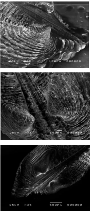
Samples investigated showed same micro-sculputure on the adult shell and on the larval shell, so is it impossible to realize the differences between the three populations (fig 3.1A, B, C).
Fig. 3.1ACobas ind
Fig. 3.1B Corti ind.
The S.E.M. technique demonstrates that it is difficult to make out prodissoconch I and prodissoconch II, also because adults have often the shell degraded or encrusted by calcareous concretions (Fig. 3.1 D, E, F).
Fig. 3.1D Cobas ind.
Fig. 3.1E Corti ind.
The prodissoconch seems to reflect a lecitotrophic or direct development (Fig. 3.1 G,H), in fact S.E.M.’s photos shows a direct passage between embryonal shell and teleoconc, without the larval shell; so we can suppose a non-planctotrophic larval development.
Fig. 3.1 G Juvenile of Asperaca nodulosa
Fig. 3.1 H Juvenile of Asperaca nodulosa
The lecitotrophic development is a development parameter that could be changed during the evolution of the species. In fact animals of Asperarca nodulosa could be entered the Gibraltar’s sill during the glacial period thanks to the pelagic larvae, than, after their settlement on the new habitat, they could have changed the development from planctotrophic to lecitotrophic; this change was possible loosing the ciliate filaments around the mouth (for feeding during the larval stage) and by a regression of the velum (Fig. 3.1.I) during the veliger stage.
If one species changes its mode of development from planctotrophic to lecitotrophic or direct, the transformation is irreversible; because it is possible too loose organs but not recreate them. So the change is unilateral, it is impossible that a lecitotrophic species becames planctotrophic.
3.2 Hystological investigation
All samples don’t have larvae (embryos) contained in the mantle cavity of the female (Fig. 3.2A), affixed or not, or in the gills filaments (Fig. 3.2B,C).
Fig. 3.2A Mantle cavity
Fig. 3.2B Gills filaments
Fig.3.2C Gill section
Another parameter was searched, i.e. the presence of hermaphroditism in gonads. Gonad sections of adult samples of Asperarca nodulosa shows female (Fig. 3.2D, E) and male (Fig. 3.2F, G) fully grown and differentiated gonad.
Fig. 3.2D Male of Asperarca nodulosa
Fig. 3.2E Cross section of male gonad
Fig 3.2F female of Asperarca nosulosa
Fig. 3.2G Cross section of female gonad oocytes in second growth period
3.3 Diversity by longitudinal gradient
During the oceanographic expeditions, shells were selected from each sample, spanning the entire range of adult and sub-adult sizes, so that the analysis include the natural range of shapes and allometries expressed by the taxon at the localities
.
Number and size of specimens coming from Eastern- basin reflects the peculiarity of the basin (especially Eastern Mediterranean due to currents and salinity. In fact the number (Fig. 3.3A) and size (Fig. 3.3B) decrease from West to East.Fig. 3.3A Numbers of A. nodulosa sampled 0 5 10 15 20 25 30 35
cobas96 cobas98 cort29 Geco2
s.number
for each station.
Fig. 3.3B Mean Size range from Western to Eastern basin 0 2 4 6 8 10 12 14 cobas geco dimension (mm.)
3.4 Genetic study
Extractions methods:
With the phenol/chlorophorm extraction method DNA solution resulted low concentrated and impure instead samples extracted by the Invitrogen tissue kit shows a clean and high concentrated template.
Mitochondrial markers:
Mitochondrial primers didn’t amplified sequences useful for the phylogenetic
study; this procedure didn’t obtain a sufficient DNA quantity to carry out molecular analyses.Specific nuclear primers study: ITS
The molecular phylogeny of Asperaca nodulosa based on Internal Trascribed Spacer 1 sequence data supports already the separation identified reflecting the subdivision in three sub-basin areas (Fig. 3.4.4). The bootstrap methods showed that these clades are confirmed with high statistical reliability.
g3.scf g2.scf g.scf 107.scf 96.scf 98.scf 109.scf 29.scf outgroup 99 99 81 97 100 100
Fig. 3.4.4 Phylogenetic relationshops among three population of Asperarca nodulosa based on MP analysis of the ITS1 sequences.
The trees produced by these methods showed the same topology with the formation of three distinct groups.
RAPD:
The Random Amplified Polymorphic DNA method, using primers of arbitrary sequences, amplified for 91 fragments, from a length of 220bp to 3975bp (Schedule 3.4.1). primer sequence Tot fragments 91 lenght 1 5'-d(GGTGCGGGAA) -3' 18 220-3975bp 2 5'-d(GTTTCGCTCC) -3' 18 260-1360bp 3 5'-d(GTAGACCCGT) -3' 14 240-890bp 4 5'-d(AAGAGCCCGT)-3' 15 230-1320bp 5 5'-d(AACGCGCAAC)-3' 15 255-1900bp 6 5'-d(CCCGTCAGCA)-3' 11 270-900bp Schedule 3.4.1
Amplified products were compared in each population, and common bands among populations, bands shared between two population and band present only in one population, consequently specific for that population were researched (Schedule 3.4.2). Statistical analysis were based on presence, co-presence or absence of bands.
Specific band population
Bands in common
Shared bands Polimorphic locy
Cobas 21 : 24% Cb/Ct 8 19 : 22% Cobas 25%
Corti 17 : 20% Ct-G 2 Corti 26%
Geco 10 : 12% G-Cb 9 Geco 22%
Schedule 3.4.2
The percentage of polymorphic locy decrease from West to Est. The analysis showed a low intra-population genetic variability and an high variability between populations coming from far zones.
The set of data gives a remarkable separation among the three populations.
Fig. 3.4.3 Phylogenetic relationships among Asperarca nodulosa populations based on RAPD data.
Dice : D=2Nab/Na+Nb
Na = Band’s numbers of population A
Nab = Numbers of fragments shared between population A and Nb = Band’s numbers of population B
Samples from Eastern-Mediterranean seems to be more genetically distant from the ones of the Western part; in according with m the previous observations.
4. New colonization in the Mediterranean sea from alien species:
Anadara demiri
From the biogeographer’s perspective the Levant Basin is a unique laboratory to study one of the most impressive on-goig marine colonization observable by man: the Lessepsian invasion (Por, 1975, 1978, 1990; Por and Dimentman, 1989; Zibrowius, 1991, 1994; Ribera, 1993;Galil, 1993).
Biodiversity in the Mediterranean Sea has undergone modifications during recent decades, following the introduction of non-indigenous species (fig. 4.1). Their entrance into the basin can be attributed to both natural phenomena and anthropogenic activities.
The opening of the Suez Canal in 1869 placed the Mediterranean in direct contact with Indo-Pacific biota and a number of species have been reported to naturally migrate from the Red Sea to the Mediterranean by a process defined as ‘lessepsian migration’ (Por, 1978). The recent expansion of shipping movements and aquaculture commerce have dramatically accelerated the exchange of biota worldwide and the number of possible introduction vectors has increased (Carlton, 1989; Ruiz et al., 2000) (Fig. 4.2).
Fig. 4.2 The inferred pathways for exotic molluscan species in the Mediterranean basin.
It is the presence of such adverse conditions that appears to have stimulated the establishment of resilient exotic organisms, giving them a competitive advantage with respect to native species and causing their demise. This is the case with the arcid bivalve, Anadara inaequivalvis (Bruguie' re, 1789) (de Zwaan et al., 1995). Its introduction into the Adriatic in the early 1970s (Rinaldi, 1972) coincided with the advent of intense eutrophication events due to extraordinary algal blooms (Fonda Umani et al., 1992).
These blooms caused cycles of normoxia interspersed with the success of A.inaequivalvis (Fig.4.1 A,B) is that, in contrast to native bivalve species, its haemolymph of A. inaequivalvis contains nucleated erythrocytes packed with
haemoglobin, which enable the binding of oxygen in oxygen-deficient conditions (Holden et al., 1994), and haematin, which is involved in the removal of sulphides (de Zwaan et al., 1995).
Fig. 4.1 A, B Anadarainaequivalvis A B
Since 2000 a new competitor has made its appearance in the Adriatic Sea: Anadara demiri (Fig. 4.2) (Piani, 1981) (Morello & Solustri, 2001). The species was probably introduced into the Mediterranean Sea either as planktonic larvae carried in the ballast waters of ships or as benthic stages within shipments of other bivalves that were the object of aquaculture (C. Smriglio, in litteram). It was first reported in the Mediterranean-sea in the bay of Izmir (Turkey) in the late 1970s (Demir, 1977) and, 20 years later, in the Gulf of Thermaikos and the bay of Thessaloniki (Aegean Sea) (Zenetos, 1994).
Fig. 4.1 C,D Anadara demiri C D
At present very little is known on the biology and ecology of this species, but, similarly to A. inaequivalvis, it appears to contain respiratory pigments with high oxygen affinity which could, under hypoxic conditions, confer it a competitive advantage with respect to autochthonous bivalves. Zenetos (1994)
hypothesized a correlation between the greatest densities of A. demiri in the bay of Thessaloniki and conditions of intense environmental pollution. A further competitive advantage could be conferred to A. demiri by its ability, retained into adult life, of attaching by means of byssus threads to all kinds of hard substrata.
The Mediterranean as a whole show an actual capacity of receiving and sustaining shallow-water benthic species with tropical or sub-tropical affinity, in particularly in the Eastern zone (Taviani, 2002).
This progressive and inexorable colonization highlight the importance of collecting information on introduced species soon after their first appearance in a new environment as a completion in the study of colonization’s mechanism. The chance of colonising new areas is an opportunity for the species involved in a Lessepsian (“migrations by man”, sensu Por, 1971; see also e.g. Por, 1975, 1978, 1990; Por & Dimentman, 1989; Zibrowius, 1991, 1994; Ribera, 1993; Galil, 1993) or introduced process to implement adaptive potentialities.
The entrance, of Anadara demiri, into the basin was attributed to anthropogenic activities of the opening of the Suez Canal; Indo-Paciphic species is migrated from the Red Sea.
This hypothesis should be wrong, because we suppose that the origin of this species can be Atlantic; seeing the similarity between the A. demiri and Anadara transversa (Say, 1822) (Fig. 4.1 E).
To investigate the real origin of A. demiri we studied the morphology and genetic variability, eventually accumulated, between the Mediterranean Anadara and the sibling Atlantic species (Fig. 4.3); using Anadara inaequivalvis as out-group the species.
Fig. 4.3 Anadara transversa Geographic range
Morphological analysis:
After having taken off the animal by a Lysis buffer, the shell was broken and ultra-sound cleaned, afterwards, it was washed with ultra-pure water. The apex was get on a stub to be photograph at S.E.M., searching micro-sculpture on prodissococh in comparison with micro-sculpture on the adult shell; and, the remarkable lines between teleoconch and prodissoconch I and II.
Anadara demiri (Fig. 4.4A) and A. transversa (Fig. 4.4B), at S.E.M., seem to have similar microsculpture as well at the teleoconch as at the prodissoconch, but adult animals have the shell degraded or encrusted by calcareous concretions that make the analysis doubtful.
This study is focusing on the new colonization of the Basin, by understanding the actual process of migration, so was important to investigate the type of development. The S.E.M. photos show an embryonal prodissoconch followed by the larval shell (Fig. 4.5 A,B)
A
B
Genetic study and Preliminary results
After the data set resulted from morphological analysis, was important to compare them with a genetic approach. The phylogenetic study was carried out, based on the nuclear protein-coding gene histone 3.
Fig. 4.7 phylogenetic tree
i1h3.scf i3.scf i4.scf d2.scf t3.scf d4.scf t4.scf t2h3.scf d3h3.scf d3h3.scf(2) outgroup 100 72 82 99 98 61 86 100 0.0 0.1 0.2 0.3 0.4 0.5 0.6
The preliminary result of the present molecular study show a genetic nearness between the Atlantic species A. transversa and the new-introduced Anadara demiri and, at the other hand, a molecular distance of this species from the Indo-pacific A. inaequivalvis.
To better understand the origin of this species it was useful to compare Anadara demiri with other Arcidae (Anadara trapezia, gb|AF033677.1; Arca imbricata, gb|AY654989.1|; Arca zebrata gb|AF416864.1); Samples of A. demiri seems to be more genetically distant from the Indo-Pacific species (A. inaequivalvis and A. zebrata); in according with the previous studies.
d3h3.scf d3h3.scf(2) t2h3.scf d4.scf d2.scf t3.scf an.trapezia i4.scf i3.scf arca imb. arca zeb. 100 100 69 66 50 34 83 91
5. Conclusion
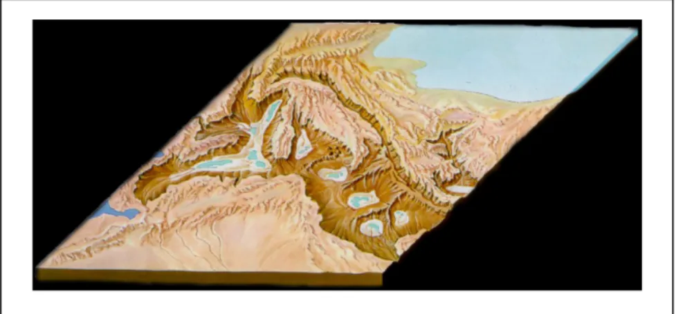
The present Mediterranean biogeography is the transitory result of climatic and geological events suffered by this semi-closed basin throughout its history. The potential consequences over stenoecious marine ecosystem of the late Miocene “Messinian Salinity Crisis” are discussed. The biogeography of the Mediterranean deep-sea benthos is affected by the geodynamic evolution of the basin and in particular by the post-Messinian establishment of shallow sills (Gibraltar and Siculo-Tunisian); since the Pleistocene at least, the sills acts as a filter biasing the flux of potential Atlantic deep-sea invaders. These peculiar biota show a distinct evolution through time, in particular we investigate this phenomenon trough the bivalves: Asperarca nodulosa (Arcidae – Mollusca). Researching the history-evolution of these species we study the colonization’s dynamics in the Mediterranean basin after the MSC during the glacial period and the adaptability faculties of invaders.
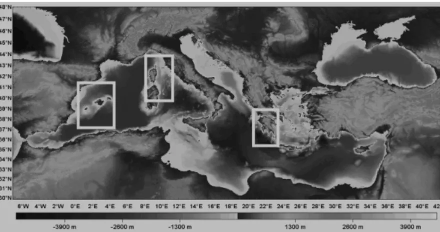
The colonization by Asperaca nodulosa was after the MSC during the following glacial periods; animals went into the basin trough the Gibraltar sill and settled in the three deep sub-basins (Fig. 5.1).
Fig. 5.1 Mediterranean-sea’s depth
The species, probably, arrived into the Mediterranean-sea getting across the currents during the larvae stage.
This study underline the isolation of the three population, that after the colonization, didn’t have no more change between the three group.
The thesis was supported by three ways of analysis: morphological, histological and molecular. All methods show results in according to each other.
The morphological analysis demonstrate a lecitotrophic or direct larval development (SEM’s photos show only the prodicconch I) and, at the other hand, also histological investigation highlight the absence of larvae in gills or in mantle and the presence of differentiated gonads with sex ratio.
The molecular study gives result in according with the other data; in fact both genetic ways, arbitrary markers (RAPDs) and specific nuclear primers (ITS), show a low intra-population variability and a high variability among populations coming from far sampling zone; pointing out the separation between the three sub-basin areas.
Possible environmental factors, including bottom currents and geological sills, acting on a common genotype to cause the observed pattern of differentiation. The species could have loosen the plancotrophy, regressing the mouth structures and the ciliate velum. The mechanism is a one-side change, probably happened for the peculiar currents, salinity and bio-geography of the basin; the result is isolation.
Nowadays the Mediterranean-sea is a water-basin in transformation, for the capacity to receive new species; especially in the Eastern-part, more warm and salted, trough the Suez canal from Indo-Pacific species, but, also, from Atlantic invaders ( the alien species Anadara demiri) carried in the ballast waters of ships and adapted in the Western-part of the sea and in the high Adriatic-sea more similar in salinity to the Atlantic Ocean.
6. References
Abelló P. & Cartes J.E. (1992).- Population characteristics of the deep-sea lobsters Polycheles
typhlops and Stereomastis sculpta (Decapoda: Polychelidae) in a bathyal mud community of
the Mediterranean Sea.- Marine Biology, New York, Vol. 114, pp. 109-117.
Allen, J . A. 1979. The adaptations andradiation of deep-sea bivalves. Sarsia 64: 19-28
Allouc J. 1987 – Les paleocommunautes profondes sur find rocheux du Pleistocena mediterranèen: Dscription et essai d’interpretation palèeocologique. Geobios, 20: 241-263.
Almeida, M.J., Machado, J. & Coimbra, J., 1996. The e¡ect of Polydora sp. infestation on the shell calcification of the oyster Crassostrea gigas. Bulletin de l’Institut Oceanographique Monaco, special no., 14, 195^202 Angel MV and Smith R (eds) (1999) Insights into thhydrodynamics and biogeochemistry of the South Aegean Sea, Eastern Mediterranean: The PELAGOS (EU) PROJECT. Progress in Oceanography 44 (special issue): 1}699.
Arvanitidis C., Bellan G., Drakopoulos P., Valavanis V., Dounas C., Koukouras A. & Eleftheriou A. (2002).- Seascape biodiversity patterns along the Mediterranean and the Black Sea : lessons from the biogeography of benthic polychaetes.- Marine Ecology Progress Series, Oldendorf (Luhe), Vol. 244, pp. 139-152.
Audeberta F., Durette-Desseta M. and Neil B. Chiltonb, 2000 - Internal transcribed spacer rDNA can be used to infer the phylogenetic relationships of species within the genus Nematodirus (Nematoda: Molineoidea)
Avise, J . C., Lansman, R. A. 1983. Polymorphism of mitochondrial DNA in populations of higher animals. In Evolution o f Genes and Proteins, ed. M. Nei. R. K: Koehn, pp. 147-90. Sunderland, Me: Sinauer. 33 1 pp.
Barash A., Danin Z. 1972 – the Indo-Pacific species of Mollusca in the Mediterranean and noteon a collection from the Suez Canal. Israel J. Zool., 21: 303-374.
Barnard, J . L. 1962. South Atlantic abyssal amphipods collected by R. V. Vema. In Abyssal
Crustacea, Vema Res. Ser. 1:l-78
Barrier P., Di Geronimo I., Montenat C., Roux M. & Zibrowius H. (1989).- Présence de faunes bathyales atlantiques dans le Pliocène et le Pléistocène de Méditerranée (détroit de Messine, Italie).- Bulletin de la Société géologique de France, Paris, (8), Vol. 5, N° 4, pp. 787-796.
Bellan-Santini D., Fredj G. & Bellan G. (1992).- Mise au point sur les connaissances concernant le benthos profond méditerranéen.- Oebalia - International Journal of Marine
Biology and Oceanography, Taranto, suppl. Vol. 17, pp. 21-36
Belayev, G. M. 1974. On the age of the deep-sea fauna of the ocean and the ultra-abyssal fauna of trenches. Bull. Mosk. Ispital. Prirordi. Otd. Biolog. 79:94-112 (In Russian)
Bellan, G. 1977. A discussion of the relationship between systematics and ecology in polychaetous annelids. In Essay on Polychaeteous Annelids, ed. D. J . Reish, K. Fauchald,
Allan Hancock Found. Spec. Publ., pp. 449-60. Los Angeles, Calif. 604 pp.
Ben Moussa A., Brebion Ph., Laurat-Rage A., Demaicq G. 1988 – Interèt paleobiologique des mollusques messiniens de Melilla. Rev. Paleobiol., 7: 335-358.
Benson, R. H. 1975. The origin of the psychrosphere as recorded in changes of deep-sea ostracode assemblages. Lethaia 8:69-83
Benson, R. H. 1979. In search of lost oceans: A paradox in discovery. In Historical
Biogeography, Plate Tectonics, and the Changing Environment, ed. J . Gray, A. J . Boucot, Proc. Biol. Colloq., Oregon State Univ. 37:379-89 sublittoral, bathyal and abyssal asteroids in
the northeast Pacific Ocean. Ophelia 10:3547
Benson R.H., Rakik-El-Bied K.R. 1991 – The Messinian parastratotype at Cuevas del Almanzora, Vera Basin SE Spain: refutation of the deep-basin, shallow-water hypothesis?. Micropaleontology, 37: 289-302.
Bemstein, B. B., Meador, J. P. 1979. Temporal persistence of biological patch structure in an abyssal benthic community. Mar. Biol. 51:179-83
Bianchi C.N. & Morri C. (2000).- Marine biodiversity of the Mediterranean Sea: Situation, problems and prospects for future research.- Marine Pollution Bulletin, Oxford, Vol. 40, pp. 367-376.
Bishop, J . D. D. 1981. Two new leuconids (Peracarida, Cumacea) of widespread occurrence in the deep Atlantic. Crusraceana 40: 144-59
Biju-Duval B., Dercourt J., Le Pichon X. 1997 – From the Tethys Ocean to the Mediterranean seas: a plate tectonic model of the evolution of the western Alpine system. In: B. Biju-Duval, L. Montadert, Structural History of the Mediterranean basins, International symposium, Editions Technip, Paris: 143-164.
Blanc P.-L. (2000).- Of sills and straits: a quantitative assessment of the Messinian salinity crisis.- Deep-Sea Research I, Oxford, 47, pp. 1429-1460.
Blanc-Valleron M.-M., Rouchy J.-M., Pierre C., Badaut-Trauth D., Schuler M. 1998 – Evidence of Messinian nonmarine deposition at Site 968. In: A.H.F. Robertson, K.C. Emeis, C. Richter, A. Camerlenghi, Proc. ODP, Scientific Results, 160: 437-445.
Bouchet, P., Waren, A. 1979. Planktotrophic larval development in deep-water gastropods. Sarsia 64:3740
Bouchet P., Taviani M. 1992 – Atlantic deep-sea gastropods in the Mediterranean: new findings. Boll. Malacol., 2: 137-148.
Bouchet p., Taviani M. 1992b – La colonizzazione dei bacini a soglia. Il caso del Mar Mediterraneo. Lav. Soc. Malacol. It., Parma, 24: 120-130.
Boudouresque C.F., Briand F., Nolan C. 1993 – Introduced species in European coastal waters. European Commision, Luxemburg, Belgium, Office for Official Publication of the
Brett. J. R. 1956. Some vrincivles in the thermal requirements of'fishes'. Q. Rev. Biol. 3 1:75-87
Brown, W. M. 1983. Evolution of aimal mitochondria1 DNA. See Ref. 2, pp. 62-88
Brunn, A. F. 1957. Deep-sea and abyssal depths. In Treatise on Marine Ecology and Paleoecology. Vol. I . Ecology, ed. J . W . Hedgepeth, Geol. Soc. Am., Mem. 67, pp. 641-72. New York: New York Lithographing. 1296 pp.
Buckeridge, J . S. 1983. Fossil barnacles (Cirrivedia: Thoracica) of New Zealand and '~ustralia. N. Z. Geol. Sum. Paleont Bull 50 1-151
Buckley H.A. & Johnson L.R. (1988).- Late Pleistocene to Recent sediment deposition in the central and western Mediterranean.- Deep-Sea Research, Oxford, Vol. 35, pp. 749-766. Bush, G L 1975 Modes of an~mal speciation. Ann. Rev. Ecol. Svst. 6:339- 84
Busson G. (1984).- Transposition des données sur les marais salants aux grandes accumulations évaporitiques du passé.- Géologie méditerranéenne, Marseille, Vol. 9, N° 4, pp. 563-591.
Busson G. 1990 – Le Messinien de la Mediterranèe: vingt ans après. Geologie de la France, voll.3-4: 3-58.
Butler R. W. H., Pedley H.M., Maniscalco R., Grasso M., McClelland E., Finegan B. 1996 – The significance of Messinian occurrences of Globorotalia margaritae and Globorotaria puncticulat in Sicily. Terra Nova, 8:59-64.
Buzas, M. A,, Culver, S. J . 1984. Species duration and evolution: Benthic Foraminifera on the Atlantic continental margin of North America. Science 225329-30
Campbell G, Jones CS, Lockyer AE, Hughes S, Brown D, Noble LR and Rollinson D (2000) Molecular evidence supports an african affinity of the neotropical freshwater gastropod,
Biomphalaria glabrata, say 1818, an intermediate host for Schistosoma mansoni. Proc Biol
Sci 267:2351-8.
Carey, A. G. 1972. Food sources of sublittoral, bathyal and abyssal asteroids in the northeast Pacific Ocean. Ophelia 10:3547
Carney, R. S., Haedrich, R. L., Rowe, G. T. 1983. Zonation of fauna in the deep sea. In Deep-Sea Biology, ed. G . T . Rowe, The Deep-Sea 8:371-98. New York: Wiley. 560 pp.
Carpine C. (1970).- Ecologie de l'étage bathyal dans la Méditerranée occidentale.- Mémoires
de l’Institut Océanographique de Monaco, Vol. 2, pp. 1-146.
Carlton, J.T., 1989. Man’s role in changing the face of the ocean:biological invasions and implications for conservation of nearshore environments. Conservation Biology, 3, 265^273. Carter, G. S. 1961. Evolution in the deep seas. In Oceanography, ed. M. Sears, Publ. Am.
Chardy, P. 1972. ~ t u d e biometrique des variations intraspecifiques chez I'isopode Janirella bonnieri Stephensen, 19 15, par l'analyse canonique. Bull. Mus. nat. Hist. nat., Paris 3' ser.36(Zool. 30):363-77
Chemello R., Oliverio M. 2001 – La distribuzione in Mediterraneo degli invasori lessepsiani; un modello per I molluschi bentonici. Biogeographia, 22: 227-237.
Chernoff, B. 1982. Character variation among populations and the analysis of biogeography. Am. Zool. 22:425-39
Christensen, A.M., 1970. Feeding biology of the sea-star Astropecten irregularis Pennant. Ophelia, 8, 1^134.
Ciesm 2002 – Alien marine organism introduced by ships in the Mediterranean and Black sea. CIESM Workshop Monographs, 20, Monaco, 136pp.
Ciesm 2000 – Atlas of Exotic Mollusc in the Mediteranean.
Cita M. B., Corselli C. 1993 – Messiniano:vent’anni dopo. Mem Soc. Geol. It., 49:145-164. Cita M. B., McKenzie J.A. 1986 – The terminal Miocene event. A.G.U., Mesozoic and Cenozoic Oceans, Geodynamics Series, vol. 15:123-140.
Cita M.B., Santambrogio S., Melillo B., Rogate F. 1990 – Messinian paleoenvironments: new evidence from the Tyrrhenian
Clark, R. B. 1977. Reproduction, speciation and polychaete taxonomy. See Ref. 5, pp. 477-501 populations in the deep sea. Deep-Sea Res. 19:6614
Clarke, K.R. & Warwick, R.M., 1994. Change in marine communities: an approach to statistical analysis and interpretation. Plymouth: Plymouth Marine Laboratory.
Clauzon G., Suc J.P., Gautier F., Berger A. & Loutre M.F. (1996).- Alternate interpretation of the Messinian salinity crisis: Controversy resolved?- Geology, Boulder, Vol. 24, N° 4, pp. 363-366.
Conti S., Fontana D. 1998 – Recognition of primary and secondary Miocene lcinid deposits in the Appennine chain. Mem. Sci. Geol., 50:101-131.
Corni, M.G. & Cattani, O., 1990. Aspects of gonadomorphogenesis and reproductive cycle of Scapharca inaequivalvis (Brug.) (Bivalvia; Arcidae). Journal of Shell¢sh Research, 8, 335^344.l
Corselli C., 2001 – Change and diversity: the Mediterranean deep corals from Miocene to the Present. In: F. M. Faranda, L. Guglielmo, G. Spezie, Mediterranean ecosystem: structures and process, Springer- Verlag Italia:361-366.
Corselli C., Basso D., 1996. First evidence of benthic communities based on chemosynthesis on the Napoli mud volcano. Mar. Geol., 132: 227-239.
Costa, R., Bisol, P. M., Sibuet, M. 1982. Genetic variability in deep-sea holothurians. In
International Echinoderms Conference, Tampa Bay, ed. J. M. Lawrence, pp. 189-91.
Rotterdam: Balkema Sea. London: Sedgwick & Jackson. 417 PP.
Cracraft, J. 1982. Geographic differentiation, cladistics, and vicariance biogeography: Reconstructing the tempo and mode of evolution. Am. Zool. 22:411-24
Crane, K.. Ballard. D. 1980. The Galapagos rift at 86' W: 4. Structure and morphology of hydrothermal fields and their relationship to the volcanic and tectonic processes of the rift valley. J. Geophys. Res. 85: 1443-54 Columbia Univ. Press. 349 pp.
Crooks, J.A., 1998. Habitat alteration and community-level e¡ects of an exotic mussel, Musculista senhousia. Marine Ecology Progress Series, 162, 137^152.
Demir, M., 1977. On the presence of Arca (Scapharca) amygdalum Philippi, 1847 (Mollusca: Bivalvia) in the harbour of Izmir,Turkey. Instanbul Universitesi Fen Fakultesi Mecmuasi, Ser. B, 42,197^202.
Dahl, E. 1979. Amphipoda Gammaridea from the deep Norwegian Sea. A preliminary report.
Sarsia 6457-60
David, B. 1983. Isolement gkographique de populations benthiques abyssales: les Pourtalesia
jeffreysi (Echinoidea, Holasteroida) en Mer de Norvkge. Oceanol. Acta 6: 13-20 Soc. Wash.
96: 160-77
Dayton, P. K., Hessler, R. R. 1972. Role of biological disturbance in maintaining diversity in the deep sea. D;;~- Sea Res. 19: 199-208
Dayton, P. K . , Newman, W. A,, Oliver, J. 1982. The vertical zonation of the deep-sea Antarctic acorn barnacle, Bathylasma corollijbrme (Hoek): Experimental transplants from the shelf into shallow water. J. Biogeogr. 9:95-
Dayton, P. K., Oliver, J. S. 1977. Antarctic soft-bottom benthos in oligotrophic and eutrophic environments. Science 197:55-8
DeJong RJ, Morgan JA, Paraense WL, Pointier JP, Amarista M, Ayeh-Kumi PF, Babiker A, Barbosa CS, Bremond P, Pedro Canese A, de Souza CP, Dominguez C, File S, Gutierrez A, Incani RN, Kawano T, Kazibwe F, Kpikpi J, Lwambo NJ, Mimpfoundi R, Njiokou F, Noel Poda J, Sene M, Velasquez LE, Yong M, Adema CM, Hofkin BV, Mkoji GM and Loker ES (2001) Evolutionary relationships and biogeography of Biomphalaria (Gastropoda, Planorbidae) with implications regarding its role as host of the human bloodfluke,
Schistosoma mansoni. Mol Biol Evol 18:2225-2239.
DeLong, E. F., Yayanos, A. A. 1985. Adaptation in the membrane lipids of a deep-sea bacterium to varying pressures. Science 228: 1101-3
Desbruyeres, D., Crassous, P., Grassle, J . , Khripounoff, A,, Reys, D., et al. 1982. Donnees ecologiques sur un noveau site d'hydrothermalisme actif de la ride du Pacifique oriental. C. R.