A
A
l
l
m
m
a
a
M
M
a
a
t
t
e
e
r
r
S
S
t
t
u
u
d
d
i
i
o
o
r
r
u
u
m
m
–
–
U
U
n
n
i
i
v
v
e
e
r
r
s
s
i
i
t
t
à
à
d
d
i
i
B
B
o
o
l
l
o
o
g
g
n
n
a
a
DOTTORATO DI RICERCA IN
SCIENZE BIOMEDICHE:PROG. N. 2 “EMATOLOGIA CLINICA
E SPERIMENTALE ED EMATOPATOLOGIA”
Settore/i scientifico-disciplinare/i di afferenza: Med 15
"Depicting the role of CDKN2A/ARF alterations in
adult BCR-ABL1-positive acute lymphoblastic
leukemia patients: from genomic deletions to
prognostic impact"
Presentata da:
Dott.ssa Anna Ferrari
Coordinatore Dottorato
Relatore
Stefano Pileri
Giovanni Martinelli
Correlatore
Ilaria Iacobucci
Esame finale anno 20112
TABLE OF CONTENTS
- RIASSUNTO pag. 5
- ABSTRACT pag. 6
1. INTRODUCTION pag. 7
1.1 Acute Lymphoblastic Leukemia (ALL)
Philadelphia Chromosome Positive (Ph+) pag. 8 1.2 Molecular basis of the
Philadelphia chromosome translocation pag. 8 1.2.1 Structure and functions of the Bcr and Abl proteins pag. 8
1.2.2 BCR-ABL fusion gene pag. 10
1.2.2.1 Breakpoints in ABL pag. 10
1.2.2.2 Breakpoints in BCR pag. 10
1.2.3 Mechanisms of BCR-ABL-mediated leukaemogenesis pag. 12
1.2.3.1 Altered cellular adhesion pag. 12
1.2.3.2 Activation of mitogenic signaling pathways pag. 12 - Ras and the mitogen-activated protein kinase pathways pag. 12 - Janus kinase–signal transducer and activator
of transcription pathway pag. 13
- Phosphatidylinositol 3 kinase pathway pag. 13
- Myc pathway pag. 14
1.2.3.3 Inhibition of apoptosis pag. 14
1.2.3.4 Proteasomal degradation pag. 14
1.3 Discovery of novel alterations in ALL Ph+ pag. 16
1.4 9p21 locus (INK4a/INK4b/ARF/ANRIL) pag. 19
1.4.1 ARF/p14ARF pag. 23
1.4.1.1 Gene structure pag. 23
1.4.1.2 ARF is a peculiar protein with unusual primary structure pag. 25
3
- Expression pag. 25
- Degradation pag. 25
1.4.1.4 Biological function pag. 26
- The ARF tumour suppressor is a critical activator
of the p53 pathway pag. 26
- Nucleolar functions of the ARF tumour suppressor pag. 27 - ARF regulates the protein turnover and function
of most of its interacting partners pag. 27 - ARF promotes the sumoylation of its binding partners pag. 31
1.4.2 CDKN2A/INK4A/p16INK4A pag. 31
1.4.3 CDKN2B/INK4B/p15INK4A pag. 34
1.4.4 ANRIL/CDKN2BAS pag. 35
1.5 CDKN2A locus alterations pag. 38
1.6 Single Nucleotide Polymorphisms (SNPs) in 9p21 locus pag. 41
1.7 Prognostic significance of 9p21 inactivation pag. 43
1.8 Treatment of Ph+ Acute Lymphoblastic Leukemia pag. 47
1.8.1 Standard chemotherapy and allogeneic
stem cell transplantation (alloSCT) pag. 47
1.8.2 Tyrosine kinase inhibitors pag. 48
1.8.2.1 Imatinib mesylate.(Gleevec, Glivec; STI571) pag. 48 1.8.2.2 Second-generation tyrosine kinase inhibitors pag. 49
2 AIMS pag. 51
3 PATIENTS AND METHODS pag. 53
4 RESULTS pag. 64
5 DISCUSSION pag. 77
6 REFERENCES pag. 81
4
5
RIASSUNTO
La tesi è basata sull’analisi dei meccanismi responsabili della patogenesi e resistenza al trattamento con inibitori tirosino-chinasici (TKI) nei pazienti con leucemia acuta linfoblastica (LAL) Philadelfia positiva (Ph+). Tale forma di leucemia rappresenta il più frequente e prognosticamente sfavorevole sottotipo di LAL dell’adulto. In particolare i suoi obbiettivi sono stati quelli di analizzare, con una metodica ad alta risoluzione (SNP array), la frequenza e l’estensione delle delezioni nel locus 9p21 in 112 pazienti adulti con LAL Ph+ e di determinare i principali meccanismi di inattivazione, al fine di definire nuovi marcatori diagnostici e prognostici ed identificare nuovi meccanismi coinvolti nella progressione leucemica. La regione cromosomica 9p21 è un sito frequentemente alterato in differenti tumori umani, sia solidi che ematologici. Questo locus codifica per importanti proteine coinvolte nella regolazione del ciclo cellulare e nell’apoptosi (ARF, CDKN2A e di CDKN2B), inoltre viene trascritto un RNA non codificante, ANRIL. L’analisi dei dati di SNP ha dimostrato alterazioni genomiche alla diagnosi in CDKN2A/ARF e in CDKN2B rispettivamente nel 29 e nel 25% dei pazienti alla diagnosi . Mentre ANRIL era deleto nel 29% dei casi. Nel 72% dei casi le delezioni erano monoalleliche. In particolare, nel 43% dei casi la delezione era limitata ai geni CDKN2A e CDKN2B, mentre nel 57% si estendeva anche ai geni vicini o all’intero cromosoma 9. Nel 28%, invece, la delezione era biallelica e nella maggior parte di questi casi era limitata ai soli geni CDKN2A e CDKN2B. Al fine di valutare se le delezioni di CDKN2A/B potessero essere coinvolte nella progressione leucemica, abbiamo analizzato le alterazioni genetiche nel locus 9p21 (30 pazienti) al momento della ricaduta. Solo in CDKN2A/ARF abbiamo riscontrato un incremento quasi significativo (p = 0.06) nella percentuale di delezione (47%) rispetto a quella della diagnosi. Sia alla diagnosi che al relapse le delezioni erano in eterozigosi nella maggior parte dei casi. Per verificare se nella nostra casistica anche le mutazioni puntiformi potessero inattivare il locus 9p21 abbiamo analizzato la sequenza di tutti gli esoni di ARF, CDKN2A e di CDKN2B. Mutazioni sono state raramente riscontrate, e solo una, ha comportato una sostituzione amminoacidica. In conclusione si può affermare che l’inattivazione degli oncosoppressori CDKN2A e ARF è un evento frequente nelle LAL Ph+, che il principale meccanismo di inattivazione è dato dalle delezioni genomiche, mentre le mutazioni che comportano la sostituzione amminoacidica sono rare, e che le delezioni sono frequentemente acquisite al momento della progressione leucemica e risultano essere un sfavorevole marcatore prognostico.
6
ABSTRACT
This 9p21 locus, encode for important proteins involved in cell cycle regulation and apoptosis containing the p16/CDKN2A (cyclin-dependent kinase inhibitor 2a) tumor suppressor gene and two other related genes, p14/ARF and p15/CDKN2B. This locus, is a major target of inactivation in the pathogenesis of a number of human tumors, both solid and haematologic, and is a frequent site of loss or deletion also in acute lymphoblastic leukemia (ALL) ranging from 18% to 45% 1. In order to explore, at high resolution, the frequency and size of alterations affecting this locus in adult BCR-ABL1-positive ALL and to investigate their prognostic value, 112 patients (101 de
novo and 11 relapse cases) were analyzed by genome-wide single nucleotide polymorphisms
arrays and gene candidate deep exon sequencing. Paired diagnosis-relapse samples were further available and analyzed for 19 (19%) cases.
CDKN2A/ARF and CDKN2B genomic alterations were identified in 29% and 25% of newly
diagnosed patients, respectively. Deletions were monoallelic in 72% of cases and in 43% the minimal overlapping region of the lost area spanned only the CDKN2A/2B gene locus. The analysis at the time of relapse showed an almost significant increase in the detection rate of
CDKN2A/ARF loss (47%) compared to diagnosis (p = 0.06). Point mutations within the 9p21
locus were found at very low level with only a non-synonymous substition in the exon 2 of
CDKN2A. Finally, correlation with clinical outcome showed that deletions of CDKN2A/B are
significantly associated with poor outcome in terms of overall survival (p = 0.0206), disease free-survival (p = 0.0010) and cumulative incidence of relapse (p = 0.0014).
The inactivation of 9p21 locus by genomic deletions is a frequent event in BCR-ABL1-positive ALL. Deletions are frequently acquired at the leukemia progression and work as a poor prognostic marker.
7
8
1.1 ACUTE LYMPHOBLASTIC LEUKEMIA (ALL) PHILADELPHIA
CHROMOSOME POSITIVE (Ph+)
The Philadelphia chromosome (Ph) is the most common cytogenetic abnormality associated with adult acute lymphoblastic leukemia (ALL). Age is an important determinant of prognosis and outcome for patients with acute lymphoblastic leukemia. Long-term survival rates approach 80% in children aged <5 years but decrease to approximately 50% to 60% in adolescents and young adults, to approximately 30% in adults ages 45 to 54 years, and rarely exceed 15% in older adults. Prognostic changes that occur with increasing age may be attributable in part to age-dependent increases in unfavorable cytogenetic abnormalities. Although Ph+ ALL occurs in only approximately 5% of patients with ALL aged <20 years, the incidence escalates to 33% in patients aged 40 years and is 49% in patients aged >40 years; the incidence decreases to 35% in patients aged >60 years 2.
1.2 MOLECULAR BASIS OF THE PHILADELPHIA CHROMOSOME
TRANSLOCATION
The Philadelphia chromosome is observed in 95% of adult chronic myeloid leukaemia (CML), 15–20% of adult ALL, 3–5% of childhood ALL and very rarely in acute myeloid leukaemia (AML)3,4. It was the first specific genetic lesion identified in a human cancer and it results from a reciprocal translocation (t) between chromosomes 9 and 22 [t(9;22)]. This translocation creates a fusion of human homologue of the Abelson Murine Leukaemia virus ABL on 9q34 with breakpoint cluster region BCR on 22q11. Bcr-Abl fusion proteins are constitutively active tyrosine kinases that can alter multiple signaling pathways, contributing to tumor growth and proliferation. The breakpoint may occur within 1 of 4 sites on the BCR gene to produce 3 proteins of different sizes: p190, p210, and p230 2. Immunophenotyping is pre-B (CD19+, CD10+), often associated to the expression of myeloid markers (CD13+, CD33+).
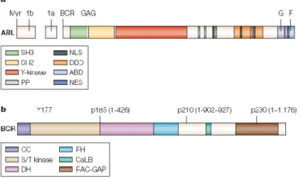
1.2.1 Structure and functions of the Bcr and Abl proteins
The BCR and ABL genes are expressed ubiquitously.
Bcr is a 160-kd cytoplasmic protein with several functional domains. The N-terminal 426 amino acids of Bcr, encoded by the first exon, are retained in all Bcr-Abl fusion protein isoforms. This region contains a serine-threonine kinase domain, whose only known substrates are Bcr and Bap-1 (a member of the Bap-14-3-3 family of proteins), and two serine/threonine–rich regions that bind
9 Src homology (SH)2 domains. The proximal SH2-binding domain is essential for transformation of rat fibroblasts by Bcr-Abl 5. The two key motifs of the first BCR exon are tyrosine 177 and the coiled-coil domain contained in amino acids 1 to 63. Phosphorylated tyrosine 177 forms a binding site for Grb-2 (an adapter molecule that links BCR to the Ras pathway) and is required for the induction of myeloid leukemia 6. The coiled-coil is crucial for dimerization of Bcr-Abl 7, which in turn is required for activation of Abl kinase activity and oncogenicity of Bcr-Abl. BCR regions of exon 1 are not essential to oncogenicity but influence the specific phenotype of the leukaemia (Fig. 1.1).
The ABL gene, the human homolog of v-abl (the oncogene of the Abelson murine leukemia virus), codes for a 145-kd nonreceptor tyrosine kinase. Two isoforms exist that differ in the first exon (1a and 1b). Only Abl type 1b protein contains a myristoylation site and, therefore, can be anchored to the plasma membrane. Three domains located toward the N-terminus of Abl are named after their homology to the respective domains in Src, the prototype non-receptor tyrosine kinase. The SH1 domain carries the tyrosine kinase function, the SH2 domain binds phosphotyrosine-containing consensus sites, and the SH3 domain binds to proline-rich consensus sequences in proteins like Crk 8 and Crkl 9. Abl differs from Src in having a long (~90-kd) C-terminal region that contains actin-and DNA-binding domains 7, three nuclear localization signals, and one nuclear export signal. Another unique feature of Abl is the N-terminal ‘‘Cap’’ region that is critical to the regulation of kinase activity. Abl is expressed predominantly in the nucleus10 but shuttles between nucleus and cytoplasm. The functions of the Abl protein are complex and include cell cycle inhibition, cellular responses to genotoxic stress 11, and signal transduction from growth factor receptors and from integrins 12 (Fig. 1.1).
Fig. 1.1 Schematic representation of the Abl (a) and Bcr (b) proteins. There are several important domains that
10
1.2.2 BCR-ABL fusion gene
1.2.2.1 Breakpoints in ABL
Breakpoints within the ABL gene can occur anywhere within a 50 segment that extends for over 300 kilobases (kb) 14. Typically, breakpoints are within intronic sequences, most frequently between the two alternative first exons of ABL. Thus, BCR-ABL fusion genes may contain both exons 1b and 1a, exon 1a alone, or neither of the alternative first exons. BCR-ABL mRNA lacks exon 1, regardless of the structure of the fusion gene, with the transcript consisting of BCR exons fused directly to ABL exon a2.
1.2.2.2 Breakpoints in BCR
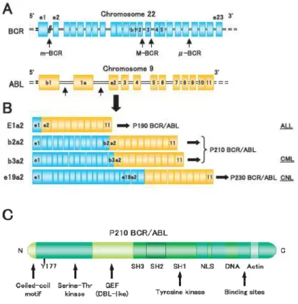
The breakpoints within the BCR gene on chromosome 22 are found within three defined regions. In 95% of patients with CML and approximately one third of patients with ALL, the BCR gene is truncated within a 5.8-kb region known as the major breakpoint cluster region. This region contains five exons, originally named b1 to b5, but now referred to as e12 to e16, according to their true positions in the gene. Most breakpoints are within introns immediately downstream of exon 13 (b2) or exon 14 (b3). Because processing of BCR-ABL mRNA results in the joining of
BCR exons to ABL exon a2, hybrid transcripts are produced that have an e13a2 (b2a2) or an
e14a2 (b3a2) junction. In both cases, the mRNA consists of an 8.5-kb sequence that encodes a 210-kd fusion protein, p210 Bcr-Abl (Fig. 2). In two-thirds of patients with Ph+ ALL and in rare cases of CML and AML, the breakpoint in BCR occurs in a region upstream of the major breakpoint cluster region known as the minor breakpoint cluster region. This region consists of the 54.4-kb intron between the two alternative second exons of the BCR gene, e20 and e2.
BCR-ABL fusion genes that have breakpoints within the minor breakpoint cluster region contain both
BCR alternative first exons (e1 and e10) together with the alternative second exon (e20). The hybrid mRNA consists of sequences that are approximately 7 kb in length in which exon e1 from
BCR is joined to exon a2 of ABL. The translated product is a 190-kd fusion protein, p190
Bcr-Abl (also referred to as p185 Bcr-Bcr-Abl). Interestingly, transcripts with an e1a2 junction are detectable at very low levels in patients with a major breakpoint cluster region rearrangement. The third defined breakpoint cluster region within the BCR gene was named ‘‘micro’’ breakpoint cluster region 15. In this case, the breaks occur within a 30 segment of the BCR gene between exons e19 and e20 (known as c3 and c4 in the original nomenclature) (Fig. 1.2). Transcription of the hybrid gene yields an e19a2 BCR-ABL fusion transcript that encodes a 230-k protein, p230
11 The p190 BCR-ABL fusion gene occurs in about 90% of children with Ph+ ALL and between 50% and 80% of adults with Ph+ ALL. The p210 BCR-ABL gene constitutes the rest of the Ph+ ALL population. The p230 BCR-ABL mutation is associated with Ph+ chronic neutrophilic leukemia 2.
Fig. 1.2 Three BCR-ABL variants and association of leukemia types. (A) Locations of the breakpoints in the
ABL and BCR genes and (B) structure of the chimeric BCR-ABL mRNA transcripts derived from the various
12
1.2.3 Mechanisms of BCR-ABL-mediated leukaemogenesis
Tyrosine kinase enzymatic activity is central to cellular signaling and growth, and constitutively elevated kinase activity has been associated with transformation in several systems. The Abl protein is a non-receptor tyrosine kinase whose enzymatic activity is under close physiologic control 16 . In contrast, Bcr-Abl proteins are constitutively active tyrosine kinases. The degree of transforming activity of Abl correlates with the degree of tyrosine kinase activity. p190 Bcr-Abl, which has higher tyrosine kinase activity, is therefore associated with the development of the more aggressive acute leukemia phenotype, while p210 Bcr-Abl plays a role in the more indolent chronic leukemia phenotype.
1.2.3.1 Altered cellular adhesion
In normal hematopoiesis, progenitor cells adhere to the stromal cells of the bone marrow and their associated extracellular matrix. The latter contains proteins such as fibronectin that function as adhesive ligands for receptors expressed on the surface of hematopoietic progenitor cells. Current thinking holds that the process of adhesion is essential for the regulation of hematopoiesis, providing a means of anchoring progenitors within the vicinity of cytokine-secreting cells 17, exposing them to specific signals that determine their fate. Ph+ progenitors exhibit reduced adhesion to stromal cells and the extracellular matrix 18, which ‘‘liberates’’ them from the regulatory signals that are supplied to normal, adherent hematopoietic progenitors. It also may explain why their homing to the bone marrow is disturbed, leading to the appearance of immature cells in the peripheral blood. There is evidence that the function of β integrins on the surface of CML progenitor cells is perturbed, the net effect being reduced adhesion and increased proliferation 17. In addition, migration, in response to certain chemokines such as MIP-1a, is abnormally high 19.
1.2.3.2 Activation of mitogenic signaling pathways
Bcr-Abl is known to activate several signaling pathways with mitogenic potential 20. It is important to remember that in many cases, the available data comes from experiments in Bcr-Abl–positive cell lines, and activation of some of these pathways in primary CML cells has yet to be verified.
Ras and the mitogen-activated protein kinase pathways
Bcr-Abl binds directly to proteins that activate Ras 21. Autophosphorylation of tyrosine 177 generates a binding site for the adapter molecule Grb-2 5. Grb- 2 associates with the Sos protein,
13 which stimulates the conversion of the inactive GDP-bound form of Ras to the active GTP-bound state 21. Ras also may be activated by two other adapter molecules, Shc and CrkL, which are substrates of Bcr-Abl 22. Although CrkL appears to be necessary for the transformation of fibroblasts by Bcr-Abl, direct binding of Crkl to Bcr-Ablis not required for the transformation of myeloid cells. Activated Ras binds to the serinethreonine kinase Raf-1, recruiting it to the plasma membrane where it is activated by tyrosine phosphorylation and initiates a signaling cascade by way of the mitogen-activated protein kinase (MAPK) pathway. Grb-2 also recruits the scaffolding adapter Gab2, which then is phosphorylated by Bcr-Abl, resulting in activation of phosphatidylinositol 3 (PI-3) kinase/Akt and Ras/Erk 23. Bcr-Abl activates different types of mitogen-activated protein kinases, including extracellular signal–related kinases (ERK)-1/2 and JNK or stress-activated protein kinase. Ultimately, these pathways regulate gene transcription.
Janus kinase–signal transducer and activator of transcription pathway.
Phosphorylation of members of the signal transducer and activator of transcription (STAT) family of transcription factors has been reported in Bcr-Abl–positive cell lines24 and in primary CML cells. Physiologically, STATs are phosphorylated by Janus kinases (Jak) that are downstream of growth factor receptors. In contrast, phosphorylation of STAT5 in Bcr-Abl– expressing myeloid cells appears to be mediated by the Src family kinase, Hck, which binds the SH2 and SH3 domains of Bcr-Abl25. There is evidence that activation of STAT5 by p210 Bcr-Abl contributes to malignant transformation of K562 cells26 and inhibits apoptosis by up-regulating the transcription of Bcl-xL27. STAT5, however, is not required for leukemia induction by Bcr-Abl in mice, casting doubt on its relevance in a more physiologic context. Interestingly, p190 Bcr-Abl differs from p210 Bcr-Abl in that it also is able to activate STAT6. It remains to be seen whether the predominantly lymphoblastic phenotype associated with p190 Bcr-Abl is related to this property of the shorter form of the oncoprotein.
Phosphatidylinositol 3 kinase pathway.
Proliferation of BCR-ABL–positive cell lines and primary cells is dependent on PI-3 kinase. Bcr-Abl apparently activates this pathway by forming a multimeric complex with PI-3 kinase, p120Cbl, and the adaptor molecules Crk and CrkL. In BCR-ABL–expressing cells, activated PI-3 kinase stimulates the serine-threonine kinase Akt 28, which in turn phosphorylates the forkhead transcription factor, FKHRL1. The net result of activating this pathway appears to be the proteasome-mediated degradation of the key cell cycle inhibitor p27Kip1, although the precise intermediates are unknown. Activated Akt may function in an antiapoptotic capacity. A key substrate of Akt is the proapoptotic protein or ‘‘death agonist’’ Bad. Bad promotes cell death by
14 binding to and thereby inactivating the antiapoptotic Bcl-2 and Bcl-xL. Thus, phosphorylation of Bad by Akt may prevent it from binding to these proteins, resulting in reduced apoptosis. Indeed, increased Bad phosphorylation was seen in BCR-ABL–positive cells; however, even with Bad completely dephosphorylated, a fraction of cells survived, indicating the existence of Bad-independent survival pathways.
Myc pathway.
Myc is classed as a proto-oncogene because it is overexpressed in many human malignancies. As a transcription factor and immediate early response gene, Myc converts mitogenic signals to alterations of gene expression. Not surprisingly, Myc targets include genes related to cell cycle and apoptosis. Within Bcr-Abl, the SH2 domain 29 and the C-terminus are required for full activation of Myc. It recently has been shown that Jak2 is involved in Myc induction by Bcr-Abl, apparently by way of induction of Myc mRNA and by stabilization of the protein.
1.2.3.3 Inhibition of apoptosis
Apoptosis caused by growth factor withdrawal is eliminated when factor dependent cell lines are transfected with exogenous BCR-ABL. The mechanisms by which Bcr-Abl inhibits apoptosis in cell lines are not well understood. The release of cytochrome C from mitochondria, a prerequisite for caspase-3 activation, apparently is blocked in BCR-ABL–expressing cell lines. Members of the Bcl-2 family of proteins may be involved in mediating the antiapoptotic effect of Bcr-Abl. Up-regulation of Bcl-2 by Bcr-Abl has been demonstrated in two different cellular contexts: one dependent on the Ras pathway and the other on the PI-3 kinase pathway. Bcl-2 targets Raf-1 to mitochondria where it inactivates the proapoptotic protein Bad by phosphorylating it on serine residues 30. Down-regulation of interferon consensus sequence binding protein (ICSBP) by Bcr-Abl also has been implicated as an important antiapoptotic event; conversely, ICSBP antagonizes Bcr-Abl by decreasing Bcl-2 expression. Another regulator of apoptosis targeted by Bcr-Abl is Bcl-xL, the expression of which is dependent of STAT5 activation. Surprisingly, a recent report has demonstrated that Bcr-Abl can actively induce apoptosis when trapped in the nucleus 31. Treatment of human and murine Bcr-Abl– positive cell lines with imatinib stimulated entry of the oncoprotein into the nucleus.
1.2.3.4 Proteasomal degradation
It recently was reported that Bcr-Abl tyrosine kinase activity induced the proteasome-mediated degradation of the ABL-interactor proteins Abi-1 and Abi-2 30. Bcr-Abl was found to cause down-regulation of the DNA repair protein DNA-PKcs in cell lines. Loss of DNA-PKcs activity
15 was correlated with impaired DNA repair and may facilitate the acquisition of additional genetic lesions that lead to disease progression. Another important degradation target is the cell p27, a crucial inhibitor of progression from the G1 to the S phase of the cell cycle. Furthermore, Bcr-Abl can stabilize the expression of Mdm2, a protein that targets the tumor suppressor p53 for ubiquitination, which also would promote genomic instability 32.
16
1.3 DISCOVERY OF NOVEL ALTERATIONS IN ALL PH+
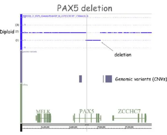
The recent coming of new “genome-wide” techniques, like gene expression profiling (GEP) and analysis of single nucleotide polymorphism (SNP) arrays has enabled to identify multiple novel genetic alterations targeting key cellular pathways, including lymphoid differentiation, cell cycle, tumor suppression, apoptosis and drug responsiveness. By GEP analysis, several down/up-expressed genes have been identified. By genome-wide SNP array analysis of leukaemia blast cells of paediatric and adult CML blastic phases and Ph+ ALL patients 33, the presence of three frequent genetic deletions affecting IKZF1 (IKAROS), PAX5 deletions (Fig. 1.3) and the
CDKN2A/B locus was identified.
Fig. 1.3 Schematic representation of PAX5 deletion in a patient with Ph+ ALL as detected by SNP array analysis. The interrupted blue line represents the diploid genome (2) in the region containing MELK, PAX5 and
ZCCHC7 genes. Deletion is represented by a stretch of SNPs, below the level of the diploid line (1). Copy number
17
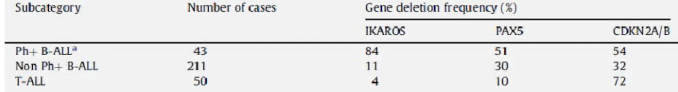
IKZF1 and PAX5 encode transcription factors required for normal lymphoid development. CDKN2A/B locus encodes three tumour suppressor genes that are widely inactivated in many
human cancers. At diagnosis, 84% of patients had already sustained IKFZ1 deletions and more than 50% of patients exhibited PAX5 loss; mono- or biallelic losses, encompassing the entire
CDKN2A (CDKN2A and ARF) and CDKN2B gene cluster were also recognized in half of the
patients (Tab. 1.1). Several other recurring deletions were identified in BCR-ABL1 ALL, albeit at a lower frequency, including C20orf94, RB1, MEF2C and EBF1.
Tab. 1.1 Frequency of deletions in Ph+ ALL (Modified from C. G. Mullighan, Gene and Development 2008) 35.
a No significant differences in gene deletion frequencies were observed between 21 paediatric and 22 adult cases
subjected to analysis. Study of the CDKN2A/B gene cluster in 41 of these cases by quantitative PCR using primers directed to each of the INK4A, ARF and INK4B exons indicated an overall deletion frequency of 64%.
IKFZ1 deletions, limited to the gene in the majority of the cases, were mostly monoallelic and
were responsible for the expression of a dominant-negative isoform 36. In T-cell ALL, the frequencies of IKFZ1 and PAX5 deletions were much lower (Tab. 1.1), consistent with findings that these two genes play key roles in regulating B-cell lineage commitment and differentiation
37
. Although differentiation arrest is a distinctive feature of ALL, the manner by which IKFZ1 and PAX5 inactivation collaborate with BCR-ABL to induce lymphoblastic leukemia is not yet understood. CDKN2A/B deletions occur in all lymphoid malignancies (Tab. 1.1), pointing to their general role in tumor suppression in both T- and B-cell ALL, as well as in many other tumor types. The fact that deletion of PAX5 and CDKN2A/B generally occurred together with
IKFZ1 loss implies that disruption of each of these genes contributes independently to Ph+
B-cell ALL 35. GEP is providing a new diagnostic marker and could identify some therapeutic targets, having a major impact on the way we diagnose and treat Ph+ leukaemia patients. Although considerable work remains to be done before these predictions are realized, our capacity to obtain these type of data continues to mature at a rapid speed. Thus, the fruits of GEP should soon help us to accurately identify specific leukaemia subtypes, abnormally expressed or
18 spliced genes and to select targeted therapies. For instance, recent GEP studies have identified a new ALL subtype with a gene-expression pattern resembling that of BCR-ABL1-positive ALL. This newly recognized group includes 15–20% of all precursor B-ALL cases and is associated with an unfavourable outcome.
19
1.4 9p21 LOCUS (INK4A/ ARF/ INK4B/ ANRIL)
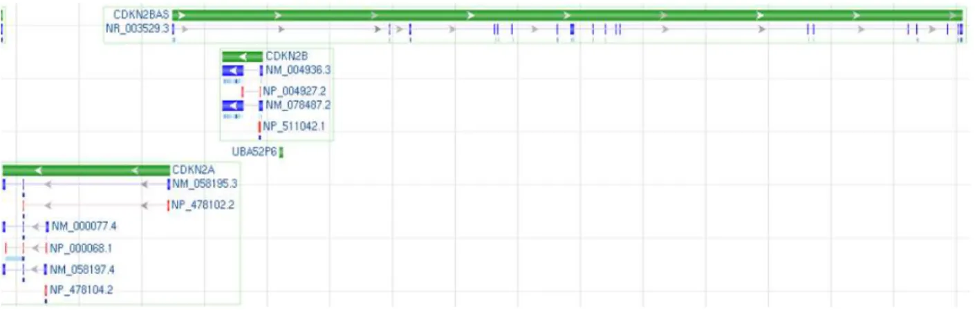
The chromosome 9p21 segment is a 40-kb region encoding two members of the INK4 family of Cyclin-Dependent Kinase inhibitors, CDKN2B (p15INK4B, ENSG00000147883) and CDKN2A (p16INK4A, ENSG00000147889), and one other related gene ARF (p14ARF), transcribed in an Alternative Reading Frame compared to CDKN2A; all of them encode critical factors for the regulation of cell cycle and for the influence on key physiological processes such as replicative senescence, apoptosis, and stem-cell self-renewal. 9p21 also contains a newly annotated non-coding RNA, termed ANRIL (CDKN2BAS, ENSG00000240498), which spans 126.3 kb and overlaps at its 5’ end with CDKN2B, and it is transcribed from the opposite strand to CDKN2A/B (Fig. 1.4).
Fig. 1.4 9p21 Locus NC_000009.11 Homo sapiens chromosome 9, GRCh37.p2 primary reference assembly
MapViewer representation. Green bars show the length and the gene transcription verse. Blu bars show exons and
red bars represent gene translation. Modified from NCBI MapViewer
http://www.ncbi.nlm.nih.gov/nuccore/224589821
CDKN2B and CDKN2A arose from a gene duplication event and are consequently very similar
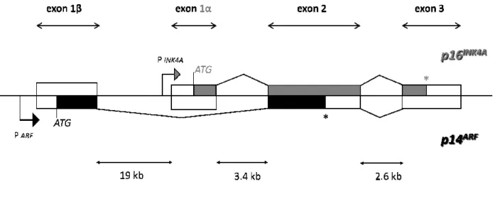
(77% amino acid sequence identity in humans). CDKN2A and ARF have different promoter and different first exons (1α and 1β, respectively) (Fig. 1.5), giving rise to products in alternate reading frames with no homology at the protein level and with distinct functions in the cells.
Fig. 1.5 Organization of the CDKN2A
p14ARF are in black. The alternative first exons are transcribed from different promoters (arrows). *Denotes the
p16INK4A and p14ARF termination codons.
(p16) gene. Intronic sizes derived from http://snpper.chip.org are indicated. Biochem Cell Biol, 2006 38.
Exons 1α, 2, and 3 encode p16 Exons 1β, 2, and 3 encode p14 Therefore, the 9p21 locus has a un
frames (ORFs) that initiate in different first exons and continue in alternative reading frames in a common second exon. This unusual utilization of overlapping exonic sequences in mammalian cells enables a single gene to encode two completely unrelated protein products
the gene CDKN2A, in a more centromeric position, two exons encode for another tumor suppressor gene: CDKN2B (p15
Therefore on chromosome 9p21.3 there are and ‘4’ (Appendix A), each conta
http://www.ncbi.nlm.nih.gov/nuccore/NM_000077.4 (http://www.ncbi.nlm.nih.gov/gene/1029
amino acids; isoform ‘1’ encodes p16
that is structurally unrelated to p16 but acts in cell cycle G1 control by stabilizing the tumor suppressor protein p53.
The chromosome band 9p21 shows a
where a multispecies sequence alignment shows 76% sequence identity and 94% sequence CDKN2A/ARF locus. The coding regions of p16INK4A are shown in
alternative first exons are transcribed from different promoters (arrows). *Denotes the termination codons. Exon 1β of the p14ARF gene is 19 kb upstream of exons 1
Intronic sizes derived from http://snpper.chip.org are indicated. Modified from Stuart J.Gallagher,
, 2, and 3 encode p16INK4A, which induces G1 cell cycle arrest via the Rb pathway. , 2, and 3 encode p14ARF, which inhibits p53 degradation via binding to
has a unique gene structure that generates two distinct open reading frames (ORFs) that initiate in different first exons and continue in alternative reading frames in a common second exon. This unusual utilization of overlapping exonic sequences in mammalian
s enables a single gene to encode two completely unrelated protein products
, in a more centromeric position, two exons encode for another tumor
p15INK4B).
on chromosome 9p21.3 there are two main transcripts of CDKN2A
, each contains three exons and spans: 7,382 (according with NCBI,
http://www.ncbi.nlm.nih.gov/nuccore/NM_000077.4) and 26740 bp
ov/gene/1029), respectively. They encode proteins of 156 and 173 amino acids; isoform ‘1’ encodes p16 (INK4A), while isoform ‘4’ encodes p14
that is structurally unrelated to p16 but acts in cell cycle G1 control by stabilizing the tumor
The chromosome band 9p21 shows a significant evolutionary conservation only in
ultispecies sequence alignment shows 76% sequence identity and 94% sequence 20
shown in gray, and those of alternative first exons are transcribed from different promoters (arrows). *Denotes the is 19 kb upstream of exons 1α of the CDKN2A Modified from Stuart J.Gallagher, Int J
, which induces G1 cell cycle arrest via the Rb pathway. , which inhibits p53 degradation via binding to Mdm2 39. ique gene structure that generates two distinct open reading frames (ORFs) that initiate in different first exons and continue in alternative reading frames in a common second exon. This unusual utilization of overlapping exonic sequences in mammalian s enables a single gene to encode two completely unrelated protein products 40. Adjacent to , in a more centromeric position, two exons encode for another tumor
CDKN2A locus, isoforms ‘1’
(according with NCBI,
and 26740 bp
respectively. They encode proteins of 156 and 173 ), while isoform ‘4’ encodes p14 (ARF), a protein that is structurally unrelated to p16 but acts in cell cycle G1 control by stabilizing the tumor
significant evolutionary conservation only in the exon 2, ultispecies sequence alignment shows 76% sequence identity and 94% sequence
similarity across six mammalian species
overlaps, the drop in conservation beyond the coding region of isoform 4 suggests that this isoform is more responsible for the observed conservation
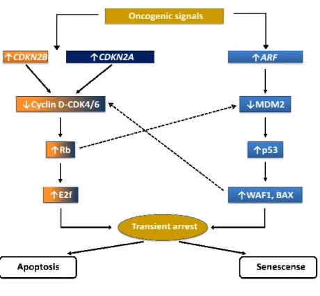
The CDKN2A/ARF locus encodes proteins that RB are at the heart of the two main tumour
to potentially oncogenic stimuli. Each pathway consists of several upstream regulators and downstream effectors. The pathways interact at several points, and
(Fig. 1.6). p53 is a transcription factor that regulates apoptosis and cellular senescence by inducing the transcription of specific genes; the RB pathway directly regulates the cell cyc hence cellular senescence, but is also important in apoptosis, probably by interacting with the p53 pathway.
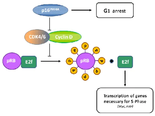
Fig. 1.6 The CDKN2A/ARF/CDKN2B
INK4 family of cyclin.dependent kina
p53 stabilization. Modified from Judith Campisi
y across six mammalian species. While exon 2 of both alternative reading frames overlaps, the drop in conservation beyond the coding region of isoform 4 suggests that this isoform is more responsible for the observed conservation 41.
locus encodes proteins that function upstream of both RB and p53. p53 and RB are at the heart of the two main tumour-suppressor pathways that control cellular responses to potentially oncogenic stimuli. Each pathway consists of several upstream regulators and
he pathways interact at several points, and cross
). p53 is a transcription factor that regulates apoptosis and cellular senescence by inducing the transcription of specific genes; the RB pathway directly regulates the cell cyc hence cellular senescence, but is also important in apoptosis, probably by interacting with the
CDKN2B locus and the p53 and RB tumour-suppressor pathways
kinase inhibitors bind to and inactivate CDK4/6. ARF inhibits MDM2, resulting in Modified from Judith Campisi, Nature Reviews Cancer, 2003 42.
21 both alternative reading frames overlaps, the drop in conservation beyond the coding region of isoform 4 suggests that this
function upstream of both RB and p53. p53 and suppressor pathways that control cellular responses to potentially oncogenic stimuli. Each pathway consists of several upstream regulators and cross-regulate each other ). p53 is a transcription factor that regulates apoptosis and cellular senescence by inducing the transcription of specific genes; the RB pathway directly regulates the cell cycle and hence cellular senescence, but is also important in apoptosis, probably by interacting with the
suppressor pathways. Members of the
22 In the p53 pathway, signals such as DNA damage induce ARF. The tumor suppressor activity of
ARF is largely ascribed to its ability to regulate p53 in response to aberrant growth or oncogenic
stresses such as c-MYC activation. ARF increases p53 levels by sequestering Mdm2, which facilitates the degradation and inactivation of p53. One mechanism that has been proposed to explain how Mdm2 regulates p53 is that it acts as an E3 ubiquitin ligase to target p53 for proteasomal degradation. Although strong biochemical and genetic evidence link ARF and p53 in tumor suppression, several p53-independent functions of ARF have also been reported. Moreover, ARF has been reported to interact with multiple proteins other than Mdm2, including E2F-1, MDMX, HIF-1α, topoisomerase I, MYC, and nucleophosmin (NPM). p53 has both transactivation and transrepression activity, and so controls the transcription of numerous genes. Among the p53 target genes are WAF1, an inhibitor of cyclin-dependent protein kinases (CDKs) that, among other activities, causes cell-cycle arrest, and BAX, which promotes apoptotic cell death.
CDKN2A/B inhibit the complex between cyclin-dependent kinases 4/6 (CDK4/6) and cyclin D
(the binding of the INK4 proteins INK4A/INK4B to CDK4/6 induces an allosteric change that abrogates the binding of these kinases to D-type cyclins) that phosphorylate, and therefore inactivate, RB during the mid-late G1 phase of the cell cycle (RB phosphorylation interrups its interactions with both histone deacetylase HDAC and E2F, enabling E2F to promote S phase entry). RB also controls the expression of numerous genes, although it does so primarily by recruiting transcription factors and chromatin remodelling proteins. One downstream consequence of RB activity is the inhibition of E2F activity, which is important for the transcription of several genes that are required for progression through the G1 and S phases of the cell cycle. RB also regulates p53 activity through a trimeric p53–Mdm2–RB complex 42,43. The ARF/CDKN2A/B proteins are established tumour suppressors altered in a range of human cancers including familial cutaneous malignant melanoma, glioma (60%), head and neck cancers
(50%) and bladder cancers (45%). High frequencies of 9p21.3 deletions have been documented in acute lymphoblastic leukemia ranging from 18% to 45% 1. Moreover, the chromosome 9p21.3 region adjacent to the loci encoding CDKN2A and CDKN2B is an important susceptibility locus for several diseases with a complex genetic background. Recent genome-wide association (GWA) studies have shown that single nucleotide polymorphisms (SNPs) in this region are associated with coronary artery disease (CAD), ischaemic stroke, aortic aneurysm, type II diabetes, glioma, and malignant melanoma. Candidate gene approaches have also reported SNPs in this region to be associated with breast, ovarian, and pancreatic carcinoma, melanoma, and
23 acute lymphoblastic leukaemia 44. Most of the risk variants in the chromosome 9p21 region identified by GWA studies are in non-coding regions, suggesting that their effects are likely to be mediated by influences on gene expression. Sequence variation can influence expression by
cis or trans mechanisms 45.
Finally, the human CDKN2A/ARF locus encodes one additional transcript, isoform ‘3’ (p12), and perhaps others as well. The p12 transcript represents a p16INK4A-isoform that shares the p16INK4A promoter, 5’ UTR, ATG and exon 1α, but then uses an alternative splice donor within the first intron of p16INK4A to splice to exon 2. The extra sequence contains a stop codon, and therefore the transcript produces a 12 kDa protein that shares the only first ankyrin repeat of p16INK4A. In non-diseased tissue, this transcript is only expressed in pancreas but not other human tissues. The significance and function of this transcript is unknown, but based on crystal structure studies of INK4 proteins, p12 would not be predicted to bind cdk4 or cdk6. Even if the p12 transcript does not encode a direct tumor suppressor protein, however, its intimate relationship with the
INK4A/ARF locus makes it likely that its transcription influences the expression of p16INK4A
and/or p14ARF46.
1.4.1 ARF/p14ARF
1.4.1.1 Gene structure
The ARF tumor suppressor transcript was first identified in humans in 1995 (p14ARF) 47,48, and its protein product confirmed in mice (p19ARF) that same year 49. Its gene locus is on the short arm of chromosome 9 in humans, and on a corresponding location on chromosome 4 in mice. ARF is an alternate reading frame (ARF) product of the CDKN2A locus. It spans approximately 26 kb of genomic DNA and comprise three exons 1β, 2, and 3 (Fig. 1.7). The p14ARF open reading frame is derived from a distinct first exon (exon 1β), originating approximately 19 kb centromeric to the first exon of p16INK4A (exon 1α) and 23 kb centromeric to exon 2. Exon 1β, under the control of its own promoter, is spliced to the second and third exons that are separated by 3 kb of intronic sequence and are shared with p16INK4A. The open reading frame of the 1.1 kb ARF transcript is terminated within exon 2, with exon 3 comprising an untranslated 3‘ exon.
Fig. 1.7 ARF NM_058195.3 MapViewer representation.
verse. Blu bars show exons and red bars represent gene translation. Modified from NCBI MapView
http://www.ncbi.nlm.nih.gov/gene/1029#
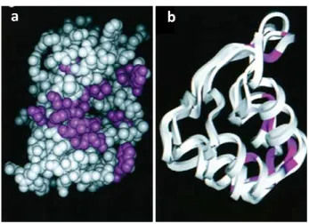
1.4.1.2 ARF is a peculiar protein with unusual primary structure
The human protein comprises 173 amino acids with a molecular weight of 13,902 Da, whereas the mouse homologue of 169 amino acids has a molecular weight of 19,238 Da. Both proteins share only limited sequence homology (50%) that could explain some of their functional differences but both are capable of inducing cell cycle arrest at the G2/M as well
cycle boundaries. The ARF proteins show significant sequence similarity within their amino terminal 14 amino acids (11/14 identity), and this region retains many of the known ARF functions, including nucleolar localization, Mdm2 binding an
The carboxy-terminus of ARF
region (amino acids 130–169) of p19 independent apoptosis. The ability of p14
also involves the C-terminal nucleolar localization sequence of p14
both composed of more than 20% arginine residues conferring them highly basic and hydrophobic properties. Interestingly, there
and the protein probably needs to form complexes with other molecules, both to be folded and for its charge to be neutralized at physiological pH. This probably explains the increasing number of yet identified ARF partners. Mouse
p14ARF has none. Mouse p19
residues (Met45 and Met48 respectively). Translational initiation from these methionine residues produces both in mouse and human a short form of the protein that, when overexpressed, localizes to mitochondria (smARF). Nevertheless, full
nucleoli thanks to nucleolar localization signals (NoLS).
NM_058195.3 MapViewer representation. Green bar shows the length and the gene transcription
verse. Blu bars show exons and red bars represent gene translation. Modified from NCBI MapView
http://www.ncbi.nlm.nih.gov/gene/1029#.
ARF is a peculiar protein with unusual primary structure
The human protein comprises 173 amino acids with a molecular weight of 13,902 Da, whereas mouse homologue of 169 amino acids has a molecular weight of 19,238 Da. Both proteins share only limited sequence homology (50%) that could explain some of their functional differences but both are capable of inducing cell cycle arrest at the G2/M as well
cycle boundaries. The ARF proteins show significant sequence similarity within their amino terminal 14 amino acids (11/14 identity), and this region retains many of the known ARF functions, including nucleolar localization, Mdm2 binding and ability to induce cell cycle arrest.
ARF also encodes functional domains. In particular, the C
169) of p19ARF, that is missing in p14ARF
independent apoptosis. The ability of p14ARF to promote the sumoylation of its binding partners terminal nucleolar localization sequence of p14ARF38.
both composed of more than 20% arginine residues conferring them highly basic and hydrophobic properties. Interestingly, there are no recognizable structural motifs in ARF proteins and the protein probably needs to form complexes with other molecules, both to be folded and for its charge to be neutralized at physiological pH. This probably explains the increasing entified ARF partners. Mouse p19ARF contains only one lysine and human p19ARF and human p14ARF contain only single internal methionine residues (Met45 and Met48 respectively). Translational initiation from these methionine residues roduces both in mouse and human a short form of the protein that, when overexpressed, localizes to mitochondria (smARF). Nevertheless, full-length ARF preferentially localizes in the nucleoli thanks to nucleolar localization signals (NoLS). p19ARF contains
24
Green bar shows the length and the gene transcription verse. Blu bars show exons and red bars represent gene translation. Modified from NCBI MapViewer
The human protein comprises 173 amino acids with a molecular weight of 13,902 Da, whereas mouse homologue of 169 amino acids has a molecular weight of 19,238 Da. Both proteins share only limited sequence homology (50%) that could explain some of their functional differences but both are capable of inducing cell cycle arrest at the G2/M as well as at G1/S cell cycle boundaries. The ARF proteins show significant sequence similarity within their amino-terminal 14 amino acids (11/14 identity), and this region retains many of the known ARF
d ability to induce cell cycle arrest. also encodes functional domains. In particular, the C-terminal is involved in p53-to promote the sumoylation of its binding partners
p14ARF and p19ARF are both composed of more than 20% arginine residues conferring them highly basic and are no recognizable structural motifs in ARF proteins and the protein probably needs to form complexes with other molecules, both to be folded and for its charge to be neutralized at physiological pH. This probably explains the increasing contains only one lysine and human contain only single internal methionine residues (Met45 and Met48 respectively). Translational initiation from these methionine residues roduces both in mouse and human a short form of the protein that, when overexpressed, preferentially localizes in the a unique NoLS in its
25 exon1β (aa 26-37) which deletion induces the nuclear translocation of the protein. The situation is more complex for p14ARF as two NoLS have been identified in the protein. The first one localized in exon 1β plays a key role in the antiproliferative function of p14ARF as its deletion inhibits the ability of p14ARF to stop the cell cycle and to bind Mdm2. The second one stands in exon 2 (aa 83-101) and is involved in the ability of p14ARF to promote the sumoylation of its binding partners 50.
1.4.1.3 Expression and turnover Expression
Normal cells contain low levels of ARF but the expression of a variety of proliferation-promoting proteins, including Myc, E2F, E1A, oncogenic Ras and v-Abl, upregulate ARF as part of a checkpoint response conveying on the well known p53-Mdm2 pathway. The discovery of multiple ARF interactors and the observation that, aside oncogenic stimuli, also viral, genotoxic, hypoxic and oxidative stresses activate an ARF-dependent response, suggest that ARF could exert a wider role to protect the cell. It is becoming clear that the ARF response is quite complex and is likely accomplished by the interaction with a multitude of different cellular partners that in part explain the p53-independent activities of ARF 51. ARF is ubiquitously expressed and is elevated in cells lacking p53. In human cells p14ARF expression levels remain low as cells near senescence and p14ARF depleted cells still undergo a senescence-like arrest when challenged with Ras 46. Not much is known about the regulation of p14ARF expression. p14ARF transcription is induced by E2F1, but not by oncogenic Ras and while p14ARF transcription is inhibited by Tbx-2, Tbx-3 and Cbx-7, it is not down-regulated by Bmi-1 38,46,52.
Degradation
ARF is a relatively stable protein, with half-life estimations ranging from approximately 1–6 h. p19ARF and p14ARF undergo amino-terminal ubiquitination and their degradation depends on the ubiquitin-proteosome pathway, and may be enhanced by Mdm2 expression. Stress induced nucleoplasmic redistribution of ARF destabilises the protein and targeting of a p14ARF fragment encompassing amino acids 2–29 to the nucleolus increased its half-life 53. Consistent with these data, ARF-B23 complex formation, which restricts ARF to the nucleolus, stabilises ARF by blocking its ubiquitination 54. In contrast, Mdm2-ARF complex formation occurs preferentially in the nucleoplasm and this may allows for enhanced ARF ubiquitination and proteasomal degradation 38.
26
1.4.1.4 Biological functions
The ARF tumour suppressor is a critical activator of the p53 pathway
The ARF tumour-suppressor protein suppresses aberrant cell growth in response to oncogene activation by inducing the p53-pathway. The ARF induction of p53 is mediated through two ubiquitin ligases, Mdm2, a RING finger oncoprotein and ARF-BP1/Mule (ARF-binding protein
1/Mcl-1 ubiquitin ligase E3), a HECT (homology to E6-AP C-terminus) containing protein. Both
Mdm2 and ARF-BP1 act as specific E3 ubiquitin ligases for p53, are highly expressed in various types of tumours, and have the potential to abrogate the tumour-suppressor functions of p53. ARF associates with Mdm2 to inhibit the ubiquitination, nuclear export and subsequent degradation of p53. It has been proposed that ARF physically sequesters Mdm2 in nucleoli, thus relieving nucleoplasmic p53 from Mdm2-mediated degradation 53. Recent data, however, suggest that nucleolar relocalization of Mdm2 is not required for p53 activation and that the redistribution of ARF into the nucleoplasm enhances its interaction with Mdm2 and its p53-dependent growth-suppressive activity 54. This current model of ARF function supports the concept that nucleolar disruption contributes to p53 signalling since many stress signals perturb the nucleolus, causing the release of nucleolar proteins (including ARF, L5, L11, L23 and B23) that activate the p53 pathway. In addition to Mdm2, ARF-BP1 is a key regulator of the p53 cell cycle regulatory pathway; ARF-BP1 directly binds and ubiquitinates p53 in an Mdm2-independent manner. Silencing of ARF-BP1 extended the half-life of p53, resulted in the transcriptional activation of the p53 targets, p21Waf1 and BAX, and activated a p53- dependent apoptotic response. Unexpectedly, ARF-BP1 also ubiquitinates and promotes the degradation of the anti-apoptotic bcl-2 family member, Mcl-1, and down-regulation of ARF-BP1 expression can also render cells more resistant to killing by genotoxic agents. Thus, ARF-BP1 has been assigned both anti-apoptotic (via p53 degradation) and pro-apoptotic (via Mcl-1 degradation) functions. Whilst the effect of ARF on ARF-BP1- mediated Mcl-1 degradation is presently unexplored, the divergent roles of ARF-BP1 may be regulated by ARF. Following aberrant oncogene activation, ARF expression is induced and inhibits ARF-BP1 activity toward p53 in the nucleus, thereby leading to p53-dependent cell cycle arrest or apoptosis. In the cytoplasm, where ARF is not abundant, oncogene activation may lead to ARF-BP1 mediated Mcl-1 degradation further promoting apoptosis. ARF also enhances p53 function by promoting the phosphorylation and inhibiting the transcriptional activity of the RelA NF-kB subunit. The NF-kB family of transcription factors display anti-apoptotic activity and antagonise the p53 pathway through induction of Mdm2 and repression of p53. Thus, by counteracting the functions of Rel A, ARF increases the effectiveness of the p53 pathway 38.
27
Nucleolar functions of the ARF tumour suppressor
ARF is predominantly a nucleolar protein and rather than residing in inactive “storage” within the nucleolus, ARF may regulate ribosome biogenesis by retarding the processing of early 47S/45S and 32S rRNA precursors 46. These effects do not depend on Mdm2 or p53 but may involve the interaction of ARF with B23. B23 is an abundant nucleolar endoribonuclease that is required for the maturation of 28S rRNA and interacts with many cellular proteins, including p53, Mdm2, ARF, NPM3 and the BARD-BRCA1 ubiquitin ligase. The interaction of ARF and B23 retains both proteins in the nucleolus, bound to the pre-60S ribosome, where they appear to influence ribosome biogenesis and/or function. The actual role of the nucleolar ARF–B23 complex remains unclear; p19ARF can promote the ubiquitination and degradation of B23, whereas p14ARF had no effect on B23 protein expression. In response to cytotoxic drugs, such as actinomycin D and DNA damaging agents, including UV light, B23 and ARF undergoes nucleoplasmic redistribution, where Mdm2 and B23 compete for ARF binding. The nucleoplasmic translocation of ARF and B23 promotes the formation of the B23-Mdm2 and the ARF-Mdm2 binary complexes and induces potent activation of the p53 pathway. Thus, ARF may directly access ribosome function to inhibit cell growth through its nucleolar association with B23, and it may regulate the p53 cell cycle pathway via its nucleoplasmic interaction with Mdm2 and ARF-BP1 38.
ARF regulates the protein turnover and function of most of its interacting partners
ARF can also suppress the proliferation of mouse cells lacking both Mdm2 and p53, implying interactions with other regulators. Consistent with these findings, the spectrum of tumours seen in mice lacking ARF and p53, with or without Mdm2, was significantly greater than that associated with animals lacking either gene 38.
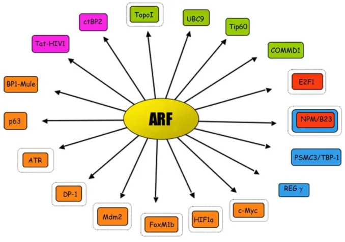
During the last years many efforts have been attempted in search of ARF partners that could partly explain the p53-Mdm2 ARF independent functions. In addition to its first ‘‘spouse’’ Mdm2, the ARF interactors ‘‘harem’’ consists of something like 30 different proteins involved in various cellular activities (Fig. 1.8, Tab. 1.2): proteins involved in transcriptional control, such as E2Fs, DP1, c-Myc, p63, Hif1a, Foxm1b, nucleolar proteins such as nucleolin/ C23 and nucleophosmin (NPM/B23), viral proteins such as HIV-1Tat, proteins involved in copper metabolism like COMMD1, proteins involved in chromosomal stability and/or chromatin structure such as Topoisomerase I, Tip60, and WRN helicase, ubiquitin ligases like Ubc9 (the E2 ligase required for sumoylation), Mdm2 and ARF-BP1/Mule, (E3-ubiquitin ligases) 51.
28
Fig. 1.8 A schematic view of the ‘‘ARF harem’’. ■ Orange is for partners whose activity is blocked by ARF. ■
Red is for partners that are induced to proteasome and ubiquitin-dependent degradation by ARF. ■ Pink is for partners that are induced to proteasome and ubiquitin-independent degradation by ARF. ■ Green is for partners whose activity or stability are positively regulated by ARF. ■ Blue is for partners that regulate ARF protein turnover. A second black circle indicate nucleolar sequestration 51.
29
Tab. 1.2 Cellular protein partners of the ARF tumour-suppressor protein, and its biological effect. Modified
from Stuart J.Gallagher, Int J Biochem Cell Biol, 2006 38.
ARF binding partner Biological effect of ARF binding
APA-1 No apparent effects
ARF-BP1/Mule1 Inhibition of ARF-BP1 ubiquitin ligase activity B23 Degradation of B23, inhibition of B23 shuttling BCL6 Inhibition of BCL6 transcriptional activity CARF Enhanced ARF-mediated cell cycle arrest c-MYC Inhibition of c-MYC transactivation COMMD1 Poly-ubiquitination of COMMD1 CtBP2 Degradation of CtBP2;
DP-1 Inhibition of ARF-induced E2F proteolysis E2F-1, -2, -3 Degradation of E2F
Foxm1b Inhibition of Foxm1b transactivation HIF-1α Inhibition of HIF-1α transactivation
HIV1-Tat protein Ubiquitin-independent degradation of the HIV1-Tat protein Mdm2 Inhibition of mdm2 ubiquitin ligase activity
MdmX Enhanced p53 transactivation
Neurabin Enhanced ARF-mediated cell cycle arrest p120E4F Enhanced ARF-mediated cell cycle arrest
Pex19p Inhibition of p19ARF, pex19p does not bind human ARF SUMO Conjugation of SUMO to p53 and MDM2
Tat-binding protein-1 Induces ARF stabilization
Topoisomerase I Enhanced topoisomerase I activity
Ubc9 Probable involvement in p14ARF-mediated sumoylation Werners helicase Nucleolar exclusion of Werners helicase
Although the actual mechanism by which ARF affects the activity of its partners is still unclear, the functional consequence is, quite invariably, inactivation.
For some targets, ARF interaction causes alteration of stability. For example, B23/NPM and E2F become degraded by the proteasome in an ubiquitin-dependent manner, while the CtBP2 antiapoptotic transcriptional co-repressor and HIV-1 Tat become degraded by the proteasome in
30 a ubiquitin-independent manner. Other targets changes their localization like E2Fs, c-Myc, Foxm1B, Mdm2, ATR, DP-1, Hif1a upon ARF expression. Only few others, like Tip60, Topo I and COMMD1 become activated or stabilized. Finally, most of the partners become sumoylated although a precise function to this modification has not yet been assigned.
Particularly interesting is the inhibitory effect that ARF exerts on oncogenes such as members of the E2F family, required both for cell-cycle progression and to mediate ARF oncogenic activation, suggesting a potential role of these interactions as being part of a negative feedback loop. In a series of reports ARF was shown to interact with E2F1, and this interaction prevented the formation of active E2F1 transcritional complexes, inhibited E2F1 transactivation potential, and promoted the proteasome-dependent degradation of E2F1, 2 and 3. In line with a role of ARF in promoting ubiquitin-dependent degradation of its partners is the observation that NPM/B23, an abundant nucleo/nucleolar multifunctional protein involved in ribosome biogenesis, is a molecular target of ARF. The vast majority of ARF appears localized in nucleoli, tightly associated with NPM/B23. There is a regulative loop between ARF and B23, in which degradation and inhibition of both proteins is finely and tightly modulated by external stimuli. In such a situation, ARF serves a dual function to restrain both growth and proliferation.
Interestingly, ARF appears to mediate also ubiquitin-independent degradation like that of the antiapoptotic transcriptional co-repressor C-terminal binding protein 2 (CtBP2) and of the HIV1-Tat protein. Interestingly, Mdm2 has been shown to ubiquitinate HIV1-HIV1-Tat, although, in this case, ubiquitination determines an increase of the Tat-mediated transactivation properties. This lead to the speculation that ARF could act on HIV-1Tat in two ways: directly mediating its degradation and inhibiting the Mdm2 activity versus Tat, thus blocking viral transcription. This hypothesis would intriguingly fit with the ARF role in viral defense.
As mentioned above, in some cases, ARF is able to stabilize its partners from proteasomal degradation. In a quite recent study, it has been described the ARF’s ability to induce a non-classical poly-ubiquitination of a new interacting partner, the COMMD1 factor, a multifunctional protein involved in copper metabolism and apoptosis. While in normal conditions COMMD1 is degraded by the proteasome, ARF coexpression leads to a stabilization of the protein through its poly-ubiquitination on K63 lysine of the ubiquitin peptide.
Altogether, although these observations reinforce the idea that the ARF antioncogenic activity could be partly exerted through the cellular degradation machinery. In this sense, ARF interaction with the proteasome could serve dual roles: on one side it is necessary to regulate ARF protein turnover, while, on the other side, it could play a role in bringing ARF interacting partners in contact with the ubiquitin/proteasome machinery 51.
ARF promotes the sumoylation of its binding partners
ARF can promote the conjugation of the small u
partners, including Mdm2 and B23. Sumoylation is analogous to ubiquitination, and is the process by which the SUMO protein is conjugated to a target protein. The effects of this modification are target specific a
structures and regulation of transcription factor activities. The diverse functional consequences of sumoylation provide a possible explanation for the versatility of ARF downstream effects. Although, ARF can promote sumoylation in
not been demonstrated for ARF binding partners and sumoylation sites on these proteins remain unidentified. However, the fact that a subset of melanoma
sumoylate Mdm2 in vivo, suggests that sumoylation may provide a mechanism for the diverse actions of the ARF tumour-suppressor protein
1.4.2 CDKN2A/INK4A/p16INK4A
CDKN2A (cyclin dependent
dependent Kinase 4 (INK4) family of proteins which also includes p15
p19INK4D. The INK4 proteins are roughly 40% homologous to one another, but individual members are highly conserved across species. Family members have in common a structural motif: a series of ankyrin repeats responsible for interaction with cyclin kinases 4/6 (CDK4/6).CDKN2A
by three exons: 1α, 2 and 3 (Fig.
Fig. 1.9 CDKN2A NM_000077.4 MapViewer representation
transcription verse. Blu bars show exons and red bars represent gene translation. Modified from NCBI MapViewer
http://www.ncbi.nlm.nih.gov/gene/1029
ARF promotes the sumoylation of its binding partners
ARF can promote the conjugation of the small ubiquitin-like protein SUMO
partners, including Mdm2 and B23. Sumoylation is analogous to ubiquitination, and is the process by which the SUMO protein is conjugated to a target protein. The effects of this modification are target specific and include control of protein stability, formation of subnuclear structures and regulation of transcription factor activities. The diverse functional consequences of sumoylation provide a possible explanation for the versatility of ARF downstream effects. Although, ARF can promote sumoylation in vivo, the biological impact of target sumoylation has not been demonstrated for ARF binding partners and sumoylation sites on these proteins remain unidentified. However, the fact that a subset of melanoma-associated p14ARF
, suggests that sumoylation may provide a mechanism for the diverse suppressor protein 38.
INK4A
ependent kinase inhibitor 2A) is a member of the inase 4 (INK4) family of proteins which also includes p15
. The INK4 proteins are roughly 40% homologous to one another, but individual members are highly conserved across species. Family members have in common a structural motif: a series of ankyrin repeats responsible for interaction with cyclin
CDKN2A gene encompasses 6.6 kb and is encoded, how previously said,
Fig. 1. 9).
_000077.4 MapViewer representation. Green bar shows the length and the gene
Blu bars show exons and red bars represent gene translation. Modified from NCBI MapViewer
m.nih.gov/gene/1029.
31 like protein SUMO-1 to its binding partners, including Mdm2 and B23. Sumoylation is analogous to ubiquitination, and is the process by which the SUMO protein is conjugated to a target protein. The effects of this nd include control of protein stability, formation of subnuclear structures and regulation of transcription factor activities. The diverse functional consequences of sumoylation provide a possible explanation for the versatility of ARF downstream effects. , the biological impact of target sumoylation has not been demonstrated for ARF binding partners and sumoylation sites on these proteins remain
ARF
mutations failed to , suggests that sumoylation may provide a mechanism for the diverse
) is a member of the INhibitor of cyclin-inase 4 (INK4) family of proteins which also includes p15INK4B, p18INK4C and . The INK4 proteins are roughly 40% homologous to one another, but individual members are highly conserved across species. Family members have in common a basic structural motif: a series of ankyrin repeats responsible for interaction with cyclin-dependent gene encompasses 6.6 kb and is encoded, how previously said,
Green bar shows the length and the gene Blu bars show exons and red bars represent gene translation. Modified from NCBI MapViewer