Scienze Nefrologiche e Uro-Andrologiche
Ciclo XXIV
Settore Concorsuale di afferenza: 06/D2
Settore Scientifico disciplinare: MED/14
TITOLO TESI
Uremic Neuropathy:
Epiemiological Study in Hemodialysis Patients
Presentata da: Dott. Davide Ricci
Coordinatore Dottorato
Relatore
Prof. Sandro Mattioli
Prof. Sergio Stefoni
INTRODUCTION PERIPHERAL NEUROPATHY
General clinical approach to a patient suspected of having a peripheral neuropathy…...pag 3 Pattern Recognition Approach to Neuropathic Disorders...pag 7 Acquired Neuropathies...pag 11
ELECTROPHYSIOLOGIC STUDIES OF MUSCLE AND NERVE
Electromyography...pag 20 Nerve Conduction Studies...pag 23
PERIPHERAL UREMIC NEUROPATHY
Historical Aspects...pag 28 Incidence and Clinical Features...pag 30 Clinical and Neurophysiological Findings in Uremic Neuropathy...pag 31 Effects of Dialysis and Transplantation on Uremic Neuropathy...pag 33 Dialyzable Toxins and the “Middle Molecule Hypothesis...pag 34
EPIDEMIOLOGICAL STUDY
Patients...pag 37 Materials and Methods...pag 38 Results...pag 40 Discussion...pag 42 Conclusions...pag 45 Tables...pag 46 Figures...pag 49 References...pag 51
INTRODUCTION
PERIPHERAL NEUROPATHY
Peripheral nerves are composed of sensory, motor, and autonomic elements. Diseases can affect the cell body of a neuron or its peripheral processes, namely the axons or the encasing myelin sheaths. Most peripheral nerves are mixed and contain sensory and motor as well as autonomic fibers. Nerves can be subdivided into three major classes: large myelinated, small myelinated, and small unmyelinated. Motor axons are usually large myelinated fibers that conduct rapidly (approximately 50 m/s). Sensory fibers may be any of the three types. Large-diameter sensory fibers conduct proprioception and vibratory sensation to the brain, while the smaller-diameter myelinated and unmyelinated fibers transmit pain and temperature sensation. Autonomic nerves are also small in diameter. Thus, peripheral neuropathies can impair sensory, motor, or autonomic function, either singly or in combination. Peripheral neuropathies are further classified into those that primarily affect the cell body (e.g., neuronopathy or ganglionopathy), myelin (myelinopathy), and the axon (axonopathy). These different classes of peripheral neuropathies have distinct clinical and electrophysiologic features.
General clinical approach to a patient suspected of having a peripheral neuropathy
In approaching a patient with a neuropathy, the clinician has three main goals: (1) identify where the lesion is, (2) identify the cause, and (3) determine the proper treatment. The first goal is accomplished by obtaining a thorough history, neurologic examination, and electrodiagnostic and other laboratory studies. Despite an extensive evaluation, in approximately half of patients no etiology is ever found; these patients typically have a predominately sensory polyneuropathy and have been labeled as having idiopathic or cryptogenic sensory polyneuropathy (CSPN).
Information from the History and Physical Examination: Seven Key Questions
What Systems Are Involved?
It is important to determine if the patient's symptoms and signs are motor, sensory, autonomic, or a combination of these. If the patient has only weakness without any evidence of sensory or autonomic dysfunction, a motor neuropathy, neuromuscular junction abnormality, or myopathy should be considered. Some peripheral neuropathies are associated with significant autonomic nervous system dysfunction. Symptoms of autonomic involvement include fainting spells or orthostatic lightheadedness; heat intolerance; or any bowel, bladder, or sexual dysfunction. There will typically be an orthostatic fall in blood pressure without an appropriate increase in heart rate. Autonomic dysfunction in the absence of diabetes should alert the clinician to the possibility of amyloid polyneuropathy. Rarely, a pandysautonomic syndrome can be the only manifestation of a peripheral neuropathy without other motor or sensory findings. The majority of neuropathies are predominantly sensory in nature.
What Is the Distribution of Weakness?
Delineating the pattern of weakness, if present, is essential for diagnosis, and in this regard two additional questions should be answered: (1) Does the weakness only involve the distal extremity or is it both proximal and distal? and (2) Is the weakness focal and asymmetric or is it symmetric? Symmetric proximal and distal weakness is the hallmark of acquired immune demyelinating polyneuropathies, both the acute form [acute inflammatory demyelinating polyneuropathy (AIDP) also known as Guillain-Barré syndrome (GBS)] and the chronic form [chronic inflammatory demyelinating polyneuropathy (CIDP)]. The importance of finding symmetric proximal and distal weakness in a patient who presents with both motor and sensory symptoms cannot be overemphasized because this identifies the important subset of patients who may have a treatable acquired demyelinating neuropathic disorder (i.e., AIDP or CIDP).
Findings of an asymmetric or multifocal pattern of weakness narrows the differential diagnosis. Some neuropathic disorders may present with unilateral extremity weakness. In the absence of sensory symptoms and signs, such weakness evolving over weeks or months would be worrisome for motor neuron disease [e.g., amyotrophic lateral sclerosis (ALS)], but it would be important to exclude multifocal motor neuropathy that may be treatable. In a patient presenting with asymmetric subacute or acute sensory and motor symptoms and signs, radiculopathies, plexopathies, compressive mononeuropathies, or multiple mononeuropathies (e.g., mononeuropathy multiplex) must be considered.
What Is the Nature of the Sensory Involvement?
The patient may have loss of sensation (numbness), altered sensation to touch (hyperpathia or allodynia), or uncomfortable spontaneous sensations (tingling, burning, or aching). Neuropathic pain can be burning, dull, and poorly localized (protopathic pain), presumably transmitted by polymodal C nociceptor fibers, or sharp and lancinating (epicritic pain), relayed by A-delta fibers. If pain and temperature perception are lost, while vibratory and position sense are preserved along with muscle strength, deep tendon reflexes, and normal nerve conduction studies, a small-fiber neuropathy is likely. This is important, as the most likely cause of small-fiber neuropathies, when one is identified, is diabetes mellitus or glucose intolerance. Amyloid neuropathy should be considered as well in such cases, but most of these small-fiber neuropathies remain idiopathic in nature despite extensive evaluation. Severe proprioceptive loss also narrows the differential diagnosis. Affected patients will note imbalance, especially in the dark. A neurologic examination revealing a dramatic loss of proprioception with vibration loss and normal strength should alert the clinician to consider a sensory neuronopathy/ganglionopathy. In particular, if this loss is asymmetric or affects the arms more than the legs, this pattern suggests a non-length-dependent process as seen in sensory neuronopathies.
If the patient presents with symmetric distal sensory symptoms and signs suggestive of a distal sensory neuropathy, but there is additional evidence of symmetric upper motor neuron involvement, the physician should consider a disorder such as combined system degeneration with neuropathy. The most common cause for this pattern is vitamin B12 deficiency, but other causes of combined system degeneration with neuropathy should be considered (e.g., copper deficiency, HIV infection, severe hepatic disease, adrenomyeloneuropathy).
What Is the Temporal Evolution?
It is important to determine the onset, duration, and evolution of symptoms and signs. Does the disease have an acute (days to 4 weeks), subacute (4–8 weeks), or chronic (>8 weeks) course? Is the course monophasic, progressive, or relapsing? Most neuropathies are insidious and slowly progressive in nature. Neuropathies with acute and subacute presentations include GBS, vasculitis, and radiculopathies related to diabetes or Lyme disease. A relapsing course can be present in CIDP and porphyria.
Is There Evidence for a Hereditary Neuropathy?
In patients with slowly progressive distal weakness over many years with very little in the way of sensory symptoms yet with significant sensory deficits on clinical examination, the clinician should consider a hereditary neuropathy (e.g., Charcot-Marie-Tooth disease or CMT). On examination, the feet may show arch and toe abnormalities (high or flat arches, hammertoes); scoliosis may be present. In suspected cases, it may be necessary to perform both neurologic and electrophysiologic studies on family members in addition to the patient.
Does the Patient Have Any Other Medical Conditions?
It is important to inquire about associated medical conditions (e.g., diabetes mellitus, systemic lupus erythematosus); preceding or concurrent infections (e.g. diarrheal illness preceding GBS); surgeries (e.g., gastric bypass and nutritional neuropathies); medications (toxic neuropathy); including
over-the-counter vitamin preparations (B6); alcohol; dietary habits; and use of
dentures (e.g., fixatives contain zinc that can lead to copper deficiency).
Pattern Recognition Approach to Neuropathic Disorders
Based upon the answers to the seven key questions, neuropathic disorders can be classified into several patterns based on the distribution or pattern of sensory, motor, and autonomic involvement (Table 384-2). Each pattern has a limited differential diagnosis. A final diagnosis is established by utilizing other clues such as the temporal course, presence of other disease states, family history, and information from laboratory studies.
Electrodiagnostic Studies
The electrodiagnostic (EDx) evaluation of patients with a suspected peripheral neuropathy consists of nerve conduction studies (NCS) and needle electromyography (EMG). In addition, studies of autonomic function can be valuable. The electrophysiologic data provides additional information about the distribution of the neuropathy that will support or refute the findings from the history and physical examination; it can confirm whether the neuropathic disorder is a mononeuropathy, multiple mononeuropathy (mononeuropathy multiplex), radiculopathy, plexopathy, or generalized polyneuropathy. Similarly, EDx evaluation can ascertain whether the process involves only sensory fibers, motor fibers, autonomic fibers, or a combination of these. Finally, the electrophysiologic data can help distinguish axonopathies from myelinopathies as well as axonal degeneration secondary to ganglionopathies from the more common length-dependent axonopathies.
NCS are most helpful in classifying a neuropathy as being due to axonal degeneration or segmental demyelination. In general, low-amplitude potentials with relatively preserved distal latencies, conduction velocities, and late potentials, along with fibrillations on needle EMG, suggest an axonal neuropathy. On the other hand, slow conduction velocities, prolonged distal latencies and late potentials, relatively preserved amplitudes, and the absence of fibrillations on needle EMG imply a primary
demyelinating neuropathy. The presence of nonuniform slowing of conduction velocity, conduction block, or temporal dispersion further suggests an acquired demyelinating neuropathy (e.g., GBS or CIDP) as opposed to a hereditary demyelinating neuropathy (e.g., CMT type 1). Autonomic studies are used to assess small myelinated (A-delta) or unmyelinated (C) nerve fiber involvement. Such testing includes heart rate response to deep breathing, heart rate, and blood pressure response to both the Valsalva maneuver and tilt-table testing, and quantitative sudomotor axon reflex testing. These studies are particularly useful in patients who have pure small-fiber neuropathy or autonomic neuropathy in which routine NCS are normal.
Other Important Laboratory Information
In patients with generalized symmetric peripheral neuropathy, a standard laboratory evaluation should include a complete blood count, basic chemistries including serum electrolytes and tests of renal and hepatic function, fasting blood glucose (FBS), HbA1c, urinalysis, thyroid function
tests, B12, folate, erythrocyte sedimentation rate (ESR), rheumatoid factor,
antinuclear antibodies (ANA), serum protein electrophoresis (SPEP), and urine for Bence Jones protein. An oral glucose tolerance test is indicated in patients with painful sensory neuropathies even if FBS and HbA1c are
normal, as the test is abnormal in about one-third of such patients. Serum and urine immunofixation electrophoresis (IFE) are necessary, rather than just an SPEP, in patients with a demyelinating neuropathy or if one suspects amyloidosis (e.g., severe autonomic symptoms) as an IFE is more sensitive at identifying a monoclonal gammopathy. A skeletal survey should be performed in patients with acquired demyelinating neuropathies and M-spikes to look for osteosclerotic or lytic lesions. Patients with monoclonal gammopathy should also be referred to a hematologist for consideration of a bone marrow biopsy. In addition to the above tests, patients with a mononeuropathy multiplex pattern of involvement should have a vasculitis workup, including antineutrophil cytoplasmic antibodies (ANCA), cryoglobulins, hepatitis serology, Western blot for Lyme disease, HIV, and occasionally a cytomegalovirus (CMV), titer.
There are many autoantibody panels (various antiganglioside antibodies) marketed for screening routine neuropathy patients for a treatable condition. These autoantibodies have no proven clinical utility or added benefit beyond the information obtained from a complete clinical examination and detailed EDx. A heavy metal screen is also not necessary as a screening procedure, unless there is a history of possible exposure or suggestive features on examination (e.g., severe painful sensorimotor and autonomic neuropathy and alopecia—thallium; severe painful sensorimotor neuropathy with or without GI disturbance and Mee's lines—arsenic; wrist or finger extensor weakness and anemia with basophilic stippling of red blood cells—lead).
In patients with suspected GBS or CIDP, a lumbar puncture is indicated to look for an elevated cerebral spinal fluid (CSF) protein. In idiopathic cases of GBS and CIDP, there should not be pleocytosis in the CSF. If cells are present, one should consider HIV infection, Lyme disease, sarcoidosis, or lymphomatous or leukemic infiltration of nerve roots. Some patients with GBS and CIDP have abnormal liver function tests. In these cases, it is important to also check for hepatitis B and C, HIV, CMV, and Epstein-Barr virus (EBV) infection. In patients with an axonal GBS (by EMG/NCS) or those with a suspicious coinciding history (e.g., unexplained abdominal pain, psychiatric illness, significant autonomic dysfunction), it is reasonable to screen for porphyria.
In patients with a severe sensory ataxia, a sensory ganglionopathy or neuronopathy should be considered. The most common causes of sensory ganglionopathies are Sjögren syndrome and a paraneoplastic neuropathy. Neuropathy can be the initial manifestation of Sjögren syndrome. Thus, one should always inquire about dry eyes and mouth in patients with sensory signs and symptoms. Further, some patients can manifest sicca complex without full-blown Sjögren syndrome. Thus, patients with sensory ataxia should have an senile systemic amyloidosis (SSA) and single strand binding (SSB) in addition to the routine ANA. To workup a possible paraneoplastic sensory ganglionopathy, anti-neuronal nuclear antibodies (e.g., anti-Hu antibodies) should be obtained. These antibodies are most commonly seen in patients with small cell carcinoma of the lung but are seen also in breast,
ovarian, lymphoma, and other cancers. Importantly, the paraneoplastic neuropathy can precede the detection of the cancer, and detection of these autoantibodies should lead to a search for malignancy.
Nerve Biopsies
Nerve biopsies are now rarely indicated for evaluation of neuropathies. The primary indication for nerve biopsy is suspicion for amyloid neuropathy or vasculitis. In most instances, the abnormalities present on biopsies do not help distinguish one form of peripheral neuropathy from another (beyond what is already apparent by clinical examination and the NCS). Nerve biopsies should only be done if the NCS studies are abnormal. The sural nerve is most commonly biopsied because it is a pure sensory nerve and biopsy will not result in loss of motor function. In suspected vasculitis, a combination biopsy of a superficial peroneal nerve (pure sensory) and the underlying peroneus brevis muscle obtained from a single small incision increases the diagnostic yield. Tissue can be analyzed by frozen section and paraffin section to assess the supporting structures for evidence of inflammation, vasculitis, or amyloid deposition. Semithin plastic sections, teased fiber preparations, and electron microscopy are used to assess the morphology of the nerve fibers and to distinguish axonopathies from myelinopathies.
Skin Biopsies
Skin biopsies are sometimes used to diagnose a small-fiber neuropathy. Following a punch biopsy of the skin in the distal lower extremity, immunologic staining can be used to measure the density of small unmyelinated fibers. The density of these nerve fibers is reduced in patients with small-fiber neuropathies in whom nerve conduction studies and routine nerve biopsies are often normal. This technique may allow for an objective measurement in patients with mainly subjective symptoms. However, it adds little to what one already knows from the clinical examination and EDx.
Acquired Neuropathies
Primary or Al Amyloidosis
Besides FAP, amyloidosis can also be acquired. In primary or AL amyloidosis, the abnormal protein deposition is composed of immunoglobulin light chains. AL amyloidosis occurs in the setting of multiple myeloma, Waldenström macroglobulinemia, lymphoma, other plasmacytomas, or lymphoproliferative disorders, or without any other identifiable disease.
Approximately 30% of patients with AL primary amyloidosis present with a polyneuropathy, most typically painful dysesthesias and burning sensations in the feet. However, the trunk can be involved and some manifest with a mononeuropathy multiplex pattern. CTS occurs in 25% of patients and may be the initial manifestation. The neuropathy is slowly progressive, and eventually weakness develops along with large-fiber sensory loss. Most patients develop autonomic involvement with postural hypertension, syncope, bowel and bladder incontinence, constipation, impotence, and impaired sweating. Patients generally die from their systemic illness (renal failure, cardiac disease).
The monoclonal protein may be composed of IgG, IgA, IgM, or only free light chain. Lambda ( ) is more common than light chain (>2:1) in AL amyloidosis. The CSF protein is often increased (with normal cell count), and thus the neuropathy may be mistaken for CIDP. Nerve biopsies reveal axonal degeneration and amyloid deposition in either a globular or diffuse pattern infiltrating the perineurial, epineurial, and endoneurial connected tissue and in blood vessel walls.
The median survival of patients with primary amyloidosis is less than 2 years, with death usually from progressive congestive heart failure or renal failure. Chemotherapy with melphalan, prednisone, and colchicine, to
reduce the concentration of monoclonal proteins, and autologous stem cell transplantation may prolong survival, but whether the neuropathy improves is controversial.
Diabetic Neuropathy
Diabetes mellitus (DM) is the most common cause of peripheral neuropathy in developed countries. DM is associated with several types of polyneuropathy: distal symmetric sensory or sensorimotor polyneuropathy, autonomic neuropathy, diabetic neuropathic cachexia, polyradiculoneuropathies, cranial neuropathies, and other mononeuropathies. Risk factors for the development of neuropathy include long-standing, poorly controlled DM and the presence of retinopathy and nephropathy.
Diabetic Distal Symmetric Sensory and Sensorimotor Polyneuropathy (DSPN)
DSPN is the most common form of diabetic neuropathy and manifests as sensory loss beginning in the toes that gradually progresses over time up the legs and into the fingers and arms. When severe, a patient may develop sensory loss in the trunk (chest and abdomen), initially in the midline anteriorly and later extending laterally. Tingling, burning, deep aching pains may also be apparent. NCS usually show reduced amplitudes and mild to moderate slowing of conduction velocities (CVs). Nerve biopsy reveals axonal degeneration, endothelial hyperplasia, and, occasionally, perivascular inflammation. Tight control of glucose can reduce the risk of developing neuropathy or improve the underlying neuropathy. A variety of medications have been used with variable success to treat painful symptoms associated with DSPN, including antiepileptic medications, antidepressants, sodium channel blockers, and other analgesics.
Diabetic Autonomic Neuropathy
Autonomic neuropathy is typically seen in combination with DSPN. The autonomic neuropathy can manifest as abnormal sweating, dysfunctional
thermoregulation, dry eyes and mouth, pupillary abnormalities, cardiac arrhythmias, postural hypotension, gastrointestinal abnormalities (e.g., gastroparesis, postprandial bloating, chronic diarrhea or constipation), and genitourinary dysfunction (e.g., impotence, retrograde ejaculation, incontinence). Tests of autonomic function are generally abnormal, including sympathetic skin responses and quantitative sudomotor axon reflex testing. Sensory and motor NCS generally demonstrate features described above with DSPN.
Diabetic Radiculoplexus Neuropathy (Diabetic Amyotrophy or Bruns-Garland Syndrome)
Diabetic radiculoplexus neuropathy is the presenting manifestation of DM in approximately one-third of patients. Typically, patients present with severe pain in the low back, hip, and thigh in one leg. Rarely, the diabetic polyradiculoneuropathy begins in both legs at the same time. Atrophy and weakness of proximal and distal muscles in the affected leg become apparent within a few days or weeks. The neuropathy is often accompanied or heralded by severe weight loss. Weakness usually progresses over several weeks or months, but can continue to progress for 18 months or more. Subsequently, there is slow recovery but many are left with residual weakness, sensory loss, and pain. In contrast to the more typical lumbosacral radiculoplexus neuropathy, some patients develop thoracic radiculopathy or, even less commonly, a cervical polyradiculoneuropathy. CSF protein is usually elevated, while the cell count is normal. ESR is often increased. EDx reveals evidence of active denervation in affected proximal and distal muscles in the affected limbs and in paraspinal muscles. Nerve biopsies may demonstrate axonal degeneration along with perivascular inflammation. Patients with severe pain are sometimes treated in the acute period with glucocorticoids, although a randomized controlled trial has yet to be performed, and the natural history of this neuropathy is gradual improvement.
The most common mononeuropathies are median neuropathy at the wrist and ulnar neuropathy at the elbow, but peroneal neuropathy at the fibular head, and sciatic, lateral femoral, cutaneous, or cranial neuropathies also occur. In regard to cranial mononeuropathies, a seventh nerve palsy is most common, followed by third nerve, sixth nerve, and, less frequently, fourth nerve palsies. Diabetic third nerve palsies are characteristically pupil-sparing.
Hypothyroidism
Hypothyroidism is more commonly associated with a proximal myopathy, but some patients develop a neuropathy, most typically carpal tunnel syndrome. Rarely, a generalized sensory polyneuropathy characterized by painful paresthesias and numbness in both the legs and hands can occur. Treatment is correction of the hypothyroidism.
Sjögren Syndrome
Sjögren syndrome, characterized by the sicca complex of xerophthalmia, xerostomia, and dryness of other mucous membranes, can be complicated by neuropathy. Most common is a length-dependent axonal sensorimotor neuropathy characterized mainly by sensory loss in the distal extremities. A pure small-fiber neuropathy or a cranial neuropathy, particularly involving the trigeminal nerve, can also be seen. Sjögren syndrome is also associated with sensory neuronopathy/ganglionopathy. Patients with sensory ganglionopathies develop progressive numbness and tingling of the limbs, trunk, and face in a non-length-dependent manner such that symptoms can involve the face or arms more than the legs. The onset can be acute or insidious. Sensory examination demonstrates severe vibratory and proprioceptive loss leading to sensory ataxia.
Patients with neuropathy due to Sjögren syndrome may have antinuclear antibodies (ANA), SS-A/Ro, and SS-B/La antibodies in the serum but most do not. NCS demonstrate reduced amplitudes of sensory studies in the affected limbs. Nerve biopsy demonstrates axonal degeneration. Nonspecific perivascular inflammation may be present, but only rarely is
there necrotizing vasculitis. There is no specific treatment for neuropathies related to Sjögren syndrome. When vasculitis is suspected, immunosuppressive agents may be beneficial. Occasionally, the sensory neuronopathy/ganglionopathy stabilizes or improves with immunotherapy, such as IVIg.
Rheumatoid Arthritis
Peripheral neuropathy occurs in at least 50% of patients with rheumatoid arthritis (RA) and may be vasculitic in nature (Chap. 326). Vasculitic neuropathy can present with a mononeuropathy multiplex, a generalized symmetric pattern of involvement, or a combination of these patterns. Neuropathies may also be due to drugs used to treat the RA (e.g., tumor necrosis blockers, leflunomide). Nerve biopsy often reveals thickening of the epineurial and endoneurial blood vessels as well as perivascular inflammation or vasculitis, with transmural inflammatory cell infiltration and fibrinoid necrosis of vessel walls. The neuropathy often is responsive to immunomodulating therapies.
Systemic Lupus Erythematosus (SLE)
Between 2 and 27% of individuals with SLE develop a peripheral neuropathy. Affected patients typically present with a slowly progressive sensory loss beginning in the feet. Some patients develop burning pain and paresthesias with normal reflexes, and nerve conduction studies suggest a pure small-fiber neuropathy. Less common are multiple mononeuropathies presumably secondary to necrotizing vasculitis. Rarely, a generalized sensorimotor polyneuropathy meeting clinical, laboratory, electrophysiologic, and histologic criteria for either GBS or CIDP may occur. Immunosuppressive therapy is beneficial in SLE patients with neuropathy due to vasculitis. Immunosuppressive agents are less likely to be effective in patients with a generalized sensory or sensorimotor polyneuropathy without evidence of vasculitis. Patients with a GBS or CIDP-like neuropathy should be treated accordingly (Chap. 385).
A distal symmetric, mainly sensory, polyneuropathy complicates 5–67% of scleroderma cases. Cranial mononeuropathies can also develop, most commonly of the trigeminal nerve, producing numbness and dysesthesias in the face. Multiple mononeuropathies also occur. The EDx and histologic features of nerve biopsy are those of an axonal sensory greater than motor polyneuropathy.
Mixed Connective Tissue Disease (MCTD)
A mild distal axonal sensorimotor polyneuropathy occurs in approximately 10% of patients with MCTD.
Sarcoidosis
The peripheral or central nervous systems are involved in about 5% of patients with sarcoidosis. The most common cranial nerve involved is the seventh nerve, which can be affected bilaterally. Some patients develop radiculopathy or polyradiculopathy. With a generalized root involvement, the clinical presentation can mimic GBS or CIDP. Patients can also present with multiple mononeuropathies or a generalized, slowly progressive, sensory greater than motor polyneuropathy. Some have features of a pure small-fiber neuropathy. EDx reveals an axonal neuropathy. Nerve biopsy can reveal noncaseating granulomas infiltrating the endoneurium, perineurium, and epineurium along with lymphocytic necrotizing angiitis. Neurosarcoidosis may respond to treatment with glucocorticoids or other immunosuppressive agents.
Celiac Disease (Gluten-Induced Enteropathy or Non-Tropical Sprue)
Neurologic complications, particularly ataxia and peripheral neuropathy, are estimated to occur in 10% of patients with celiac disease. A generalized sensorimotor polyneuropathy, pure motor neuropathy, multiple mononeuropathies, autonomic neuropathy, small-fiber neuropathy, and neuromyotonia have all been reported in association with celiac disease or antigliadin/antiendomysial antibodies. Nerve biopsy may reveal a loss of large myelinated fibers. The neuropathy may be secondary to malabsorption
of vitamins B12 and E. However, some patients have no appreciable vitamin
deficiencies. The pathogenic basis for the neuropathy in these patients is unclear but may be autoimmune in etiology. The neuropathy does not appear to respond to a gluten-free diet. In patients with vitamin B12 or vitamin E deficiency, replacement therapy may improve or stabilize the neuropathy.
Inflammatory Bowel Disease
Ulcerative colitis and Crohn's disease may be complicated by GBS, CIDP, generalized axonal sensory or sensorimotor polyneuropathy, small-fiber neuropathy, or mononeuropathy. These neuropathies may be autoimmune, nutritional (e.g., vitamin B12 deficiency), treatment related (e.g., metronidazole), or idiopathic in nature. An acute neuropathy with demyelination resembling GBS may occur, particularly in patients treated with tumor necrosis factor blockers.
Uremic Neuropathy
Approximately 60% of patients with renal failure develop a polyneuropathy characterized by length-dependent numbness, tingling, allodynia, and mild distal weakness. Rarely, a rapidly progressive weakness and sensory loss very similar to GBS can occur that improves with an increase in the intensity of renal dialysis or with transplantation. Mononeuropathies can also occur, the most common of which is carpal tunnel syndrome. Ischemic monomelic neuropathy (see below) can complicate arteriovenous shunts created in the arm for dialysis. EDx in uremic patients reveals features of a length-dependent, primarily axonal, sensorimotor polyneuropathy. Sural nerve biopsies demonstrate a loss of nerve fibers (particularly large myelinated nerve fibers), active axonal degeneration, and segmental and paranodal demyelination. The sensorimotor polyneuropathy can be stabilized by hemodialysis and improved with successful renal transplantation.
A generalized sensorimotor neuropathy characterized by numbness, tingling, and minor weakness in the distal aspects of primarily the lower limbs commonly occurs in patients with chronic liver failure. EDx studies are consistent with a sensory greater than motor axonopathy. Sural nerve biopsy reveals both segmental demyelination and axonal loss. It is not known if hepatic failure in isolation can cause peripheral neuropathy, as the majority of patients have liver disease secondary to other disorders, such as alcoholism or viral hepatitis, which can also cause neuropathy.
Critical Illness Polyneuropathy
The most common causes of acute generalized weakness leading to admission to a medical intensive care unit (ICU) are GBS and myasthenia gravis. However, weakness developing in critically ill patients while in the ICU is usually caused by critical illness polyneuropathy (CIP) or critical illness myopathy (CIM), or much less commonly, by prolonged neuromuscular blockade. From a clinical and EDx standpoint, it can be quite difficult to distinguish these disorders. Most specialists suggest that CIM is more common. Both CIM and CIP develop as a complication of sepsis and multiple organ failure. They usually present as an inability to wean a patient from a ventilator. A coexisting encephalopathy may limit the neurologic exam, in particular the sensory examination. Muscle stretch reflexes are absent or reduced.
Serum creatine kinase (CK) is usually normal; an elevated serum CK would point to CIM as opposed to CIP. NCS reveal absent or markedly reduced amplitudes of motor and sensory studies in CIP, while sensory studies are relatively preserved in CIM. Needle EMG usually reveals profuse positive sharp waves and fibrillation potentials, and it is not unusual in patients with severe weakness to be unable to recruit motor unit action potentials. The pathogenic basis of CIP is not known. Perhaps circulating toxins and metabolic abnormalities associated with sepsis and multiorgan failure impair axonal transport or mitochondrial function, leading to axonal degeneration.
Leprosy, Lyme disease, Diphtheritic neuropathy, HIV, HVZ,
ELECTROPHYSIOLOGIC STUDIES OF MUSCLE AND NERVE
The motor unit is the basic element subserving motor function. It is defined as an anterior horn cell, its axon and neuromuscular junctions, and all the muscle fibers innervated by the axon. The number of motor units in a muscle ranges from approximately 10 in the extraocular muscles to several thousand in the large muscles of the legs. There is considerable variation in the average number of muscle fibers within the motor units of an individual muscle, i.e., in the innervation ratio of different muscles. Thus the innervation ratio is <25 in the human external rectus or platysma muscle and between 1600 and 1700 in the medial head of the gastrocnemius muscle. The muscle fibers of individual motor units are divided into two general types by distinctive contractile properties, histochemical stains, and characteristic responses to fatigue. Within each motor unit, all of the muscle fibers are of the same type.
Electromyography
The pattern of electrical activity in muscle [i.e., the electromyogram (EMG)], both at rest and during activity, may be recorded from a needle electrode inserted into the muscle. The nature and pattern of abnormalities relate to disorders at different levels of the motor unit.
Relaxed muscle normally is electrically silent except in the end plate region, but abnormal spontaneous activity occurs in various neuromuscular disorders, especially those associated with denervation or inflammatory changes in affected muscle. Fibrillation potentials and positive sharp waves (which reflect muscle fiber irritability) and complex repetitive discharges are most often—but not always—found in denervated muscle and may also occur after muscle injury and in certain myopathic disorders, especially inflammatory disorders such as polymyositis. After an acute neuropathic lesion, they are found earlier in proximal rather than distal muscles and sometimes do not develop distally in the extremities for 4–6 weeks; once present, they may persist indefinitely unless reinnervation occurs or the muscle degenerates so completely that no viable tissue remains. Fasciculation potentials (which reflect the spontaneous activity of individual
motor units) are characteristic of slowly progressive neuropathic disorders, especially those with degeneration of anterior horn cells (such as amyotrophic lateral sclerosis). Myotonic discharges—high-frequency discharges of potentials derived from single muscle fibers that wax and wane in amplitude and frequency—are the signature of myotonic disorders such as myotonic dystrophy or myotonia congenita but occur occasionally in polymyositis or other, rarer, disorders.
Slight voluntary contraction of a muscle leads to activation of a small number of motor units. The potentials generated by any muscle fibers of these units that are within the pickup range of the needle electrode will be recorded. The parameters of normal motor unit action potentials depend on the muscle under study and age of the patient, but their duration is normally between 5 and 15 ms, amplitude is between 200 V and 2 mV, and most are bi- or triphasic. The number of units activated depends on the degree of voluntary activity. An increase in muscle contraction is associated with an increase in the number of motor units that are activated (recruited) and in the frequency with which they discharge. With a full contraction, so many motor units are normally activated that individual motor unit action potentials can no longer be distinguished, and a complete interference pattern is said to have been produced.
The incidence of small, short-duration, polyphasic motor unit action potentials (i.e., having more than four phases) is usually increased in myopathic muscle, and an excessive number of units is activated for a specified degree of voluntary activity. By contrast, the loss of motor units that occurs in neuropathic disorders leads to a reduction in number of units activated during a maximal contraction and an increase in their firing rate, i.e., there is an incomplete or reduced interference pattern. The configuration and dimensions of the potentials may also be abnormal, depending on the duration of the neuropathic process and on whether reinnervation has occurred. The surviving motor units are initially normal in configuration but, as reinnervation occurs, they increase in amplitude and duration and become polyphasic.
Action potentials from the same motor unit sometimes fire with a consistent temporal relationship to each other, so that double, triple, or multiple discharges are recorded, especially in tetany, hemifacial spasm, or myokymia.
Electrical silence characterizes the involuntary, sustained muscle contraction that occurs in phosphorylase deficiency, which is designated a contracture.
EMG enables disorders of the motor units to be detected and characterized as either neurogenic or myopathic. In neurogenic disorders, the pattern of affected muscles may localize the lesion to the anterior horn cells or to a specific site as the axons traverse a nerve root, limb plexus, and peripheral nerve to their terminal arborizations. The findings do not enable a specific etiologic diagnosis to be made, however, except in conjunction with the clinical findings and results of other laboratory studies.
The findings may provide a guide to the severity of an acute disorder of a peripheral or cranial nerve (by indicating whether denervation has occurred and the completeness of the lesion) and whether the pathologic process is active or progressive in chronic or degenerative disorders such as amyotrophic lateral sclerosis. Such information is important for prognostic purposes.
Various quantitative EMG approaches have been developed. The most common is to determine the mean duration and amplitude of 20 motor unit action potentials using a standardized technique. The technique of macro-EMG provides information about the number and size of muscle fibers in a larger volume of the motor unit territory and has also been used to estimate the number of motor units in a muscle. Scanning EMG is a computer-based technique that has been used to study the topography of motor unit action potentials and, in particular, the spatial and temporal distribution of activity in individual units. The technique of single-fiber EMG is discussed separately below.
Nerve Conduction Studies
Recording of the electrical response of a muscle to stimulation of its motor nerve at two or more points along its course permits conduction velocity to be determined in the fastest-conducting motor fibers between the points of stimulation. The latency and amplitude of the electrical response of muscle (i.e., of the compound muscle action potential) to stimulation of its motor nerve at a distal site are also compared with values defined in normal subjects. Sensory nerve conduction studies are performed by determining the conduction velocity and amplitude of action potentials in sensory fibers when these fibers are stimulated at one point and the responses are recorded at another point along the course of the nerve. In adults, conduction velocity in the arms is normally between 50 and 70 m/s, and in the legs is between 40 and 60 m/s.
Nerve conduction studies complement the EMG examination, enabling the presence and extent of peripheral nerve pathology to be determined. They are particularly helpful in determining whether sensory symptoms are arising from pathology proximal or distal to the dorsal root ganglia (in the former instance, peripheral sensory conduction studies will be normal) and whether neuromuscular dysfunction relates to peripheral nerve disease. In patients with a mononeuropathy, they are invaluable as a means of localizing a focal lesion, determining the extent and severity of the underlying pathology, providing a guide to prognosis, and detecting subclinical involvement of other peripheral nerves. They enable a polyneuro-pathy to be distinguished from a mononeuropathy multiplex when this is not possible clinically, an important distinction because of the etiologic implications. Nerve conduction studies provide a means of following the progression and therapeutic response of peripheral nerve disorders and are being used increasingly for this purpose in clinical trials. They may suggest the underlying pathologic basis in individual cases. Conduction velocity is often markedly slowed, terminal motor latencies are prolonged, and compound motor and sensory nerve action potentials may be dispersed in the demyelinative neuropathies (such as in Guillain-Barré syndrome, chronic inflammatory polyneuropathy, metachromatic
leukodystrophy, or certain hereditary neuropathies); conduction block is frequent in acquired varieties of these neuropathies. By contrast, conduction velocity is normal or slowed only mildly, sensory nerve action potentials are small or absent, and there is EMG evidence of denervation in axonal neuropathies such as occur in association with metabolic or toxic disorders.
The utility and complementary role of EMG and nerve conduction studies are best illustrated by reference to a common clinical problem. Numbness and paresthesias of the little finger and associated wasting of the intrinsic muscles of the hand may result from a spinal cord lesion, C8/T1 radiculopathy, brachial plexopathy (lower trunk or medial cord), or a lesion of the ulnar nerve. If sensory nerve action potentials can be recorded normally at the wrist following stimulation of the digital fibers in the affected finger, the pathology is probably proximal to the dorsal root ganglia (i.e., there is a radiculopathy or more central lesion); absence of the sensory potentials, by contrast, suggests distal pathology. EMG examination will indicate whether the pattern of affected muscles conforms to radicular or ulnar nerve territory, or is more extensive (thereby favoring a plexopathy). Ulnar motor conduction studies will generally also distinguish between a radiculopathy (normal findings) and ulnar neuropathy (abnormal findings) and will often identify the site of an ulnar nerve lesion. The nerve is stimulated at several points along its course to determine whether the compound action potential recorded from a distal muscle that it supplies shows a marked alteration in size or area or a disproportionate change in latency, with stimulation at a particular site. The electrophysiologic findings thus permit a definitive diagnosis to be made and specific treatment instituted in circumstances where there is clinical ambiguity.
F-Wave Studies
Stimulation of a motor nerve causes impulses to travel antidromically (i.e., toward the spinal cord) as well as orthodromically (to the nerve terminals). Such antidromic impulses cause a few of the anterior horn cells to discharge, producing a small motor response that occurs considerably later than the direct response elicited by nerve stimulation. The F wave so elicited
is sometimes abnormal (absent or delayed) with proximal pathology of the peripheral nervous system, such as a radiculopathy, and may therefore be helpful in detecting abnormalities when conventional nerve conduction studies are normal. In general, however, the clinical utility of F-wave studies has been disappointing, except perhaps in Guillain-Barré syndrome, where they are often absent or delayed.
H-Reflex Studies
The H reflex is easily recorded only from the soleus muscle (S1) in normal adults. It is elicited by low-intensity stimulation of the tibial nerve and represents a monosynaptic reflex in which spindle (Ia) afferent fibers constitute the afferent arc and alpha motor axons the efferent pathway. The H reflexes are often absent bilaterally in elderly patients or with polyneuropathies and may be lost unilaterally in S1 radiculopathies.
Muscle Response to Repetitive Nerve Stimulation
The size of the electrical response of a muscle to supramaximal electrical stimulation of its motor nerve relates to the number of muscle fibers that are activated. Neuromuscular transmission can be tested by several different protocols, but the most helpful is to record with surface electrodes the electrical response of a muscle to supramaximal stimulation of its motor nerve by repetitive (2–3 Hz) shocks delivered before and at selected intervals after a maximal voluntary contraction.
There is normally little or no change in size of the compound muscle action potential following repetitive stimulation of a motor nerve at 2–3 Hz with stimuli delivered at intervals after voluntary contraction of the muscle for about 20–30 s, even though preceding activity in the junctional region influences the release of acetylcholine and thus the size of the end-plate potentials elicited by a test stimulus. This is because more acetylcholine is normally released than is required to bring the motor end-plate potentials to the threshold for generating muscle fiber action potentials. In disorders of neuromuscular transmission this safety factor is reduced. Thus in myasthenia gravis, repetitive stimulation, particularly at a rate of between 2
and 5 Hz, may lead to a depression of neuromuscular transmission, with a decrement in size of the response recorded from affected muscles. Similarly, immediately after a period of maximal voluntary activity, single or repetitive stimuli of the motor nerve may elicit larger muscle responses than before, indicating that more muscle fibers are responding. This postactivation facilitation of neuromuscular transmission is followed by a longer-lasting period of depression, maximal between 2 and 4 min after the conditioning period and lasting for as long as 10 min or so, during which responses are reduced in size.
Decrementing responses to repetitive stimulation at 2–5 Hz are common in myasthenia gravis but may also occur in the congenital myasthenic syndromes. In Lambert-Eaton myasthenic syndrome, in which there is defective release of acetylcholine at the neuro-muscular junction, the compound muscle action potential elicited by a single stimulus is generally very small. With repetitive stimulation at rates of up to 10 Hz, the first few responses may decline in size, but subsequent responses increase. If faster rates of stimulation are used (20–50 Hz), the increment may be dramatic so that the amplitude of compound muscle action potentials eventually reaches a size that is several times larger than the initial response. In patients with botulism, the response to repetitive stimulation is similar to that in Lambert-Eaton myasthenic syndrome, although the findings are somewhat more variable and not all muscles are affected.
Single-Fiber Electromyography
This technique is particularly helpful in detecting disorders of neuromuscular transmission. A special needle electrode is placed within a muscle and positioned to record action potentials from two muscle fibers belonging to the same motor unit. The time interval between the two potentials will vary in consecutive discharges; this is called the neuromuscular jitter. The jitter can be quantified as the mean difference between consecutive interpotential intervals and is normally between 10 and 50 s. This value is increased when neuromuscular transmission is disturbed for any reason, and in some instances impulses in individual muscle fibers
may fail to occur because of impulse blocking at the neuromuscular junction. Single-fiber EMG is more sensitive than repetitive nerve stimulation or determination of acetylcholine receptor antibody levels in diagnosing myasthenia gravis.
Single-fiber EMG can also be used to determine the mean fiber density of motor units (i.e., mean number of muscle fibers per motor unit within the recording area) and to estimate the number of motor units in a muscle, but this is of less immediate clinical relevance.
Blink Reflexes
Electrical or mechanical stimulation of the supraorbital nerve on one side leads to two separate reflex responses of the orbicularis oculi—an ipsilateral R1 response having a latency of approximately 10 ms and a bilateral R2 response with a latency in the order of 30 ms. The trigeminal and facial nerves constitute the afferent and efferent arcs of the reflex, respectively. Abnormalities of either nerve or intrinsic lesions of the medulla or pons may lead to uni- or bilateral loss of the response, and the findings may therefore be helpful in identifying or localizing such pathology.
PERIPHERAL UREMIC NEUROPATHY
Historical Aspects
The possibility of peripheral neuropathy in patients treated with hemodialysis was first raised shortly after the introduction of the first formal hemodialysis program.[1] The first clinical documentation of neuropathywas provided in 1961 in two young male patients with hereditary interstitial nephritis and deafness. The development of neuropathy in these cases, how-ever, was attributed to the underlying hereditary disorder, rather than viewed as a complication of ESKD. Following this report, Asbury et al.[2] provided extensive clinical and pathological findings in four men who developed neuropathy as a consequence of ESKD of varying etiologies. All four patients had clinical features of renal disease for many years before the development of neuropathy, which manifested as a symmetrical length-dependent sensorimotor neuropathy. Nerve biopsies established axonal degeneration, maximal distally, with sparing of proximal nerve segments and nerve roots. Moreover, there was no evidence to suggest nerve compression, inflammation, or the superimposition of a systemic disease process, such as diabetes or amyloid, leading to the conclusion that the development of neuropathy was a consequence of the underlying renal disorder.
Early clinical neurophysiological investigations in ESKD patients demonstrated reductions in motor nerve conduction velocity in symptomatic and asymptomatic patients.[3,4] Jebsen et al.,[5] studying the natural history of uremic neuropathy, compared clinical and nerve conduction findings in patients treated conservatively to those receiving dialysis therapy. Whereas the development of neuropathy in the conservativel treated group was related to deteriorating renal function, those patients treated with long-term dialysis manifested improvement in both clinical and neurophysiological parameters.
Following these early reports and in light of the increasing use of dialysis and renal transplantation therapies, greater attention has been focused on uremic neuropathy, with numerous studies reporting high rates of
neuropathy in ESKD patients, generally relating the development of neuropathy to the severity of renal failure. Of particular note, studies by Nielsen [6, 7-10] and Bolton et al.[11,12] in the 1970s demonstrated nerve conduction slowing in clinically unaffected nerve segments, with correlation between the extent of renal impairment and degree of conduction slowing, as well as improvement in neurophysiological parameters following renal transplantation. These studies provided clinical data to suggest that a uremic toxin was responsible for the development of neuropathy in ESKD patients, a hypothesis that was to become a major focus of future neurophysiological research in this condition.
Incidence and Clinical Features
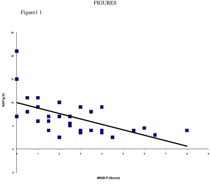
Peripheral neuropathy in ESKD generally presents as a distal symmetrical polyneuropathy with greater lower-limb than upper-limb involvement. The condition is of insidious onset, progressing over months, and has been noted to have a male predominance. It generally only develops at glomerular filtration rates less than 12 ml/min. [13] The most frequent clinical features are those of large-fiber involvement, with paresthesias, reduction in deep tendon reflexes, impaired vibration sense, weakness, and muscle wasting (Fig. 1). In the 1970s, Nielsen[6] demonstrated the presence of neuropathic symptoms in over 50% of patients with ESKD. Other studies have demonstrated prevalence rates varying from 60% to 100%, depending on the diagnostic criteria applied. [14,15,16,17] Laaksonen et al.[16]122 staged the clinical severity of uremic neuropathy in 21 ESKD patients, using a modified version of the neuropathy symptom score (NSS) developed by Dyck et al.,[18] and combined this assessment with results of nerve conduction studies. The NSS quantified symptoms that were grouped into three categories to reflect alteration in motor, sensory, and autonomic systems. Within each group, further subsets were used to group symptoms according to the region affected and the presence of positive or negative symptoms. Using the NSS and the staging procedure previously used in studies of diabetic patients, 81% of ESKD patients received a diagnosis of neuropathy. Stage 1 neuropathy (asymptomatic neuropathy) was diagnosed in 19%, stage 2 neuropathy (symptoms nondisabling) was present in 48%, and stage 3 neuropathy (disabling symptoms) was noted in 14%. In a more recent study,[19] 93% of ESKD patients had neuropathic symptoms on NSS testing, with 72% diagnosed with stage 2 neuropathy and 21% with stage 3 neuropathy, despite all patients meeting currently accepted guidelines of dialysis adequacy.143
Clinical and Neurophysiological Findings in Generalized Uremic Neuropathy
Early studies of uremic neuropathy utilizing nerve biopsy techniques revealed prominent axonal degeneration, most severe in the distal parts of nerve trunks. Although initial studies suggested that demyelination was a significant feature of uremic neuropathy, subsequent reviews demonstrated that demyelination was secondary to axonal loss and that proximal segments of the nerves were relatively spared.[20-22] These findings supported the concept that uremic neuropathy was a dying-back neuropathy, with metabolic failure of the neuron causing distal axonal degeneration.55 Numerous neurophysiological series have been undertaken in patients with uremic neuropathy and have demonstrated findings consistent with a generalized neuropathy of the axonal type. Early studies focused on motor nerve conduction parameters and demonstrated slowing of conduction velocity in patients prior to the development of clinical neuropathy. Subsequent studies demonstrated abnormalities of nerve conduction with generalized slowing in both sensory and motor nerves, accompanied by reduction in sensory response amplitudes. Motor response amplitudes tend to remain relatively preserved, although abnormalities in lower-limb motor nerves were noted in some patients, accompanied by neurogenic changes in distal lower-limb muscles on electromyography. In a recent study, amplitude of the sural sensory nerve action potential was found to be the most sensitive indicator of uremic neuropathy, being reduced in 50% of ESKD patients.[19] Other groups have confirmed similar findings, demonstrating reductions in sensory and motor response amplitudes in addition to abnormalities of late responses.[16,23]
Reduction in peroneal nerve motor conduction velocity[24]137 and prolongation of tibial F-wave minimum latencies[16]122 have been established as sensitive indicators of neuropathy in ESKD patients. Prolongation of soleus H reflexes has also been demonstrated in patients without clinical evidence of neuropathy, suggesting that this parameter may be more sensitive in detecting early neuropathy.[17]
Studies of quantitative sensory testing in ESKD patients have demonstrated increased vibratory perception thresholds, most marked in the lower limbs,[25] whereas somatosensory-evoked potentials in ESKD patients demonstrate abnormalities of conduction along both the distal and proximal segments of peripheral somesthetic pathways, but less commonly along intracranial sensory pathways.[26,27] A study of single-fiber electromyography demonstrated normal fiber densities in motor units of ESKD patients.186 This finding suggested that reinnervation, characterized by increased fiber density, had failed to occur. However, this was accompanied by increased jitter, possibly reflecting peripheral demyelination in the setting of axonal degeneration. Early studies of nerve excitability, utilizing a limited range of excitability parameters, demonstrated an elevated threshold for excitation even when nerve conduction values were normal, in addition to demonstrating prolongation of absolute and relative refractory periods.[26,28] As a consequence, it was concluded that the safety factor for neural transmission at the nodes of Ranvier would be lowered.
In addition to the slowly progressive sensorimotor axonal neuropathy, a more rapidly progressive motor neuropathy has been described. A small number of ESKD patients with diabetes have also been shown to develop a subacute neuropathy progressing over a few months, with severe muscle weakness. In this group of patients, nerve conduction studies may demonstrate features of either a demyelinatingor axonal neuropathy. Although the presence of diabetes complicates assessment of nerve conduction data, the absence of preexisting neuropathic symptoms and the clinical improvement noted following dialysis or renal transplantation suggest a metabolic basis for the neuropathy, related to the underlying ESKD. Analysis of cerebrospinal fluid (CSF) is rarely helpful, as CSF protein concentration is frequently elevated in ESKD patients and may simulate the albuminocytologic dissociation that is characteristic of Guillain–Barre´ syndrome.[15] Small-fiber neuropathy may develop as a clinical entity in ESKD patients. Lindblom and Tegner[29] demonstrated abnormalities of thermal sensation in 30% of ESKD patients and concluded that smallfiber neuropathy may exist as a distinct entity in these patients.
Effects of Dialysis and Transplantation on Uremic Neuropathy
Early reports investigating the effects of hemodialysis on uremic neuropathy suggested that some patients with mild neuropathy recovered completely with adequate dialysis.[30] In fact, failure to improve was considered to be an indictor of insufficient dialysis. These reports, however, did emphasize that the extent of improvement was likely to be related to the severity of neuropathy and that patients with severe neuropathy wer unlikely to experience any significant recovery. More recent studies, however, have demonstrated that improvement in neuropathy with dialysis is an uncommon event. [31,32,9] Although these studies suggest that dialysis retards the progressionof neuropathy in most patients, in some cases a gradual deterioration of neuropathy may occur. A comparison of hemodialysis and peritoneal dialysis with regard to neuropathy progression has demonstrated no significant difference between the two dialysis forms.[4] Renal transplantation remains the only known cure for uremic neuropathy,[11,12] with clinical improvement in sensory and to a lesser extent, motor function occurring within a few days of transplantation. [33] Serial nerve conduction studies following transplantation demonstrated a correlation between the improvement in nerve conduction and biochemical parameters, suggesting that metabolic phenomena may underlie the rapid improvement.156 Recent evidences suggests that treatment with erythropoietin (EPO) may prove beneficial in ESKD patients with neuropathy[34] as well as for patients with neuropathy due to other etiologies.[35,36] Treatment with EPO improved motor nerve conduction velocity in ESKD patients, but had no effect on sensory indices. In vitro studies have shown that EPO receptors are present on Schwann cells and in dorsal root ganglion neurons. Upregulation of EPO receptors occurs after axonal injury, mediated by release of nitric oxide, and administration of exogenous EPO is associated with reduction in limb weakness and neuropathic pain behavior.[37]
Dialyzable Toxins and the “Middle Molecule Hypothesis”
Hegstrom et al.[38] postulated that uremic neuropathy occurred due to accumulation of a dialyzable substance on the basis of their observational studies that demonstrated improvement in neuropathy in two subjects with long-standing ESKD following commencement of dialysis therapy. Later studies demonstrated that patients treated with peritoneal dialysis had lower rates of uremic neuropathy despite the fact that these patients frequently had higher blood urea and creatinine concentrations.[39] The lower neuropathy rate in the peritoneal dialysis group was thought to indicate that the substance responsible for neuropathy was better dialyzed by the peritoneum than by the cellophane membranes used in hemodialysis. On this basis, the most likely group of substances was thought to be the “middle molecules,” substances with a molecular weight of 300–12,000
A study using a hemodialysis membrane highly permeable to middle molecules also demonstrated a dramatic reduction in the development of neuropathy.[40] These early studies, however, were hampered by a number of difficulties, not least of which was the inability to measure middle molecule levels.10 Another major shortcoming of the hypothesis has been the lack of conclusive evidence that any single molecule in the middle molecular range is actually neurotoxic. The only middle molecule for which some evidence of neurotoxicity exists is PTH, with some studies suggesting a link between PTH and the neurological complications of ESKD. PTH has been shown to prolong motor nerve conduction velocities in animal studies,[41] although human studies of the effect of PTH on peripheral nerves have yielded conflicting results, with variable changes in motor nerve conduction velocity in patients with ESKD. Despite the shortcomings of the middle molecule hypothesis, the hypothesis that a dialyzable toxin may be involved in the pathophysiology of this condition remains prevalent. More recently, it has been suggested that the following criteria should be met in order for a substance to be truly regarded as a uremic neurotoxin: (1) it must be an identifiable chemical; (2) it should be elevated in the blood of uremi patients; (3) there should be a direct positive relationship between
blood level and neurologicaldysfunction; (4) it should cause neurological dysfunction in animals at appropriate blood levels; and (5) its removal from the blood should abolish the dysfunction.38 The middle molecule hypothesis fails to satisfy a number of these criteria, most importantly criterion 3, as there is very little evidence to suggest that such molecules are actually neurotoxic. Despite the evidence that a dialyzable toxin may underlie the development of uremic neuropathy, the mechanism of this neurotoxicity remained unclear.
Nerve excitability techniques provide an indirect assessment regarding the activity of a variety of axonal ion channels, energydependent pumps, and ion exchange processes activated during the process of impulse conduction. Over recent years, nerve excitability properties have been explored in a diverse range of neurological conditions including length-dependent uremic neuropathy. These studies have examined changes in nerve function that occurred in ESKD patients before, during, and after a single session of hemodialysis. Measures of nerve function were also assessed in relation to changes in serum levels of potential neurotoxins, including K+, urea, and ‘‘middlemolecules’’ such as parathyroid hormone and b2-microglobulin. Using these novel excitability techniques, predialysis excitability abnormalities were established to be strongly correlated with serum K+ in all studies. Excitability studies also demonstrated that abnormalities of nerve function occurred at a level much lower than that required for cardiac toxicity, with patients manifesting axonal changes with serum K+ concentrations in the high normal range (i.e., 4.9–5.0 mmol ⁄ l). Following dialysis there was significant improvement although minor excitability abnormalities persisted, suggesting that dialysis alone is insufficient in normalizing nerve function. In contrast, the strong correlations noted with serum K+ were not demonstrated for any of the ‘‘middle molecules.’’ These findings are also supported by studies that investigated the causes of weakness and fatigue in ESKD, and demonstrated that abnormalities of K+ regulation may underlie muscle fatigue and thereby contribute to exercise limitation in ESKD. Potassium satisfies all the criteria that have been recently proposed for a substance to be truly regarded as a uremic neurotoxin. It is an identifiable chemical that is elevated in the serum of
ESKD patients and causes neurological dysfunction in both humans and experimental animals. It is also a critical determinant of axonal resting membrane potential. Furthermore, excitability studies have demonstrated that a direct relationship exists between serum levels of K+ and neurophysiological abnormalities and that removal of K+ leads to improvement in nerve function. What is the relationship between transient changes in nerve function and the chronic irreversible axonal lossthat occurs in length-dependent uremic neuropathy? It is important to recognize that alterations in axonal membrane potential are unlikely to be transient in ESKD patients, particularly given themarked elevations in total body K+, which may cause a postdialysis rebound in serum K+ concentration. Such persistent elevation in serum K+ would lead to chronic membrane depolarization. It is well known that chronic changes in membrane potential are harmful to axons and may trigger reverse activation of the Na+⁄Ca2+ exchanger, leading to an influx of Ca2+. Such processes may initiate a cascade that eventually induces axonal death.[42]
EPIDEMIOLOGICAL STUDY
Patients
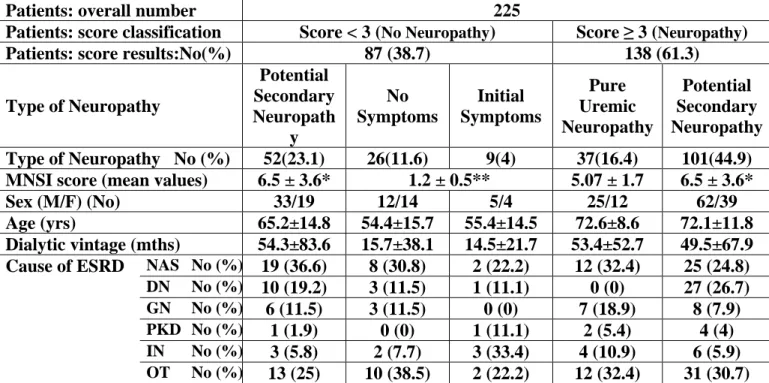
We investigated all the 225 patients (138 males and 87 females) on RDT in two Italian Dialysis Centres (Nephrology, Dialysis and Hypertension - Policlinico S. Orsola-Malpighi - Bologna and Nephrology and Dialysis Unit - Ospedale S. Croce e Carle - Cuneo).
Their mean age was 67.8 ± 14.2 years (median age 70 years) with a mean dialysis vintage of 56 ± 51.5 months.
We excluded from the study only 2 amputee patients and 4 patients who were affected by severe vascular encephalopathy because of their unreliability to the answers to the specific test performed.