18 Copyright © 2013 by Korean Society of Otorhinolaryngology-Head and Neck Surgery.
This is an open-access article distributed under the terms of the Creative Commons Attribution Non-Commercial License (http://creativecommons.org/licenses/by-nc/3.0) which permits unrestricted non-commercial use, distribution, and reproduction in any medium, provided the original work is properly cited.
Preliminary Findings from Our Experience in
Anterior Palatoplasty for the Treatment of
Obstructive Sleep Apnea
Andrea Marzetti1 ∙ Massimiliano Tedaldi2 ∙ Francesco Maria Passali31Head and Neck Surgery Division, San Carlo Hospital, Rome; 2Department of Neurosciences, La Sapienza University of Rome, Rome; 3ENT Clinic, Department of Surgical Sciences, University of Rome “Tor Vergata”, Rome, Italy
Original Article
INTRODUCTION
Obstructive sleep apnea (OSA) is a common disorder affecting at least 2% to 4% of adult population characterized by the col-lapse of the pharyngeal airway resulting in repeated episodes of
airflow cessation, oxygen desaturation and sleep disruption [1]. According to the National Commission on Sleep Disorders, more than 40 million people in the United States are affected by sleep-disordered breathing until; in spite of these epidemio-logical data, until the year 2000 it was estimated that up to 93% of females and 82% of males with moderate-severe OSA re-mained undiagnosed and untreated [2,3]. Clinically, OSA is de-fined as the occurrence of daytime sleepiness, loud snoring, wit-nessed breathing interruptions, or awakenings due to gasping or choking in the presence of at least 5 obstructive respiratory events (apneas, hypopneas or respiratory effort related arousals) per hour of sleep.
Objectives. Obstructive sleep apnea (OSA) is a common disorder affecting at least 2% to 4% of adult population charac-terized by the collapse of the pharyngeal airway. It is well established that retropalatal region is the most common site of obstruction. Consequently, many surgical techniques have been introduced. The purpose of this study is to present our preliminary results in the anterior palatoplasty (AP) compared with results of uvulopalatal flap (UPF). Methods. Thirty-eight consecutive patients with mild-moderate OSA were prospectively enrolled into a randomised
surgi-cal protocol. Surgisurgi-cal success was measured primarily by satisfactory reduction in snoring, as reported by snoring as-sessment questionnaire (SQ) of sleep partners. Secondary outcomes measures included improvement in the Epworth Sleepiness Scale (ESS) scores, changes in the magnitude of pharyngeal collapse, and postoperative pain intensity. Results. The ESS after AP improved from a preoperative value 8.5±3.7 to a postoperative mean of 4.9±3.2 (P<0.001)
af-ter UPF improved from a preoperative value of 8.1±3.5 to 5.2±3.2 postoperatively (P<0.001). The results of satis-factory reduction in the volume of snoring and response at polysomnographic data were also similar in both proce-dures. We reported a statistically significant difference of the collapse noted at Müller manoeuvre that improved from 2.7±1.0 on average, to 1.1±0.9 (P<0.001) after AP and with a lesser extent, (from 2.8±1.1 on average to 1.8±1.1;
P<0.05), after UPF. The mean duration of pain was 10.8 days for UPF patients and 7.1 days for AP patients. The mean
pain score in the first 3 days, was 6.8 in UPF patients and 5.1 in AP patients.
Conclusion. The subjective and objective improvements evidenced may suggest how AP is far superior to other techniques aimed at creating a palatal fibrotic scar. In the light of these results we can suggest AP procedure as more practical and comfortable when compared to UPF.
Keywords. Sleep apnea, Obstructive sleep apnea, Snoring surgery
• Received April 30, 2012 Revision June 4, 2012 Accepted June 25, 2012
• Corresponding author: Francesco Maria Passali
ENT Clinic, Department of Surgical Sciences, University of Rome “Tor Vergata”, Via Anagnina 718, 00118 Morena, Rome, Italy
Tel: +39-3483934793, Fax: +39-0679844154 E-mail: [email protected]
It is well established that retropalatal region is the most com-mon site of obstruction in patients with snoring and OSA syn-drome [4]. Consequently, since the introduction of uvulopalato-pharyngoplasty (UPPP) [5] many surgical techniques have been introduced aimed at creating scar tissue, inciting fibrosis and stiffening the palate. Among the proposed techniques [6,7], the reversible uvulopalatal flap (UPF) introduced by Powell et al. [8] also achieves the same results as the UPPP, but with less post-operative discomfort, less risk of developing velopalatal insuffi-ciency, and fewer complaints of a thickened secretion or foreign body sensation.
The purpose of this study is to present the preliminary results of our experience in the modified cautery-assisted palatal stiff-ening operation (CAPSO) first introduced by Pang and Terris [9] in 2007 and renamed thereafter by the same authors, as anteri-or palatoplasty (AP).
Patients with mild-moderate sleep apnea were randomised to undergo either UPF or AP. Two specific outcomes were identi-fied and addressed: 1) the efficacy of UPF and AP for mild-mod-erate OSA, 2) the morbidity associated with these two techni-ques.
MATERIALS AND METHODS
The present study is an unfounded, with no monetary support from any source, study and was approved by the Hospital Ethics and Research Committee (San Carlo Hospital, Rome).
Thirty-eight consecutive patients (25-male, 13-female) with mild-moderate OSA were prospectively enrolled into a rando-mised surgical protocol. Inclusion criteria were: 20 years of age or older, no evidence of morbid obesity (body mass index [BMI] <33), presence of mild-moderate OSA (apnea-hypopnea index [AHI] >5 and <30) as documented by polysomnography; tonsil size grades 1, 2 and 3, elongated uvula, minimal tongue base collapse as seen at the Müller manoeuvre, Fujita I (retropalatal obstruction), no prior surgical intervention for sleep apnea and no contraindications to surgery. Informed consent was given by each patient.
The parameters considered in this study were the evaluation of OSA severity and postoperative pain. OSA severity was eval-uated analysing the subjective and objective assessment of
snor-ing by: 1) patients’ roommates completed snorsnor-ing assessment questionnaire (SQ) (Table 1); 2) the evaluation of daytime sleep-iness by the Epworth Sleepsleep-iness Scale (ESS) completed by pa-tients before and two months after surgery and 3) the registra-tion of retropalatal Müller grade according to a 5-point scale from 0 to 4 (Table 2). The patients also completed a visual ana-logue scale (VAS) for postoperative pain from day 1 to 14 after surgery.
The patients were evaluated during in and out visits scheduled on the first and third days, 1 and 2 weeks and 2 months postop-eratively. The postoperative assessment included a complete head and neck examination, with emphasis on the endoscopic evalu-ation of the posterior nasal space, and the oropharyngeal area, soft palatal redundancy, uvula size and Müller manoeuvre. Surgical success was measured primarily by satisfactory re-duction in snoring, as reported by SQ of sleep partners complet-ed before and two months after surgery (Table 3). Secondary outcomes measures included improvement in the Epworth scale scores, changes in the magnitude of pharyngeal collapse and pal-atal length, and postoperative pain intensity.
The polysomnography was repeated between 6 and 9 months after surgery and surgical response was determined by criteria proposed by Sher et al. [10] of a 50% reduction in the AHI and a postoperative AHI of less than 10.
Surgical technique
Uvulopalatal flap
UPF was performed as describe originally by Powell et al. [8] in general anaesthesia. The mucosa, submucosa with glands, and fat on the lingual surface of the uvula and soft palate were re-moved with scalpel. The uvular tip was amputated and bleeding was controlled by bipolar electrocoagulation. The uvula was re-flected back toward the soft palate and fixed into its new posi-tion with multiple sutures of 3/0 Vicryl.
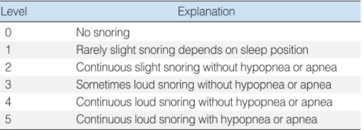
Table 1. Snoring assessment questionnaire
Level Explanation 0 No snoring
1 Rarely slight snoring depends on sleep position 2 Continuous slight snoring without hypopnea or apnea 3 Sometimes loud snoring without hypopnea or apnea 4 Continuous loud snoring without hypopnea or apnea 5 Continuous loud snoring with hypopnea or apnea
Table 2. Retropalatal Müller grade
Level Collapse grade 0 No collapse 1 Collapse <25% 2 Collapse >25% <50% 3 Collapse >50% <75% 4 Collapse >75%
Table 3. Pre- and postoperative results of SQ
SQ Level Preoperative After 2 months
0 14 UPF 12 AP
1 2 UPF 2 AP 2 3 UPF 2 AP 2 UPF 1 AP 3 10 UPF 11 AP 1 UPF
4 6 UPF 2 AP
SQ, snoring assessment questionnaire; UPF, uvulopalatal flap; AP, anterior palatoplasty.
Anterior palatoplasty
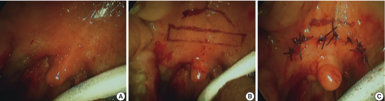
This procedure was performed as describe by Pang and Terris [9], according the modification in which general anaesthesia is used instead of local anaesthesia. As first step an uvulectomy may be performed, followed by vertical cuts on either side of the uvula through both soft palatal arches. A horizontal rectangular strip of mucosa (40-50 mm in length and 7-10 mm in width) was thereafter removed from the soft palate 10 mm to the limit of soft palatal arches, down to the muscular layer. No less than 9 sutures were used to suture the horizontal stripped area with a Vicryl 3/0 round body-curved needle (Fig. 1).
In both surgical procedures a tonsillectomy was first performed if the patient had relatively large tonsils (tonsil size 2 and 3). Post-operative medications included antibiotic suspension for 7 days and acetaminophen as needed for pain relief.
Statistical analysis
The results were statistically evaluated by comparing mean val-ues of each preoperative parameter (ESS, palatal collapse, palatal length and SQ) together with postoperative pain evaluation in the two surgical techniques groups using the unpaired test. A t-test was used also to determine if there was any difference be-tween the 2 groups with respect to age, sex, BMI or OSA severity.
RESULTS
Thirty-four (98.4%) out of thirty-eight patients completed all pre- and postoperative protocol. No statistically significant dif-ferences were noted between the two groups with regards to any demographic or disease severity parameter (BMI, age, sex, AHI, ESS) used in this trial. Fifteen patients were randomised to AP, 19 to UPF.
In AP group on 15 patients 3 had prior surgical intervention in young age, in 10 we performed tonsillectomy (six patients with tonsils size grades 2 and four with grades 3) and in 2 patients with tonsil size grades 1 we didn’t performed tonsillectomy. In UPF group on 19 patients 4 had prior surgical intervention in young age, in 10 we performed tonsillectomy (five patients
with tonsils size grades 2 and five with grades 3) and in 5 patients with tonsil size grades 1 we didn’t performed tonsillectomy. The mean operative time was 35±15 minutes for AP patients and 37±18 minutes for UPF patients. There were no complica-tions in both groups; specifically, no patients with velopharyn-geal incompetence, oronasal fistula or primary or secondary hae-morrhage.
Anterior palatoplasty
This group patients had a mean age of 48.3±10.2 years, BMI at the time of surgery ranged from 25.2 to 28.2 (mean, 26.5±2.4) and there were no significant BMI changes after surgery. Preop-erative mean of AHI was of 22±12.5; the ESS mean was preop-erative 8.5±3.7 and postoppreop-erative 4.9±3.2 (P<0.001). Twelve (80%) out of 15 patients achieved a satisfactory reduction in the volume of snoring, as reported by the sleep partner SQ, 2 months postoperatively (Table 3). The collapse noted at Müller manoeu-vre during upper airway examination improved from 2.7±1.0 on average to 1.1±0.9 (P<0.001). A representative postopera-tive picture is shown in Fig. 2. The polysomnography showed that AHI in 13 of the 15 patients (86%) improved from the mean of 22±12.5 to 8.6±6.8 (P<0.001) reaching a surgical response
A B C
Fig. 1. Anterior palatoplasty intraoperative pictures.
according the criteria established by Sher et al. [10].
Uvulopal atal flap
This group of patients had a mean age of 46.2±11.2 years, BMI at the time of surgery ranged from 24.8 to 28.4 (mean, 26.6± 2.3) and there was no significant difference between pre- and postoperative BMI values. The mean AHI was 23±12.3 preop-eratively, whereas the EES mean was preoperatively 8.1±3.5 and 5.2±3.2 postoperatively (P<0.001). Fourteen out of 19 pa-tients (73.6%) achieved a satisfactory reduction in the volume of snoring, as reported by sleep partner SQ 2 months postopera-tively (Table 3). The collapse noted at Müller manoeuvre im-proved from 2.8±1.1 on average to 1.8±1.1 (P<0.05). A re-presentative postoperative picture is shown in Fig. 3. The poly-somnography showed that 16 of the 19 patients (84%) achieved a surgical response when applying criteria established by Sher et al. [10], the AHI mean decrease from 23±12.3 to 9.6±7.8 (P< 0.001).
Pain assessment
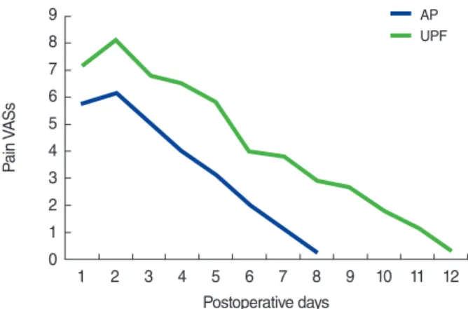
Variation of pain with VAS was determined at postoperative fol-low-up. Postoperative oedema was present in all cases. Duration of oedema varied from 3 to 14 days (mean, 8 days). Complaints of pain (beginning in the early postoperative period, peaking on the 2nd day, and lasting from 3rd to 14th day) and difficulty in swallowing were evaluated all along this period of time. None of the patients needed analgesics or re-hospitalization for intrac-table pain. The mean duration of pain was 10.8 days for UPF patients and 7.1 days for AP patients. This difference was statisti-cally significant (P<0.05). The mean pain score was 6.8 for UPF patients and 5.1 for AP patients for the first 3 days. VASs for pain are shown in Fig. 4.
DISCUSSION
Continuous positive airway pressure (CPAP) is the treatment of choice for adult patients with mild-moderate OSA. However, adherence to CPAP is insufficient in 17% to 54% of patients [11]. Therefore, over the last three decades various operation have been developed at the level of palate aimed at improving airway patency by stiffening the pharyngeal wall. Palatal surgery is now the largest established surgical approach to snoring and mild-moderate OSA [12].
The UPF procedure described by Powell et al. [8] provides the same results as the UPPP introduced in 1985 [5] as already de-monstrated in several studies but with less postoperative pati-ents’ discomfort. Pang and Terris [9] in 2007 introduced AP as alternative surgical procedure to stiffen the palate surface. According to the literature, short-term improvements occur in 88% of UPF treated patients [13]. In this preliminary study the AP results were comparable to UPF subjective results: in fact the ESS after AP improved from a preoperative value 8.5±3.7 to a postoperative mean of 4.9±3.2 (P<0.001). In a similar way, the ESS values after UPF improved from a preoperative value of 8.1±3.5 to 5.2±3.2 postoperatively (P<0.001).
The results of satisfactory reduction in the volume of snoring, as reported by sleep partner SQ 2 months postoperatively, were also similar in both surgical procedures. In fact partners’ SQ rea-ched 0 level in 12 (80%) of AP cases. When compared with UPF postoperative partners’ SQ—improved until level 0, in fourteen (73.6%) cases—no significant difference was found between the two groups. And compared to a similar response at polysomno-graphic data of the two surgical techniques the real superior effi-cacy came from the data of palatal collapse during Müller ma-noeuvre and pain assessment.
We reported a statistically significant difference of the collapse noted at Müller manoeuvre that improved from 2.7±1.0 on av-erage, to 1.1±0.9 (P<0.001) after AP and with a lesser extent, (from 2.8±1.1 on average to 1.8±1.1; P<0.05), after UPF pro-Fig. 4. Mean VASs for pain. VAS, visual analog scale; UPF, Uvulopal-atal flap; AP, anterior palatoplasty.
AP UPF 1 2 3 4 5 6 7 8 9 10 11 12 Pa in V A S s Postoperative days 9 8 7 6 5 4 3 2 1 0
bably because UPF although produced with the reposition and stabilization of uvula on the soft palate the wide opening of ret-ropalatal airway space, therefore narrowing the lateral distance between the tonsillar pillars. Moreover during the postoperative objective examination (Figs. 2, 3) we noted a major anterior ro-tation of the palatal wall in AP than in UPF that also showed a higher thickness and alteration of normal anatomy (absence of uvula).
The pain was significantly more intense and prolonged in the UPF group compared with the AP group. The mean duration of pain was 10.8 days for UPF patients and 7.1 days for AP pati-ents. This difference was statistically significant (P<0.05). The mean pain score in the first 3 days, was 6.8 in UPF patients and 5.1 in AP patients.
The subjective and objective improvements evidenced in this study may suggest how AP is far superior to other techniques aimed at creating a palatal fibrotic scar confirming the advantag-es already dadvantag-escribed by Pang et al. [14].
In short term, the effectiveness of both UPF and AP proce-dures was very high. Post operative pain was mostly seen in UPF lasting longer than in AP cases. In the light of these results and in spite of similar success rates we can suggest AP procedure as more practical and comfortable when compared to UPF.
CONFLICT OF INTEREST
No potential conflict of interest relevant to this article was re-ported.
REFERENCES
1. Epstein LJ, Kristo D, Strollo PJ Jr, Friedman N, Malhotra A, Patil SP, et al. Clinical guideline for the evaluation, management and long-term care of obstructive sleep apnea in adults. J Clin Sleep Med. 2009 Jun;5(3):263-76.
2. Young T, Evans L, Finn L, Palta M. Estimation of the clinically diag-nosed proportion of sleep apnea syndrome in middle-aged men and women. Sleep. 1997 Sep;20(9):705-6.
3. National Commission on Sleep Disorders Research. Wake up Amer-ica: a national sleep alert. Washington, DC: National Commission on Sleep Disorders Research; 1995.
4. Yagi H, Nakata S, Tsuge H, Yasuma F, Noda A, Morinaga M, et al. Morphological examination of upper airway in obstructive sleep ap-nea. Auris Nasus Larynx. 2009 Aug;36(4):444-9.
5. Fujita S, Conway WA, Zorick FJ, Sicklesteel JM, Roehrs TA, Wittig RM, et al. Evaluation of the effectiveness of uvulopalatopharyngo-plasty. Laryngoscope. 1985 Jan;95(1):70-4.
6. Ellis PD. Laser palatoplasty for snoring due to palatal flutter: a fur-ther report. Clin Otolaryngol Allied Sci. 1994 Aug;19(4):350-1. 7. Mair EA, Day RH. Cautery-assisted palatal stiffening operation.
Otolaryngol Head Neck Surg. 2000 Apr;122(4):547-56.
8. Powell N, Riley R, Guilleminault C, Troell R. A reversible uvulopala-tal flap for snoring and sleep apnea syndrome. Sleep. 1996 Sep;19(7): 593-9.
9. Pang KP, Terris DJ. Modified cautery-assisted palatal stiffening oper-ation: new method for treating snoring and mild obstructive sleep apnea. Otolaryngol Head Neck Surg. 2007 May;136(5):823-6. 10. Sher AE, Schechtman KB, Piccirillo JF. The efficacy of surgical
modi-fications of the upper airway in adults with obstructive sleep apnea syndrome. Sleep. 1996 Feb;19(2):156-77.
11. Weaver TE, Grunstein RR. Adherence to continuous positive airway pressure therapy: the challenge to effective treatment. Proc Am Tho-rac Soc. 2008 Feb;5(2):173-8.
12. Vicini C, editor. Chirurgia della roncopatia. Lucca: Eureka; 2007. 13. Neruntarat C. Uvulopalatal flap for snoring on an outpatient basis.
Otolaryngol Head Neck Surg. 2003 Oct;129(4):353-9.
14. Pang KP, Tan R, Puraviappan P, Terris DJ. Anterior palatoplasty for the treatment of OSA: three-year results. Otolaryngol Head Neck Surg. 2009 Aug;141(2):253-6.