O P E N A C C E S S
GYNAECOLOGY
Review Article
Caterina Pizzicaroli
1*, Ioannis Malandrenis
2, Giovanni Larciprete
2, Federica Rossi
2, Carlotta Montagnoli
2and
Edoardo Valli
1-21Department of Obstetrics and Ginecology, Tor Vergata University, Rome, Italy
2Department of Obstetrics and Gynecology, Fatebenefratelli Isola Tiberina Hospital, Rome, Italy
Received: January 14, 2016; Published: January 20, 2016
*Corresponding Author: Caterina Pizzicaroli , Department of Obstetrics and Gynecology, Tor Vergata Univerity Hospital, Rome, Italy.
Clinical Management of Ectopic Pregnancy
Abstract
Introduction
The early diagnosis of ectopic pregnancy, made before the occurrence of complications linked to an hemodinamic instability, leads to a reduction of mortality from 35.5. to 3.8 per 1000 ectopic pregnancies .When the diagnosis is made, there are different therapeutic options that depend on the conditions of the patient, the β-hCG levels, the dimension of the adnexal mass, the condition of emergency, the site of the ectopic pregnancy and the compliance of the patient. The management can be surgical, medical and observational. Surgery can be performed in symptomatic patients with hemodynamic instability and clinical contraindications to Methotrexate therapy. Available surgical options for tubal pregnancy are: conservative treatment (salpingotomy), intermediate surgery (partial salpingectomy), and radical treatment (salpingectomy), usually performed by laparoscopy. Medical treatment by Methotrexate can be performed in stable, asymptomatic patient, with β-hCG values ≤ 3000-5000 mIU/mL and without ultrasonographic evidence of haemoperitoneum or fetal cardiac activity. Methotrexate therapy can be administered locally or systemically, with a fixed multiple dose or single dose regimen. The follow up of patients undergoing medical therapy consists of β-HCG evaluation until its serum level is undetectable. The proposal of a wait-and-see attitude can be made in absence of clinical symptoms, adnexal mass less than 4 cm at ultrasonographic evaluation, endopelvic free fluid less than 50 mL, low hCG levels (≤ 2000 mU/mL) and patient compliance in ac-cepting potential complications like tubal rupture and haemorrhage. Other kinds of specific treatments are available for non tubal ectopic pregnancy.
In the 70’s, less than 20% of ectopic pregnancies were diagnosed before rupture, so there was a high rate of morbidity and mortality linked to this disease. Nowadays, there is a more valid vigilance by clinicians. Thanks to the help of transvaginal ultrasounds and serum β-hCG levels, that lead to a progress in the diagnosis, more than 80% of ectopic pregnancies are diagnosed as intact and this let it possible to follow a conservative management. The early diagnosis made before the occurrence of complications linked to a hemodinamic instabil-ity, has lead to a reduction of mortality from 35.5. to 3.8 per 1000 ectopic pregnancies [1-5].
When the diagnosis is made, there are different therapeutic options that depend on the condition of the patient, the β-hCG level, the dimension of the adnexal mass, the condition of emergency, the site of the ectopic pregnancy and the compliance of the patient. The in-cidence of recurrent ectopic pregnancy is between 5% and 20% and rises up to 32% following 2 ectopic pregnancies [5]. The expected resolution time of the ectopic pregnancy is between 3 to 7 weeks after MTX application [1].
Observation, Surgery and Medical therapy
Guidelines provided by the Royal College of Obstetricians and Gynaecologists [6] stated that tubal pregnancy can be managed by laparotomy, operative laparoscopy, medically and occasionally by observation alone.
In 1982, Mashiach., et al. [7] basing on the concept that early ectopic pregnancy can result in spontaneous reabsorption or tubal abortion, suggested the possibility of a self-limiting disease that can be faced with an expectant management. The first to practice it in patients with suspicion of ectopic pregnancy on admission was Lund, in 1995[8]. Accordind to RCOG guidelines [6] expectant man-agement is an option for clinically stable asymptomatic women with an ultrasound diagnosis of ectopic pregnancy and a decreasing serum levels of β-hCG, initially less than 1000 IU/L (Grade of Recommendation C; Evidence level III). So, parameters that can lead to the proposal of a wait-and-see attitude are: absence of clinical symptoms, adnexal mass less than 4 cm at ultrasonographic evaluation, en-dopelvic free fluid less than 50 mL, low β-hCG levels (≤ 2000 mU/mL) that shows rapidly declining at the 48 hours re-evaluation [1], and patient compliance in accepting potential complications like tubal rupture and internal haemorrhage [9-10]. When proposing expectant management attention must be given in those patients who fail to resolve, because the risk of morbidity remains high [11] . In fact more than 90% of women with ectopic pregnancies undergoes an operative intervention because of increasing and endangering symptoms [1,5,12].
Indications for surgery include: symptomatic patients with clear free fluid in the Douglas space, hemodynamic instability, rupture of the ectopic mass, clinical contraindications to Methotrexate (MTX) medical therapy, very high β-hCG levels, a coexistence of an intra-uterine pregnancy, failed medical therapy or poor compliance with follow-up aftercare, lack of timely access to a medical institution for the management of tubal rupture and desire for permanent contraception [13-16]. The kind of surgery (laparotomic or laparoscopic) depends on the surgical anamnesis of the patient (previous laparotomic surgery) and her haemodynamic condition, and on the endo-scopic skills of the surgeon. The adequate surgical option depends on the site of ectopic pregnancy.
In recent years the surgical treatment of ectopic pregnancy has shifted from an often urgent laparotomy to a more conservative laparoscopy [13]. Many other studies demonstrate that in hemodynamic stable patients, laparoscopy offers many advantages over lapa-rotomy (less costs, hospitalization, surgery time, blood loss, analgesics and recovery time) [13,17,18]. At present, laparoscopy is the preferred treatment because almost all patients can be referred to it, it is safe and less expensive than laparotomy beacuse of reduced morbidity and shorter hospital stay [11,19]. In 1973 in the United States, Hope suggested the use of laparoscopy in the diagnosis of ec-topic pregnancy [20], then laparoscopy began to be widely used in the diagnostic management of ecec-topic pregnancy because it allowed an earlier diagnosis, solved the dilemma of prolonged clinical observation and avoided the risk of performing a not useful laparotomy [11,17,18].
RCOG guidelines stated that: a laparoscopic approach to the surgical management of tubal pregnancy, in the haemodynamically stable patient, is preferable to an open approach (Grade of Recommendation A; Evidence level Ia). Infact, a comparison of the two surgi-cal techniques in 228 women in three randomized controlled trials showed that even if laparoscopic approach presented a greater risk of persistent trophoblast than laparotomy, the endoscopic procedure was associated with shorter operation times, less intraoperative blood loss, shorter hospital stays and lower analgesic requirement [6]. Usually laparotomy is performed in women with extensive intra-peritoneal bleeding, in case of tubal rupture, if there is a poor visualization of the pelvis during laparoscopy or if the patient is hemodi-namically instable or presenting an high adherence status [21,22]. An isthmic location of an ectopic pregnancy, with a high intramural proportion, can be an indication for primary laparotomy because of the severe risk for uncontrollable intraoperative bleeding, even if these situations can often be treated laparoscopically. In all other circumstances, the operative management should be laparoscopic, if the hemodynamic status of the patient, the surgeon’s experience and the availability of endoscopic treatment allow it [1-22].
Treatment
Observation - expectant management
Surgical Treatment
Conservative treatment (salpingotomy), intermediate surgery (partial salpingectomy), and radical treatment (salpingectomy) are the available surgical options for the treatment of a tubaric pregnancy [1-21]. Paramount importance has the so called “case to case” evaluation on the more appropriate surgery to be performed for that specific patient. Success rate depends on the permeability of the involved tube and on the status of the contra lateral tube.
A very important matter of debate is which kind of laparoscopic treatment (conservative-salpingotomy or radical-salpingectomy) should be performed in women wishing to preserve their reproductive capacity. There is no consensus in literature regarding the opti-mal treatment of tubal pregnancy for the manteinance of the fertility. The most important factor for future reproductive outcome after surgery for ectopic tubal pregnancy seems to be the status of contralateral tube [23]. In a randomized controlled trial of Mol F [11-24], where he compares salpingotomy and salpingectomy in women with a tubal pregnancy with a safe contra lateral tube, it is shown that salpingotomy doesn’t improve time to develop spontaneous pregnancy and it leads more often to persistent trophoblast. In other papers it’s reported that salpigotomy is associated with higher subsequent intrauterine pregnancy than salpingectomy, but that there is also a higher risk to develop recurrent ectopic pregnancy [23]. In case of tubal ectopic pregnancy in a patient who expresses the desire for future pregnancy and presents with a damage in contra lateral tube or in the absence of the contra-lateral tube, the treatment of choice is salpingotomy [25] if a salpingectomy has to be performed, the subsequent loss of fertility leads to the in vitro fertilization (IVF) as an appropriate treatment [11] for a future pregnancy.
Therefore in young patients (less than 30 years) without previous infertility and with a normal contralateral tube, the fertility rate after salpingectomy and after salpingotomy is comparable. In the presence of a healthy contralateral tube there is no clear evidence that salpingotomy should be used in preference to salpingectomy (Grade of Recommendation B; Evidence level IIa) [6]. Laparoscopic salpin-gotomy should be considered as the primary treatment when managing tubal pregnancy in presence of contralateral tubal disease and/ or desire for future fertility (Grade of Recommendation B; Evidence level IIa) [6]. There are cases (previous ipsilateral extrauterine preg-nancy, contralateral severe peri-annexial adherent condition) in which bilateral salpingectomy can be considered in order to improve the success rate of medical assisted reproduction techniques [21]. Obviously, after each conservative tubaric treatment, there is the need to control serum levels of β-hCG control to exclude the condition of a persistent trophoblast [21].
Tubal pregnancy
Conservative surgery
The success rate of medical and conservative surgical treatment of ectopic pregnancies, with no requirement for further treatment, is comparable [13,26,27,28,29,30]. Different conservative surgical options have been suggested in literature:
a. Transampullary aspiration and “Ampullary milking”: the first one consists in the aspiration of trophoblastic tissue from the tube without incision. The second one, “ampullary milking”, is possible just in selected cases, when the product of conception is located near the outer region of the tube or the fimbrial end and it can be removed by “milking” ipsilateral tube, pushing the product of conception until it is extruded into the abdominal cavity. This operation is organ saving but it has a high rate of incomplete removal, recurrence and trophoblast residuals [1,5,22].
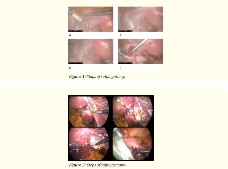
b. Salpingotomy: usually performed in hemodinamically stable women with desire for future pregnancies, without contralateral tubal pathology and adhesions in the tubal wall, and in case of ampullar, infundibular or isthmic mass, less than 5 cm in size [5,22]. One of the most common complications of laparoscopic tubal salpingotomy in the treatment of tubal ectopic pregnancy is the incomplete removal of the products of conception as a persistent ectopic pregnancy requiring additional therapy [22]. This has to be kept in mind and be a fundamental part of the preoperational evaluation [3]. Twenty UI Vasopressin, in 100 mL of normal saline, is injected in the mesosalpinx near the tubal location of the gestational products before tubal preparation. Vasopressine allows a better operative management, presents a positive side effect on the prognosis and improves hemostasis. In addiction Vasopressin leads to a local hypoxia and causes a limitation of the metabolic rate of ectopic tissue and put it at risk [1,4,31].
Before salpingectomy is performed due to a failure of a salpingotomy, or in case of tubal rupture, ectopic isthmic pregnancy, or a recurrent tubal pregnancy, there is the possibility to try an intermediate surgical option. It is called partial salpingectomy and consists in a segmental tubaric resection [1,2].
No desire to maintain fertility, recurrence of tubal pregnancies or of extrauterine pregnancy after a failed sterilization, persistent bleeding after a conservative tubal procedure, previously reconstructed tube, tubal rupture, severe adhesions, hydrosalpinx and tubal pregnancy size more than 5 cm, represent the main indications for salpingectomy [1,22].
According to RCOG guidelines [6], medical therapy should be offered to suitable women, and units should have treatment and follow-up protocols for the use of methotrexate in the management of ectopic pregnancy (Grade of Recommendation B; Evidence level IIa).
Intermediate surgery: partial salpingectomy
Radical surgery: salpingectomy
Non tubal pregnancy
Medical Treatment
The treatment for many locations of ectopic pregnancy is usually surgical:
a. Ovarian pregnancy can be treated laparoscopically because the possibility to perform a mini-invasive surgery by simple tion of the gestational product or ovarian wedge resection that let the remaining ovarian tissue be protected for the widest sible extention. Oophorectomy is not commonly needed [1,13,32].
b. Abdominal Pregnancy: if detected at early stage can be treated laparoscopically. But, because of the possibility of vessels invasion by the tumour that can cause an important bleeding, and due to the usually late diagnosis of this condition, the difficulty in cental removal, and the high risk for maternal life, the management of this kind of ectopic pregnancy almost always requires laparotomy [5]. However no consensus is nowadays reached in literature about the best treatment (medical or surgical) to form in this kind of ectopic pregnancy.
c. Interstitial or Cornual Pregnancy: hemodynamic maternal instability or signs of ongoing rupture of the mass and, finally, dications in performing MTX therapy are the most common indications to perform surgical therapy. Laparoscopic cornual tion or salpingotomy can be performed [33]. Intramural pregnancy presents an increased risk of traumatic rupture with hemor rhagic shock and maternal death. Cornual pregnancy implants in the same anatomical area of the tube but opens into the uterine cavity. In case of larger pregnancies that can result in uterine perforation, curettage under laparoscopic guidance can be formed [2,5,31]. Hysteroscopic removal of cornual pregnancy has also been described but with an unclear efficacy [13].
d. Cesarean scar ectopic pregnancy: There are many different kinds of treatment described in literature: hysteroscopic management [34] laparoscopy [35], selective transarterial embolization with subsequent dilatation and curettage and local or systemic tion of MTX [21,36]. Expectant management has been described but it presents with a poor prognosis due to the high risk of ture [37]. Finally, in literature, the first trimester termination of pregnancy is suggested as the treatment of choice [1].
e. Cervical and Vaginal Pregnancy: Conservative therapy usually works just if the diagnosis is made before 12 weeks of gestation. Infact in this kind of ectopic pregnancy the throphoblast invasion can cause erosive lesions of uterine vessels leading to a sive hemorrage requiring surgical action by means of uterine artery embolisation or even hysterectomy [1,21]. In order to execute dilatation and curettage and to reduce the bleeding, helpfull preparatory treatment can be MTX injections, uterine artery bolization, vaginal ligation of the descending branch of uterine arteries, intracervical prostaglandin injection, cervical cerclage and balloon tamponade of the cervical canal [1,13].
f. Heterotopic Pregnancy: laparoscopic salpingectomy is the most used treatment [5].
The possibility of persistence of products of conception is the same after laparoscopy and laparotomy (5-15%) [1]. The preferential site where trophoblastic tissue can outlast is the medial portion of the tube. This is responsible for elevated serum β-hCG levels in the follow up [2,5,10].
Methotrexate (MTX), a folinic acid antagonist, works by blocking DNA and partly also RNA synthesis and cell division. So, tropho-blast, that is a rapid cellular turnover tissue, is particularly affected by its action [38,39]. Other systemic drugs like actinomycin D, pros-taglandins, and RU-486 have also been suggested to be used in the medical treatment of an ectopic pregnancy [1].
In literature the first description of a successful treatment of a tubal pregnancy by systemic MTX dates back to 1985 [40]. Then, to reduce the side effects and the costs of the therapy and to improve the compliance of the patients, some authors suggested the use of individualized doses of of this drug [41].
Medical MTX therapy presents with some side effects such as stomatitis, conjunctivitis, rash, alopecia, gastritis-enteritis, elevat-ed liver enzymes, bone marrow suppression, photosensitivity, pleuritis and dermatitis [1,11]. Even if Methotrexate therapy has been shown to be safe a careful follow-up is recommended for women presenting with abdominal-pelvic pain in the few days following MTX therapy, to evaluate if it is required a surgical intervention because of a possible failure of medical treatment [42]. So, indications for treatment should be carefully evaluated, due to the possible failure of the therapy and the possible side effects [1].
Medical therapy with MTX can be performed in patient that are stable, asymptomatic, with ß-hCG value ≤ 3000-5000 mIU/mL and without ultrasonographic evidence of haemoperitoneum or fetal cardiac activity [6,14,17]. According with RCOG guidelines, women most suitable for MTX therapy present with a serum β-hCG below 3000 IU/L and minimal symptoms (Grade of Recommendation B; Evidence level IIa) [6]. Some papers report that MTX therapy can be successful also with serum β-hCG levels higher than 3000 IU/L [17], but according to the paper of Sowter et al, quality-of-life data suggest that methotrexate is only an attractive option for women with an β-hCG below 3000 IU/L [6,43]. Even if this is not considered as a reliable predictor of therapeutic medical success and in literature there’s not a clear concern about it, some authors suggest, as a patient selection criterion for MTX therapy, a dimension of the mass less than 3-4 cm, maybe because women with larger adnexal masses are more likely to have them already ruptured [13-14-44]. Patient’s high operative risk or conditions that represents contraindications to anesthesia are also considered indications to try medical therapy [1].
Breastfeeding, immunodeficiency, alcoholism, chronic liver disease, preexisting blood dyscrasia, known ipersensitivity to MTX, ac-tive pulmonary or peptic ulcer disease and hepatic, renal or hemathologic dysfunctions, represent contraindications to MTX therapy. Some authors report cases of association between renal failure and MTX-associated death [13,45]. Relative contraindications to MTX therapy are : the presence of fetal cardiac activity, an ectopic mass with a diameter of more than 3 cm and β-hCG levels greater than 5000 IU/L [46].
MTX can cause some teratogenic effects on fetal neural tube, cardiovascular system and urinary tract. For this reason, it’s very im-portant, before starting therapy, to be sure that there is not the concomitant presence of an intrauterine on going pregnancy [13,47].
If medical therapy is offered, women should be given clear informations (preferably written) about the possible need for further treatment and adverse effects following treatment. Women should be able to return easily for assessment at any time during follow-up (Grade of Recommendation B; Evidence level IIa) [6].
Side effects
Indications
Contraindications
Teratogenicity
Regimen
MTX therapy can be administered:
b. Systemically (intramuscular way): in his paper, Stucki reported that if the patient present with β-hCG value less than 5000 UI/L and a pregnancy diameter under 3 cm, a 88% success percentage rate accompanies systemic MTX administration [17]. Another important predictor of the outcome of MTX treatment seems to be the serum progesterone level. Infact if the serum progesterone level is more than 7–10 ng/mL there is a greater risk of failure with single-dose methotrexate [46]. A randomized study reports that patients who present with progesterone levels more than 10ng can equally profit from an oral treatment of 600 mg stone [48].
Two kinds of regimen are available:
b1. Fixed multiple dose regimen (MTX 1.0 mg/kg i.m daily 1,3,5,7 alternated with folinic acid 0.1 mg/kg orally on days 2,4,6,8) 42]. The combination with folinic acid is used to reduce chemotherapy toxicity [49]. This regimen, when used in women with serum β-hCG levels below 3.000 UI/L, has been proven to be effective compared to laparoscopic salpingotomy [19].
b2. Single dose methotrexate calculated as 1.0 mg/kg or 50 mg/m2 i.m (without folinic acid) [42]. For most women MTX dose this is between 75 mg and 90 mg [6-29-30]. In women with β-hCG <1.500 IU/L, this regimen has been shown to be cost effective pared to laparoscopic salpingotomy [43]. In addiction, it seems to offer the greatest benefits in terms of efficacy and tolerability, it has a very good patient compliance and it has also proved to be a good alternative to laparoscopy in some cases [42]. Women who present with β-hCG value of 3000- 4000 mIU/mL have a greater probability to undergo surgery or multiple dose treatment [50]. Infact serum β-hCG levels values are evaluated on days four and seven and a further MTX dose is given if there’s not a fall by more than 15% between these two checks [6,29,30].
There are just little data about the comparison between the fixed multiple dose methotrexate regimen and the single dose regimen and the authors report different conclusions about which one of the two regimen is preferred. Lipscomb in 1998 reported that multi-dose regimen was more successful (93%) when compared with single multi-dose regimen (88%) but the second one, requiring not folate in-tegration, presented less side effects (28% vs 48%) and was cheaper than the first one [51]. A randomized trial about the treatment of unruptured tubal ectopic pregnancy in patients with β-hCG levels of about 2000 IU/L, has shown that the effect of the multiple (90%) and the single dose regimen (80%) were similar and that even if the first kind of regimen was associated with more side-effects, the time for serum β-hCG values to be less than 5 mU/mL was shorter than in single dose regimen (18 versus 22 days) [11-52]. Srivichai et al [53] reported a high success rate for single-dose MTX therapy (90%) but suggested to prefer multi-dose regimen or surgery in pa-tients with big ultrasonographic adnexal masses or high β-hCG levels at the moment of the diagnosis. Other randomized trials pointed that the two kinds of regimen were comparable in terms of effectiveness [38,52,54,55].
The follow up of a patient undergoing medical MTX therapy consists of β-hCG evaluation until serum level is undetectable. Infact, close β-hCG monitoring allows to diagnose treatment failure if there is an inadequate reduction of serum β-hCG concentration [11-56]. Some patients (15-20%) may require a second MTX dose, but less than 1% needs 3 doses or more [11-57]. To decide if it’s necessary to administer a second MTX, protocols can be useful. One protocol [41] suggests to consider day 1 as the day of MTX injection and then to evaluate the level of ß-HCG on days 4 and 7. If the decrease in serum levels is less than 15%, so a second intramuscular MTX dose (50mg/m2) has to be given. Another protocol [14] suggests to evaluate β-hCG just on day 7. If its serum level is declined less than 25% when compared with day 1 serum level, a second MTX dose has to be administered.
The follow up after an operative treatment of an ectopic pregnancy requires to evaluate the reduction in β-hCG levels. It should be of at least 70% after 48 hours, and after a week should result in a decrease of another 70% [1]. Large uncontrolled studies have reported that less than 10% of women that underwent MTX therapy required surgery after medical approach, and that 14% of women nedeed 2 or more doses of MTX [6,44]. Outpatient medical therapy with single-dose methotrexate is associated with a saving in treat-ment costs (Grade of Recommendation A; Evidence level Ib) [6] .
Single versus multiple dose regimen
Success rate
MTX vs surgery
Persistent Throphoblast
Appendix: Images and Video
Papers report a success percentage of MTX therapy of 74-100% [58]. The success rate of MTX therapy is closely related to:
a. β-hCG levels (the lower the serum level at the beginning of the therapy, the higher the success rate) [59-60]. Menon et al in a tematic review of 503 patients treated with MTX, stated that if the initial β-hCG values were more than 5,000 mIU/mL, the percentage rate of treatment failure were statistically greater [61-62]. Another non-randomized clinical trial on 238 patients treated with MTX and elaborated with a logistic regression analysis showed that levels of β-hCG greater than 2,000 mIU/mL increased the odds of failure by about 4.5 times [61,63].
b. -Size of the ectopic mass. There is no consensus in literature about the maximum size of the ectopic mass that can be treated with medical MTX therapy, varying from 3,5 and 5 cm from different authors [9,19,30,61,64].
c. Endometrial thickness: recent papers related endometrial thickness with the indices of success or failure of MTX therapy. ing from the concept that endometrial thickness reflects the hormonal levels and because of that the higher the level of β-hCG, the worse the prognosis of MTX therapy, so the authors stated that high values of endometrial thickness (in particular above 12 mm) are linked with significantly higher failure rates [61,64,66].
d. So, embryonic heart activity, mass greater than 4 cm, β-hCG values greater than 5,000 mIU/mL, presence of blood in the neal cavity, increasing rate of β-hCG above 50% within 48 h prior to MTX, rapid and continuous increase of the β-hCG level during MTX therapy, ultrasonographic sign of a tubal ring or the visualization of a yolk sac and high endometrial tickness are considered as risk factors for faiulure of the medical therapy [9,38,64,67,68,69].
So, the two ways to treat ectopic pregnancy are medical therapy or surgery that differentiate quite considerably for resource con-sumption. Site of the ectopic mass, clinical conditions of the patient and medical skills affect the choice of the treatment [70]. In a paper of Lecuru et al. [71] it was reported that even if MTX therapy presented with shorter hospital stay and lower hospitalization rate, it presented a longer follow up with more visits and biological test when compared with laparoscopy. Dhar in his paper [42] stated that in patient with low β-hCG levels and adnexal mass less than 4 cm, MTX medical management seems to offer benefits over surgical treat-ment because it is less invasive and less expensive and it doesn’t require surgical skills. In addiction, he reported that future reproduc-tive expectations are better with methotrexate with higher intrauterine pregnancy rates and lower ectopic rates subsequently. On the other side, due to the need of a prolonged follow up and because of the risk of a tubal rupture after MTX therapy, patient’s compliance to therapy represents an important selection criterion in the choice of the kind of therapy [42].
According to RCOG guidelines [6], when salpingotomy is used for the management of tubal pregnancy, protocols should be in place for the identification and treatment of women with persistent trophoblast. Post-operative β-hCG evaluation represents a good option for detection and treatment of persistent trophoblast after salpingotomy. To prevent persistent trophoblast, phrophylactic single dose of systemic Metotrexate is reported to be more successful that salpingotomy alone [19].
The first time is the injection of a vasoconstrictor substance in the mesosalpinx before salpingotomy, to increase hemostasis re-ducing the subsequent need of coagulation. Many surgeons perform bipolar coagulation on the free tubal surface before performing incision. The incision can be performed with a monopolar needle or a cold scissor possibly in the proximal third of the hematosalpinx. This is often sufficient to obtain the spontaneous exit of blood clots and trophoblastic tissue from the salpinx. The extraction can be completed by aspirating the residual trophoblastic tissue from the tubal incision.
Figure 1: Steps of salpingotomy.
Figure 2: Steps of salpingectomy.
Salpingectomy is often performed with bipolar forceps cause it is simply and cheap. Ultracision, ligasure and other more techno-logical instruments can be used depending on the availability and the budget of the structure. Salpingectomy can be performed either from the uterine horn to the fimbrial portion (anterograde) or from the fimbrial portion to the uterine horn (retrograde). It is impor-tant to resect the tube as close as possible to uterus, to avoid cornual pregnancies in further in vitro fertilization pregnancies.When tubo-ovarian or fimbrial adhesions are detectable, the need to remove them and mobilize the ovary is mandatory. The tube is then trac-tioned and the tubal artery coagulated after the bifurcation from the ovarian artery. The coagulation and dissection of the mesosalpinx is performed with bipolar forceps and cold scissors near the tube to minimize the vascular damage to the ovary until the excision is completed. The tube is finally extracted using an endoscopic bag to minimize spread of trophoblastic tissue
Video 1: Short cutted video with the whole procedure of salpingectomy by laparoscopy. http://youtu.be/bSovaTPnnVo
Bibliography
1. Alkatout I., et al. “Clinical diagnosis and treatment of ectopic pregnancy. Obstetrical and Gynaecological Survey 68.8 (2013): 571-581.
2. Alcatout I. “Ectopic Prgnancy. In: Schollmeyer T., et al. eds. Practical Manual for Laparoscopic & Hysteroscopic Gynecological Surgery”. (2013):169-89.
3. Murray H., et al. “Diagnosis and treatment of ectopic pregnancy”. Canadian Medical Association Journal 173 (2005): 905-912. 4. Barbosa C., et al. “Laparoscopic management of ectopic pregnancy”. Manual of Gynecological Laparoscopic Surgery (2010): 115-123.
5. Hucke J and Füllers U. “Extrauterine Schwangerschaft”. Der Gynäkologe 6 (2005):535-552.
6. “The Management of tubal pregnancy (Reviewed 2010)”. Royal college of obstetricians and gynaecologists guidelines 21 (2004): 1-10.
7. Mashiach S., et al. “Non operative management of ectopic pregnancy: a preliminary report”. JRM-The Journal of Reproductive cine 27 (1982): 127.
8. Lund J. “Early ectopic pregnancy: comments on conservative treatment”. Journal of Obstetrics and Gynecology of the British Empire 62.1 (1995): 70-76.
9. “American College of Obstetricians and Gynecologists. Medical management of ectopic pregnancy”. Obstetrics & Gynecology 111.6 (2008): 1479-1485.
10. Hoower KW., et al. “Trends in the diagnosis and treatment of ectopic pregnancy in the United States”. Obstetrics & Gynecology 115.3 (2010): 495-502.
11. Van Mello NM., et al. “Ectopic pregnancy: how the diagnostic and therapeutic management has changed”. Fertility and sterility 98.5 (2012): 1066-1073.
12. Lipscomb GH., et al. “Nonsurgical treatment of ectopic pregnancy”. The New England Journal of Medicine 343.18 (2000): 1325-1329.
13. Oron G and Tulandi T. “A pragmatic and evidence-based management of ectopic pregnancy”. Journal of minimally invasive ogy 20.4 (2013): 446-454.
14. Tulandi T. “Ectopic pregnancy. 2013. UpToDate.
15. Stock L and Milad M. “Surgical management of ectopic pregnancy. Clinical Obstetrics and Gynecology 55.2 (2012): 448-454. 16. Nowak-Markwitz E., et al. “Cutoff value of human chorionic gonadotropin in realtion to the number of methotrexate cycles in the successful treatment of ectopic pregnancy”. Fertil and Steril 92.4 (2009): 1203-1207.
17. Kadar N. “Early recourse to laparoscopy in the management of suspected ectopic pregnancy. Accuracy and morbidity”. JRM-The Journal of Reproductive Medicine 35.12 (1990): 1153-1156.
18. Samuelsson S and Sjovall A. “Laparoscopy in suspected ectopic pregnancy”. Acta Obstetricia et Gynecologica Scandinavica 51.1 (1972): 31-35.
19. Hajenius PJ., et al. “Interventions for tubal ectopic pregnancy”. Cochrane Database of Systematic Reviews 24.1 (2007): CD000324. 20. Hope R. “The differential diagnosis of ectopic pregnancy by peritoneoscopy”. Surgery, gynecology & obstetrics 64 (1937): 229-234. 21. Stucki D and Buss J. “The ectopic pregnancy, a diagnostic and therapeutic challenge”. Journal of medicine and life 1.1 (2008): 40-48.
22. Saranovic M., et al. “Ectopic pregnancy and laparoscopy”. Clinical and experimental obstetrics and gynecology 41.3 (2014): 276-279.
23. Juneau C and Wright Bates G. “Reproductive outcomes after medical and surgical management of ectopic pregnancy”. Clinical Obstetrics and Gynecology 55.2 (2012): 455-460.
24. Mol F., et al. “Salpingotomy versus salpingectomy in women with tubal pregnancy (ESEP study): an open-label, multicentre, domised controlled trial”. Lancet 383.9927 (2014):1483-14899.
25. [25] Bouyer J, Job-Spira N, Puoly JL, Coste J, Germain E, Fernandez H. Fertility following radical, conservative-surgical or medical treatment for tubal pregnancy: a population-based study. BJOG 2000;107:714-21.
26. Hajenius PJ., et al. “Randomised trial of systemic methotrexate versus laparoscopic salpingectomy in tubal pregnancy”. Lancet 350.9080 (1997): 774-779.
27. Fernandez H., et al. “Fertility after ectopic pregnancy: the DEMETER randomized trial”. Human Reproduction 28.5 (2013): 1247-1253.
28. Nieuwkerk PT., et al. “Systemic methotrexate therapy versus laparoscopic salpingostomy in patients with tubal pregnancy. Part I. Impact on patients’ health-related quality of life”. Fertil and Steril 70.3 (1998): 511-517.
29. Saraj A, Wilcox J, Najmabadi S, Stein S, Johnson M, Paulson R. Resolution of hormonal markers of ectopic gestation: a randomized trial comparing single-dose intramuscular methotrexate with salpingostomy. Obstet Gynecol.1998; 92:989–94.
30. Sowter MC., et al. “A randomized trial comparing single dose systemic methotrexate and laparoscopic surgery for the treatment of unruptured tubal pregnancy”. BJOG: An International Journal of Obstetrics & Gynaecology 108.2 (2001): 192-203.
31. Nezhat C., et al. “Ectopic pregnancy. Operative Gynecologic Laparoscopy: principles and techniques”. (1995): 107-120.
32. Alcatout I., et al. “Organ-preserving management of ovarian pregnancies by laparosocpic approach”. Fertil and Steril 95.8 (2011): 2467-2470.
33. Tulandi T and Al Jarouidi D. “Interstitial pregnancy: results generated from the Society of Reproductive Surgeon Registry”. rics & Gynecology 103.1 (2004): 47-50.
34. Deans R and Abbot J. “Hysteroscopic management of caesarean scar ectopic pregnancy”. Fertil and Steril 93.6 (2009): 1735-1740. 35. Lee CL., et al. “Laparoscopic management of an ectopic pregnancy in a previous caesarean section scar”. Human Reproduction 14.5 (1999): 1234-1236.
36. Sugawara J., et al. “Successful conservative treatment of a caesarean scar pregnancy with uterine artery embolization”. Tohoku Journal Of Experimental Medicine 206.3 (2005): 261-265.
37. Ash A., et al. “Cesarean scar pregnancy”. BJOG: An International Journal of Obstetrics & Gynaecology 114.3 (2007): 253-263. 38. Balci O., et al. “The efficacy of multiple-dose methotrexate treatment for unruptured tubal ectopic pregnancy and conversion rate to surgery: a study on 294 cases”. Fertil and Steril 93.7 (2010): 2415-2417.
39. Farquhar CM. “Ectopic pregnancy”. Lancet 366.9484 (2005): 583-591.
40. Chotiner HC. “Nonsurgical management of ectopic pregnancy associated with severe hyperstimulation syndrome”. Obstetrics & Gynecology 66.5 (1985): 740-743.
41. Stovall TG., et al. “Single-dose methotrexate for treatment of ectopic pregnancy”. Obstetrics & Gynecology 168 (1993): 1759-1765. 42. Dhar H., et al. “Methotrexate treatment of ectopic pregnancy: experience at Nizwa hospital with literature review”. Oman Medical Journal 26.2 (2011): 94-98.
43. Sowter MC., et al. “An economic evaluation of single dose systemic methotrexate and laparoscopic surgery for the treatment of unruptured ectopic pregnancy”. British Journal of Obstetrics and Gynaecology 108.2 (2001): 204-212.
44. Lipscomb G., et al. “Predictors of success of methotrexate treatment in women with tubal ectopic pregnancies”. The New England Journal of Medicine 341.26 (1999): 1974–1978.
45. Kelly H., et al. “A cautionary tale: fatal outcome of methotrexate therapy given for management of ectopic pregnancy”. Obstetrics & Gynecology 107.2 (2006): 439-441.
46. Kirk E., et al. “The non-surgical management of ectopic pregnancy”. Ultrasound in Obstetrics & Gynecology 27.1 (2006): 91–100. 47. Hernández-Diaz S., et al. “Folic acid antagonists during pregnancy and the risk of birth defects”. The New England Journal of cine 343 (2000) : 1608-1614.
48. Rozenberg P., et al. “Medical treatment of ectopic pregnancies: a randomized clinical trial comparing methotrexate-mifepristone and methotrexate-placebo”. Human Reproduction 18.9 (2003): 1802–1808.
49. Bagshawe KD., et al. “The role of lowdose methotrexate and folinic acid in gestational trophoblastic tumours (GTT)”. British nal of Obstetrics and Gynaecology 96.7 (1989): 795-802.
50. Soliman KB., et al. “Safety and efficacy of systemic methotrexate in the treatment of unruptured tubal pregnancy”. Saudi Medical Journal 27.7 (2006): 1005-1010.
51. Lipscomb GH., et al. “Analysis of three hundred fifteen ectopic pregnancies treated with single-dose methotrexate”. American nal of Obstetrics & Gynecology 178.6 (1998): 1354–1358.
52. Guvendag Guven ES., et al. “Comparison of single and multiple dose methotrexate therapy for unruptured tubal ectopic cy: a prospective randomised study”. Acta Obstetricia et Gynecologica Scandinavica 89.7 (2010): 889-895.
53. Srivichai K., et al. “Medical treatment of ectopic pregnancy: a ten-year review of 106 cases at Maharaj Nakorn Chiang Mai Hospi- tal”. Journal of the Medical Association of Thailand 89.10 (2006): 1567–1571.
54. Alleyassin A., et al. “Comparision of success rates in the medical management of ectopic pregnancy with single-dose and dose administration of methotrexate: a prospec- tive, randomized clinical trial”. Fertility and Sterility 85.6 (2006): 1661–1666. 55. Hamed HO., et al. “Comparison of double and single-dose methotrexate protocols for treatment of ectopic pregnancy”. tional Journal of Gynecology & Obstetrics 116.1 (2012): 67-71.
56. Natale A., et al. “Pre and post treatment oatterns of human chorionic gonadotropin for early detection of persistence after a single dose of methotrexate for ectopic pregnancy”. European Journal of Obstetrics and Gynecology 117.1 (2011): 87-92.
57. Barnhart KT., et al. “The medical management of ectopic pregnancy: a meta- analysis comparing ‘‘single dose’’ and ‘‘multidose’’ regimens”. Obstetrics & Gynecology 101.4 (2003): 778–784.
58. Condous G., et al. “Should an ectopic pregnancy always be diagnosed using trans-vaginal ultrasonography in the first trimester prior to surgery?” Ultrasound in Obstetrics & Gynecology 22.S1 (2003): 227-236.
59. Kirk E and Bourne T. “The nonsurgical management of ectopic pregnancy”. Current Opinion in Obstetrics and Gynecology 18.6 (2006): 587–593.
60. Cho GJ., et al. “Predictors of success of repeated injection of single-dose methotreaxate regimen for tubal ectopic pregnancy”. Journal of Korean Medical Science 21.1 (2006): 86–89.
61. Cecchino GN., et al. “Methotrexate for ectopic pregnancy: when and how”. Archives of Gynecology and Obstetrics 290.3 (2014): 417-422.
62. Menon S., et al. “Establishing a human chorionic gonadotropin cutoff to guide methotrexate treatment of ectopic pregnancy: a systematic review”. Fertility and Sterility 87.3 (2007): 481–484.
63. Sagiv R., et al. “The optimal cutoff serum level of human chorionic gon- adotropin for efficacy of methotrexate treatment in women with extrauterine pregnancy”. International Journal of Gynecology & Obstetrics 116.2 (2012): 101–104.
64. Cobellis G., et al. “Methotrexate treatment for tubal pregnancy. Criteria for medical approach”. Minerva Ginecologica 55.6 (2003): 531–535.
65. Da Costa Soares R., et al. “Endometrial thickness as an orienting factor for the medical treatment of unruptured tubal pregnancy”. Acta Obstetricia et Gynecologica Scandinavica 83.3 (2004): 289–292.
66. Takacs P., et al. “Evaluation of the relationship between endometrial thickness and failure of single-dose methotrexate in ectopic pregnancy”. Archives of Gynecology and Obstetrics 272.4 (2005): 269–272.
67. Moon MH., et al. “Outcome prediction for treatment of tubal preg- nancy using an intramuscular methotrexate proto- col”. Journal of Ultrasound in Medicine 27.10 (2008): 1461–1467.
68. Potter MB., et al. “Predictors of success with methotrexate treatment of tubal ec- topic pregnancy at Grady Memorial Hospital”. American Journal of Obstetrics & Gynecology 188.5 (2003): 1192–1194.
69. American Society for Reproductive Medicine. “Medical treatment of ectopic pregnancy: a committee opinion”. Fertility and ity 100.3 (2013): 638–644.
70. Zargar M., et al. “Comparison of single and multidose of methotrexate in medical treatment of ectopic pregnancy”. Pakistan nal of Medical Sciences 24.4 (2008): 586-589.
71. Lecuru F., et al. “Hospital Resources Used for Ectopic Pregnancy Treatment by Laparoscopy and Methotrexate”. Journal of the Society of Laparoendoscopic Surgeons 5.2 (2001): 117-122.