Myocardial Ischemia
Coronary Microcirculatory
Vasoconstriction During Ischemia
in Patients With Unstable Angina
Mario Marzilli, MD, Gianmario Sambuceti, MD, Silvio Fedele, MD, Antonio L’Abbate, MD, FACC Pisa, Italy
OBJECTIVE To verify the behavior of coronary microvascular tone during spontaneous ischemia in patients with unstable angina (UA).
BACKGROUND In UA, the pathogenetic role of vasoconstriction is classically confined at the stenotic coronary segment. However, microcirculatory vasoconstriction has been also suggested by previous experimental and clinical studies.
METHODS The study included 10 patients with UA (recent worsening of anginal threshold and appearance of angina at rest) and single-vessel CAD. Blood flow velocity was monitored by a Doppler catheter in the diseased artery. Transstenotic pressure gradient was monitored by aortic and distal coronary pressure monitoring. Stenosis resistance was calculated as the ratio between pressure gradient and blood flow, microvascular resistance as the ratio between distal pressure and blood flow. Measurements were obtained at baseline, following intracoronary adenosine (2 mg) and during transient ischemia. Aortic and distal coronary pressures were also measured during balloon coronary occlusion.
RESULTS Adenosine did not affect stenosis resistance, while it decreased (p ⬍ 0.05) microvascular resistance to 52⫾ 22% of baseline. Angina and ischemic ST segment shift were associated with transient angiographic coronary occlusion in 7 of 10 patients; however, in no case was ischemia associated with interruption of flow. Despite markedly different flow values, distal coronary pressure was similar during adenosine and during spontaneous ischemia (48⫾ 15 vs. 46⫾ 20 mm Hg, respectively, NS). During ischemia, a marked increase in the resistance of both coronary stenosis and coronary microcirculation was observed (to 1,233%⫾ 1,298% and 671%⫾ 652% of baseline, respectively, p ⬍ 0.05). Distal coronary pressure was markedly reduced during balloon coronary occlusion (14⫾ 7 mm Hg, p ⬍ 0.05 vs. both adenosine and ischemia), suggesting the absence of significant collateral circulation.
CONCLUSIONS In patients with UA, transient myocardial ischemia is associated with vasoconstriction of both stenotic arterial segment and downstream microcirculation. (J Am Coll Cardiol 2000;35: 327–34) © 2000 by the American College of Cardiology
The pathophysiology of unstable angina (UA) has not been fully elucidated. Ischemic attacks are associated with reduc-tion in coronary blood flow and occlusion of a major coronary branch (1,2) attributed to thrombosis and/or related vasoconstriction at the site of “active” (3) atheroscle-rotic plaques that have been observed in a high prevalence of patients with unstable conditions (41% to 75%) (4 –7).
According to classical concepts, focal increases in coro-nary resistance should be associated with compensatory vasodilation at the level of microcirculation (8). However, experimental studies have shown that this is not the case during prolonged severe ischemia (9,10). Moreover, indirect evidence suggests the occurrence of a microvascular vaso-constriction in patients with stable or unstable angina
(11–13). Finally, elegant studies documented that sub-stances released during platelet aggregation can constrict coronary microvasculature in the presence of atherosclerotic endothelial dysfunction (14).
These observations emphasize the need for a better understanding of the role of microcirculatory vasomotor tone during episodes of UA. To this purpose, simultaneous evaluation of blood flow changes together with proximal and distal coronary pressures are required. This information can now be obtained by intracoronary Doppler technology and pressure tip guidewires (15), and the vasomotor tone of coronary microcirculation can be characterized during acute ischemia in patients with UA.
METHODS
Study population. Ten patients with Braunwald class III B
UA (16), candidates for coronary angioplasty (mean age,
From the CNR Institute of Clinical Physiology, Pisa, Italy.
Manuscript received March 12, 1999; revised manuscript received September 10, 1999, accepted October 21, 1999.
61⫾ 11 years), were included in the study. Inclusion criteria were as follows: 1) recent appearance of angina pectoris on effort and at rest (average duration, 26 ⫾ 15 days); 2) electrocardiographic documentation of transient ischemia in the coronary care unit (CCU) in the day preceding revas-cularization; 3) no previous myocardial infarction; 4) angio-graphic documentation of one-vessel coronary artery disease (left anterior descending, left circumflex or right coronary artery in seven, two and one patient, respectively); 5) preserved global left ventricular (LV) function; and 6) absence of arterial hypertension and/or LV hypertrophy. Exercise stress test, performed before admission in the CCU, was positive in all patients. All patients agreed to participate in the study after being informed of the partially investigative nature of the protocol, which was approved by the local ethical committee.
Study protocol. All patients were studied after an
over-night fast, under active treatment with oral diltiazem 60 mg three times a day, isosorbide mononitrate 20 mg three times a day (every 8 h) and aspirin. In patients with stenosis of the left circumflex or right coronary artery, a 5F unipolar pacing catheter was placed in the right ventricle. In all patients, a bolus of heparin (10,000 IU) was injected intravenously (IV) and a 0.014-in. angioplasty guidewire was advanced distally to the stenosis through an 8F guiding catheter. Thereafter, a 0.014-in. fiberoptic pressure-monitoring guidewire (Pres-sureguide; Radi Medical Uppsala; Sweden) was set at zero, calibrated and positioned distal to the stenosis as previously described (17). Finally, a 2.5F Doppler-tip catheter (Millar instruments, Inc.; Houston, Texas) was placed by means of a 0.014 in. guidewire in the prestenotic segment. Care was taken not to have visible side branching between the tip and the stenosis and to place the catheter in the center of the lumen to maintain a stable flow-velocity signal.
The following signals were monitored continuously and recorded on paper: 1) four electrocardiographic leads (D1
-D2-D3, V4); 2) phasic and mean aortic pressure; 3) phasic
and mean distal coronary pressure; and 4) phasic and mean coronary blood flow velocity. Stable blood flow and hemo-dynamics were verified for at least 5 min before baseline recordings were obtained.
Adenosine (2 mg) was selectively injected into the dis-eased artery through the Doppler catheter. Patients were then observed for 30 min; within this time frame, six patients developed angina and ST segment elevation. In three and three patients ischemia, defined as ST-T segment shift, occurred 5 to 10 and 15 to 20 min, respectively, following adenosine infusion. In the remaining patients,
transient ischemic attack was induced by hyperventilation. All ischemic episodes were reverted by intracoronary injec-tion of isosorbide dinitrate 0.4 to 0.6 mg.
Before angioplasty, Doppler catheter was removed while pressure wire was maintained in place. In all patients, coronary angioplasty was successful; placement of a coronary stent was needed in 7 of 10 patients for an optimal angiographic result. All patients became asymptomatic thereafter.
Data analysis. Recordings (2.5 cm/s) were obtained at the
following times: 1) baseline; 2) 30 s after adenosine; 3) appearance of ST segment shift and/or angina; 4) maximum ST segment shift; 5) after nitrates; 6) following restoration of baseline coronary hemodynamics; and 7) during the first balloon inflation.
Vessel diameter at the catheter tip and stenosis severity were measured by quantitative angiography as previously described (18) at baseline, following intracoronary adeno-sine, during ischemia, during balloon inflation and after recovery. Lesion configuration was evaluated according to the criteria of Ambrose et al. (19).
Coronary stenosis resistance index was calculated as the ratio between mean transstenotic pressure gradient and mean blood flow velocity. Coronary microvascular resistance index was calculated as the ratio between mean distal coronary pressure and mean blood flow velocity. Blood flow and resistance indexes were expressed as percent of baseline values.
Statistical analysis. All data are expressed as mean values
⫾1 SD. Analysis of variance, followed by Newman-Keuls procedure for multiple comparisons and repeated measures, was used to compare heart rate, arterial and distal coronary pressures, raw values of both mean blood flow and coronary resistance indexes at the various stages of the protocol. Linear regression analysis was performed by least squares method. A probability p value⬍0.05 was considered sig-nificant.
RESULTS
Electrocardiogram and hemodynamics. Heart rate,
sys-tolic and diassys-tolic aortic pressure remained stable through-out the study (Fig. 1). Baseline electrocardiogram was normal in all patients; angina was associated with ST segment elevation in V4 in four of seven patients with
stenosis of the left anterior descending coronary artery and in D3 in one patient with stenosis of the left circumflex
artery; ST segment depression was observed in the remain-ing five patients. In all patients, electrocardiographic alter-ations during the study were similar to those observed in the coronary care unit and were reproduced by balloon coronary occlusion in all patients but two who showed ST segment elevation during angioplasty and depression during angina. Left ventriculography showed mild-to-moderate dyssyn-ergy in jeopardized regions in 4 of 10 patients. Left
Abbreviations and Acronyms CCU ⫽ coronary care unit LV ⫽ left ventricular UA ⫽ unstable angina
ventricular ejection fraction was 0.52 ⫾ 0.05. Coronary artery stenosis showed complex configuration in 7 of 10 patients; average area reduction was 91% ⫾ 5%. At basal angiography, no patient had complete coronary occlusion or visible collaterals.
Coronary diameter at the Doppler tip did not show significant variations throughout the study. During isch-emia, an angiographic pattern suggestive of complete or incomplete coronary occlusion was observed at the site of epicardial stenosis in 6 and 4 of 10 patients, respectively. Average duration of ischemia (from the appearance of ST
segment shift to its recovery) was 148 ⫾ 62 s; the first balloon inflation was prolonged to 120 s in all patients.
Blood flow velocity and distal pressure. Flow velocities
are shown in Figure 2. At baseline, average blood flow
Figure 1. Behavior of heart rate (top), systolic and diastolic arterial pressure (bottom) at baseline, during adenosine (ADN) in the first 30 s following the appearance of ST segment shift (early ischemia) at maximum ST displacement (max ischemia), early after intra-coronary administration of nitrates (early rec), at restoration of baseline hemodynamics (full rec) and during balloon coronary occlusion. Circles connected by the thicker line represent average,
vertical lines show SDs. Heart rate and arterial pressure remained
relatively stable throughout the study. Figure 2. Transstenotic pressure gradient (top), distal coronary pressure (middle) and percent blood flow velocity (bottom) values at the various steps of the protocol as indicated in Figure 1. Blood flow velocity was assumed to be zero during balloon coronary occlusion (dashed line). Despite very different flow values, distal coronary pressure was similar following adenosine and during maximal ST shift; by contrast, it was markedly lower during balloon coronary occlusion (*⫽ p ⬍ 0.05 vs. baseline; † ⫽ p ⬍ 0.05 vs. adenosine; §⫽ p ⬍ 0.05 vs. max ischemia).
velocity was 3.21 ⫾ 2.67 cm/s. Following intracoronary adenosine, flow velocity increased to 189⫾ 93% of baseline (p ⬍ 0.05 vs baseline). The correlation between angio-graphic severity of epicardial obstruction and residual cor-onary reserve did not reach the statistical significance (r ⫽ ⫺0.32, NS).
A progressive decline in flow velocity preceded ST segment displacement and angina, reaching its minimum, at maximum ST segment shift (23%⫾ 21% of baseline, p ⬍ 0.05 vs. baseline and adenosine, Figs 2 to 4). In no case was complete cessation of forward blood flow observed. At maximum ST changes, flow velocity was similar in patients with or without transient coronary occlusion (18%⫾ 21% vs. 12%⫾ 10% of baseline, respectively, NS). After nitrates, flow velocity normalized in all patients (104% ⫾ 17% of baseline, p⬍ 0.05 vs. adenosine and ischemia).
Mean distal coronary pressure was 60 ⫾ 15 mm Hg at baseline and decreased to 48 ⫾ 15 mm Hg (p ⬍ 0.05 vs. baseline) during adenosine, and to 46⫾ 20 mm Hg during ischemia (p ⬍ 0.05 vs. baseline, NS vs. adenosine). Distal coronary pressure declined at the beginning of ischemic episode and remained stable afterwards despite the further progressive decline of blood flow. Balloon inflation was associated with an abrupt fall in distal coronary pressure that remained stable throughout coronary occlusion; average
value was 14 ⫾ 7 mm Hg and was markedly (p ⬍ 0.05)
lower than at baseline, during adenosine vasodilation and, more importantly, during spontaneous ischemia (Fig. 2). When repeated inflations were performed, distal pressure consistently dropped to a level similar to the one measured during the first inflation. Fractional flow reserve (16) was not correlated with the angiographic severity of coronary stenosis (r⫽ 0.1, NS).
Coronary resistance indexes. Intracoronary adenosine did
not significantly affect stenosis resistance, which decreased slightly to 76% ⫾ 24% of baseline (NS) (Fig. 5). During ischemia, the decrease in coronary flow velocity was paral-leled by an increase in transstenotic pressure gradient; accordingly, the stenosis resistance increased to 1,233% ⫾ 1,298% of baseline (p⬍ 0.05 vs. both baseline and adeno-sine). Intracoronary nitrates restored the stenosis resistance to 101%⫾ 26% of baseline (p ⬍ 0.05 vs. ischemia).
The increase in blood flow following intracoronary aden-osine was associated with a parallel decrease in microvascu-lar resistance to 52% ⫾ 22% of baseline (p ⬍ 0.05 vs. baseline) (Fig. 5). By contrast, during ischemia, the decrease in blood flow was not associated with the expected micro-circulatory vasodilation. Under this condition, coronary microvascular resistance markedly increased up to 671%⫾ 652% of baseline (p ⬍ 0.05 vs. both baseline and adeno-sine), and distal pressure was maintained at a similar value as during adenosine administration. Intracoronary nitrates re-stored baseline microvascular resistance value (to 104% ⫾ 20% of baseline, p ⬍ 0.05 vs. ischemia, p ⬍ 0.05 vs. adenosine).
DISCUSSION
The major finding of the present study is the recognition of an increased coronary microvascular resistance during isch-emic attacks in patients with unstable conditions. This finding is not in agreement with the traditional view of a maximal compensatory vasodilation in the ischemic territo-ries. By contrast, this observation strongly supports the hypothesis that spontaneous ischemia can be associated with a microvascular constriction and suggests abnormalities in
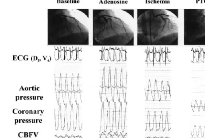
Figure 3. Coronary angiography, ECG (D3), aortic and distal coronary pressure and coronary blood flow velocity (CBFV) in a patients with stenosis and transient occlusion of the left circumflex coronary artery. Adenosine increased transstenotic pressure gradi-ent and blood flow. During ischemia and ST segmgradi-ent elevation in D3, distal coronary pressure was similar to that observed following adenosine. During balloon occlusion (PTCA) distal pressure was markedly lower, while blood flow velocity was not monitored.
Figure 4. Coronary angiography, electrocardiogram (D3, V4),
aortic and distal coronary pressure and coronary blood flow velocity (CBFV) in patients with stenosis and transient occlusion of the left anterior descending coronary artery. Adenosine increased transste-notic pressure gradient and blood flow. During ischemia and ST segment elevation in V4, distal coronary pressure was similar to
that observed following adenosine. During balloon occlusion (PTCA) distal pressure was markedly lower, while blood flow velocity was not monitored.
distal coronary vasomotion can contribute to the precipita-tion and maintenance of ischemia in UA.
Coronary blood flow reduction during spontaneous isch-emia. In the present study, blood flow velocity markedly
decreased during ischemia in all patients. Since arterial diameter at the tip of Doppler catheter did not significantly
change throughout the study, velocity changes reflected blood flow variations. Thus, in agreement with previous studies (20 –22), the present data document a severe flow reduction during transient ischemia in patients with UA.
During ischemia, distal pressure was only slightly re-duced. Elevated poststenotic pressure despite a marked reduction in flow might reflect either feeding of ischemic territory by collateral flow (23) or increased microvascular resistance (24). However, the contribution of collateral circulation is challenged by the fact that distal pressure decreased at a very lower level during balloon coronary occlusion and related ischemia. Moreover, the low value of distal pressure during balloon inflation, which is indicative of vascular closing pressure at zero flow, makes unlikely the possibility that the observed increase in resistance during ischemic episodes would have been produced by an increase in tissue pressure due to ischemic wall distortion and increased LV end-diastolic pressure (24).
Obviously, the increased transstenotic pressure gradient, associated with a decreased flow, indicates a rise in resis-tance of epicardial obstruction (25). Although the mecha-nisms (active vasoconstriction, intermittent platelet aggre-gation or thrombosis) remain elusive, the present study document that this phenomenon “per se” cannot fully explain the pathophysiology of resting angina. According to the classical concept of autoregulation, the pressure drop caused by the increase in stenosis resistance should have been couterbalanced by a compensatory dilation of coronary microvessels and thus by a reduction of distal resistance (8). By contrast, transient ischemia was associated with an increase in distal resistance up to 8 times the baseline and almost 13 times the minimal (adenosine) values. Thus, distal vasoconstriction explained almost half of the increase in global resistance (Fig. 5). The driving pressure in distal coronary bed was similar during ischemia and during vasodilation. Nevertheless, following adenosine, the same pressure was able to push forward approximately 13 times more blood volume into microcirculation than during isch-emia. Thus, together with the very low value of coronary pressure during coronary angioplasty, the increase in calcu-lated distal resistance strongly suggests the presence of an intense active microvascular vasoconstriction during isch-emia. The transmural distribution of the vasoconstriction remains undefined.
Mechanisms underlying coronary vasoconstriction dur-ing ischemia. The evidence of a microcirculatory
vasocon-striction during ischemia is not entirely new. An intense microvessel constriction during prolonged severe ischemia (9,10) has been observed in canine myocardium. In humans, an increase in coronary resistance has also been documented during ischemia induced by atrial pacing tachycardia (13). In patients with UA and intracoronary thrombi, Wilson et al. (11) observed the occurrence of ischemia and slow run-off of contrast medium immediately after coronary angioplasty; since no transstenotic gradient was measured,
Figure 5. Coronary resistance at the level of epicardial stenosis
(top) and in distal microcirculation (middle) at the various steps of
the protocol as indicated in Figure 1. Bottom panel shows the behavior of total coronary resistance and the contribution of stenosis (black) and microvascular resistance (white columns). Coronary resistance was not calculated during balloon coronary occlusion. During ischemia, both coronary stenosis and distal microcirculation showed a significant increase in resistance to flow (*⫽ p ⬍ 0.05 vs. baseline, † ⫽ p ⬍ 0.05 vs. adenosine; § ⫽ p ⬍ 0.05 vs. max ischemia).
the authors could only suspect a microvascular vasoconstric-tor response to thrombus-derived agents. Actually, it is now well recognized that substances such as serotonin vasodilate the normal coronary circulation, while this effect is turned to vasoconstriction in the presence of atherosclerotic endothe-lial damage (14). In this line, intermittent ischemia might reflect mural thrombosis and release of substances able to vasoconstrict microvessels (26,27).
Alternatively, vasoactive signals not linked to coronary thrombosis might be responsible for coronary vasoconstric-tion. Actually, endothelial dysfunction impairs microvascu-lar adaptation to ischemia (28), and constrictor response to reduced intraluminal pressure has been described in isolated microvessels (29). Similarly, ischemia mediated by activa-tion of postsynaptic alpha2-receptors has been documented
in anesthetized dogs (30). Potent coronary vasoconstrictors such as neuropeptide-Y (31) and endothelin (32) are more effective on distal vessels than on large arteries. In this line, Zeiher et al. (33) recently documented a greater endothelin-I immunoreactivity in unstable rather than sta-ble coronary plaques. The vasoconstrictor response to these substances might have been exaggerated by atherosclerotic endothelial dysfunction at the level of both coronary plaque and distal microvasculature (34) through a reduced NO release further aggravated by the decreased flow velocity and shear stress (35).
Finally, whether a distal vasoconstriction was present also during “balloon ischemia” remains an open question. In fact, due to the method used, the absence of flow during balloon inflation prevents the possibility to assess microvascular tone through computation of vascular resistance; however, the low distal pressure value during balloon coronary artery occlusion reflects only the integrated effect of collateral circulation and tissue pressure and cannot exclude vasocon-striction in the absence of flow.
Study limitations. Several limitations of the present study
deserve further discussion. All patients were evaluated during active treatment with aspirin, diltiazem, nitrates and heparin. This procedure, imposed by ethical considerations, might have interfered with blood flow regulation. However, although the present data cannot be directly extended to untreated patients, they point out that coronary vasocon-striction is not fully prevented by the conventional treat-ment of UA (36,37), while it promptly recedes following intracoronary nitrates.
The observation of ischemic ST segment elevation in 5 of 10 patients might indicate the presence of a highly selected population of patients with Prinzmetal angina (38). How-ever, inclusion criteria were in line with the definition of unstable angina (16) and all patients had a severe stenosis and positive exercise stress test. Moreover, the same behav-ior of microvascular resistance was observed regardless of the type of electrocardiographic alterations.
Theoretically, the presence of guidewires across the ste-nosis and of two catheters in the proximal vessel might have
contributed to the precipitation of coronary spasm. How-ever, this kind of instrumentation was not associated with the development of transient ischemia in 13 patients with stable angina pectoris evaluated by De Bruyne et al. (15) at the time of coronary angioplasty. Similarly, in our experi-ence, no transient ischemia was observed in nine patients with stable angina evaluated with a similar protocol (39). However, the functional severity of the stenosis might have been overestimated by the presence of guidewires.
As opposed to previous studies, a poor correlation was observed between angiographic and functional estimates of stenosis severity (coronary flow reserve or fractional flow reserve) (17,40,41). This finding might be ascribed to the selection of patients with a very severe stenosis characterized by a significant pressure gradient at rest. The skewness of stenosis severity in so small a patient sample distribution might have hampered the recognition of a correlation. Alternatively, it should also be considered that measure-ments of either coronary reserve or parameters related to transstenotic pressure gradient describe the severity of a coronary stenosis only in the presence of normal maximal distal vasodilation (16). In the present study, the microvas-cular dysfunction either primary (42) or caused by repetitive ischemia (43) might have affected vasodilating capability altering maximal flow capacity.
CONCLUSIONS
The present study documents the relevance of the micro-circulatory vasomotor tone in the pathogenesis of myocar-dial ischemia. Because of the small number of patients and possible selection biases, this study does not allow verifica-tion of the exact prevalence of the microvascular vasocon-striction in general populations of patients with UA. Thus, these findings need further confirmation by different tech-niques and laboratories. However, in agreement with pre-vious studies (11–13,42,44), these data challenge the com-mon view that exclusively attributes the genesis and course of myocardial ischemia to phenomena confined at the level of epicardial obstruction. The relevance of coronary stenosis as a major factor in the pathogenesis of myocardial ischemia is unquestioned. Procedures that remove its impact—such as coronary artery bypass grafting or coronary angioplasty— are strongly effective in reducing the occurrence of ischemic episodes. However, it is worthwhile to note that the altered control of distal coronary tone may aggravate the effects of epicardial obstruction to such an extent that, according to the present study, is equivalent to a second stenosis of similar severity. The present data do not allow verification of the mechanisms underlying this paradoxical behavior; how-ever, they document the lack of the physiologic autoregu-latory response worsening the severity of flow impairment in the ischemic vascular bed. Identifying the mechanisms of the altered control of vasomotor tone, during ischemia, might allow a more accurate identification of patients who
will develop UA and the development of more specific forms of anti-ischemic therapy. At present the most used vasoactive drugs are most effective on large epicardial arteries; in fact, direct arteriolar vasodilators may precipitate ischemia by coronary steal. The identification of the mech-anisms underlying this paradoxical microvascular vasocon-striction and thus of specific substances able to prevent it might provide a more efficient pharmacologic strategy especially for those patients who cannot benefit from coro-nary revascularization.
Acknowledgments
The authors are indebted to the patients who volunteered to experience angina and ischemia for research purposes in the catheterization room, to the staff of catheterization labora-tory for their skillfull cooperation and to Mrs. Ilaria Citti for her careful secretarial assistance.
Reprint requests and correspondence: Dr. Gianmario
Sam-buceti, CNR Institute of Clinical Physiology, Via P. Savi, 8, 56100, Pisa, Italy. E-mail: [email protected].
REFERENCES
1. Epstein SE, Talbot TL. Dynamic coronary tone in precipitation, exacerbation and relief of angina pectoris. Am J Cardiol 1981;48:797– 803.
2. Bertrand ME, LaBlanche LJM, Tilmant PY, et al. Frequency of provoked coronary arterial spasm in 1,089 consecutive patients under-going coronary arteriography. Circulation 1982;65:1299 –306. 3. Ambrose JA. Plaque disruption and the acute coronary syndromes of
unstable angina and myocardial infarction: if the substrate is similar, why is the clinical presentation different? J Am Coll Cardiol 1992;19: 1653– 8.
4. De Feyter PJ, Ozaki Y, Baptista J, et al. Ischemia-related lesion characteristics in patients with stable or unstable angina: a study with intracoronary angioscopy. Circulation 1995;92:1408 –13.
5. Waxman S, Sassower MA, Mittleman MA, et al. Angioscopic predictors of early adverse outcome after coronary angioplasty in patients with unstable angina and non-Q-wave myocardial infarction. Circulation 1996;93:2106 –13.
6. Thieme T, Wernecke KD, Meyer R, et al. Angioscopic evaluation of atherosclerotic plaques: validation by histomorphologic analysis and association with stable and unstable coronary syndromes. J Am Coll Cardiol 1996;28:1– 6.
7. Van Belle E, Lablanche JM, Bauters C, et al. Coronary angioscopic findings in the infarct-related vessel within 1 month of acute myocar-dial infarction. Circulation 1998;97:26 –33.
8. Gould KL, Lipscomb K, Calvert C. Compensatory changes of the distal coronary vascular bed during progressive coronary vasoconstric-tion. Circulation 1975;51:1085–94.
9. Guyton RA, McClenathan JH, Newman GE, Michaelis LL. Evolu-tion of regional ischemia distal to a proximal coronary stenosis: self-propagation of ischemia. Am J Cardiol 1977;40:381–92. 10. Gorman MW, Sparks HV. Progressive coronary vasoconstriction
during relative ischemia in canine myocardium. Circ Res 1982;51:411– 20.
11. Wilson RF, Lesser JF, Laxson DD, White CW. Intense microvascular constriction after angioplasty of acute thrombotic arterial lesions. Lancet 1989;1:807–11.
12. Pupita G, Maseri A, Kaski JC, et al. Myocardial ischemia caused by distal coronary artery constriction in stable angina pectoris. N Engl J Med 1990;323:514 –20.
13. Sambuceti G, Marzilli M, Marraccini P, et al. Coronary vasoconstric-tion during myocardial ischemia induced by rises in metabolic demand in patients with coronary artery disease. Circulation 1997;95:2652–9. 14. Golino P, Piscione F, Willerson JT, et al. Divergent effects of
serotonin on coronary-artery dimensions and blood flow in patients with coronary atherosclerosis and control patients. N Engl J Med 1991;324:641– 8.
15. DeBruyne B, Bartunek J, Sys SU, et al. Simultaneous coronary pressure and flow velocity measurements in humans. Circulation 1996;64:1842–9.
16. Braunwald E. Unstable angina: a classification. Circulation 1989;80: 410 – 4.
17. Pijls NHJ, De Bruyne B, Peels K, et al. Measurement of fractional flow reserve to assess the functional severity of coronary artery stenoses. N Engl J Med 1996;334:1703– 8.
18. Spears JR, Sandor T, Als AV, et al. Computerized image analysis for quantitative measurement of vessel diameter from cineangiograms. Circulation 1983;68:453– 61.
19. Ambrose JA, Winters SL, Stern A, et al. Angiographic morphology and the pathogenesis of unstable angina pectoris. J Am Coll Cardiol 1985;5:609 –16.
20. Maseri A, Parodi O, Severi S, et al. Transient transmural reduction of myocardial blood flow, demonstrated by thallium-201 scintigraphy as a cause of variant angina. Circulation 1976;54:280 – 8.
21. Fuster V, Badimon L, Badimon JJ, Chesebro JH. The pathogenesis of coronary artery disease and the acute coronary syndromes: part 1. N Engl J Med 1992;326:242–50.
22. Maseri A, Crea F, Cianflone D. Myocardial ischemia caused by distal vasoconstriction. Am J Cardiol 1992;70:1602–5.
23. Pijls NHJ, Bech GJW, Gamal MIH, et al. Quantification of re-cruitable coronary collateral blood flow in conscious humans and its potential to predict future ischemic events. J Am Coll Cardiol 1995;25:1522– 8.
24. Ellis AK, Klocke FJ. Effects of preload on the transmural distribution of perfusion and pressure-flow relationships in the canine coronary vascular bed. Circ Res 1979;46:68 –77.
25. Gould KL. Coronary Artery Stenosis. New York: Elsevier, 1990;79 – 91.
26. Willerson JT, Golino P, Eidt J, et al. Specific platelet mediators and unstable coronary lesions: experimental evidence and potential clinical implications. Circulation 1989;80:198 –205.
27. Rubanji GM, Frye RL, Holmes DR Jr, Van Houtte PM. Vasocon-strictor activity of coronary sinus plasma from patients with coronary artery disease. J Am Coll Cardiol 1987;9:1243–9.
28. Smith TP, Canty JM. Modulation of coronary autoregulatory re-sponses by nitric oxide. Circ Res 1993;73:232– 40.
29. Chilian WM, Layne SM. Coronary microvascular responses to reduc-tions in perfusion pressure: evidence for persistent arteriolar vasomotor tone during coronary hypoperfusion. Circ Res 1990;66:1227–38. 30. Heusch G, Deussen A. The effects of cardiac sympathetic nerve
stimulation on perfusion of stenotic coronary arteries in the dog. Circ Res 1983;53:8 –15.
31. Clarke JG, Davies GJ, Kerwin R, et al. Coronary artery infusion of neuropeptide Y in patients with stable angina pectoris. Lancet 1987; 1:1057–9.
32. Larkin SW, Clark JG, Keogh BE, et al. Intracoronary endothelin induces myocardial ischemia by small vessel constriction in the dog. Am J Cardiol 1989;64:956 – 8.
33. Zeiher AM, Goebel H, Schachinger V, Ihling C. Tissue endothelin-I immunoreactivity in the active coronary atherosclerotic plaque: a clue to the mechanism of increased vasoreactivity of the culprit lesion in unstable angina. Circulation 1995;91:941–7.
34. Meredith IT, Yeung AC, Weidinger FF, et al. Role of impaired endothelium-dependent vasodilation in ischemic manifestations of coronary artery disease. Circulation 1993;87 suppl V:V56 – 66. 35. Cox DA, Vita JA, Treasure CB, et al. Impairment of flow mediated
coronary dilation by atherosclerosis in man. Circulation 1989;80:458 – 65.
36. Horowitz JD. Role of nitrates in unstable angina pectoris. Am J Cardiol 1992;70 suppl B:64B–71.
37. Prisant LM, von Dohlen T, Rogers W, et al. Pharmacotherapy of unstable angina. J Clin Pharmacol 1992;32:390 –9.
38. Maseri A, Severi S, DeNes M, et al. Variant angina: one aspect of a continuous spectrum of vasospastic myocardial ischemia: pathogenetic mechanisms, estimated incidence and clinical and coronary arterio-graphic findings in 138 patients. Am J Cardiol 1978;42:1019 –35. 39. Sambuceti G, Marzilli M, Fedele S, et al. Microcirculatory
tachy-cardia in patients with coronary artery disease (abstr). Circulation 1998;98 suppl I:I487.
40. Uren NG, Melin JA, De Bruyne B, et al. Relation between myocardial blood flow and the severity of coronary artery stenosis. N Engl J Med 1994;330:1782– 8.
41. Kirkeeide RL, Gould KL, Parsel L. Assessment of coronary stenoses by myocardial perfusion imaging during pharmacologic vasodilation: VII. Validation of coronary flow reserve as a single integrated func-tional measurement of stenosis severity reflecting all its geometric dimensions. J Am Coll Cardiol 1986;7:103–13.
42. Sambuceti G, Marzullo P, Giorgetti A, et al. Global alteration in perfusion response to increasing oxygen consumption in patients with single vessel coronary artery disease. Circulation 1994;90: 1696 –705.
43. Bolli R, Triana JF, Jeroudi MO. Prolonged impairment of coronary vasodilation after reversible ischemia: evidence for microvascular stun-ning. Circ Res 1990;67:332–7.
44. Selwyin AP, Forse G, Fox K, et al. Patterns of disturbed myocardial perfusion in patients with coronary artery disease: regional myocardial perfusion in angina pectoris. Circulation 1981;64:83–90.