Research Article
Quantitative Determination of 18-
𝛽-Glycyrrhetinic
Acid in HepG2 Cell Line by High Performance Liquid
Chromatography Method
Giuseppina Nocca
,
1,2Cinzia Callà
,
3Stefano Angelo Santini,
3Adriana Amalfitano,
1Luca Marigo,
4Diana Valeria Rossetti,
1Gianrico Spagnuolo
,
5,6and Massimo Cordaro
41Istituto di Biochimica e Biochimica Clinica, Universit`a Cattolica del Sacro Cuore Rome, Italy
2Istituto di Chimica del Riconoscimento Molecolare, CNR, c/o Universit`a Cattolica del Sacro Cuore, L.go F. Vito 1, I-00168 Rome, Italy
3UOC Chimica, Biochimica e Biologia Molecolare, Dip. Scienze di Laboratorio e Infettivologiche,
Fondazione Policlinico Universitario A. Gemelli, IRCCS, Universit`a Cattolica del Sacro Cuore Rome, Italy
4UOC Odontoiatria Generale e Ortodonzia, Dip Scienze dell’Invecchiamento, Neurologiche, Ortopediche e della Testa Collo.
Fondazione Policlinico Universitario A. Gemelli, IRCCS, Universit`a Cattolica del Sacro Cuore Rome, Italy
5Department of Neurosciences, Reproductive and Odontostomatological Sciences University of Naples “Federico II”, Italy
6I.M. Sechenov First Moscow State Medical University, Institute of Dentistry, Moscow, Russia
Correspondence should be addressed to Giuseppina Nocca; [email protected] Received 6 July 2018; Accepted 4 November 2018; Published 13 November 2018 Academic Editor: Valentina Venuti
Copyright © 2018 Giuseppina Nocca et al. This is an open access article distributed under the Creative Commons Attribution License, which permits unrestricted use, distribution, and reproduction in any medium, provided the original work is properly cited.
A reverse phase high performance liquid chromatographic (RP-HPLC) method was developed for identification and estimation of 18-𝛽-glycyrrhetinic acid (GA) in HepG2 cell line. The analysis was carried out using a JASCO HPLC system with a C-18 (3 𝜇m) Supelco reversed phase column (150 x 4.7 mm) using a mobile phase of 80% CH3OH and 20% of CH3CN: tetrahydrofuran: water (10:80:10, v/v/v). The method was linear in the concentration range of 1.5–120𝜇g /mL (n = 5). The LOD and LOQ were determined based on standard deviation of the y-intercept and the slope of the calibration curve. The LOD and LOQ values were found to be 11.46𝜇g/mL and 34.72 𝜇g/mL, respectively. The mean percentage recovery by standard addition experiments of GA is 92.4 %± 5.2%. The intracellular GA concentration value, obtained as mean of five different determinations, was 45.8± 7.45 𝜇g/mL. We have developed a HPLC-UV method for quantitative determination of GA inside cells, with advantages in the cost reduction and economy of the analytical process.
1. Introduction
The use of natural substances as adjuvants in many drug therapies is a new important trend in modern medicine, due to a satisfactory clinical efficacy and a low degree of toxicity [1–3]. Liquorice is a perennial plant with well-known pharmacological properties that is largely employed in the cosmetic and pharmacological fields, due to its several bio-logical effects: antimicrobial, antiulcer, immunomodulatory, anti-inflammatory, etc. [4–9].
One of the most active compounds of liquorice is the triterpenoid glycyrrhizic acid (glycyrrhizin, GZ) and
the main product of its metabolism: the aglycone 18-𝛽-glycyrrhetinic acid (GA) [5, 6]. GA exhibits corticosteroid and mineral-corticoid activity due to the presence of the 𝛼,𝛽-unsaturated ketone group: in fact, GA is able to interact with mineral-corticoid and glucocorticosteroid receptors and exhibits anti-inflammatory properties [7]. Several studies [10, 11] have reported that inappropriate use of licorice can produce pseudoaldosteronism, by inactivating 11beta-hydroxysteroid-dehydrogenase [11-𝛽HSD] and by binding to mineralocorticoid receptors. 11-𝛽HSD catalyzes the oxida-tion of the active mineralocorticoid, cortisol, to the inac-tive cortisone and 11alpha-hydroxysteroiod-dehydrogenase is Volume 2018, Article ID 5673186, 5 pages
responsible for the reduction reaction. Thus, GA potenti-ates the anti-inflammatory activity of cortisol by inhibiting its intracellular inactivation. Moreover, GA is involved in strengthen red blood cell membrane integrity against both oxidative and proteolytic damage [12].
Moreover, in vitro studies showed that GA inhibits the proliferation of different cancer cells [13–15], without affecting normal cells [16–18]. This effect is probably due to different mechanisms, such as downregulation of glutathione (GSH) [17] and production of reactive oxygen species (ROS) [18, 19]. However, all of the above reported actions are depen-dent on the solubility of the compound in the hydrophilic medium and, over all, on its ability to penetrate into cells because its interactions with receptors and with enzymes occur intracellularly. Given the lipid nature of GA, in in vitro experiments, it has to be previously dissolved in DMSO [20, 21]; this solvent has a polar domain and two nonpolar groups, making GA soluble in both aqueous and organic media [22]. Moreover, DMSO can induce water pores in dipalmitoylphosphatidylcholine bilayers and this is a possi-ble mechanism to increase penetration of active molecules through lipid membranes [23].
Several analytical methods have been developed with different techniques to evaluate the concentration of the var-ious components of licorice in the preparations for cosmetic and biomedical application. In several publications, analyti-cal methods have been developed using High-Performance Liquid Chromatography (HPLC) that is a very sensitive and reproducible technique [24–29]. On these bases, the aim of the present study was to develop a HPLC-UV method able to carry out a quantitative determination of GA inside HepG2 cells. This human hepatocellular cell line was chosen because these cells are able to metabolize GA [30]. To our knowledge, in this paper a method for detection of GA inside cells is reported, for the first time.
2. Materials and Methods
2.1. Chemicals and Reagents. 18-𝛽-glycyrrhetinic acid (GA)
was purchased from Acros Organics (VWR International Srl, Milan, Italy). Cell culture medium and reagents, DMSO, ethanol (EtOH), tetrahydrofuran (HPLC grade), and
ace-tonitrile (CH3CN, HPLC grade) were purchased from Sigma
Chemical (Milan, Italy). Methyl alcohol (CH3OH, HPLC
grade, Prolabo, France) and ultrapure water (obtained by a P.Nix Power System apparatus, Human, Seoul, Korea) were used for HPLC analyses.
2.2. HPLC Conditions. Standard solutions were analyzed
using a JASCO HPLC system (2 PU-980 pumps, UV-970 UV/ VIS detector and AS-1555 autosampler). The analyses were
performed at a wavelength of 254 nm with a C-18 (3𝜇m)
Supelco reversed phase column (150 x 4.7 mm) using a mobile
phase of 80% CH3OH (A) and 20% of CH3CN:
tetrahydro-furan: water (10:80:10, v/v/v) (B) [modified from Chamoli
[31] (15 min), 1.0 mL/min flow, 50𝜇L injected volume]. Each
analysis was performed five times.
2.3. Cell Culture. HepG2, human liver carcinoma cells
(Isti-tuto Zooprofilattico, Brescia, Italy) were grown in a 5% CO2
atmosphere at 37∘C in IMDM (Iscove’s Modified Dulbecco’s
Medium) with HEPES (10 mM), glucose (4.5 g/L), NaHCO3
(3.7 g/L), penicillin (100 units/mL), streptomycin (100 g/mL), 1% nonessential amino acids, and 10% fetal calf serum.
2.4. Cells Treatments. Stock solution of GA (200.0 mmol/L)
was prepared immediately before use in DMSO, to obtain a final concentration of 0.1% (v/v) DMSO, which does not induce any alteration in cell vitality [32, 33]. The final
concentration of GA was 200𝜇mol/L (94.14 𝜇g/mL).
2.5. Determination of Intracellular Concentrations of GA.
HepG2 (5 mL) were plated in 25 cm2 flasks at a density of
approximately 25,000 cells/cm2and cultured to subconfluent
monolayers; GA (200𝜇mol/L) was then added and the cells
and incubated for 2 h at 37∘C. Cell monolayers without GA
were used as controls. After incubation, the cells were washed
in PBS solution and lysed by freezing (-80∘C). Cellular lysates
were resuspended in 1 mL of EtOH and centrifuged (20,000
x g, 15 min, 4∘C) and the supernatants were collected,
evapo-rated, and finally resuspended in 500𝜇L of EtOH. Samples
were analyzed as reported in HPLC conditions paragraph. The concentration of GA in each sample was quantified using the calibration curve performed with standard solutions before each analysis. Each determination was repeated three times and each experiment was performed 5 times (n=5)
2.6. Determination of Limit of Quantitation (LOQ) and Limit of Detection (LOD). According to the ICH recommendations
[34], LOD and LOQ were determined based on standard deviation of the y-intercept and the slope of the calibration curve (n=5). Calibration curves were constructed from GA standard solutions at 5 different concentrations within the
range of 1.5-120𝜇g/mL (1.5, 15, 30, 60 and 120 𝜇g/mL).
The following equations were used for calculating LOD and LOQ: LOD= (3, 3 × SD slope) LOQ= (10 × SD slope) (1)
2.7. Recovery Studies. The efficiency of GA recovery from cell
lysates was evaluated adding two different concentrations (20
and 40𝜇g/mL) into control cellular lysates. Two replicates
of each concentration were prepared for each lysate. The absolute recovery was evaluated as the ratio between the experimentally observed concentration and the theoretical concentration.
3. Results and Discussion
GA is a compound with several biological activities; for this reason, it is widely used both in cosmetic and in medical preparations. Since the biological effects of GA depend on its intracellular concentration and not only its concentration
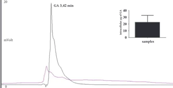
in tr acel lu la r g o f GA samples mVolt GA , min 20 0
Figure 1: Chromatographic profiles of HepG2 lysates. In intracellular sample, incubated for 2h with GA, a signal that corresponds to a substance with the same elution time of GA was found (black); the described signal was not present in the untreated samples (pink). Inset: Intracellular GA amount.
in culture medium, a simple HPLC-UV method for the intracellular GA determination was developed and described in this paper.
To be sure to be able to detect the exact intracellular GA concentration, the method was first tested for linearity, using calibrating standard solution mixtures of the GA
in concentration range 1.5–120𝜇g/mL. Each determination
was repeated three times and each calibration curve was performed 5 times (n=5). The obtained regression equation
was Y = 22017∗X – 21465 with R2= 0.9946, confirming the
linearity of the response under the conditions used. The LOD and LOQ were determined based on standard deviation of the y-intercept and the slope of the calibration curve. The
LOD and LOQ values were found to be 11.46𝜇g/mL and
34.72𝜇g/mL, respectively.
We were then able to apply our analytical method to identify and quantify the intracellular GA in cells treated with
a concentration of 200𝜇mol/L of GA in cell culture medium
(94.14𝜇g/mL; 5 mL). In these experimental conditions, no
cytotoxic effects were observed after two hours (data not shown). In this preliminary phase of the study, this is very important, since intracellular concentration of GA could affect cellular vitality.
As shown in Figure 1 a signal corresponding to a sub-stance with the same elution time of GA was found in cellular lysates after incubation for 2 h with GA as shown in materials and methods; the described signal was not present in the untreated samples (controls).
The intracellular GA concentration value, obtained as
mean of five different experiments, was 45.8± 7.45 𝜇g/mL.
Considering that the final volume of samples resuspended in EtOH was 0.5 mL (see materials and method) the true
amount of GA inside the cells was about 23𝜇g (Figure 1,
insert).
The GA percentage mean recovery by standard addition
experiments is92.4% ± 5.2% (data not shown). These results
indicate the reliability of our analytical method.
4. Conclusions
Our HPLC-UV method for identification and quantification of GA inside HepG2 cells, reliable in linearity and recovery, could be used in different fields of pharmacology research where the measurement of intracellular concentrations of GA is mandatory.
Data Availability
The datasets generated during and/or analysed during the current study are available from the corresponding author on reasonable request.
Conflicts of Interest
The authors declare that they have no conflicts of interest.
References
[1] J. Li, H. Cao, P. Liu et al., “Glycyrrhizic acid in the treatment of liver diseases: literature review,” BioMed Research International, vol. 2014, Article ID 872139, 15 pages, 2014.
[2] Y. Arase, K. Ikeda, N. Murashima et al., “The long term efficacy of glycyrrhizin in chronic hepatitis C patients,” Cancer, vol. 79, no. 8, pp. 1494–1500, 1997.
[3] K. Ikeda, “Glycyrrhizin injection therapy prevents hepatocel-lular carcinogenesis in patients with interferon-resistant active chronic hepatitis C,” Hepatology Research, vol. 37, no. 2, pp. S287–S293, 2007.
[4] A. Roohbakhsh, M. Iranshahy, and M. Iranshahi, “Gly-cyrrhetinic acid and its derivatives: Anti-cancer and can-cer chemopreventive properties, mechanisms of action and structure-cytotoxic activity relationship,” Current Medicinal
Chemistry, vol. 23, no. 5, pp. 498–517, 2016.
[5] G. A. Tolstikov, L. A. Baltina, and N. G. Serdyuk, “Glycyrrhetic acid (a review),” Pharmaceutical Chemistry Journal, vol. 32, pp. 5–14, 1998.
[6] B. Ploeger, T. Mensinga, A. Sips, W. Seinen, J. Meulenbelt, and J. DeJongh, “The pharmacokinetics of glycyrrhizic acid evaluated by physiologically based pharmacokinetic modeling,”
Drug Metabolism Reviews, vol. 33, no. 2, pp. 125–147, 2001.
[7] D. Armanini, I. Karbowiak, and J. W. Funder, “Affinity of liquorice derivatives for mineralocorticoid and glucocorticoid receptors,” Clinical Endocrinology, vol. 19, no. 5, pp. 609–612, 1983.
[8] F. Capasso, N. Mascolo, G. Autore, and M. R. Duraccio, “Glycyrrhetinic acid, leucocytes and prostaglandins,” Journal of
Pharmacy and Pharmacology, vol. 35, no. 5, pp. 332–335, 1983.
[9] W. Logemann, F. Lauria, G. Cudkowicz, and J. Franceschini, “Antileukæmic activity of glycyrrhetinic acid,” Nature, vol. 187, no. 4737, pp. 607-608, 1960.
[10] D. Armanini, C. Fiore, M. J. Mattarello, J. Bielenberg, and M. Palermo, “History of the endocrine effects of licorice,”
Experimental and Clinical Endocrinology & Diabetes, vol. 110,
no. 6, pp. 257–261, 2002.
[11] R. A. Isbrucker and G. A. Burdock, “Risk and safety assessment on the consumption of Licorice root (Glycyrrhiza sp.), its extract and powder as a food ingredient, with emphasis on the pharmacology and toxicology of glycyrrhizin,” Regulatory
Toxicology and Pharmacology, vol. 46, no. 3, pp. 167–192, 2006.
[12] C. Fiore, L. Bordin, D. Pellati, D. Armanini, and G. Clari, “Effect of glycyrrhetinic acid on membrane band 3 in human erythrocytes,” Archives of Biochemistry and Biophysics, vol. 479, no. 1, pp. 46–51, 2008.
[13] Z.-H. Tang, T. Li, Y.-G. Tong et al., “A systematic review of the anticancer properties of compounds isolated from Licorice (Gancao),” Planta Medica, vol. 81, no. 18, pp. 1670–1687, 2015. [14] H. Yamaguchi, T. Noshita, T. Yu et al., “Novel effects of
glycyrrhetinic acid on the central nervous system tumorigenic progenitor cells: Induction of actin disruption and tumor cell-selective toxicity,” European Journal of Medicinal Chemistry, vol. 45, no. 7, pp. 2943–2948, 2010.
[15] G. Sharma, S. Kar, S. Palit, and P. K. Das, “18𝛽-glycyrrhetinic acid (concur) induces apoptosis through modulation of Akt/FOXO3a/Bim pathway in human breast cancer MCF-7 cells,” Journal of Cellular Physiology, vol. 227, no. 5, pp. 1923– 1931, 2012.
[16] T. Yu, H. Yamaguchi, T. Noshita, Y. Kidachi, H. Umetsu, and K. Ryoyama, “Selective cytotoxicity of glycyrrhetinic acid against tumorigenic r/m HM-SFME-1 cells: Potential involvement of H-Ras downregulation,” Toxicology Letters, vol. 192, no. 3, pp. 425–430, 2010.
[17] H. Yamaguchi, T. Yu, Y. Kidachi et al., “Selective toxicity of glycyrrhetinic acid against tumorigenic r/m HM-SFME-1 cells is potentially attributed to downregulation of glutathione,”
Biochimie, vol. 93, no. 7, pp. 1172–1178, 2011.
[18] I. Cacciotti, L. Chronopoulou, C. Palocci et al., “Controlled release of 18-𝛽-glycyrrhetic acid by nanodelivery systems increases cytotoxicity on oral carcinoma cell line,”
Nanotechnol-ogy, vol. 29, no. 28, Article ID 285101, 2018.
[19] K.-W. Lin, A.-M. Huang, T.-C. Hour, S.-C. Yang, Y.-S. Pu, and C.-N. Lin, “18𝛽-Glycyrrhetinic acid derivatives induced mitochondrial-mediated apoptosis through reactive oxygen species-mediated p53 activation in NTUB1 cells,” Bioorganic &
Medicinal Chemistry, vol. 19, no. 14, pp. 4274–4285, 2011.
[20] A. Li, N. Ma, Z. Zhao, M. Yuan, H. Li, and Q. Wang, “Gly-cyrrhetinic acid might increase the nephrotoxicity of bakuchiol by inhibiting cytochrome P450 isoenzymes,” PeerJ, Article ID e2723, 2016.
[21] N. Papaevgeniou, M. Sakellari, S. Jha et al., “18𝛼-Glycyrrhetinic acid proteasome activator decelerates aging and Alzheimer’s disease progression in caenorhabditis elegans and neuronal cultures,” Antioxidants & Redox Signaling, vol. 25, no. 16, pp. 855–869, 2016.
[22] N. C. Santos, J. Figueira-Coelho, J. Martins-Silva, and C. Sal-danha, “Multidisciplinary utilization of dimethyl sulfoxide: pharmacological, cellular, and molecular aspects,” Biochemical
Pharmacology, vol. 65, no. 7, pp. 1035–1041, 2003.
[23] R. Notman, M. Noro, B. O’Malley, and J. Anwar, “Molecular basis for dimethylsulfoxide (DMSO) action on lipid mem-branes,” Journal of the American Chemical Society, vol. 128, no. 43, pp. 13982-13983, 2006.
[24] T.-H. Tsai and C.-F. Chen, “Determination of three active prin-ciples in licorice extract by reversed-phase high-performance liquid chromatography,” Journal of Chromatography A, vol. 542, no. C, pp. 521–525, 1991.
[25] H. Meena, M. Mohsin, H. K. Pandey, P. S. Negi, and Z. Ahmed, “Estimation of cordycepin by improved HPLC method in the natural and cultured mycelia of high medicinal value Himalayan entomogenous fungus Cordyceps sinensis,”
Elec-tronic Journal of Environmental, Agricultural and Food Chem-istry , vol. 9, no. 10, pp. 1598–1603, 2010.
[26] L. Wang, W. Kong, M. Yang, J. Han, and S. Chen, “Safety issues and new rapid detection methods in traditional Chinese medicinal materials,” Acta Pharmaceutica Sinica B (APSB), vol. 5, no. 1, pp. 38–46, 2015.
[27] K. Shanker, A. Fatima, A. S. Negi et al., “RP-HPLC method for the quantitation of glabridin in Yashti-madhu (Glycyrrhiza glabra),” Chromatographia, vol. 65, no. 11-12, pp. 771–774, 2007. [28] S. Liu, L.-Z. Yi, and Y.-Z. Liang, “Traditional Chinese medicine
and separation science,” Journal of Separation Science, vol. 31, no. 11, pp. 2113–2137, 2008.
[29] V. Andrisano, D. Bonazzi, and V. Cavrini, “HPLC analysis of liquorice triterpenoids - applications to the quality control of pharmaceuticals,” Journal of Pharmaceutical and Biomedical
Analysis, vol. 13, no. 4-5, pp. 597–605, 1995.
[30] Y. Satomi, H. Nishino, and S. Shibata, “Glycyrrhetinic acid and related compounds induce G1 arrest and apoptosis in human hepatocellular carcinoma HepG2,” Anticancer Reseach, vol. 25, no. 6 B, pp. 4043–4047, 2005.
[31] A. Chamoli, M. Ahmad, M. Hasan, and B. Panda, “Simul-taneous determination of 18𝛼-glycyrrhetinic acid and 18𝛽-glycyrrhetinic acid in Glycyrrhiza glabra root by reversed phase high-performance liquid chromatography,” Drug, Design,
Development and Therapy, vol. 7, pp. 59–62, 2016.
[32] G. Nocca, C. Call`a, G. E. Martorana et al., “Effects of dental methacrylates on oxygen consumption and redox status of human pulp cells,” BioMed Research International, vol. 2014, Article ID 956579, 10 pages, 2014.
[33] G. Radicioni, A. Stringaro, A. Molinari et al., “Characterization of the cell penetrating properties of a human salivary proline-rich peptide,” Biochimica et Biophysica Acta (BBA) -
Biomem-branes, vol. 1848, no. 11, pp. 2868–2877, 2015.
[34] ICH, “Validation of Analytical Procedure,” in Proceedings of the
International Conference on Harmonization, IFPMA, Geneva,
Tribology
Advances in Hindawi www.hindawi.com Volume 2018 Hindawi www.hindawi.com Volume 2018International Journal ofInternational Journal of
Photoenergy
Hindawi www.hindawi.com Volume 2018 Journal ofChemistry
Hindawi www.hindawi.com Volume 2018 Advances inPhysical Chemistry
Hindawi www.hindawi.com Analytical Methods in Chemistry Journal of Volume 2018 Bioinorganic Chemistry and Applications Hindawi www.hindawi.com Volume 2018Spectroscopy
International Journal ofHindawi
www.hindawi.com Volume 2018
Hindawi Publishing Corporation
http://www.hindawi.com Volume 2013 Hindawi www.hindawi.com
The Scientific
World Journal
Volume 2018Medicinal ChemistryInternational Journal of Hindawi www.hindawi.com Volume 2018
Nanotechnology
Hindawi www.hindawi.com Volume 2018 Journal ofApplied Chemistry
Journal of Hindawi www.hindawi.com Volume 2018 Hindawi www.hindawi.com Volume 2018 Biochemistry Research International Hindawi www.hindawi.com Volume 2018Enzyme
Research
Hindawi www.hindawi.com Volume 2018 Journal ofSpectroscopy
Analytical Chemistry International Journal of Hindawi www.hindawi.com Volume 2018Materials
Journal of Hindawi www.hindawi.com Volume 2018 Hindawi www.hindawi.com Volume 2018 BioMedResearch International
Electrochemistry
International Journal of Hindawi www.hindawi.com Volume 2018