Joint PhD Program in Crop Science (XXV Cycle)
PhD School in Agricultural and VeterinarySciences
Grapevine rupestris stem pitting-associated virus (GRSPaV) and vein
necrosis: effect of genetic variability in symptoms expression
SSD AGR/12
Candidate:
Michele Della Bartola
Supervisor:
Dr. Alberto Materazzi
PhD School Director:
Prof. Alberto Pardossi
INDEX
Abstract
031.
General introduction
05 1.1 Vein Necrosis 06 - Diagnosis - Aetiology1.2 Grapevine Rupestris Stem Pitting-associated Virus 09
- Association with RSP
- Association with Syrah decline
- Association with Vein Necrosis
1.3 Overall objectives 16
2.
Molecular detection of Grapevine Rupestris Stem Pitting-associated
Virus in grapevine accessions
2.1 Introduction 17
2.2 Materials and Methods 18
2.3 Results 23
2.4 Discussion 30
3. Molecular and serological characterization of ORF 5 (CP gene)
3.1 Introduction 31
3.1.1 Molecular characterization 31
3.1.2 Serological characterization 33
3.2 Materials and Methods 36 3.2.1 Molecular characterization 36 3.2.2 Serological characterization 41 3.3 Results 43 3.3.1 Molecular characterization 43 3.3.2 Serological characterization 51 3.4 Discussion 52
4.
Molecular characterization of a fragment of ORF 1 (replicase gene)
4.1 Introduction 54
4.2 Materials and Methods 56
4.3 Results 60
4.4 Discussion 66
5. Biological indexing for Vein Necrosis Disease
5.1 Introduction 68
5.2 Materials and Methods 70
5.3 Results 75
5.4 Discussion 79
Abstract
Vein necrosis (VN) is a virus-like disease of grapevine that is latent in V. vinifera and in most of rootstock and hybrids, with the only exception of rootstock 110 Richter (V.
berlandieri x V. rupestris). Despite its ubiquitous presence in many grape growing
regions, its economic impact on production is still largely unknown, because of its latency in V. vinifera. Though the causal agent of VN has not been identified, as the disease is able to be transmitted by grafting, VN is then considered a virus-like disease. Recent studies suggested a correlation between VN and the virus Grapevine rupestris stem pitting-associated virus (GRSPaV).
In this research, the presence and molecular variability of GRSPaV in grapevine accessions from Tuscany and California was studied through RT-PCR and sequence analyses. RT-PCR analyses with group-specific primer sets distinguished three molecular groups of GRSPaV variants. All of the three groups were detected both in Tuscan and Californian grapevine accessions.
Amplification, cloning and sequencing of two distinct viral genomic regions were carried out in order to obtain a finer molecular characterization of GRSPaV isolates. Depending on the genomic region analyzed, five (ORF5) to six (ORF1) phylogenetic groups of virus variants were observed. Notably, the sixth phylogenetic group identified according to sequence analysis of a 299 nucleotides fragment of ORF1 comprises only GRSPaV variants derived from Tuscan grapevine accessions, that show low nucleotide identity with any other GRSPaV sequence deposited in GenBank. This could represent a new and possibly yet unknown phylogroup of GRSPaV variants, that has named “group 5”.
Linking the results of GRSPaV molecular characterization with those of VN biological indexing previously conducted on the accessions object of the study, a strong correlation emerged between VN positive plants and infections by GRSPaV variants belonging to phylogenetic groups 2a and 2b.
In order to further assess this correlation, a biological indexing trial was performed for 37 grapevine accessions from Italy (Tuscany and Apulia), USA, Portugal and Japan. Results obtained confirmed the hypothesis that only phylogenetic groups 2a and 2b of the virus are able to induce VN symptoms on indicator host 110 Richter.
These findings show evidence of a different role of GRSPaV variants in VN determinism; only phylogenetic groups 2a and 2b appear to be able to induce symptoms of VN. This could suggest the possibility to diagnose VN by RT-PCR assays with group-specific primers, representing a much faster, cheaper and more simple way to monitor the diffusion and incidence of this disease.
1. General introduction
Grapevine (Vitis spp.) is the most widely cultivated fruit crop worldwide, encompassing about 8 million hectares of arable land (Vivier and Pretorius, 2002) with about 67200 kilotons produced in 2007 (FAOSTAT, 2009). Since their first appearance over 65 million years ago in Eurasia, south western Asia, Mediterranean region and central Europe (de Saporta, 1879), grapes from V. vinifera, have been used extensively for making wine throughout the world (This et al., 2006). Other members of the family Vitaceae used in wine grape breeding programs, especially for breeding rootstocks and inter-specific hybrids, include Muscadinia rotundifolia, V. aestivalis, V. amurensis, V. berlandieri, V.
candicans, V. caribaea, V. champinii, V. cinerea, V. cordifolia, V. labrusca, V. longii, V. riparia, V. rupestris and V. simpsonii (This et al., 2006). Wine, the main product of
wine-grapes, has been made for millennia. Besides their economic importance as alcoholic beverages, wines also have ancient historical connections with the development of human culture (McGovern, 2004) and, in recent years, the health benefits of wine consumption in moderate amounts have been recognized (German and Walzem, 2000). Italy is ranked 2nd internationally in grape production behind China. In Italy, grape
production is the 1st most economically important agricultural crop. (FAOSTAT, 2011).
Although the grape berry is used for multiple purposes, wine production from cultivars of V. vinifera have the highest economic impact (Mullins et al., 1992).
In 2012, 4089000 tons of wine have been produced in Italy, 2385904 of which were destined to export, for an economic value 6075404 $ (FAOSTAT, 2012).
Tuscany has a long and well-established wine-makers tradition: its wines are known and appreciated in the whole world. In the last decades, the industry has strongly expanded in the region, significantly contributing to the regional economy. Currently, wine grapes are grown in about 60327 hectares (ha) and wine production reach 2097621 tons (ISTAT, 2012).
Because grapevines are commonly vegetatively propagated, spread of many debilitating viruses occurs through cuttings, resulting in economic losses to growers. Since virus
diseases cannot be controlled by economically feasible chemical agents similar to fungicides, strategies aimed at the management of grapevine viruses are largely directed at preventing virus spread by utilizing virus-tested planting material.
********
On a worldwide basis, the grapevines appear to be infected with more viruses than any other perennial woody species. Currently, the International Council for the Study of Virus and Virus-like Diseases of the Grapevine (ICVG) recognized more 60 viruses reported on genera Vitis and Muscadinia, belonging to 27 genera and exhibiting the whole set of genome types: single-stranded DNA, stranded DNA, double-stranded RNA, single-double-stranded negative-sense RNA, single-double-stranded positive-sense RNA (Martelli, 2012).
The ‘traditional’ virus diseases such as fanleaf, leafroll, rugose wood and fleck represent a group of well-known disorders in several grape-growing countries around the world (Hewitt, 1954; Goheen et al., 1958; Savino et al., 1989; Martelli, 1993), while many of the other viral and virus viral-like disorders are of limited geographic distribution. The aetiology of many of these diseases remains largely unsolved.
1.1 Vein necrosis
Vein necrosis (VN) is a virus-like disease, reported for the first time in France in 1973 (Legin and Vuittenez), and later found in some regions of the former Soviet Union (Milkus and Schterenberg, 1978), in Italy and Bulgaria (Martelli et al., 1978). To date it is widespread in several European countries and the basin of the Mediterranean, where, at times, has reached high levels of incidence (Credi et al. 1985). Surveys conducted in Tuscany on "Sangiovese", "Aleatico”, "Mammolo", “Prugnolo gentile” and “Vermentino bianco” have confirmed the very high presence even in this region, reaching frequency percentages ranging from 61.0% to 90.0%. (Triolo and Materazzi, 2000; Triolo and
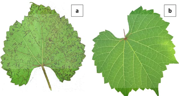
Figure 1. VN symptoms on the underside of 110R leaf blades. Pictures of symptomatic (a)
and symptomless (b) 110R leaves.
reductions in growth accompanied by the appearance, from the late spring, of necrosis on veinlets and veins on the underside of the leaf blade (Fig. 1). With time, necrotic spots also appear on the upper side of the leaf. Symptoms generally appear first on the leaves at the base of the shoots, and then gradually affect younger leaves, causing a premature yellowing and fall. Severe strains may induce necrosis of tendrils and dieback of green shots, with an almost complete cessation of growth and, eventually, death of the 110 R host plant.
In a classification concerning the economic importance of the most common systemic diseases of grapevine, vein necrosis is part of the third group (Tab. 1) (Martelli, 2002). In fact, despite its wide diffusion, its economic impact on production is still largely unknown, because of its latency in V. vinifera and in most rootstock hybrids.
Table. 1. Classification of the main systemic diseases of grapevine based on their
economic importance. (Martelli , 2002).
VN diagnosis
Since the etiology of VN is still unknown, biological assay with indicator species of the genus Vitis is the only diagnostic method available. The diagnosis can be easily carried out through indexing with rootstock hybrid 110 R, with symptoms appearing about 2-3 months after the graft. Diagnosis time can be reduced micrografting shoots of the indicator onto the plants to be tested by green grafting (Walter et al., 1990). With this technique, symptoms appear within 30-60 days.
VN aetiology
Though the causal agent VN has not been identified, as the disease is able to be Group I
- Grapevine degeneration (fanleaf and other nepoviruses)
- Leafroll complex
- Rugose wood complex:
Rupestris stem pitting
Kober stem grooving Corky bark LN 33 stem grooving - Fleck - Phytoplasma: Flavescence Dorée Bois Noir Group II - Enations
Group III - Vein necrosis
Recent studies carried out in Apulia allowed to consider a close correlation between VN and the virus Grapevine rupestris stem pitting-associated virus (GRSPaV) (Bouyahia et al., 2005), a foveavirus initially isolated from plants affected by "Rupestris stem pitting", one of the syndromes of the "Rugose Wood” (RW) complex.
The spread of VN, at the moment, seems to be linked exclusively to the marketing of infected propagating material. In fact, the disease can not be transmitted by mechanical inoculation on herbaceous plants. No alternate hosts or vectors are known.
1.2 Grapevine rupestris stem pitting-associated virus
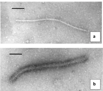
Grapevine rupestris stem pitting-associated virus [GRSPaV; genus Foveavirus; family Betaflexiviridae (King et al., 2012)] is a graft-transmissible virus (Fig, 2) widely distributed
in many grape-growing regions around the world (Minafra and Boscia, 2003).
The positive-sense, single-stranded RNA of GRSPaV is 8725 nucleotides (nt) in length, excluding the poly A tail, and encodes five open reading frames (ORFs) (Meng et al. 1998; Zhang et al. 1998) (Fig. 3). The 3'-most ORF (ORF 5) encodes a 28 kiloDalton (kDa) coat protein (CP), whereas the 5'-most ORF (ORF 1) encodes the viral replicase
Figure 2. Particles of non-decorated (a) and decorated (b) Grapevine
Rupestris Stem Pitting-associated virus observed by Immunosorbent electron microscopy (Petrovic et al., 2003)
a
polyprotein of 244 kDa. ORFs 2, 3 and 4 encode polypeptides of 24, 13 and 8 kDa, respectively, which together constitute the ‘triple gene block’ (TGB) proteins TGBp1, TGBp2 and TGBp3 (Meng et al., 1998). Recent studies have shown that TGBp1 has both a cytosolic and nuclear distribution, whereas both TGBp2 and TGBp3 are associated with the endoplasmic reticulum (Rebelo et al., 2008). A putative ORF6 encoding a 14kDa protein of unknown functions, partially overlapping the CP, has been reported in some variants of GRSPaV (Meng et al., 1998; Zhang et al., 1998; Lima et al., 2006a).
Grapevine (Vitis spp.) is the only known natural host of GRSPaV. The virus can be spread via vegetative propagation and grafting (Minafra and Boscia, 2003) and possibly through seeds of infected grapevine (Lima et al., 2006a). Although GRSPaV has been detected in the pollen of infected grapevines (Rowhani et al., 2000), its spread through pollen is not confirmed. The virus is not transmissible by mechanical inoculations and no biological vector has been reported. Studies have indicated the biological association of GRSPaV with rupestris stem pitting (RSP), one of the 4 disorders of the rugose wood complex (Meng et al., 1998; Zhang et al., 1998). RSP shows pitting symptoms on the woody cylinder below the graft union in ‘St. George’ grapevines (V. rupestris Scheele).
GRSPaV occurs in nature as a family of molecular variants (Martelli and Boudon Padieu, ORF 1
ORFs 2-3-4
ORF 5
Figure 3. Genome organization of GRSPaV. Each block arrow represents an Open Reading Frame (ORF).
ORFs with same colour have similar functions. Domains of the replicase (M= Methyltransferase, A= AlkB, O= Out-like peptidase, P= Papain-like protease, H= RNA helicase, R= RNA-dependent RNA polymerase) are shown. The map was drawn based on the complete genome sequence available on GenBank (AF057136).
al. 2003; Terlizzi and Credi 2003; Lima et al. 2006a; Habili et al., 2005; Nolasco et al.,
2006; Nakaune et al., 2008).
Recently, new groups of GRSPaV variants have been described. Goszczynski (2010), analyzing a 199 base pairs (bp) fragment of the RdRP domain in ORF 1, clustered GRSPaV variants from South Africa in 5 groups. Terlizzi et al. (2011), using a new broad-spectrum primer set designed in the polymerase domain of ORF 1, described 7 groups.
Moreover, several studies of different grapevine diseases suggested that such variants might have a diverse pathological role in RSP (Meng et al., 2005), VN (Bouyahia et al., 2006) and Syrah decline (Lima et al., 2006b) aetiology, some of which may not elicit RSP symptoms when graft-inoculated on the indicator ‘St George’ (Meng et al., 2005). In addition, some variants that are latent in Vitis vinifera cultivars and in most American
Vitis species and hybrids can produce necrosis of the veinlets when graft-inoculated on
110R (Bouyahia et al., 2005). In recent years, GRSPaV, along with a novel marafivirus and several viroids, has been documented in Californian Syrah grapevines showing decline symptoms (Al Rwahnih et al., 2009), although the role of GRSPaV in Syrah decline is yet to be resolved.
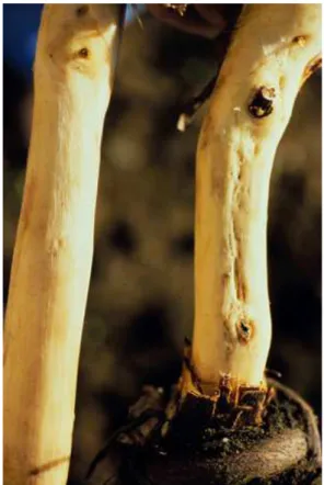
Association with RSP
Rupestris stem pitting (RSP) is a syndrome of the RW complex, which causes a distinctive basipetal pitting in the wood extending downward from the graft union (visible after the removal of the cortex), with consequent reduction of vigor of the plant (Fig. 4).
Symptoms occur only on V. rupestris St. George and a few other rootstocks related to V.
rupestris. Despite RSP is the most widespread RW syndrome, information about its
etiology were rather scarce until a few years ago.
Although the transmissibility of RSP for grafting let assume the viral nature of the disease, only researches conducted in 1998 permitted to make a significant step forward, with the identification of a virus closely associated with RSP, called Grapevine Rupestris Stem Pitting-associated Virus (GRSPaV), described for the first time as the probable causal agent of this syndrome (Zhang et al., 1998).
The cause-effect relationship between GRSPaV and RSP remains, however, still to be completely elucidated. In recent years, thanks to the development of more powerful diagnostic protocols for the detection of GRSPaV by RT-PCR and Western blot, many authors detected GRSPaV in plants that indexed negative to RSP. In detail, the use of the two diagnostic techniques, singularly or in combination, on accessions negative to the biological indexing, allowed to highlight that a significant percentage of these plants, between 22.0% and 66.0%, were infected by GRSPaV. (Meng et al., 1999a; Meng et al., 2000; Nolasco et al., 2000; Meng and Gonsalves, 2003; Bouyahia et al., 2005).
The finding, by Meng et al. (2005), that a variant of GRSPaV, detected on V. rupestris St.
Figure 4. Rupestris stem pitting symptoms.
Healthy (left) and RSP-positive (right) trunks; bark was removed to reveal pitting and grooving symptoms (photo by University of California, Integrated Viticulture).
Association with “Syrah decline”
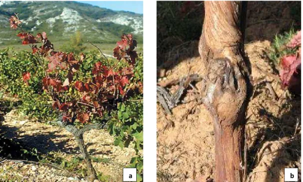
The " Syrah decline" is a disease that was firstly described in France (Renault-Spilmont et
al., 2002), the symptoms of which appear on vines of the variety "Syrah" or "Shiraz",
causing leaf reddening, scorching, swollen graft unions, and stem necrosis symptoms (Fig. 5); generally the scion dies after one or two years, while the rootstock continues to grow.
Nowadays the aetiology of these symptoms has not yet been clarified. In the last years, a growing number of vineyards in many grape-growing countries have been reporting disorders with vines of the variety "Syrah". The symptoms exhibited by plants are similar to those of the "Syrah decline", with abnormalities at the grafting point, early reddening of the leaves and slow maturation of the fruits, which have a low concentration of sugars.
In Australia (Habili et al., 2005) and California (Lima et al., 2006b), GRSPaV was detected in plants of V. vinifera “Syrah” presenting symptoms of "Syrah Decline".
Figure 5. Syrah decline symptoms: (a) leaf reddening affecting the entire canopy of an infected
accession and (b) swelling and cracking at the graft union (Battany et al., 2004)
Analysis conducted by RT-PCR on extracts of leaf petioles of “Syrah” vines from California to verify the presence of all the major grapevine-infecting viruses have indicated the presence of only GRSPaV (Lima et al., 2006b).
The genome of this variant of GRSPaV , called GRSPaV-SY , was composed of 8725 nucleotides and organized in 6 ORFs.
The nucleotide sequence of GRSPaV-SY had a homology of 77.0% compared to the sequences of 4 reference variants already known (GRSPaV , GRSPaV-1, GRSPaV-SG1, GRSPaV-BS), and these 4 isolates are much more related to each other than with GRRPaV-SY.
The capsid protein gene (ORF 5) of GRSPaV-SY is the region of the genome that has the highest nucleotide and aminoacidic homology (83.0-84.0% and 91.0-92.0%, respectively) with the sequence of other variants of GRSPaV, while the replicase gene (ORF 1) is the more variable region (75.0-76.0% nucleotide and 85.0% aminoacidic homology).
A similar survey was conducted in Australia, on plants of V. vinifera ‘Syrah’ presenting symptoms similar to those of the "Syrah decline" (Habili et al., 2005). Even in this case, by RT-PCR was rediscounted the presence of a variant of GRSPaV in symptomatic plants. The analysis and comparison of the nucleotide sequence of a region of 628 bp of the gene for replicase indicates a low nt identity between this variant and the other GRSPaV isolates previously reported.
Nonetheless, recent papers suggest that frequent presence of this variant in Syrah decline-affected plants, reported by Habili et al. (2005) and Lima et al. (2006b), could may be only accidental and seem to reject the association of GRSPaV and GRSPaV-SY variant in the aetiology of Syrah decline (Goszczynski , 2010; Beuve et al., 2013).
Association with vein necrosis disease
Studies carried out in Apulia in 2005, showed a high presence of GRSPaV in infected plants by VN , suggesting that this foveavirus may be the correlated to the aetiology of
Immunosorbent Assays (ELISA tests) to check the presence of the 8 main grapevine viruses: grapevine fanleaf nepovirus (GFLV), grapevine leafroll associated ampelovirus 1 (GLRaV-1), grapevine leafroll associated closterovirus 2 (GLRaV-2), grapevine leafroll associated ampelovirus 3 (GLRaV-3), grapevine leafroll associated closteroviridae 7 (GLRaV-7), grapevine vitivirus A (GVA), grapevine vitivirus B (GVB) and grapevine fleck maculavirus (GFkV). Only 16 (7.3%) of 218 samples tested were found to be infected by at least one of the 8 viruses and, moreover, the ELISA-positive samples were equally divided between plants positive or not to VN. Further investigations, carried out by RT-PCR, allowed to detect an extremely high presence of GRSPaV. In detail, among 174 plants surveyed , 93 of which indexed positive and 81 of which indexed negative to VN , GRSPaV was founds in as many as 91 (97.8 %) of the 93 VN-positive plants and only in one of the negative ones, therefore showing a close association between the foveavirus GRSPaV and VN.
Recently, studies about the association of VN and GRSPaV were carried out in our department, conducted on grapevine accessions collected in Tuscany during clonal and sanitary improvement programs. Each of these accessions were previously tested by ELISA for the 9 main viruses infecting grapevine (GFLV, arabis mosaic nepovirus (ArMV), GFkV, GVA, GVB, GLRaV-1, GLRaV-2, GLRaV-3 and GLRaV-7) and biologically indexed on specific Vitis indicators for the presence of grapevine degeneration, leafroll, fleck, Kober stem grooving, LN33 stem grooving, Rupestris stem pitting, corky bark, enation, vein necrosis and vein mosaic diseases.
Molecular assay (RT-PCR) for GRSPaV detected the virus in 53 out of 62 plants, 38 (71.7%) of which presented specific VN symptoms. In order to understand the reason of the VN-negative, but GRSPaV-positive accessions, each sample was tested with PCR primers able to amplify 3 distinct molecular groups of the virus, named G I, G II, and G III (Rowhani et al., 2000).
The results obtained suggested a differential role of GRSPaV variants in VN aetiology, with all of the 38 plants infected by G I variants that were positive to the VN indexing (Bouyahia et al., 2006).
These findings, besides confirming the relationship between GRSPaV and VN, let hypothesize that only a group a GRSPaV variants could be involved in the aetiology of this disease. Since then, studies about GRSPaV molecular characterization and this virus-disease association were carried out, and are actually ongoing, on a larger sample size of different geographical origin, in order to better clarify the role of GRSPaV in the determinism of VN disease.
1.3 Overall objectives
The aims of this research comprise the detection of GRSPaV and its genetic variants in grapevine accessions from Tuscany and California, their molecular and serological characterization and the investigation of the pathogenetic role of different virus variants in VN symptoms expression.
This has been achieved through the following steps:
- Detection of different GRSPaV variants in the grapevine accessions object of the
study.
- Amplification, cloning and sequencing of ORF5 and a portion of ORF1 and
phylogenetic analysis.
- Serological detection of different virus variants by Western Blot.
- Biological indexing of grapevine accessions infected by different groups of
2. Molecular detection of Grapevine Rupestris Stem
Pitting-associated Virus in grapevine accessions
2.1 Introduction
GRSPaV is a member of the genus Foveavirus in the newly established family
Betaflexiviridae (King et al., 2012). It is not mechanically transmissible (Martelli and Jelkmann, 1998) and no natural vector has been found until now. GRSPaV has been reported from almost all vine growing areas in the world, where it seems to have a high incidence. The aetiological role of GRSPaV, however, remains to be elucidated.
Data regarding the genomic variability of RSPaV can be found in several papers (Meng et
al., 1999b; Meng et al., 2000; Rowhani et al., 2000; Soares et al., 2000; Casati et al.,
2003; Lima et al., 2003; Santos et al., 2003; Terlizzi and Credi 2003). Although different primers operating in different genomic regions of the virus were adopted for these studies, many authors quite consistently clustered GRSPaV variants in 3 or 4 groups. Rowhani et al. (2000) designed, in the CP gene of GRSPaV, 3 sets of group-specific primers able to amplify different virus variants.
The availability, in our department, of an experimental collection field of grapevine accessions collected throughout Tuscany, made it possible to further investigate the relationship between GRSPaV and VN disease.
In the present study, we performed two-step RT-PCR diagnosis of GRSPaV on RNA extracted from phloem scrapings with both universal and group-specific primer sets. The aims of the work were:
a) to check the efficiency of universal primers for GRSPaV detection;
b) to discover if one or more GRSPaV variants groups infect grapevine accessions in Tuscany;
c) to select “pure sources” grapevine accessions infected by only one GRSPaV group, in order to study the biological properties of single virus variants.
2.2 Materials and Methods
Plant materialObject of the study were 99 grapevine accessions, comprising both V. vinifera and rootstock varieties (Tab. 2).
62 of these accessions were collected in different locations of Tuscany within clonal and sanitary improvement programmes and are being held in a collection field in the Department of Food Agriculture and Environment (DAFE) at the University of Pisa. The collection comprises rootstocks V. rupestris, V. berlandieri x V. riparia Kober 5BB and V.
vinifera varieties Sangiovese, Aleatico, Ciliegiolo, Tenerone, Mammolo, Colorino,
Verdello, Prugnolo gentile, Canaiolo.
All of these grapevine plants previously tested negative to ELISA tests for the presence of 9 viruses associated with major detrimental diseases: GFLV, ArMV, GFkV, GLRaV-1, GLRaV-2, GLRaV-3, GLRaV-7, GVA and GVB. Each putative clone was also indexed on 6 to 8 replicates of each of the following 6 indicators: (I) V. vinifera cv. Cabernet franc for leafroll; (II) LN 33 (Couderc 1613 x V. berlandieri), for corky bark, LN 33 stem grooving and enation disease; (III) Kober 5BB (V. berlandieri x V. riparia), for Kober stem grooving; (IV) V. rupestris cv. St. George, for fanleaf, fleck and rupestris stem pitting; (V) 110R for VN; (VI) V. riparia Gloire de Montpellier for vein mosaic (Triolo and Materazzi, 2000; Triolo and Materazzi, 2004).
No symptomatic responses from any of the indexed accession was recorded for leafroll, grapevine degeneration, corky bark, LN33 stem grooving, Kober stem grooving, rupestris stem pitting, enation disease and vein mosaic, but when indexed on 110 Richter, 38 of the 62 accessions showed VN symptoms. (Triolo and Materazzi, 2000; Triolo and Materazzi, 2004).
Table 2. List of the grapevine accessions object of the study, species/hybrid and the place of the
collection field where they have been maintained.
Accession Species / hybrid Collection at:
5V-20-1 V. rupestris S. Piero a Grado, Pisa
5V-20-2 V. rupestris S. Piero a Grado, Pisa
5V-20-3 V. rupestris S. Piero a Grado, Pisa
5V-20-4 V. rupestris S. Piero a Grado, Pisa
5V-20-5 V. rupestris S. Piero a Grado, Pisa
RUP1 V. rupestris S. Piero a Grado, Pisa
BSK3-1 V. berlandieri x V. riparia S. Piero a Grado, Pisa
BSK3-2 V. berlandieri x V. riparia S. Piero a Grado, Pisa
BSK3-3 V. berlandieri x V. riparia S. Piero a Grado, Pisa
BSK1 V. berlandieri x V. riparia S. Piero a Grado, Pisa
1/18 V. vinifera S. Piero a Grado, Pisa
2/1 V. vinifera S. Piero a Grado, Pisa 5/1 V. vinifera S. Piero a Grado, Pisa
34/46 V. vinifera S. Piero a Grado, Pisa
AL.CAV.1 V. vinifera S. Piero a Grado, Pisa
AL.CAV.4 V. vinifera S. Piero a Grado, Pisa
AL.CAV.5 V. vinifera S. Piero a Grado, Pisa
CA V. vinifera S. Piero a Grado, Pisa CC4 V. vinifera S. Piero a Grado, Pisa CC5 V. vinifera S. Piero a Grado, Pisa CC6 V. vinifera S. Piero a Grado, Pisa CC8 V. vinifera S. Piero a Grado, Pisa
CC10 V. vinifera S. Piero a Grado, Pisa
CC13 V. vinifera S. Piero a Grado, Pisa
CC17 V. vinifera S. Piero a Grado, Pisa
CC19 V. vinifera S. Piero a Grado, Pisa
CC21 V. vinifera S. Piero a Grado, Pisa
CC24 V. vinifera S. Piero a Grado, Pisa
CC25 V. vinifera S. Piero a Grado, Pisa
CC26 V. vinifera S. Piero a Grado, Pisa
CC29 V. vinifera S. Piero a Grado, Pisa
CHI8 V. vinifera S. Piero a Grado, Pisa
CIL.SIRIO2 V. vinifera S. Piero a Grado, Pisa
COR20 V. vinifera S. Piero a Grado, Pisa
COR33 V. vinifera S. Piero a Grado, Pisa
COR35 V. vinifera S. Piero a Grado, Pisa
COR36 V. vinifera S. Piero a Grado, Pisa
FA V. vinifera S. Piero a Grado, Pisa
GALLO5 V. vinifera S. Piero a Grado, Pisa
GALLO7 V. vinifera S. Piero a Grado, Pisa
GALLO15 V. vinifera S. Piero a Grado, Pisa
KA V. vinifera S. Piero a Grado, Pisa
LUM01 V. vinifera S. Piero a Grado, Pisa
MSAS1 V. vinifera S. Piero a Grado, Pisa
MSAS3 V. vinifera S. Piero a Grado, Pisa
MLOB7 V. vinifera S. Piero a Grado, Pisa
MLOB9 V. vinifera S. Piero a Grado, Pisa
MLOC2 V. vinifera S. Piero a Grado, Pisa
MLOC15 V. vinifera S. Piero a Grado, Pisa
Accession Species / hybrid Collection at:
SGSAS2 V. vinifera S. Piero a Grado, Pisa
SGSAS3 V. vinifera S. Piero a Grado, Pisa
SGSAS4 V. vinifera S. Piero a Grado, Pisa
SG.SIRIO1 V. vinifera S. Piero a Grado, Pisa
SM6/10 V. vinifera S. Piero a Grado, Pisa
SME9 V. vinifera S. Piero a Grado, Pisa
SMF13 V. vinifera S. Piero a Grado, Pisa
SMH22 V. vinifera S. Piero a Grado, Pisa
TENSAS1 V. vinifera S. Piero a Grado, Pisa
V6F12 V. vinifera S. Piero a Grado, Pisa
WA V. vinifera S. Piero a Grado, Pisa YA V. vinifera S. Piero a Grado, Pisa
Albillo Mayor 01 V. vinifera FPS Davis , California
Albillo Real 01 V. vinifera FPS Davis , California
Arinarnoa 01 V. vinifera FPS Davis , California
B2R24P69 01 V. vinifera FPS Davis , California
B2R45P72 01 V. vinifera FPS Davis , California
Barbera V. vinifera FPS Davis , California
Cabernet Franc 14 V. vinifera FPS Davis , California
Cabernet Sauvignon 31 V. vinifera FPS Davis , California
Cabernet Sauvignon 43 V. vinifera FPS Davis , California
Chardonnay 68 V. vinifera FPS Davis , California
Chardonnay 72 V. vinifera FPS Davis , California
Chardonnay 83 V. vinifera FPS Davis , California
Diamond Muscat V. vinifera FPS Davis , California
Green Hungarian V. vinifera FPS Davis , California
Inzolia V. vinifera FPS Davis , California
Kandhara V. vinifera FPS Davis , California
Madrone Moon Farm V. vinifera FPS Davis , California
Malaga V. vinifera FPS Davis , California
Merlot 18 V. vinifera FPS Davis , California
Mourvedre V. vinifera FPS Davis , California
Negrette V. vinifera FPS Davis , California
Olmo 1266 V. vinifera FPS Davis , California
Pinot Noir 22 V. vinifera FPS Davis , California
Pinot Noir 54 V. vinifera FPS Davis , California
Pinot Noir 78 V. vinifera FPS Davis , California
Pinot Noir 85 V. vinifera FPS Davis , California
Refosco 03 V. vinifera FPS Davis , California
Ribolla Gialla V. vinifera FPS Davis , California
Riesling V. vinifera FPS Davis , California
Sangiovese 13 V. vinifera FPS Davis , California
Schioppettino V. vinifera FPS Davis , California
Thomcord 02 V. vinifera FPS Davis , California
Tinta Amarella V. vinifera FPS Davis , California
Trousseau V. vinifera FPS Davis , California
Sampling, RNA extraction and cDNA synthesis
Mature canes were collected from different part of the canopy in order to minimize possible variations due to uneven distribution of the virus in infected tissues.
Total RNA was extracted according to MacKenzie et al. (1997), with slight modifications. Briefly, 0.5 g of plant material was macerated in a plastic bag in presence of 5 ml grinding buffer containing 4 M guanidine isothiocyanate, 0.2 M sodium acetate pH 5,2, 5 mM EDTA, 2.5% (wt/vol) PVP-40 and 1% (vol/vol) 2-mercaptoethanol (added just before use). 2 ml of the homogenate was recovered and centrifuged at full speed for 5 minutes. 1 ml of the lysate was transferred into a new tube, mixed with 100 µl of 20% (wt/wol) sarkosyl and incubated at 70 °C for 10 minutes with intermittent shaking. Approximately 600 to 650 µl of homogenate was transferred to a QIAShredder column (RNeasy Plant Mini Kit, Qiagen) and the RNA extraction procedure continued according to the kit manufacturer’s protocol. Total RNA was eluted from the RNeasy column using 50 µl of RNase-free sterile water and stored at -20 °C.
For synthesis of first-strand cDNA, 10 μl of RNA extract were mixed with 2 μl of random hexamer primers (Roche) (0.5 μg/μl), denatured at 95 °C for 5 min and quickly cooled on ice for 2 min. Reverse transcription was done for 1 h at 37 °C by adding 10 μl AMV Reverse Transcriptase 5X Reaction Buffer (Promega), 2.5 μl of 10 mM dNTPs mix, and 5 units Avian Myeloblastosis Virus (AMV) reverse transcriptase (Promega, USA) in a final volume of 50 μl.
GRSPaV diagnosis with broad spectrum primers
Molecular GRSPaV detection was performed by PCR using 2 sets of broad spectrum primers, aiming to understand if they would produce identical results, or if one primer set would be able to better detect GRSPaV.
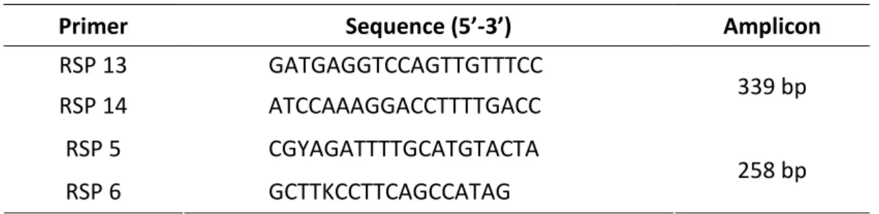
Primers RSP13/RSP14 amplify a portion of the helicase domain in ORF1 (Meng et al., 1999a), while RSP 5/6 (Santos et al., 2006) are designed on ORF5, encoding the CP (Tab. 3).
Table 3. Broad-spectrum primers for the detection of GRSPaV used in this study, 5’-3’ sequences and
amplicon length.
Primer Sequence (5’-3’) Amplicon
RSP 13 GATGAGGTCCAGTTGTTTCC
RSP 14 ATCCAAAGGACCTTTTGACC 339 bp
RSP 5 CGYAGATTTTGCATGTACTA
RSP 6 GCTTKCCTTCAGCCATAG 258 bp
5 µl of cDNA were added to PCR mixture yielding final concentrations of 1x GoTaq Buffer
(Promega, USA), 1.5 mM MgCl2, 0.4 mM each deoxynucleotide triphosphate, 0.2 µM
each primer and 1 U Taq DNA Polymerase (Promega, USA) per reaction, in a total volume of 25 µl.
PCR reactions were performed with the following parameters: 3 min at 95 °C followed by 35 cycles of 30 sec at 94 °C, 30 sec at 52 °C, and 35 sec at 72 °C, followed by a final extension of 7 min at 72 °C. PCR products (10 μl) were analyzed on a 1.5% agarose gel, stained with GelRed (Biotium, USA) and visualized under UV light.
GRSPaV diagnosis with groups specific primers
Following GRSPaV diagnosis, all of the 99 accessions were tested by PCR with GRSPaV group-specific primers (Rowhani et al., 2000), whose sequences were kindly supplied by Prof. Rowhani (University of California - Davis, USA). These degenerated primers, designed on ORF 5, are able to specifically amplify 3 distinct groups of molecular variants.
5 µl of cDNA were used in 25 µl PCR reactions prepared as previously described, with thermal cycle as follows: 3 min at 95 °C followed by 35 cycles of 30 sec at 94 °C, 30 sec at 50 °C, and 35 sec at 72 °C, followed by a final extension of 7 min at 72 °C.
2.3 Results
GRSPaV diagnosis with broad spectrum primers
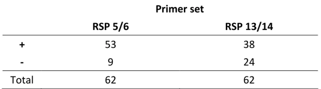
Products of the expected size, 258 bp and 339 bp, were observed in agarose gels using primers RSP 5/6 and RSP 13/14, respectively.
In total, 99 samples from V. vinifera varieties and/or rootstocks were assayed by two-step RT-PCR.
- Samples from Tuscany
Samples were tested with both primer pair RSP 13/14 and RSP 5/6. When using primers RSP 13/14, GRSPaV was detected in 38 accessions (61.2%), while primer set RSP 5/6 was able to detect the virus in 53 (85.5%) samples, including all of the 38 previously positive to RSP 13-14. The remaining 9 accessions (14.5%) provided negative results with both primer pairs (Fig. 6; Tab. 4).
Interestingly, all of the accessions previously symptomatic to VN, tested positive to GRSPaV with primers RSP 13/14. Also, negative results were obtained with primers RSP 13/14 when testing accessions that resulted negative to VN indexing.
a b c d e f g
Figure 6. Results of RT-PCR with primer pairs RSP 13/14 (up) and RSP 5/6 (down) on agarose gel
visualized by UV light. Samples, from left to right: a=Gallo7; b=Gallo15; c=SMH22; d=MLOB9; e=COR24;
Primer set
RSP 5/6 RSP 13/14
+ 53 38
- 9 24
Total 62 62
- Samples from California
Accessions were tested with primer set RSP 5/6. All of the 37 accessions, as expected, showed the specific amplicon of 258 bp, resulting infected by GRSPaV.
- Overall results of GRSPaV detection
In total, GRSPaV was detected in 90 of the 99 tested plants, both in rootstock hybrids and V. vinifera varieties.
GRSPaV diagnosis with groups specific primers
Results obtained with group-specific primers matched those achieved with primer pair RSP 5/6. In fact, all of the plants tested positive with RSP 5/6 showed positive reaction with at least one of the group-specific primer pairs, while no one of the GRSPaV-negative accessions produced amplicons of the expected size.
All of the 3 different GRSPaV groups were observed in the accessions tested. GRSPaV variants detected by G I-specific primers were the most present and were detected in 53 accessions (58.9 %). Both group G II and G III variants were present in 46 (51.1%) of the 90 GRSPaV-infected plants (Fig. 7). The 3 groups, however, were quite evenly distributed (Fig. 8).
Table 4. Results obtained with “broad-spectrum” primer sets RSP 5/6 and RSP
0% 10% 20% 30% 40% 50% 60%
- Samples from Tuscany
Limited to the 53 GRSPaV-positive accessions from Tuscany, group G I and G III were the most widespread infecting, respectively, 38 (71.7 %) and 35 (66.0 %) plants. Group G II
M a b c d e f g h i j k l m n
M a b c d e f g h i j k l m n
Figure 8. Incidence of the 3 groups of GRSPaV variants amplified by each
group-specific primer set on the total amount of GRSPaV-infected plants
Figure 7. Results of RT-PCR with G I, G II and G III group-specific primer pairs on agarose gel visualized by
UV light. Samples, from left to right: M=marker a=Pinot Noir85, b=Sangiovese13, c=Refosco03,
d=Arinarnoa01, e=B2R24P69 01, f=Pinot Noir22, g=Zinfandel09, h=Olmo1266, i=Schioppettino, j=Thomcord02, k=Albillo Mayor01, l=Albillo Real01, m=Cabernet Franc14, n=Cabernet Sauvignon43.
G I F/R G II F/R G III F/R
G I
G II
0% 10% 20% 30% 40% 50% 60% 70% 80% 20% 30% 40% 50% 60%
variants were detected in a lower number of samples (24), representing 45.3% of the infected plants (Fig. 9).
It is interesting to observe that group G I variants were detected, singularly or in mixed infection, in all and only the 38 samples providing positive results to the biological indexing for VN and that previously tested positive to GRSPaV primer set RSP13/14.
- Samples from California
The distribution of GRSPaV variants in California differed from the situation in Tuscany, with group G II being predominant. It was detected in 21 out 37 accessions (56.8 %), while groups G I and G III were less widespread and were detected in 15 (40.5%) and 11 (29.7%) samples respectively (Fig. 10).
Figure 9. Incidence of group G I, G II, and G III infections on the
total amount of GRSPaV infected accessions from Tuscany
- Mixed infections
The occurrence of mixture of GRSPaV variants in the same isolate, as already described by Nolasco et al. (2006), was frequently observed in 44 (48.9 %) infected accessions. Mixed infection by variants of G I and G III was the most common, as it was detected in 19 (21.1%) out of 90 GRSPaV-infected accessions and a considerable amount of 11 samples (12.2 %) showed mixed infection by variants belonging to all of the 3 groups. Groups G II + G III and G I + G II infections were detected in 8 (8.9 %) and 6 (6.6 %) plants, respectively (Fig. 11).
Details of the results obtained with each primer pair are listed in table 5.
Figure 11. Incidence of different single and/or mixed infection combinations on the total
amount of GRSPaV infected plants.
G I (18.9%) G II (23.3%) G III (8.9%) G I + G II (6.6%) G I + G III (21.1%) G II + G III (8.9%) G II + G III (12.2%)
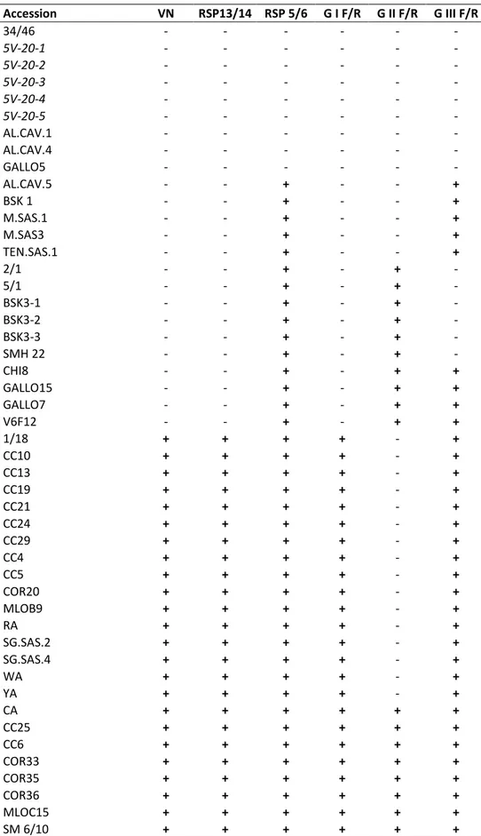
Accession VN RSP13/14 RSP 5/6 G I F/R G II F/R G III F/R 34/46 - - - - 5V-20-1 - - - - 5V-20-2 - - - - 5V-20-3 - - - - 5V-20-4 - - - - 5V-20-5 - - - - AL.CAV.1 - - - - AL.CAV.4 - - - - GALLO5 - - - - AL.CAV.5 - - + - - + BSK 1 - - + - - + M.SAS.1 - - + - - + M.SAS3 - - + - - + TEN.SAS.1 - - + - - + 2/1 - - + - + - 5/1 - - + - + - BSK3-1 - - + - + - BSK3-2 - - + - + - BSK3-3 - - + - + - SMH 22 - - + - + - CHI8 - - + - + + GALLO15 - - + - + + GALLO7 - - + - + + V6F12 - - + - + + 1/18 + + + + - + CC10 + + + + - + CC13 + + + + - + CC19 + + + + - + CC21 + + + + - + CC24 + + + + - + CC29 + + + + - + CC4 + + + + - + CC5 + + + + - + COR20 + + + + - + MLOB9 + + + + - + RA + + + + - + SG.SAS.2 + + + + - + SG.SAS.4 + + + + - + WA + + + + - + YA + + + + - + CA + + + + + + CC25 + + + + + +
Table 5. Results of RT-PCR with each broad-spectrum and group-specific primer pair,
Accession VN RSP13/14 RSP 5/6 G I F/R G II F/R G III F/R SME 9 + + + + + + SMF 13 + + + + + + CC17 + + + + - - CC8 + + + + - - CIL.SIRIO2 + + + + - - MLOC2 + + + + - - LUM01 + + + + - - RUP 1 + + + + - - SG.SAS.3 + + + + - - SG.SIRIO1 + + + + - - CC26 + + + + + - FA + + + + + - KA + + + + + - MLOB7 + + + + + - Mourvedre n.d. n.d. + - - + Diamond muscat n.d. n.d. + - - + Chardonnay 72 n.d. n.d. + - - + Barbera n.d. n.d. + - + + Trousseau Gris n.d. n.d. + - + + Ribolla gialla n.d. n.d. + - + + B2R24P69 01 n.d. n.d. + - + + Negrette n.d. n.d. + - + - Tinta Amarella n.d. n.d. + - + - Malaga n.d. n.d. + - + - Green Hungarian n.d. n.d. + - + - Kandhara n.d. n.d. + - + -
Madrone Moon Farm n.d. n.d. + - + -
Cabernet sauvignon 31 n.d. n.d. + - + - B2R45P72 01 n.d. n.d. + - + - Pinot noir 78 n.d. n.d. + - + - Zinfandel 09 n.d. n.d. + - + - Olmo 1266 n.d. n.d. + - + - Schioppettino n.d. n.d. + - + - Thomcord 02 n.d. n.d. + - + - Albillo mayor 01 n.d. n.d. + - + - Cabernet sauvignon 43 n.d. n.d. + - + - Merlot 18 n.d. n.d. + + - + Sangiovese 13 n.d. n.d. + + - + Arinarnoa 01 n.d. n.d. + + - + Riesling n.d. n.d. + + + + Inzolia n.d. n.d. + + - - Zinfandel n.d. n.d. + + - - Pinot noir 54 n.d. n.d. + + - - Chardonnay 68 n.d. n.d. + + - - Barbera 07 n.d. n.d. + + - - Chardonnay 83 n.d. n.d. + + - - Pinot noir 85 n.d. n.d. + + - - Refosco 03 n.d. n.d. + + - - Pinot noir 22 n.d. n.d. + + - - Albillo real 01 n.d. n.d. + + + - Cabernet franc 14 n.d. n.d. + + + -
2.4 Discussion
Based on the results obtained by GRSPaV detection with broad-spectrum and group-specific primers, we confirm the previously reported association between VN and the foveavirus Grapevine rupestris stem pitting associated virus.
GRSPaV is very common in grapevine, and accessions from Tuscany showed an infection rate by GRSPaV of 85.5 %.
The results obtained with the group-specific primers show that all of the 3 molecular groups of the virus are present in Tuscany and in the accessions held at Foundation Plant Services (University of California – Davis).
Interestingly, combining the results previously obtained with VN indexing of the Tuscan grapevine accessions (Triolo and Materazzi, 2000; Triolo and Materazzi, 2004) with the outcome of RT-PCR analyses, only virus isolates belonging to group G I appear closely related to VN. In fact, group-specific primer set G I detected the virus in all and only the 38 VN+ accessions.
Infections with groups G II and/or G III, recorded in vein necrosis free accessions, suggest that such groups are unable to induce vein necrosis. Their presence in vein necrosis infected accessions was always associated to the presence of group G I.
About the possibility to diagnose VN by RT-PCR, it could be proposed to use the pair of primers RSP 13/14 or G I, as they were able to specifically detect the “pathological strain” of GRSPaV in VN infected accessions.
On the other side, primers RSP 5/6 were more effective in the detection of all GRSPaV molecular variants; they could be employed as universal primers for the diagnosis of all the virus variants. However, positive samples obtained with these primers will include both VN-infected samples and accessions positive to Group II and III.
Although other elements are needed to obtain further and conclusive information about the aetiology and the pathological role of the GRSPaV molecular groups, these results
3. Molecular and serological characterization
of ORF 5 (CP gene)
3.1 Introduction
3.1.1 Molecular characterization
A high degree of sequence diversity was documented since when the first GRSPaV isolates were being sequenced. Meng et al. (1999b) found GRSPaV to consist of a heterogeneous population of sequence variants sharing between 75-99% identities, still having identical genome structures. It was also found that this heterogeneous population separated into distinct groups of viral variants and that the incidence of mixed infection in a single vine with different viral variants of GRSPaV is high. By looking at several sources of V. vinifera, it was discovered that the presence of sequence variants is independent of genotype or geographic origin of the host plant, which raised further questions in terms of transmission and dissemination of this ubiquitous virus. The findings outlined above stimulated further investigation into the genetic variability of GRSPaV from researchers around the globe.
The use of different nomenclature systems for GRSPaV molecular groups and the different genome region analyzed by independent research groups often made it difficult to compare and harmonize the phylogenetic analyses from different studies. Using primers designed to the CP of GRSPaV, several research groups identified the existence of 3 distinct groups of viral variants obtained from different geographic regions, based on coat protein sequences alone (Rowhani et al., 2000; Casati et al., 2003; Terlizzi and Credi, 2003). More recent and extensive analyses of GRSPaV genetic variability revealed the presence of 4 groups of sequence variants (Santos et al., 2003; Meng et al., 2006; Nolasco et al., 2006).
Based on the complete CP coding sequence of GRSPaV, most of the studies revealed that GRSPaV variants clustered in 4 groups, often differently named from different authors, but each of them co-incidentally comprising at least one of the full genome sequences available.
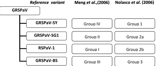
A comparison of the two most adopted nomenclatures proposed by Meng et al. (2006) and Nolasco et al. (2006) is given in Figure 12.
In recent years, the full genome of more GRSPaV isolates have been completely sequenced. Up to now, 11 full genome GRSPaV isolates, from different geographical origin, have been fully sequenced and are available in GenBank (GenBank accession
numbers: NC_001948.1; AY881626.1; AY881627.1; AF026278.1; AY368590.1;
AY368172.2; KC427107.1; JQ922417.1; FR691076.1; HE591388.1; JX559646.1), including the variant named “GRSPaV-PN” (Lima et al., 2009), which notably presents significant variability when compared with reference variants for each of the 4 GRSPaV molecular groups. GRSPaV-PN was proposed to constitute a separate group of GRSPaV variants (Terlizzi et al., 2011).
Reference variant Meng et al.,(2006) Nolasco et al. (2006)
Group IV Group II Group I Group III Group 1 Group 2a Group 2b Group 3
Figure 12. Comparison of the two main nomenclature systems for the classification of GRSPaV
variants GRSPaV GRSPaV-SY GRSPaV-SG1 RSPaV-1 GRSPaV-BS
Reference variant Meng et al.,(2006) Nolasco et al. (2006)
Group IV Group II Group I Group III Group 1 Group 2a Group 2b Group 3
3.1.2 Serological characterization
The great value of serological methods for plant virus identification is based on the specific reaction between the viral antigens and their specific antibodies. An antigen is a molecule that, when injected into a vertebrate animal (usually a mammal or a bird), it can trigger an immune response in the animal which results in production of specific antibodies that can combine with the foreign antigen (Naidu and Hughes, 2001; Purcifull
et al., 2001; Lima et al., 2005; Astier et al., 2007).
Virus particles themselves and their proteins have several antigenic determinants (epitopes) which vary in their amino acid sequence and have the properties of inducing the production of specific antibodies. The virus particles, their coat proteins and the other types of virus induced proteins can function as antigens (Hiebert et al., 1984; Naidu and Hughes, 2001; Purcifull et al., 2001; Lima et al., 2005; Astier et al., 2007; Lima
et al., 2012).
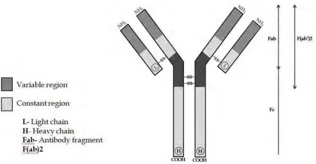
Antibodies are also proteins of the immunoglobulin group (Ig) produced against specific antigenic determinants and are present in the animal blood. The immunoglobulin G (IgG) is the most common type of Ig produced and, consequently most commonly involved in the serological tests for plant virus identification. This type of antibody is composed of four linked polypeptides with Y-shape of approximately 150 Kd, with two identical heavy chains and two identical light chains of polypeptides (Fig. 13). The IgG has two identical
combining sites specific for antigenic determinants called paratopes in the NH2 terminal
regions of the heave and light chains. These two identical combining sites have highly variable aminoacid sequence, which permit the production of specific IgGs for the different virus epitopes. The C-terminal regions of the heavy chains are linked together by sulfur bridges to produce the fragment crystallizable (Fc) fraction of the antibody (Fig. 13) which links specifically with protein A or cell membranes (Almeida et al., 2001; Purcifull et al., 2001; Lima et al., 2012).
Generally the methods that involve the antigen antibody reactions in vitro are simple and do not require sophisticated and expensive apparatus. The most serious limitation to using serology for plant virus identification and detection is the difficulty in producing a good virus-specific antiserum. Most antisera used for plant virus identification and detection are usually prepared by immunizing mammals with purified plant virus or their different types of proteins. However several other methods have been used to produce very specific antibodies, including monoclonal antibody (Mab) which consists of a single type of antibody that reacts with only one specific epitope of a virus protein.
The production of antiserum for some plant viruses is, mainly, limited by the difficulty in purifying such viruses free from plant protein contaminants and in appropriate concentration to be used as antigen. Several other methods have been developed for plant virus antigen production, including the transformation of bacterial cells with the virus coat protein gene. Although not very specific for virus species/strains
Figure 13. Diagrammatic representation of the structure of an immunoglobulin G (IgG)
molecule. Fab, F(ab)2, and Fc represent fragments obtained by enzyme cleavage of IgG. (Lima et al., 2012)
Up to date, two polyclonal antibodies to GRSPaV have been produced (Minafra et al., 2000; Meng et al., 2003), both of them against a recombinant coat protein of GRSPaV. Minafra et al. (2000) reported the use of the antibody to effectively detect GRSPaV by western blot, but not by ELISA, possibly because of a low efficiency of the antiserum in recognizing the native viral coat protein from tissue samples.
In our study, western blot assays were performed to determine antibody specificity to GRSPaV variants belonging to 4 different molecular groups.
In western blot the virus protein antigens are transferred from polyacrylamide gels in which they were previously separated by electrophoresis, to nitrocellulose or nylon membranes.
Several methods can be used to transfer the virus protein and the electro-blotting is the most used system. Similar to ELISA techniques, the proteins are detected in the membrane by the use of specific enzyme labelled antibodies (Almeida et al., 2001; Purcifull et al., 2001).
3.2 Materials and Methods
3.2.1 Molecular characterizationThe viral sources used in this study came from grapevine plants selected through the 99 accessions from Tuscany and California (Tab. 5), listed in chapter 2. A total number of 22 accessions, representative of all the possible combinations of both single and mixed GRSPaV variants infections (Tab. 6) were investigated.
Accession name G I G II G III Collected at
CC 8 + - - San Piero a Grado – Italy
MLOC2 + - - San Piero a Grado – Italy
CC17 + - - San Piero a Grado – Italy
5/1 - + - San Piero a Grado – Italy
SMH 22 - + - San Piero a Grado – Italy
MSAS1 - - + San Piero a Grado – Italy
MSAS3 - - + San Piero a Grado – Italy
Barbera 07 + - - FPS UCDavis – California
Pinot Noir 54 + - - FPS UCDavis – California
Refosco 03 + - - FPS UCDavis – California
Chardonnay 68 + - - FPS UCDavis – California
Chardonnay 83 + - - FPS UCDavis – California
Inzolia + - - FPS UCDavis – California
Cabernet Sauvignon 43 - + - FPS UCDavis – California
Thomcord 02 - + - FPS UCDavis – California
Tinta Amarella - + - FPS UCDavis – California
Mourvedre - - + FPS UCDavis – California
Diamond Muscat - - + FPS UCDavis – California
Albillo Real 01 + + - FPS UCDavis – California
Arinarnoa 01 + - + FPS UCDavis – California
B2R24P69 - + + FPS UCDavis – California
Riesling + + + FPS UCDavis – California
RNA extraction from phloem scrapings and cDNA synthesis was performed according to the protocol previously described in Chapter 2.
Primers RSP 52/53 (Rowhani et al., 2000) were used to amplify a 905 bp amplicon,
Table 6. List of the 22 grapevine accessions representative of every possible
each primer and 1 U Taq DNA Polymerase (Promega, USA) per reaction, in a total volume of 50 µl.
PCR reactions were performed with the following parameters: 3 min at 95 °C followed by 40 cycles of 30 sec at 94 °C, 45 sec at 52 °C and 1 min 30 sec at 72 °C, followed by a final extension of 10 min at 72 °C. 10 µl of the PCR products were analyzed on a 1.5% agarose gel, stained with GelRed (Biotium, USA) and visualized under with light.
Samples from Tuscany
Bands of the expected size (905 bp) were excised from the gel and PCR products were purified with Wizard SV Gel and PCR Clean-Up System kit (Promega, USA) according to the manufacturer’s protocol.
The resulting DNA solution was used in cloning reactions, using pGEM®-T Easy vector System II (Promega, USA) (Fig. 14).
Ligation reactions were set up by mixing 3 μl of DNA purified from agarose gel, 5 μl of 2x Ligation Buffer (60 mM Tris-HCl, pH 7.8, 20 mM MgCl2, 20 mM DTT, 2 mM ATP, 10%
polyethylene glycol), 1 μl pGEM®-T Easy Vector (50 ng), 1 μl of T4 DNA Ligase enzyme (3 units/µl) and sterile water to a final volume of 10 μl. The reactions were incubated overnight at 4°C.
2 µl of each ligation reaction were added into a vial containing 50 μl of thawed JM 109 High Efficiency Competent Cells (Promega, USA), gently mixed and incubated for 20 min on ice. The cells were then heat-shocked for 50 sec at 42°C and put on ice for 2 min. 950 μl of room temperature SOC medium (20 g/L Tryptone; 5 g/L Yeast Extract; 4.8 g/L MgSO4; 3.603 g/L dextrose; 0.5g/L NaCl; 0.186 g/L KCl) was added, and the cells were
incubated shaking (150 rpm) for 1.5 h at 37 ºC. After a brief centrifuge spin, 800 µl of the supernatant was discarded and the cells were gently resuspended in the remaining SOC medium.
100 µl of the transformed cells from each sample were plated on 2 LB/ampicillin/IPTG/X-Gal plates and incubated overnight at 37 °C.
6 to 10 white colonies for each sample were selected from the transformants growing on the plates, picked up with a sterile toothpick, plated on a new LB/ampicillin masterplate, incubated overnight at 37 °C and stored for 4 weeks at 4 °C.
The toothpicks were then inserted in eppendorf tubes containing 50 µl of molecular biology grade water and incubated 10 minutes at 99 °C. 1 µl of this solution was used in PCR reaction with primers RSP 52/53 in order to assess the presence of the PCR product insert in the colonies.
6 to 10 clones for each GRSPaV isolate were tested by Single Strand Conformation Polymorphism (SSCP) analysis, in order to choose the clones to be sequenced, excluding the identical ones. The protocol as described by Nolasco et al. (2006) was adopted, with slight modifications.
Briefly, the acrylamide gel was prepared with 1 ml of 10x TBE, 7 ml of H2O, 2.5 ml of 40%
acrylamide/bis-acrylamide (29:1), 75 µl of ammonium persulfate (0.1 g/ml) and 12.5 µl of tetramethylethylenediamine (TEMED). The clones to test were amplified by PCR with primers RSP 52/53 and 5 µl of the PCR product was mixed with 15 µl of denaturing solution (95 % formamide, 0.05% xylencyanol, 0.05% bromophenol blue, 20 mM EDTA ), incubated for 10' at 100 °C and placed on ice.
After the electrophoresis, gels were incubated for 30’ in a 10% acetic acid solution, followed by 3 washing steps with distilled water; then incubated for 30’ in a 1% nitric acid solution and washed again 3 times with distilled water.
Silver staining was performed incubating the gel for 30’ in a the silver nitrate solution (0,1% silver nitrate, 0,0015% formaldehyde), rinsed with distilled water and immersed in a sodium carbonate solution (3% sodium carbonate, 150 µl of 37% formaldehyde, 1% sodium thiosulfate). Once the bands appeared sharply, the staining reaction was stopped incubating the gel in a 10% acetic acid solution.
Selected clones were picked up from the masterplate and placed in 3 ml of LB medium containing ampicillin (final concentration 0,05 mg/ml) and grown overnight at 37 ºC while shaking at 150 rpm.
The cultures were then centrifuged at 10.000 g for 5 m to pellet the cells, the supernatant was discarded, and plasmids were purified using Wizard Plus SV Minipreps DNA Purification System (Promega, USA) according to manufacturer’s instructions. Plasmids were finally eluted in 100 µl of molecular biology grade water and shipped for sequencing (Eurofins MWG, Germany).
Samples from California
Bands of the expected size (905 bp) were excised from the gel and PCR products were purified with Zymoclean kit (Zymo Reasearch, USA) according to the manufacturer’s protocol.
Ligation reactions were set up by mixing 2 μl of DNA purified from agarose gel, 5 μl of 2x Ligation Buffer (60 mM Tris-HCl, pH 7.8, 20 mM MgCl2, 20 mM DTT, 2 mM ATP, 10%
polyethylene glycol), 0,5 μl pGEM®-T Easy Vector (50 ng), 1 μl of T4 DNA Ligase enzyme (3 units/µl) and sterile water to a final volume of 10 μl. The reactions were incubated overnight at 4 °C.
DH5α competent cells were prepared (protocol in appendix 1) and transformed by electroporation with Gene Pulser apparatus (Bio-Rad).
In a cold eppendorf tube, 100 µl of competent cells were mixed with 3 µl of ligation mixture and kept on ice for 5 minutes. The mix was then transferred in a cold
electroporation cuvette, electroporated (2.50 kV, 1 pulse; EC2 setting on Bio-Rad Gene Pulser) and added with 900 µl of SOC medium. The cell suspension was transferred in a 12 ml tube and incubated for 1,5 h at 37 °C, shaking at 250 rpm. Two 2YT/ampicillin/IPTG/X-Gal plates were inoculated with 100ul and 200 µl of transformed cells suspension for each sample and incubated overnight at 37 °C.
6 to 12 white colonies for each sample were picked up using a sterile toothpick and transferred to a 96 wells culture plate containing 200 µl of LB medium + 0,8% glycerol + ampicillin (1 µl ampicillin/1 ml LB). Culture plates were incubated overnight at 37 °C and stored at -80 °C.
6 to 10 clones for each sample were directly sequenced from culture at UC-Davis Sequencing facility.
Contigs were created using Vector NTI 11 software (Invitrogen, USA) and alignments of nucleotide sequences were done using CLUSTALW (Thompson et al., 1994) with default settings.
In addition to the sequences obtained in our work, corresponding sequences of GRSPaV isolates available in GenBank (Table 7) were included in the subsequent analyses.
Nucleotide and aminoacidic identities between sequences were computed using the p-distance model included in the MEGA5 software (Tamura et al., 2011) and plotted using Microsoft Excel spreadsheet (Microsoft Office 2003, Microsoft Corporation, USA).
Sequences with nucleotide identity above 99% between clones of the same isolate were considered as a single sequence, while sequences derived from additional clones of an isolate sharing with nucleotide differences greater than 1% were considered as different independent clones.
Evolutionary relationships were inferred from multiple sequence alignments calculated by CLUSTALW using the Neighbour Joining (NJ) method (Saitou and Nei, 1987) with 1000 bootstrap replications. The evolutionary distances were computed using the Kimura