C
ANDIDATAU
NIVERSITÀ DIP
ISAD
OTTORATO DIR
ICERCA INM
ICROBIOLOGIA EG
ENETICASETTORE SCIENTIFICO DISCIPLINARE MED/07
T
ESI DID
OTTORATOM
OLECULAR MECHANISMS OF DRUG RESISTANCE INC
ANDIDAALBICANS
:
ROLE OF TRANSCRIPTION FACTORSM
CM1
ANDA
DA2
R
ELATOREP
ROF.
SSAS
ONIAS
ENESIP
RESIDENTE DELC
ORSOP
ROF.
M
ARIOC
AMPAC
ORSO DID
OTTORATO2008-2010
A
A
BBSSTTRRAACCTTCandida albicans is an opportunistic fungal pathogen responsible for localized as
well as disseminated infections. C. albicans is the most commonly isolated specie from
blood cultures, accounting for over 60% of all Candida isolates. The fungistatic drug
fluconazole is the most widely used to treat fungal infections, thanks to its favorable
bio-availability and to its low toxicity. The frequent and widespread use of this antifungal has
led to the outcome of drug resistant clinical isolates. Various drug resistance mechanisms
are known that render C. albicans resistant to fluconazole, among those, the
overexpression of efflux pumps, which extrude the drug out of the fungal cell, and the
overexpression of the fluconazole target, the Erg11 enzyme. Overexpression of drug
resistance genes is often associated to mutations in trans-acting transcription factors. The
present study aimed at better understanding the role of known transcription factors
involved in fluconazole resistance in C. albicans. In particular the role of TFs Mcm1 and
Ada2 in resistance gene promoter activation has been evaluated. The transcription factors
Mrr1 and Cap1 mediate MDR1 upregulation in response to inducing stimuli, and
gain-of-function mutations in Mrr1 or Cap1, which render the transcription factors hyperactive,
result in constitutive MDR1 overexpression. The essential MADS box transcription factor
Mcm1 also binds to the MDR1 promoter, but its role in inducible or constitutive MDR1
upregulation is unknown. Using a conditional mutant in which Mcm1 can be depleted from
the cells, the importance of Mcm1 for MDR1 expression was investigated. The results
obtained indicated that Mcm1 was dispensable for MDR1 upregulation by H2O2, but
required for full MDR1 induction by benomyl. A C-terminally truncated, hyperactive Cap1
could upregulate MDR1 expression both in the presence and absence of Mcm1. In contrast,
iii Abstract,
MDR1 overexpression. These results demonstrate a differential requirement of the
co-regulator Mcm1 for Cap1- and Mrr1-mediated MDR1 upregulation. When activated by
oxidative stress or a gain-of-function mutation, Cap1 could induce MDR1 expression
independently of Mcm1, whereas Mrr1 required either Mcm1 or an active Cap1 to cause
overexpression of the MDR1 efflux pump.
Other transcription factors that mediate drug resistance gene regulation are Tac1,
which regulates CDR1 and CDR2 expression, and Upc2, regulating ERG11 gene
expression. Also in this case, gain-of-function mutations, which render these transcription
factors hyperactive, result in constitutive overexpression of the target genes.
Ada2 is part of the SAGA/ADA complex and has been shown to be recruited to 200
promoters upstream of genes involved in different stress-response functions and metabolic
processes. As for Mcm1, the importance of Ada2 for MDR1 expression was investigated,
as well as for CDR2 and ERG11 expression. Ada2 was found to be dispensable for MDR1
upregulation by H2O2, but required for MDR1 activation by hyperactive Cap1. When
activated by benomyl or a gain-of-function mutation, Mrr1 induced MDR1 expression even
better in the absence of Ada2. CDR2 expression by hyperactive Tac1 was facilitated in the
presence of Ada2 and an opposite behaviour was observed when CDR2 expression was
stimulated by the presence of fluphenazine in the medium. Finally, further experiments are
required to better understand the role of Ada2 in the expression of ERG11.
Overall, these findings provide a more detailed insight into the molecular
C
C
OONNTTEENNTTSS1 Introduction ... 1
1.1 Candida albicans ... 1
1.2 Antifungal drug resistance mechanisms ... 2
1.2.1 5-Flucytosine ... 2
1.2.2 Polyenes ... 3
1.2.3 Echinocandins ... 4
1.2.4 Azoles ... 5
1.2.5 Transcriptional regulation of azole resistance ... 8
1.3 Molecular tools available for Candida albicans ... 13
1.3.1 Transformation vectors ... 13
1.3.2 Gene inactivation ... 14
1.3.3 Reporter genes ... 16
1.4 Aims of the study ... 17
2 Materials and Methods ... 19
2.1 Bacterial strain ... 19
2.2 Primers ... 19
2.3 Plasmids ... 20
2.4 C. albicans strains ... 21
2.5 Growth and maintenance of C. albicans strains ... 25
2.6 Selection media for C. albicans transformants ... 25
2.7 Growth and maintenance of E. coli strains ... 26
v Contents,
2.9 Polymerase Chain Reaction (PCR) ... 27
2.10 Plasmid DNA digestion with restriction enzymes ... 27
2.11 Gel electrophoresis and gel elution of DNA fragments ... 28
2.12 Cloning gene of interest in vectors ... 28
2.12.1 Ligation ... 28
2.12.2 Preparation of E. Coli DH5α competent cells ... 29
2.12.3 Transformation of competent cells ... 29
2.12.4 Screening of recombinants ... 29
2.13 C. albicans transformation ... 30
2.14 Genomic DNA isolation from C. albicans ... 31
2.15 Southern hybridization ... 31
2.16 Analysis of MDR1 gene expression by quantitative real-time reverse transcription PCR ... 33
2.16.1 RNA isolation from C. albicans ... 33
2.16.2 DNase treatment ... 34
2.16.3 RNA quantity and quality controls ... 34
2.16.4 Conversion of RNA into cDNA ... 35
2.16.5 Real Time PCR ... 35
2.17 Analysis of MDR1 promoter activity by FACS analysis in the conditional mcm1 mutants ... 35
2.18 Analysis of MDR1, CDR2 and ERG11 promoter activity by FACS analysis in the ΔΔada2 mutants ... 36
2.19 Confirmation of doxycycline-induced MCM1 repression by Western immunoblotting ... 36
vi Contents,
2.20 MIC test ... 37
2.21 Spot assays ... 38
3 Results ... 39
3.1 Role of Mcm1 in the regulation of MDR1 ... 39
3.1.1 Introduction of a PMDR1-GFP reporter fusion ... 39
3.1.2 Confirmation of Mcm1 depletion ... 40
3.1.3 Determination of MDR1 promoter activity by FACS analysis ... 41
3.1.4 Introduction of hyperactive transcription factors for constitutive MDR1 overexpression ... 42
3.1.5 Quantitative Real-Time PCR for determining MDR1 expression ... 44
3.2 Role of Ada2 in the regulation of MDR1, CDR2 and ERG11 ... 45
3.2.1 Knock out of ADA2 gene ... 46
3.2.2 Complementation of ADA2 gene in ΔΔada2 mutants ... 49
3.2.3 Introduction of hyperactive alleles in the ΔΔada2 mutants ... 50
3.2.4 Introduction of GFP tagged promoter fusions ... 52
3.2.5 Matched mutants in the SC5314 background ... 54
3.2.6 FACS analysis for determining promoter activity ... 55
3.2.7 Analysis of fluconazole and cerulenin susceptibility by MIC tests ... 60
3.2.8 Confirmation of ADA2 complementation by spot assay ... 64
4 Discussion ... 66
4.1 Differential requirement of the transcription factor Mcm1 for activation of the Candida albicans multidrug efflux pump MDR1. ... 66
4.2 Differential requirement of the transcription factor Ada2 ... 70
vii Contents,
4.2.2 Role of Ada2 in the activation of CDR2 expression ... 73
4.2.3 Role of Ada2 in the activation of ERG11 expression ... 74
4.2.4 Effect of Ada2 absence on fluconazole and cerulenin resistance ... 75
4.3 Concluding remarks ... 76
1
1
I
I
NNTTRROODDUUCCTTIIOONN1.1
Candida albicans
The pathogenic fungus Candida albicans belongs to a genus composed of at least
150 other species, of whom only a few are able to cause pathology in the human host [1].
Those can cause superficial as well as invasive fungal infections. Candida spp. are the
most common cause of invasive or systemic mycoses, which have dramatically increased
over the last 3 decades, particularly in intensive care units (ICUs), but in the outpatient or
non-hospital setting as well. Over the last 2 decades, the proportion of non-C. albicans
infections has raised, nevertheless C. albicans remains the most commonly isolated specie
from blood cultures, accounting for over 60% of all Candida isolates. These trends have
heightened interest in newer antifungal agents, such as those of the echinocandin class, that
exhibit activity against a broad range of Candida spp., including non-albicans species with
reduced sensitivity or resistance to traditional first-line agents such as fluconazole and
amphotericin B [2].
One of the reasons that the consequences of invasive candidiasis or candidemia are
so severe is that the diagnosis is often made late in the course of the infection, thereby
delaying initiation of appropriate treatment. By the time a patient with invasive candidiasis
develops a positive blood culture or a person at risk has yeast in his or her bloodstream, the
patient is at high risk for metastatic infection of visceral organs that is associated with
increased morbidity/mortality. Because definitive diagnosis of candidemia is often
delayed, most patients with suspected systemic candidiasis are treated empirically (e.g.
prior to culture results when the patient has early signs/symptoms that are not specific for
2 Introduction,
disease). The potential drawback of this strategy is that many patients will receive
unnecessary courses of antifungal therapy, and this can lead to increased risks for drug
toxicity and antifungal resistance [2].
Different classes of antifungal drugs are currently available to treat fungal
infections, each of them having a specific mode of action [3] [4] [5]. Among these,
flucytosine inhibits fungal replication since it is a base analogue that is converted into
5-fluorouracil by fungus-specific enzymes, cytosine deaminase and uracil
phosphoribosyltransferase, and this leads to the formation of non functional DNA and
RNA [6] [7] [8]. The polyenes target ergosterol by binding to it and creating a pore in the
fungal membrane, eventually resulting in cell death [9]. The azoles (imidazoles, such as
ketoconazole or miconazole, and triazoles such as fluconazole or voriconazole) interrupt
the conversion of lanosterol to ergosterol resulting in a sterol-depleted membrane and in
the accumulation of toxic compounds [10]. The echinocandins (caspofungin, micafungin,
anidulafungin) work by inhibiting β-1,3-glucan synthase, thus preventing the synthesis of
glucan, an important component of Candida cell wall [7] [11].
1.2
Antifungal drug resistance mechanisms
1.2.1 5-Flucytosine
Fungi can become resistant to each of the above mentioned antifungal drugs by
specific mechanisms. 5-Flucytosine (5FC) is an antifungal drug that targets nucleic acid
synthesis. The drug is fungus specific, since fungi and plants have a cytosine deaminase
that converts 5FC into 5-fluorouracil, which is incorporated into DNA and RNA and
inhibits cellular function and division. Mammalian cells do not have cytosine deaminase,
3 Introduction,
polyenes or other antifungal agents in the treatment of fungal infections since it has a high
frequency of inducing drug resistance [5] [12].
Common resistance mutations include mutation of the cytosine deaminase, which
converts 5FC to 5-fluorouracil, and mutation in the uracil phosphoribosyltransferase
(UPRT), an enzyme important for nucleic acid synthesis. In a study performed by Hope et
al. [13], primary resistance to 5FC in 25 C. albicans strains was investigated by identifying
and sequencing the genes FCA1, FUR1, FCY21, and FCY22, which code for cytosine
deaminase, UPRT, and two purine-cytosine permeases, respectively. These proteins are
involved in pyrimidine salvage and 5FC metabolism. An association between a
polymorphic nucleotide and resistance to 5FC was found within FUR1, where the
substitution of cytosine for thymine at nucleotide position 301 results in the replacement of
arginine with cysteine at amino acid position 101 in UPRT. Isolates that were homozygous
for this mutation display increased levels of resistance to 5FC, whereas heterozygous
isolates showed reduced susceptibility. A single resistant isolate, lacking the above
polymorphism in FUR1, had a homozygous polymorphism FCA1 that results in a
glycine-to-aspartate substitution at position 28 in cytosine deaminase [13]. Another resistance
mutation can occur in the cytosine permease, which imports cytosine and 5FC into the cell.
Recently, mutation in the permease demonstrated a correlation between permease mutation
and fluconazole uptake, perhaps the competition between 5FC and fluconazole [12] [14].
1.2.2 Polyenes
The polyenes are a class of compounds with an amphipatic nature (one hydrophilic
charged side of the molecule, and one hydrophobic uncharged side of the molecule). The
polyenes target ergosterol, a sterol related to cholesterol, in fungal membranes. Ergosterol
is the primary sterol in these fungal membranes, while cholesterol is the primary sterol in
4 Introduction,
allows small molecules to diffuse across the membrane, resulting in cell death. Hence, the
polyenes are usually fungicidal at physiologic concentrations. There are two main
polyenes: amphotericin B (AmB) and nystatin. AmB is the gold standard in the treatment
of most fungal infections (Candida, Cryptococcus, and Aspergillus), especially in severe
invasive infections where rapid response is needed. However, AmB has significant
problems. Large doses are associated with nephrotoxicity, which limits its use. In addition,
AmB is insoluble and so suspensions must be delivered intravenously. Use of nystatin is
also limited by solubility issues. Both drugs can be used in a suspension for the treatment
of oral fungal infections. Recently, lipid formulations of AmB have been developed which
combine AmB with lipid structures to increase solubility and decrease toxicity. These
formulations can be administered at higher AmB concentrations without toxicity, but they
have a considerable expense [12].
Resistance to AmB has been identified in some clinical isolates, although the
mechanism by which these isolates became resistant is not known. In addition, some
laboratory strains with AmB resistance have been constructed [15]. Most of these isolates
(clinical or laboratory) have a significant reduction of ergosterol in their plasma
membrane, thus contributing to AmB resistance since the AmB target is absent [12].
1.2.3 Echinocandins
The echinocandins are the newest category of antifungal drug. The drugs work by inhibiting β-1,3-glucan synthase in the plasma membrane of the fungal cells [16]. Glucan
synthase is important for the production of glucan, an important component of the fungal
cell wall [17] [18]. Caspofungin (Cancidas), the first class member, received FDA
approval in 2002 for treatment of Candida and Aspergillus infections, although it is not
effective for Cryptococcus infections. Micafungin and anidulafungin followed in 2005 and
5 Introduction,
reported [19]. However, as patient exposure to echinocandin drugs broadens, the number
of infecting strains with reduced susceptibility is expected to rise. Unfortunately, the
relationship between reduced in vitro susceptibility to echinocandin drugs and clinical
failure is ambiguous [20].
Early genetic studies by Myra Kurtz and Cameron Douglas (Merck Research Labs)
with caspofungin in S. cerevisiae [21] [22] and C. albicans [23] indicated that Fks1, the
major subunit of glucan synthase, is the presumed target of the echinocandins. These
genetic studies suggested that target-site modification was a likely cause of reduced
susceptibility. In fact, mutations that confer reduced echinocandin susceptibility in S.
cerevisiae and C. albicans mapped to FKS1 [23] [24]. Clinical isolates of C. albicans
displaying highly elevated MIC values for caspofungin were found to contain FKS1
mutations. Specific amino acid substitutions helped define two regions termed “hot-spot” 1
and 2 (HS1/HS2) that confer reduced susceptibility to caspofungin [25]. These regions are
highly conserved amongst the Fks family [11].
1.2.4 Azoles
The azoles are the most important class of Ergosterol Biosynthesis Inhibitors (EBI).
EBI are a diverse group of antifungal agents that include azoles, morpholines,
thiocarbamates and allylamines. All of these drugs work by inhibiting the biosynthesis of
ergosterol. Lack of ergosterol in the plasma membrane results in loss of membrane
function and loss of fluidity that inhibits cell growth and division. Azoles work by
inhibiting the product of the ERG11 gene, which demethylates lanosterol, an intermediate
in the pathway [26]. Inhibition of Erg11 results in the replacement of ergosterol with
methylated sterols in the plasma membrane. There are two classes of azole drugs. The
imidazoles, including ketoconazole, miconazole, and clotrimazole, have limited use for
6 Introduction,
The triazoles, including fluconazole, voriconazole, and itraconazole, are now used
primarily for systemic infections. Because of its ability to cross the blood-brain barrier,
fluconazole remains the best treatment for Cryptococcus infections of the brain and is
important for long term prophylaxis against reactivation of those infections [12].
Yeast and fungal pathogens have developed several mechanisms enabling
resistance to the azoles class of antifungal. These mechanisms involve at least four
different types of alterations.
1.2.4.1 Alteration of antifungal transport enhanced ATP-dependent efflux
Failure to accumulate azole antifungals has been identified as a cause of azole
resistance in several post-treatment clinical fungal isolates. These isolates include yeast
species such as C. albicans, C. glabrata, C. krusei, C. dubliniensis, and Cryptococcus
neoformans. In azole-resistant isolates, genes encoding ATP binding cassette (ABC)
transporters were upregulated compared to the corresponding azole-susceptible isolates.
ABC transporter genes that have been identified as upregulated in azole-resistant isolates
include CDR1 (for “Candida drug resistance 1”) and CDR2 in C. albicans, CdCDR1 in C.
dubliniensis, a CDR1 homologue in C. tropicalis, CgCDR1 and CgCDR2 in C. glabrata,
and CnAFR in C. neoformans. In Aspergillus fumigatus, itraconazole is able to induce an
ABC transporter gene, atrF [12] [27] [28].
1.2.4.2 Alteration of antifungal transport by enhanced efflux dependent on the membrane
proton gradient
Besides upregulation of ABC transporter genes, a multidrug transporter gene
CaMDR1 (for “multidrug resistance 1”), belonging to the family of major facilitators, is
upregulated in some C. albicans azole-resistant yeast clinical isolates. In C. albicans and
7 Introduction,
deletion of CaMDR1 or CdMDR1 results in a sharp increase in azole susceptibility, thus
supporting the involvement of this specific gene in azole resistance [29] [30]. Deletion of
CaMDR1 in an azole-susceptible laboratory strain does not result in a significant increase
in azole susceptibility, consistent with the fact that CaMDR1 is almost never expressed in
this type of strain and more generally in azole-susceptible clinical isolates [12] [31].
1.2.4.3 Alteration of the target enzyme
Azole resistance can also be the result of alterations in the target enzyme, Erg11,
which demethylates lanosterol in the biosynthesis of ergosterol. Three types of alterations
can occur with Erg11. First, the gene can be overexpressed. In one study, 35% of the
resistant isolates had increased levels of ERG11 mRNA [32]. Second, the gene can be
mutated so that azoles are less active against the enzyme. Point mutations have been
identified in three “hot spot” regions in the enzyme, such as those surrounding the active
site [33]. A small number of those mutations have been proven to cause resistance by
alteration in Candida or expression in other systems. Third, a gene conversion can occur in
which allelic differences in ERG11 are eliminated as resistance develops [34] [35]. It is
postulated that this gene conversion occurs after one allele has been mutated to a resistant
phenotype. Then drug pressure would select for a gene conversion event that resulted in
the point mutation in both alleles of ERG11 [12].
1.2.4.4 Alteration of the ergosterol biosynthetic pathway
Analysis of the sterol composition of some azole-resistant yeasts has revealed some
alterations of enzymes involved in the ergosterol biosynthetic pathway. Accumulation of
ergosta-7,22-dienol-3β-ol was observed in two separate azole-resistant C. albicans clinical isolates, which is a feature consistent with an absence of sterol Δ5,6
-desaturase activity
8 Introduction,
AmB, which was expected because of the absence of ergosterol. The role of ERG3 in azole
resistance is suggested by the observation that exposure of yeast to azoles inhibits Erg11
and thus results in accumulation of 14α-methylated sterols and
14α-methylergosta-8,24(28)-dien-3β,6α-diol. Formation of this latter sterol metabolite is thought to be
catalyzed by the ERG3 gene product, and thus inactivation of ERG3 suppresses toxicity
and causes azole resistance. Loss-of-function mutations in ERG3 alleles from the C.
albicans azole-resistant Darlington strain have been characterized [37]. The deletion of ERG3 in a C. albicans laboratory strain results in azole resistance [15], and thus one can
assume that azole resistance in the Darlington strain is due to inactive ERG3 alleles.
1.2.5 Transcriptional regulation of azole resistance
Antifungal resistance is often associated with changes in the transcription of genes,
some of them being involved directly in resistance. For example, azole resistance is linked
to the upregulation of multidrug transporters belonging to the class of ABC transporters
and major facilitators and also to the upregulation of ERG11 encoding the azole target. The
molecular analysis of several azole-resistant isolates has revealed that these genes are in
most cases not coordinately regulated but that each is regulated differentially [34] [35] [38]
[39] [40]. This suggests that these genes are regulated by distinct regulatory pathways.
1.2.5.1 Regulation of ABC transporters
As mentioned above, CDR1 and CDR2 are major ABC transporters involved in
azole resistance in Candida albicans. TAC1, a C. albicans transcription factor situated near
the mating-type locus on chromosome 5, is necessary for the upregulation of the above
mentioned ABC transporters mediating azole resistance [41]. It has been shown that a
clinical azole-resistant strain that is homozygous at the mating-type locus contains a TAC1
9 Introduction,
This type of allele was defined as “hyperactive” because it caused constitutive high
expression of CDR1 and CDR2 in a tac1Δ/Δ mutant. In contrast, TAC1 alleles of the
matched azole-susceptible clinical strain or of a laboratory strain, heterozygous at the
mating-type locus, were not able to confer azole resistance to a tac1Δ/Δ mutant. These alleles were defined as “wild-type” alleles. A wild-type TAC1 allele also drives a high
expression of CDR1/2 in response to inducers. For instance, exposure to fluphenazine
results in a specific upregulation of CDR1 and CDR2 [42].
1.2.5.2 Regulation of Mdr1 efflux pump
Constitutive overexpression of the MDR1 gene, which encodes a multidrug efflux
pump of the major facilitator superfamily, is a frequent cause of resistance to fluconazole
and other toxic compounds in clinical Candida albicans strains [32] [34] [38] [39] [43]
[44].
MRR1, a zinc cluster transcription factor, is responsible for increased expression of MDR1 in drug-resistant clinical C. albicans isolates. Inactivation of MRR1 in drug resistant
isolates abolished both MDR1 expression and multidrug resistance. Sequence analysis of
the MRR1 alleles of matched drug-sensitive and drug-resistant C. albicans isolate pairs
showed that the resistant isolates had become homozygous for MRR1 alleles that contained
single nucleotide substitutions, such as a P683S change in one isolate or a G997V
substitution in another isolate, but other substitutions have also been described [45]. The
introduction of these mutated alleles into a drug-susceptible C. albicans strain resulted in
constitutive MDR1 overexpression and multidrug resistance [46] [47].
Another transcription factor that has been identified as a regulator of MDR1
expression is the CAP1 gene, which encodes for a bZip transcription factor. This TF has
been shown to be involved in multidrug resistance and oxidative stress response in C.
10 Introduction,
yeast genome results in resistance of the cell to many toxic compounds such as
fluconazole, cerulenin, brefeldin A. The hyper-resistant phenotype displayed in the
transformants was found to correlate with the overexpression of a number of potential
CAP1 transcriptional targets, including MDR1.
MDR1 expression can also be induced in the presence of certain chemical
compounds, such as benomyl or hydrogen peroxide [49] [50]. Deletion constructs of the
MDR1 promoter helped identifying cis-acting elements in the MDR1 promoter responsible
for induction by benomyl [51]. These elements were localized between -399/299 upstream
of the start codon. The cis-acting elements responsible for MDR1 induction by peroxide
were localized between -601/500 upstream of the start codon [49].
1.2.5.3 Regulation of Erg11 expression
In Candida albicans, the zinc cluster transcription factor Upc2 has been shown to
regulate the expression of ERG11 and other genes involved in ergosterol biosynthesis upon
exposure to azole antifungals [52]. ERG11 encodes lanosterol demethylase, the target
enzyme of this antifungal class. Overexpression of UPC2 reduces azole susceptibility,
whereas its disruption results in hypersusceptibility to azoles and reduced accumulation of
exogenous sterols. Overexpression of ERG11 leads to the increased production of
lanosterol demethylase, which contributes to azole resistance in clinical isolates of C.
albicans. UPC2 was found to be coordinately upregulated with ERG11 in a
fluconazole-resistant clinical isolate compared with a matched susceptible isolate from the same
patient. Sequence analysis of the UPC2 alleles of these isolates revealed that the resistant
isolate contained a single nucleotide substitution in one UPC2 allele that resulted in a
G648D change in the encoded protein. Introduction of the mutated allele into a
drug-susceptible strain resulted in constitutive upregulation of ERG11 and increased resistance
11 Introduction,
ERG11 expression can also be upregulated by the presence of azoles and other
sterol biosynthesis inhibitors. In vitro studies demonstrate that azoles are nonfungicidal,
with continued growth at strain-dependent rates even at high azole concentrations. RNA
analysis revealed that ERG11 expression in C. albicans is maximal during
logarithmic-phase growth and decreases as the cells approach stationary logarithmic-phase. Incubation with
fluconazole, however, results in a two- to five fold increase in ERG11 RNA levels within 2
to 3 hours, and this increase is followed by resumption of culture growth. ERG11
upregulation also occurs following treatment with other azoles (itraconazole, ketoconazole,
clotrimazole, and miconazole) [53].
1.2.5.4 Mcm1
CaMCM1, a homologue of S. cerevisiae MCM1, is an essential gene of the MADS
box transcription factor family involved in a variety of cellular processes, including
chromatin remodelling, arginine response and mating as well as cell cycle regulation [54]
[55]. In C. albicans, CaMCM1 is an essential gene as well. Its protein levels are crucial for
the determination of cell morphology and are regulated by an autoregulatory feedback
mechanism. Depletion of CaMcm1 leads to constitutive induction of hyphae. However,
overexpression under specific conditions also results in enhanced hyphae formation.
CaMcm1 acts as a mediator recruiting regulatory factors required for morphogenesis in C.
albicans. In a study that has been published in 2006 by Riggle and Kumamoto [56], a
35-bp MDR1 promoter element has been identified, termed the MDRE, that mediates
high-level MDR1 transcription. The MDRE promotes transcription in an
orientation-independent and dosage-dependent manner. Deletion of the MDRE in the full-lenght
promoter does not abolish MDR1 trans-activation, indicating that elements upstream of the
MDRE also contribute to transcription of MDR1 in overexpressing strains. Analysis of the
12 Introduction,
shift analysis demonstrated that both wild-type, FLC-sensitive and MDR1 trans-activated
FLC-resistant strains contain a factor that binds the MDRE. Depletion of Mcm1, by use of
strain in which MCM1 expression in under the control of a regulated promoter [57],
resulted in a loss of MDRE binding activity. Thus, the general transcription factor Mcm1
participates in the regulation of MDR1 expression.
1.2.5.5 Ada2
The SAGA/ADA coactivator complex, which regulates numerous cellular
processes by coordinating histone acetylation, is widely conserved in eukaryotes, and
analysis of the Candida albicans genome identifies the components of this complex in the
fungal pathogen [58]. Sellam et al. in 2009 investigated the multiple functions of
SAGA/ADA in C. albicans by determining the genome-wide occupancy of Ada2 using
chromatin immuneprecipitation (ChIP) [59]. Ada2 is recruited to 200 promoters upstream
of genes involved in different stress-response functions and metabolic processes.
Phenotypic and transcriptomic analysis of ΔΔada2 mutant showed that Ada2 is required
for the responses to oxidative stress, as well as to treatments with tunicamycin and
fluconazole. Ada2 recruitment to the promoters of oxidative resistance genes is mediated
by the transcription factor Cap1, and coactivator function were also established for Gal4,
which recruits Ada2 to the promoters of glycolysis and pyruvate metabolism genes.
Co-occupancy of Ada2 and the drug resistance regulator Mrr1 on the promoters of core
resistance genes characterizing drug resistance in clinical strains was also demonstrated.
Ada2 recruitment to the promoters of these genes were shown to be completely dependent
on Mrr1. Furthermore, ADA2 deletion causes a decrease in H3K9 acetylation levels of
13 Introduction,
1.3
Molecular tools available for Candida albicans
Genetic manipulation of C. albicans has been hindered by some of its own
attributes. The construction of mutant strains is relatively laborious in comparison to S.
cerevisiae due to the lack of an exploitable sexual cycle and the diploid nature of C. albicans. The expression of heterologous genes, like reporter genes, is also problematic
because of the non-canonical decoding of the CUG codon [60] [61]. Until recently, genetic
engineering in C. albicans largely depended on the use of auxotrophic strains because no
dominant markers were available for the selection of transformants. Despite these
limitations, powerful molecular methods for transformation, gene disruption and gene
expression have been developed in recent years [60] [61]. This was facilitated by the recent
availability of the complete C. albicans genome sequence. An optimized tool for gene
disruption has been described by Reuß et al. in 2004 which has been used in this study
[62].
1.3.1 Transformation vectors
The use of episomal plasmids results in relatively high transformation frequencies
because they do not require a homologous recombination event. However, these plasmids
need to contain one of the three autonomous replicating sequences identified for C.
albicans. In most cases, transformants obtained by this method will contain both episomal
and integrated plasmid copies in variable numbers [63]. This genetic variability is not
conducive to accurate molecular manipulation. Specific integration vectors that allow
high-frequency transformation into neutral chromosomal sites have been described [64] [65]
[66]. These vectors rely on the homologous recombination between a C. albicans gene or a
14 Introduction,
frequency is improved dramatically by linearising the plasmid within the region of
homology to generate recombinogenic ends [63].
1.3.2 Gene inactivation
In general, specific deletions of C. albicans genomic regions are constructed by
using a disruption cassette that contains a selectable marker sequence and flanking regions
of homology to the target locus that promote genomic integration by homologous
recombination [63]. The URA-blaster strategy was the most extensively used method for
the sequential disruption of both copies of a gene in C. albicans [67]. This strategy is based
on a hisG-URA-hisG cassette, which contains the C. albicans URA3 gene flanked by direct
repeats of Salmonella typhimurium hisG. Insertion is achieved by homologous
recombination between the gene-specific regions flanking this disruption cassette and the
chromosomal copy of the gene. Subsequent intrachromosomal homologous recombination
between the hisG repeats results in the loss of the URA3 marker. These Ura-revertants can
be selected on medium containing 5-fluoroorotic acid (5-FOA). The same cassette can then
be used to inactivate the second copy of the gene [67] [68]. More recently, several
observations revealed that the use of the URA3 marker in gene disruptions may lead to
inaccurate interpretation of the results. Proper control strains expressing the URA3 marker
from the same locus as the mutant strains are required to exclude these “URA3-effects”
[69] [70] [71] [72].
A Polymerase Chain Reaction-based disruption method employs long
oligonucleotides with homology to the target gene [73] [74]. These oligonucleotides are
used as primers to amplify the selectable marker from a vector, in order to create a
disruption cassette comprising the selectable marker flanked by short regions of homology
to the target locus. The disruption cassette is transformed directly into C. albicans. This
15 Introduction,
construction of the disruption cassette by cloning. However, a double auxotrophic strain is
required because each allele must be disrupted with a different marker, since they cannot
be recycled [68]. Alternative gene disruption methods have been developed more recently
to allow marker recycling, make the process less time-consuming and avoid the use of
5-FOA, which might induce chromosome alterations [75]. In the URA-flipper method the
URA3 marker becomes excised by the Flp recombinase, which is also part of the disruption
cassette [76]. The Cre-Lox system also relies on a recombinase, the site-specific (Cre)
recombinase, to excise the selectable markers and allow further deletions of other genes in
the same strain [77].
The SAT1 flipping method relies on the use of a cassette that contains a dominant
nourseothricin resistance marker (caSAT1) for the selection of integrative transformants
and a C. albicans-adapted FLP gene that allows the subsequent excision of the cassette,
which is flanked by FLP target sequences, from the genome [62]. Two rounds of
integration/excision generate homozygous mutants that differ from the wild-type parent
strain only by the absence of the target gene, and reintegration of an intact gene copy for
complementation of mutant phenotypes is performed in the same way. Transformants are
obtained after only one day of growth on a selective medium, and integration into the
target locus occurs with high specificity after adding homologous flanking sequences on
both sides of the cassette. FLP-recombinase is put under the control of the MAL2 promoter
and FLP-mediated excision of the SAT1 flipper cassette can be achieved by simply
growing the transformants for a few hours in medium containing maltose without selective
pressure, and nourseothricin-sensitive (Nous) derivatives can easily be identified by their slower growth on indicator plates containing a low concentration of nourseothricin.
Another way of inducing the expression of the FLP-recombinase is by putting this gene
16 Introduction,
previous one since the PMAL2 can be induced also in complete medium not containing
maltose. In this way, the FLP-recombinase expression can be induced by the presence of
BSA in the growth medium.
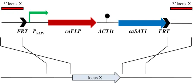
Fig. 1 shows a schematic model of the SAT1 flipper cassette used in this study.
Fig. 1. Structure of the SAT1 flipper cassette.
The C. albicans-adapted FLP gene (caFLP) is represented by the red arrow, the nourseothricin resistance marker (caSAT1) by the blue arrow, the SAP2 promoter (PSAP2) by the bent green arrow, and the transcription termination sequence of the C.
albicans ACT1 gene (ACT1t) by the filled circle. The 34-bp FLP recombination target
sequences (FRT, black arrows) (5‟-GAA GTT CCT ATA CTT TCT AGA GAA TAG GAA CTT C-3‟) flanking the cassette are not drawn to scale. The figure has been modified from Reuß et al. [62].
1.3.3 Reporter genes
Reporter genes are useful tools to study gene regulation, although they do not
necessarily reflect transcript stability of the gene of interest [60] [63]. The LAC4 gene of
the yeast Kluveromyces lactis, which encodes β-galactosidase, was the first functional
heterologous reporter gene for C. albicans [78]. The moderate sensitivity initially achieved
with colorimetric substrates was dramatically increased using a bioluminescent substrate
[79]. A second reporter system based on the enzymatic activity of the β-galactosidase was
developed by Uhl et al. in 2001 [80]. In this case, the lacZ gene of Streptococcus
thermophilus was cloned downstream of three different promoters, a constitutive one and
FRT PSAP2 FRT
5‟ locus X 3‟ locus X
caFLP ACT1t caSAT1
17 Introduction,
two inducible ones. Expression was readily detected in liquid assays using permeabilised
cells and in situ colorimetric assays of colonies growing on plates. The luciferase gene
from the sea pansy Renilla reniformis (RrLUC) does not contain CTG codons, and thus has
been exploited as a useful reporter gene in C. albicans [81]. The bioluminescence based-
assay proved an effective tool to study the activity of inducible, constitutive and
phase-specific (white or opaque) promoters. However, the efficiency of this system with intact
cells is significantly lower than with protein extracts. Reporter systems based on modified
versions of the green fluorescent protein (GFP) gene from the jellyfish Aequorea victoria
are also available [82] [83]. They have been genetically engineered to replace the
non-canonical CTG serine codon for the TTG leucine codon and to carry mutations in the
chromophore that enhance GFP fluorescence. These yEGFP (yeast enhanced GFP) genes
offer major advantages in comparison to the β-galactosidase and luciferase enzymatic
systems: no cofactors are required for the expression of GFP and the resulting fluorescence
can be detected in living organisms, even at the level of single cells [84] [85]. This makes
yEGFP an optimal reporter for studying the sub-cellular localization of a protein of interest
[60]. More recently, cassettes for the PCR-mediated construction of green, yellow and
cyan fluorescent protein fusions have also been developed [86].
1.4
Aims of the study
Fluconazole is the most widely used antimycotic to treat infections caused by C.
albicans, the most common agent of human fungal infections, but the frequent and
widespread use of this drug has caused an increased frequency of drug resistant clinical C.
albicans strains. The present study was aimed to investigate new aspects of the underlying
mechanisms that lead to fluconazole resistance in C. albicans. Previous studies have
illustrated that common fluconazole resistance mechanisms in C. albicans are the
18 Introduction,
of the target gene ERG11, which are regulated by known transcription factors (CAP1,
MRR1, TAC1, UPC2). Recently, new transcription factors (TFs), MCM1 and ADA2 have
been shown to be involved in drug resistance gene regulation. On this ground, the present
study was focused to define the role of those two transcription factors in the azole
resistance. To this aim, MCM1 conditional mutants [57] and ADA2 knock-out mutants,
constructed in this study, were used to analyze the expression of drug resistance related
genes in response to inducing chemicals. In addition, expression of drug resistance related
genes has also been evaluated in MCM1 or ADA2 mutants containing hyperactive
transcription factors alleles.
The present PhD project was carried out at the “Dipartimento di Biologia, sezione di Microbiologia” of the University of Pisa, Via San Zeno 35-39, under the supervision of
Prof. ssa Sonia Senesi, and at the mycology unit of the “Institut für Molekulare Infektionsbiologie”, Josef-Schneider Straße 2, Bau D15, 97080 Würzburg, under the
2
2
M
M
AATTEERRIIAALLSSAANNDDM
M
EETTHHOODDSSFor all microbiology and molecular biology procedures standard protocols were
followed from Sambrook et al., (1989) [87] or Asubel et al., (1989) [88] with minor
modifications.
All solutions and media were made in double distilled and distilled water,
respectively. All solutions and media were sterilized by autoclaving or filter sterilized by
passing through a 0.22 micron Millipore filter.
2.1
Bacterial strain
Escherichia coli K12: E. coli strain DH5α (F-, endA1, hsdR17 [rk-, mk-], supE44,
thi-1, recA1, gyrA96, relA1, Δ[argF-lac]U169, λ-, Φ80dlacZΔM15) (Bethesda Research
Laboratories, 1986) was used for bacterial cloning experiments.
2.2
Primers
Details of primers used in this study are shown in Table 1. Primers were obtained
from MWG (Ebersberg, Germany).
Table 1. List of primers used in this study
Primer
name Sequence (5'-3') Gene
Gene sequences
Introduced restriction sites/ position ACT1RT AGTGTGACATGGATGTTAGAAAAGAATTATACGG ACT1 +1512 +1545
ACT2RT ACAGAGTATTTTCTTTCTGGTGGAGCA ACT1 +1677 +1651
ADA2-3 GGGATGGTGTTCTCGAGGGGTAAATGTAG ADA2 +1317 +1344 XhoI +1328 ADA2-4 GATCGGGCCCTACTTGATCGGTCATACTGGAGC ADA2 +1753 +1721 ApaI +1748 ADA2-5 AACCTAGCAAACGAGCTCACGTGATGTAAGTG ADA2 -529 -498 SacI -513 ADA2-6 GATAATCATGCCGCGGTTTATGATCTCCAG ADA2 +160 +131 SacII +148 ADA2-7 GCAGGATTGACTACTGGAGCTCATAAACCATG ADA2 +118 +149 SacI +138 ADA2-8 GGTAATGGTATATTTTTCCGCGGTTCTAATCGTTC ADA2 +452 +418 SacII +433 ADA2-9 CCAAACACCAGGATTCTCGAGTGGTAATTCTT ADA2 +987 +1018 XhoI +1002 ADA2-10 CTGAGAACACGGGCCCATATGCACAAA ADA2 +1332 +1306 ApaI +1321
20 Materials and Methods,
Primer
name Sequence (5'-3') Gene
Gene sequences
Introduced restriction sites/ position ADA2-11 ATATGAGCTCGGGCCCACTCACGTGATGTAAGTGC ADA2 -518 -497 SacI-ApaI ADA2-12 CTCCATCTCAACCGCGGCCAGAAGTATTGCC ADA2 +1902 +1872 SacII +1889 CDR2-5 AGATGGTACCAGGTAGTGATAGTTAACACACC CDR2 -464 -433 KpnI -456 CDR2-6 ATATGTCGACATTGTATGTGTTAATTAGTGAAATA CDR2 +2 -33 SalI -8 EFB1A ATTGAACGAATTCTTGGCTGAC EF1B +42 +63
EFB1B CATCTTCTTCAACAGCAGCTTG EF1B +960 +939 MDR5RT ATTTGTTCAGATCAGTCATTGCTTCAGTGT MDR1 +1499 +1528 MDR6RT GGTCCGTTCAAGTAAAACAAAACTGGAATA MDR1 +1661 +1632
Restriction sites introduced into the primers are underlined.
2.3
Plasmids
The following abbreviations are used in the description of plasmids: caFLP: C.
albicans-adapted FLP gene encoding the site specific recombinase FLP; caSAT1: C. albicans-adapted nourseothricin resistance marker (dominant selection marker); FRT:
(FLP-recognition target) minimal recombination target sites of the FLP recombinase;
GFP: Green fluorescent protein gene; Px: Promoter of the given gene (X) ; Ptet:
rtTA-dependent promoter; ACT1t: Transcription termination sequence of the actin (ACT1) gene.
Table 2. Plasmids used in this study
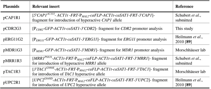
Plasmids Relevant insert Reference
Plasmids used for the MCM1 study pCAP1R1 [5'CAP1
ΔC333
-ACT1t-FRT-PMAL2-caFLP-ACT1t-caSAT1-FRT-3'CAP1]- fragment
for introduction of hyperactive CAP1 allele
Schubert et al., submitted pMPG2S [3'ACT1-PMDR1-GFP-ACT1t-caSAT1-PACT1]- fragment for MDR1 promoter
analysis, integration on ACT1 promoter
Morschhäuser et al., 2007 [46] pMRR1R3 [MRR1
P683S
-ACT1t-FRT-PMAL2-caFLP-ACT1t-caSAT1-FRT-3'MRR1]- fragment
for introduction of hyperactive MRR1 allele
Schubert et al., submitted Plasmids used for the ADA2 study
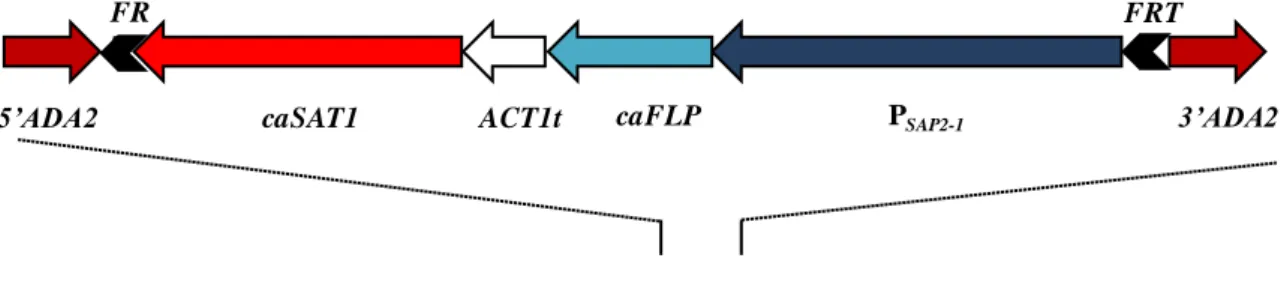
pADA2K1 [ADA2-FRT-PSAP2-1-caFLP-ACT1t-caSAT1-FRT-3'ADA2]- fragment for gene
reintegration This study
pADA2M1 [5'CAP1-FRT-PSAP2-1-caFLP-ACT1t-caSAT1-FRT-3'ADA2]- fragment This study
pADA2M2 [5'ADA2-FRT-PSAP2-1-caFLP-ACT1t-caSAT1-FRT-3'ADA2]- fragment for gene
deletion (allele 1) This study
pADA2M3 [5'ADA2-FRT-PSAP2-1-caFLP-ACT1t-caSAT1-FRT-3'ADA2]- fragment This study
pADA2M4 [5'ADA2-FRT-PMAL2-caFLP-ACT1t-caSAT1-FRT-3'ADA2]- fragment for gene
21 Materials and Methods,
Plasmids Relevant insert Reference
pCAP1R1 [5'CAP1
ΔC333- ACT1t -FRT-P
MAL2-caFLP-ACT1t-caSAT1-FRT-3'CAP1]-
fragment for introduction of hyperactive CAP1 allele
Schubert et al., submitted pCDR2G3 [PCDR2-GFP-ACT1t-caSAT1-3'CDR2]- fragment for CDR2 promoter analysis This study
pERG11G2 [PERG11-GFP-ACT1t-caSAT1-3'ERG11]- fragment for ERG11 promoter analysis
Heilmann et al., 2010 [89] pMDR1G3 [PMDR1-GFP-ACT1t-caSAT1-3'MDR1]- fragment for MDR1 promoter analysis Morschhäuser lab
pMRR1R3 [MRR1
P683S
-ACT1t-FRT-PMAL2-caFLP-ACT1t-caSAT1-FRT-3'MRR1]- fragment
for introduction of hyperactive MRR1 allele
Schubert et al., submitted pTAC1R3 [3'TAC1
G980E
-ACT1t-FRT-PMAL2-caFLP-ACT1t-caSAT1-FRT-3'TAC1]- fragment
for introduction of TAC1 hyperactive allele Morschhäuser lab pUPC2R1 [UPC2
G648D
-ACT1t-FRT-PMAL2-caFLP-ACT1t-caSAT1-FRT-3'UPC2]- fragment
for introduction of UPC2 hyperactive allele
Heilmann et al., 2010 [89]
2.4
C. albicans strains
Parental and mutant Candida albicans strains used in this study are listed in Table
3. Some mutants used in this study where already present in the collection of Prof. Morschhäuser‟s group. MRcan42 and MRcan43 where kindly provided by Prof. Steffen
Rupp (University of Stuttgart, Germany) and SC5314 is the Candida albicans reference
strain.
Table 3. Strains used in this study
Strain Parent Relevant genotype Reference
SC5314 Wild-type reference strain Gillum et al., 1984 [90] SCMPG2A SC5314 ACT1/act1::PMDR1-GFP-caSAT1
Morschhäuser et al., 2007 [46] SCMPG2B SC5314 ACT1/act1::PMDR1-GFP-caSAT1
Morschhäuser et al., 2007 [46] MRcan42 SC531 pTR-MCM1-myc/mcm1Δ Rottmann et al.,
2003 [57] MRcan43 SC5314 pTR-MCM1/MCM1-myc Rottmann et al.,
2003 [57] can42MPG2A MRcan42 ACT1/act1::PMDR1-GFP-caSAT1 this study
can42MPG2B MRcan42 ACT1/act1::PMDR1-GFP-caSAT1 this study
can43MPG2A MRcan43 ACT1/act1::PMDR1-GFP-caSAT1 this study
can43MPG2B MRcan43 ACT1/act1::PMDR1-GFP-caSAT1 this study
can42MRR1R31A MRcan42 MRR1/MRR1P683S-SAT1-FLIP this study can42MRR1R31B MRcan42 MRR1/MRR1P683S-SAT1-FLIP this study can42MRR1R32A can42MRR1R31A MRR1/MRR1P683S-FRT this study can42MRR1R32B can42MRR1R31B MRR1/MRR1P683S-FRT this study can42MRR1R33A can42MRR1R32A MRR1P683S-SAT1-FLIP/MRR1P683S-FRT this study
22 Materials and Methods,
Strain Parent Relevant genotype Reference
can42MRR1R33B can42MRR1R32B MRR1P683S-SAT1-FLIP/MRR1P683S-FRT this study can42MRR1R34A can42MRR1R33A MRR1P683S-FRT/MRR1P683S-FRT this study can42MRR1R34B can42MRR1R33B MRR1P683S-FRT/MRR1P683S-FRT this study can43MRR1R31A MRcan43 MRR1/MRR1P683S-SAT1-FLIP this study can43MRR1R31B MRcan43 MRR1/MRR1P683S-SAT1-FLIP this study can43MRR1R32A can43MRR1R31A MRR1/MRR1P683S-FRT this study can43MRR1R32B can43MRR1R31B MRR1/MRR1P683S-FRT this study can43MRR1R33A can43MRR1R32A MRR1P683S-SAT1-FLIP/MRR1P683S-FRT this study can43MRR1R33B can43MRR1R32B MRR1P683S-SAT1-FLIP/MRR1P683S-FRT this study can43MRR1R34A can43MRR1R33A MRR1P683S-FRT/MRR1P683S-FRT this study can43MRR1R34B can43MRR1R33B MRR1P683S-FRT/MRR1P683S-FRT this study can42CAP1R11A MRcan42 CAP1ΔC333-SAT1-FLIP/CAP1-2 this study
can42CAP1R11B MRcan42 CAP1-1/CAP1ΔC333-SAT1-FLIP this study
can42CAP1R12A can42CAP1R11A CAP1ΔC333-FRT/CAP1-2 this study
can42CAP1R12B can42CAP1R11B CAP1-1/CAP1ΔC333-FRT this study
can42CAP1R13A can42CAP1R12A CAP1ΔC333-FRT/CAP1ΔC333-SAT1-FLIP this study can42CAP1R13B can42CAP1R12B CAP1ΔC333-SAT1-FLIP/CAP1ΔC333-FRT this study
can42CAP1R14A can42CAP1R13A CAP1ΔC333-FRT/CAP1ΔC333-FRT this study
can42CAP1R14B can42CAP1R13B CAP1ΔC333-FRT/CAP1ΔC333-FRT this study
can43CAP1R11A MRcan43 CAP1-1/CAP1ΔC333-SAT1-FLIP this study
can43CAP1R11B MRcan43 CAP1-1/CAP1ΔC333-SAT1-FLIP this study
can43CAP1R12A can43CAP1R11A CAP1-1/CAP1ΔC333-FRT this study
can43CAP1R12B can43CAP1R11B CAP1-1/CAP1ΔC333-FRT this study
can43CAP1R13A can43CAP1R12A CAP1ΔC333-SAT1-FLIP/CAP1ΔC333-FRT this study
can43CAP1R13B can43CAP1R12B CAP1ΔC333-SAT1-FLIP/CAP1ΔC333-FRT this study can43CAP1R14A can43CAP1R13A CAP1ΔC333-FRT/CAP1ΔC333-FRT this study
can43CAP1R14B can43CAP1R13B CAP1ΔC333-FRT/CAP1ΔC333-FRT this study
SCADA2M1A SC5314 ada2Δ::SAT1-FLIP/ADA2 this study SCADA2M1B SC5314 ada2Δ::SAT1-FLIP/ADA2 this study SCADA2M2A SCADA2M1A ada2Δ::FRT/ADA2 this study SCADA2M2B SCADA2M1B ada2Δ::FRT/ADA2 this study SCADA2M3A SCADA2M2A ada2Δ::FRT/ada2Δ::SAT1-FLIP this study SCADA2M3B SCADA2M2B ada2Δ::FRT/ada2Δ::SAT1-FLIP this study SCADA2M4A SCADA2M3A ada2Δ::FRT/ada2Δ::FRT this study SCADA2M4B SCADA2M3B ada2Δ::FRT/ada2Δ::FRT this study SCADA2MK1A SCADA2M4A ADA2::SAT1-FLIP/ada2Δ::FRT this study SCADA2MK1B SCADA2M4B ADA2::SAT1-FLIP/ada2Δ::FRT this study SCADA2MK2A SCADA2MK1A ADA2::FRT/ada2Δ::FRT this study SCADA2MK2B SCADA2MK1B ADA2::FRT/ada2Δ::FRT this study
23 Materials and Methods,
Strain Parent Relevant genotype Reference
SCΔada2CG3A SCADA2M4A CDR2/cdr2::PCDR2-GFP-caSAT1 this study
SCΔada2CG3B SCADA2M4B CDR2/cdr2::PCDR2-GFP-caSAT1 this study
SCΔada2EG2A SCADA2M4A ERG11/erg11::PERG11-GFP-caSAT1 this study
SCΔada2EG2B SCADA2M4B ERG11/erg11::PERG11-GFP-caSAT1 this study
SCΔada2MG3A SCADA2M4A MDR1/mdr1::PMDR1-GFP-caSAT1 this study
SCΔada2MG3B SCADA2M4B MDR1/mdr1::PMDR1-GFP-caSAT1 this study
SCΔada2CAP1R11A SCADA2M4A CAP1ΔC333-SAT1-FLIP/CAP1-2 this study SCΔada2CAP1R11B SCADA2M4B CAP1-1/CAP1ΔC333-SAT1-FLIP this study SCΔada2CAP1R12A SCΔada2CAP1R11A CAP1ΔC333-FRT/CAP1-2 this study SCΔada2CAP1R12B SCΔada2CAP1R11B CAP1-1/CAP1ΔC333-FRT this study SCΔada2CAP1R13A SCΔada2CAP1R12A CAP1ΔC333-FRT/CAP1ΔC333-SAT1-FLIP this study SCΔada2CAP1R13B SCΔada2CAP1R12B CAP1ΔC333-SAT1-FLIP/CAP1ΔC333-FRT this study SCΔada2CAP1R14A SCΔada2CAP1R13A CAP1ΔC333-FRT/CAP1ΔC333-FRT this study SCΔada2CAP1R14B SCΔada2CAP1R13B CAP1ΔC333-FRT/CAP1ΔC333-FRT this study SCΔada2CAP1R14MG3A SCΔada2CAP1R14A MDR1/mdr1::PMDR1-GFP-caSAT1 this study
SCΔada2CAP1R14MG3B SCΔada2CAP1R14B MDR1/mdr1::PMDR1-GFP-caSAT1 this study
SCΔada2MRR1R31A SCADA2M4A MRR1P683S-SAT1-FLIP/MRR1 this study
SCΔada2MRR1R31B SCADA2M4B MRR1P683S-SAT1-FLIP/MRR1 this study
SCΔada2MRR1R32A SCΔada2MRR1R31A MRR1P683S-FRT/MRR1 this study SCΔada2MRR1R32B SCΔada2MRR1R31B MRR1P683S-FRT/MRR1 this study SCΔada2MRR1R33A SCΔada2MRR1R32A MRR1P683S-FRT/MRR1P683S-SAT1-FLIP this study SCΔada2MRR1R33B SCΔada2MRR1R32B MRR1P683S-FRT/MRR1P683S-SAT1-FLIP this study SCΔada2MRR1R34A SCΔada2MRR1R33A MRR1P683S-FRT/MRR1P683S-FRT this study SCΔada2MRR1R34B SCΔada2MRR1R33B MRR1P683S-FRT/MRR1P683S-FRT this study SCΔada2MRR1R34MG3A SCΔada2MRR1R34A MDR1/mdr1::PMDR1-GFP-caSAT1 this study
SCΔada2MRR1R34MG3B SCΔada2MRR1R34B MDR1/mdr1::PMDR1-GFP-caSAT1 this study
SCΔada2TAC1R31A SCADA2M4A TAC1G980E-SAT1-FLIP/TAC1-2 this study SCΔada2TAC1R31B SCADA2M4B TAC1-1/TAC1G980E-SAT1-FLIP this study SCΔada2TAC1R32A SCΔada2TAC1R31A TAC1G980E-FRT/TAC1-2 this study SCΔada2TAC1R32B SCΔada2TAC1R31B TAC1-1/TAC1G980E-FRT this study SCΔada2TAC1R33A SCΔada2TAC1R32A TAC1G980E-FRT/TAC1G980E-SAT1-FLIP this study SCΔada2TAC1R33B SCΔada2TAC1R32B TAC1G980E-SAT1-FLIP/TAC1G980E-FRT this study SCΔada2TAC1R34A SCΔada2TAC1R33A TAC1G980E-FRT/TAC1G980E-FRT this study SCΔada2TAC1R34B SCΔada2TAC1R33B TAC1G980E-FRT/TAC1G980E-FRT this study
SCΔada2TAC1R34CG3A SCΔada2TAC1R34A CDR2/cdr2::PCDR2-GFP-caSAT1 this study
SCΔada2TAC1R34CG3B SCΔada2TAC1R34B CDR2/cdr2::PCDR2-GFP-caSAT1 this study
SCΔada2UPC2R11A SCADA2M4A UPC2G648D-SAT1-FLIP/UPC2 this study SCΔada2UPC2R11B SCADA2M4B UPC2G648D-SAT1-FLIP/UPC2 this study SCΔada2UPC2R12A SCΔada2UPC2R11A UPC2G648D-FRT/UPC2 this study
24 Materials and Methods,
Strain Parent Relevant genotype Reference
SCΔada2UPC2R12B SCΔada2UPC2R11B UPC2G648D-FRT/UPC2 this study
SCΔada2UPC2R13A SCΔada2UPC2R12A UPC2G648D-FRT/UPC2G648D-SAT1-FLIP this study
SCΔada2UPC2R13B SCΔada2UPC2R12B UPC2G648D-FRT/UPC2G648D-SAT1-FLIP this study SCΔada2UPC2R14A SCΔada2UPC2R13A UPC2G648D-FRT/UPC2G648D-FRT this study SCΔada2UPC2R14B SCΔada2UPC2R13B UPC2G648D-FRT/UPC2G648D-FRT this study SCΔada2UPC2R14EG2A SCΔada2UPC2R14A ERG11/erg11::PERG11-GFP-caSAT1 this study
SCΔada2UPC2R14EG2B SCΔada2UPC2R14B ERG11/erg11::PERG11-GFP-caSAT1 this study
SCCG3A SC5314 CDR2/cdr2::PCDR2-GFP-caSAT1 this study
SCCG3B SC5314 CDR2/cdr2::PCDR2-GFP-caSAT1 this study
SCEG2A SC5314 ERG11/erg11::PERG11-GFP-caSAT1
Heilmann et al., 2010 [89] SCEG2B SC5314 ERG11/erg11::PERG11-GFP-caSAT1
Heilmann et al., 2010 [89] SCMG3A SC5314 MDR1/mdr1::PMDR1-GFP-caSAT1 Morschhäuser lab
SCMG3B SC5314 MDR1/mdr1::PMDR1-GFP-caSAT1 Morschhäuser lab
SCCAP1R12A SC5314 CAP1ΔC333-FRT/CAP1-2 Schubert et al., in revision
SCCAP1R12B SC5314 CAP1-1/CAP1ΔC333-FRT Schubert et al., in revision
SCCAP1R14A SCCAP1R12A CAP1ΔC333-FRT/CAP1ΔC333-FRT Schubert et al., in revision
SCCAP1R14B SCCAP1R12B CAP1ΔC333-FRT/CAP1ΔC333-FRT Schubert et al., in revision
SCCAP1R14MG3A SCCAP1R14A MDR1/mdr1::PMDR1-GFP-caSAT1 this study
SCCAP1R14MG3B SCCAP1R14B MDR1/mdr1::PMDR1-GFP-caSAT1 this study
SCMRR1R32A SC5314 MRR1P683S-FRT/MRR1 Schubert et al., in revision
SCMRR1R32B SC5314 MRR1P683S-FRT/MRR1 Schubert et al., in revision
SCMRR1R34A SCMRR1R32A MRR1P683S-FRT/MRR1P683S-FRT Schubert et al., in revision
SCMRR1R34B SCMRR1R32B MRR1P683S-FRT/MRR1P683S-FRT Schubert et al., in
revision SCMRR1R34MG3A SCMRR1R34A MDR1/mdr1::PMDR1-GFP-caSAT1 this study
SCMRR1R34MG3B SCMRR1R34B MDR1/mdr1::PMDR1-GFP-caSAT1 this study
SCTAC1R32A SC5314 TAC1G980E-FRT/TAC1-2 Morschhäuser lab
SCTAC1R32B SC5314 TAC1-1/TAC1G980E-FRT Morschhäuser lab
SCTAC1R34A SCTAC1R32A TAC1G980E-FRT/TAC1G980E-FRT Morschhäuser lab
SCTAC1R34B SCTAC1R32B TAC1G980E-FRT/TAC1G980E-FRT Morschhäuser lab SCTAC1R34CG3A SCTAC1R34A CDR2/cdr2::PCDR2-GFP-caSAT1 this study
SCTAC1R34CG3B SCTAC1R34B CDR2/cdr2::PCDR2-GFP-caSAT1 this study
SCUPC2R12A SC5314 UPC2G648D-FRT/UPC2 Heilmann et al., 2010 [89] SCUPC2R12B SC5314 UPC2G648D-FRT/UPC2 Heilmann et al.,
2010 [89] SCUPC2R14A SCUPC2R12A UPC2G648D-FRT/UPC2G648D-FRT Heilmann et al.,
2010 [89] SCUPC2R14B SCUPC2R12B UPC2G648D-FRT/UPC2G648D-FRT Heilmann et al.,
25 Materials and Methods,
Strain Parent Relevant genotype Reference
SCUPC2R14EG2A SCUPC2R14A ERG11/erg11::PERG11-GFP-caSAT1
Heilmann et al., 2010 [89] SCUPC2R14EG2B SCUPC2R14B ERG11/erg11::PERG11-GFP-caSAT1
Heilmann et al., 2010 [89]
2.5
Growth and maintenance of C. albicans strains
All strains were stored as frozen stocks with 15% glycerol at -80°C and sub
cultured on YPD agar plates (10 g yeast extract, 20 g peptone, 20 g dextrose, 15 g agar per
liter) at 30°C. Strains were routinely grown in YPD liquid medium at 30°C in a shaking
incubator at 250 rpm.
2.6
Selection media for C. albicans transformants
For selection of nourseothricin-resistant transformants, 200 μg/ml nourseothricin
(Werner Bioagents, Jena, Germany) was added to YPD agar plates. To obtain
nourseothricin sensitive derivatives in which the SAT1 flipper cassette was excised by
FLP-mediated recombination, transformants were grown overnight in either YPM medium
(10 g yeast extract, 20 g peptone, 20 g maltose per liter) without selective pressure to
induce the MAL2 promoter, or in YCB-YE-BSA medium (23.4 g Yeast Carbon Base, 2 g
Yeast Extract, 4 g Bovine Serum Albumin, pH 4) to induce the SAP2-1 promoter that
controls the expression of the caFLP gene in the SAT1 flipper cassette. One hundred to 200 cells were then spread on YPD plates containing 10 μg/ml nourseothricin and grown for 2
days at 30°C. Nourseothricin-sensitive clones were identified by their small colony size and confirmed by restreaking on YPD plates containing 200 μg/ml nourseothricin. For the
ΔΔada2 mutants, cells were spread on normal YPD agar plates since they resulted
hypersensitive to nourseothricin. In this case no difference in size was observed between
26 Materials and Methods,
2.7
Growth and maintenance of E. coli strains
Recombinant E. coli strains were routinely grown in LB liquid medium (1%
Peptone, 0.5% Yeast extract, 0.5% NaCl) under the selection pressure (100 μg/ml
ampicillin). For growth on plates, 1.5% agar [DifcoTM agar granulated, BD] was added to
the media. The cells were grown at 37°C for 16-18 hours with shaking at 200 rpm and the
plates were incubated at 37°C until the colonies appeared. While liquid cultures were
processed to isolate plasmid DNA, the plates with streaked colonies were stored at 4ºC.
2.8
Small scale plasmid DNA isolation (Miniprep)
Miniprep was carried out using a commercial kit by Macherey-Nagel
(Nucleospin® Plasmid) according to manufacturer‟s guideline. After cultivation of liquid
cultures, cells were harvested by spinning at 11,000 x g for 30 sec. The surnatant was
carefully removed and the pellet vigorously resuspended in 250 μl Buffer A1. 250 μl of
Buffer A2 was then added and the mixture gently mixed by inverting the tube 6-8 times.
The mixture was incubated at room temperature for 5 min until the lysate appeared clear.
300 μl of Buffer A3 was added to the lysate and the tube gently inverted 6-8 times. The
lysate was so centrifuged for 5 min at 11,000 x g and the clearified supernatant was loaded
on the kit columns placed on a collection tube. The column was spinned for 1 min at
11,000 x g, the flowthrough discarded and the column placed back on the collection tube.
The column‟s silica membrane was then washed by adding 600 μl Buffer A4, the column
spinned for 1 min at 11,000 x g and the flowthrough discarded. The silica membrane was
then dried by spinning the column for further 2 minutes in the same collection tube. The
plasmid was eluted by transferring the column on a clean 1.5 ml microcentrifuge tube,
adding 50 μl of double distilled water directly on the membrane, incubating for 1 minute at